Article

Genetic Diversity and Population Structure of Leishmania infantum in Morocco as Revealed by Multilocus Sequence Typing (MLST) Approach

1
Laboratory of Parasitology and Vector-Borne-Diseases, Institut Pasteur du Morocco, Casablanca 20360, Morocco
2
Laboratory of Microbial Biotechnology and Bioactive Molecules, Faculty of Sciences and Technologies, Sidi Mohammed Ben Abdellah University, Fes 30000, Morocco
3
Molecular Epidemiology and Experimental Pathology (MEEP)/ LR16IPT04, Institut Pasteur de Tunis, Université de Tunis El Manar, B.P. 74, Tunis 1068, Tunisia
4
Laboratory of Cellular and Molecular Pathology, Research Team on Immunopathology of Infectious and Systemic Diseases, Faculty of Medicine and Pharmacy, Hassan II University of Casablanca, Casablanca 21100, Morocco
*
Author to whom correspondence should be addressed.
Pathogens 2023, 12(6), 785; https://doi.org/10.3390/pathogens12060785
Submission received: 18 April 2023 / Revised: 23 May 2023 / Accepted: 26 May 2023 / Published: 31 May 2023
(This article belongs to the Special Issue Parasites: Epidemiology, Treatment and Control)

Abstract

:
Leishmania infantum is endemic in Morocco, and it causes both visceral (VL) and cutaneous leishmaniasis (CL). In this study, the multilocus sequence typing (MLST) approach was used to investigate the phylogeny and population structure of Leishmania infantum strains isolated from CL and VL patients and the canine reservoir in different leishmaniasis endemic foci in Morocco. For this purpose, eight loci (pgm, alat, me, fh, g6pd, pgd, gpi and cytb) were amplified in 40 samples, out of which 31 were successfully sequenced. The genetic diversity analysis detected a high degree of intraspecific genetic variability among the studied strains. The phylogenetic and the haplotype analyses showed that most of the strains from the same geographical areas clustered together. The recombination among Leishmania infantum strains was revealed through a splits tree analysis and the number of recombination events. Moreover, the assessment of the gene flow between Leishmania infantum and Leishmania tropica through phylogenetic analysis and haplotype diversity in two endemic foci where the two species were sympatric showed no genetic exchange between the two species.

1. Introduction

Globally, over 20 species of the genus Leishmania are causative agents of a broad spectrum of diseases called leishmaniasis, ranging from self-limiting localized cutaneous lesions to visceral leishmaniasis with a fatal outcome [1]. Leishmaniasis are endemic in 98 countries, and about 350 million people are at risk of acquiring them [2].
Visceral leishmaniasis (VL) and cutaneous leishmaniasis (CL) are endemic in Morocco. The species Leishmania (L.) infantum can cause both forms. The first case of VL due to zymodeme MON-1 was reported in 1920 in Tangier province [3]. VL is mainly distributed in the north of the country where the disease was originally described, with cases being reported in the center and the south [4].
The early clinical symptoms of VL include intermittent fever, malaise and shivering. The overt disease manifests by striking splenomegaly and is either accompanied or not by hepatomegaly. The hyperplasia of the reticuloendothelial system is accompanied by wasting and pallor of the mucous membranes [5,6].
As for the cutaneous form caused by L. infantum MON-24, El Mazini et al. (2022) reported that it was first identified in 1996 in the north in Taounate province [7]. Thereafter, many cases were notified in diverse areas throughout the country, and most of them were foci where L. tropica was endemic or where this species was reported, indicating a change in Leishmania distribution with L. infantum invading L. tropica endemic areas such as, for instance, Sefrou [8] and Taza [9].
The cutaneous lesions caused by L. infantum are usually represented as a small single ulcerated or lupoid lesion on the face that lastsup to three years [10,11,12]. Nevertheless, clinical polymorphism has been observed with erythematous, papular or nodular lesions [13,14].
Dogs are the main reservoir of zymodeme MON-1 [15], and zymodeme MON-24 was also isolated from a domestic dog [16]. Moreover, rodents were found to be infected by Leishmania infantum in endemic cutaneous leishmaniasis zones [17]. Three proven phlebotomine species of the Larroussius subgenus act as Leishmania infantum vectors: Phlebotomus ariasi, Phlebotomus longicuspis and Phlebotomus perniciosus [9,18,19].
Different methods to identify, classify and characterize Leishmania species were developed, and each one has its advantages and disadvantages. Multilocus enzyme electrophoresis (MLEE), long considered as the gold standard for species and strain typing of Leishmania, is based on 15 enzymatic systems analyzed by starch gel electrophoresis [20]. However, this method is quite slow, laborious, costly, necessitates large amounts of in vitro parasite culture and it has a low discriminatory power due to its inability to detect silent nucleotide substitutions [21].
For these reasons, PCR-based assays were developed and proved their usefulness for the description of species and even strain diversity [22]. Multilocus sequence typing (MLST), first proposed for bacterial molecular epidemiology investigations, has powerful phylogenetic potential and is likely to become the new gold standard method for Leishmania species characterization [23] due to the complex genomic composition of protozoan parasites such as species of the Leishmania genus. These parasites display a high nucleotide diversity among markers corresponding to housekeeping gene coding regions [24].
El Hamouchi et al. (2018) reported many changes in the eco-epidemiological profile of L. infantum in Morocco, such as the southward spread of the parasite and the occurrence of treatment-resistant L. infantum [25]. Moreover, the emergence of new cases of CL due to L. infantum was reported in different parts of the country where this form of leishmaniasis has never been notified [7]. We still need to better understand the dynamics of leishmaniasis due to L. infantum and the population structure of this species through genetic analysis. This could help us to assess the risk of spread of both CL and VL caused by L. infantum. To date, few studies have been conducted on the population genetics of L. infantum in Morocco despite the importance of this species.
In this survey, we used the MLST approach to study the genetic diversity, phylogeny and population structure of L. infantum strains isolated from CL and VL patients and the canine reservoir in different endemic foci in Morocco, including those where L. infantum was co-sympatric with L. tropica. To evaluate the possible gene flow between these two species, we also studied the genetic diversity of L. tropica strains collected in these co-endemic areas.
For this purpose, we used a scheme of seven housekeeping genes used in MLEE and as DNA markers in MLST studies [24,26,27] in addition to the kinetoplast marker cytochrome b (Cyt b) [28].

2. Materials and Methods

2.1. Ethics Statement

Informed consent was obtained from all adults who participated in this study. Consent for the inclusion of young children was obtained from their parents or guardians.

2.2. Samples

The human visceral leishmaniasis (VL) samples were collected by iliac crest puncture by a pediatrician from patients hospitalized at the university hospital in Fes that came from VL endemic regions in Morocco (Table 1, Figure 1).
The cutaneous leishmaniasis (CL) samples of L. infantum, including one HIV+/CL and L. tropica, were collected by dermal scraping from the edge of the lesions in northern Morocco (Table 1, Figure 1).
As for the canine VL samples, they were collected in the framework of the surveys we conducted in northern Morocco from lymph nodes of suspected dogs. We also used Moroccan L. infantum canine strains kindly provided by Pr. J.P. Dedet (the CHU Montpellier, centre national de référence des leishmanioses) (Table 1, Figure 1). All human and dog samples were microscopically examined on slide smears after fixation with absolute methanol and Giemsa staining using a ×100 immersion objective.

2.3. DNA Extraction and Leishmania Species Identification

For DNA extraction from L. infantum cultures, we used the phenol-chloroform method as described in [29]. From CL samples, the DNA was extracted from dermal scraping using an ISOLATEII Genomic DNA commercial kit (Bioline, London, UK) following the manufacturer’s instructions.
To detect and identify Leishmania species, the ITS1 coding region was first amplified using the primers LITSR and L5.8S following the protocol of Schonian et al. (2003) [30]. Then, the positive samples were digested by Hae III restriction enzyme for 2 h at 37 °C. RFLPs were visualized by electrophoresis on 3% agarose gel containing ethidium bromide along with the weight marker HyperLadder 100 bp (Bioline, London, UK). The profiles assigned the DNA samples to the Leishmania species.

2.4. PCR Amplification of the Eight Markers

Seven metabolic enzyme-coding genes, namely malic enzyme (me), fumarate hydratase (fh), glucose-6-phosphate dehydrogenase (g6pdh), glucose-6-phosphate isomerase (gpi), phosphogluconate dehydrogenase (pgd), alanine aminotransferase (alat), phosphoglucomutase (pgm) and mitochondrial cytochrome b gene (Cyt b), were amplified from each sample (Table 2).
The optimized PCR reactions for the amplification of these eight DNA markers were carried out in reaction volumes of 25 μL containing 1× PCR reaction buffer, 1.5 mM MgCl2, 1.5 U Taq DNA polymerase (Bioline, London, UK), 0.4 mM primers (Table 2), 3 μL template DNA and distilled water to reach 25 μL. The amplification reactions were conducted in an S1000 thermal cycler (Bio Rad, Hercules, CA, USA) following a cycling program for denaturation at 95 °C for 4 min, then 30 amplification cycles (94 °C for 30 s, 58 °C for 30 s and 72 °C for 90 s) and a final elongation at 72 °C for 10 min. This protocol was optimized for the amplification of the eight markers. The PCR products were visualized in 1% agarose gel containing ethidium bromide along with molecular weight marker HyperLadder 100 bp (Bioline, London, UK).

2.5. PCR Products Sequencing

The amplified PCR products were purified using an Ex’S-Pure™ enzymatic PCR cleanup kit (NimaGen, Nijmegen, The Netherlands). Then, using the same sets of primers, they were sequenced in both directions using a BrillantDye Terminator Cycle Sequencing Kit v.1.1 (NimaGen, Nijmegen, The Netherlands) and an ABI PRISM 3130xL DNA automated sequencer (Applied Biosystems, Waltham, MA, USA).

2.6. Genetic Diversity and Phylogeny Analyses

The sequences of each locus amplified were aligned and compared to entries retrieved from GenBank, inspected to check the heterozygous sites, edited and trimmed using the Chromas v.2.6.2 (Technelysium) and MEGA software v.11 [31].
We used the software Dnasp v6 [32] to calculate the number of polymorphic/heterozygous sites (S), number of haplotypes (H), haplotype diversity (Hd), nucleotide diversity (Pi), the average number of nucleotide differences (K) and the ratio of synonymous and non-synonymous substitutions (dN/dS).
To estimate population differences, the analysis of molecular variance (AMOVA) was performed using the Arlequin software v.3.5.2.2 [33] to calculate the level of genetic diversity among different populations. It indicates how much of this variance is due to differences between or within populations. We performed the AMOVA analysis among the strains based on the disease form VL, CL forms and CanL.
Along with AMOVA, we calculated the differentiation index (Fst), a common measure of the degree of genetic differentiation. This index varies from zero (no genetic differentiation) to one (the population has reached fixation for the different alleles).
Tajima’s D neutrality test [34] is based on the distribution of allele frequency of segregating nucleotide sites. Positive values indicate a bias toward intermediate-frequency alleles, while negative values indicate a bias toward rare alleles, which signifies a recent population expansion or selective sweep. Tajima’s D neutrality test was calculated for all groups.
We concatenated the sequences using the MLSTest software v.1.0.1.23 [35]. The same software was used to estimate the genotypic variation where the allele types (ATs) were calculated, and each unique sequence was given an allelic profile, based on which strains were assigned a sequence type (ST) number. The typing efficiency (TE) and discriminatory power (DP) were calculated for each locus.
We used the MLSTest software v.1.0.1.23 [35] to carry out a congruence test of all the loci with 10,000 replicates, which suggested a homogeneity of all the loci with a p value of 1, and to create the phylogenetic trees using the bio-neighbor joining (BIONJ) algorithm. The heterozygous sites were established as average states in order to avoid possible phylogenetic signal misinterpretation during the analysis, and 1000 replications were used to evaluate the significance of the nodes.
We visualized and edited the resulting trees with TreeGraph2 [36]. We used the population genetics software PopART v.1.7 to build the haplotype network using the median joining network algorithm and to visualize it [37].

2.7. Recombination Evaluation

The recombination amongst L. infantum isolates was evaluated through split decomposition analysis using the neighbor-net method implemented in SplitsTree4 v4.18.3 software [38]. The splits tree analysis was performed using alignments of the concatenated sequences of all seven loci of L. infantum isolates. The software Dnasp v6 was used to calculate the estimate of recombination (R) per gene and the minimum number of recombination events [39,40].

2.8. Temporal Variation Analysis

We assessed the temporal variation in the different sequence types (STs) across the years of sampling using Excel 2013.

2.9. Analysis of the Genetic Exchange between L. infantum and L. tropica

To assess the possibility of gene flow between the two endemic Leishmania species, L. infantum and L. tropica, in Morocco, a set of 12 L. tropica sequences were retrieved from Genbank (https://www.ncbi.nlm.nih.gov/genbank/) accessed on 12 August 2022 (Table 3); the same samples were amplified for the markers alanine aminotransferase (alat) and phosphoglucomutase (pgm). In addition, two L. tropica strains from northern Morocco were amplified for the same loci (Table 1, Figure 1).

3. Results

3.1. Leishmania infantum Identification and Markers Amplification

Based on the ITS1-PCR-RFLP analysis, all the 40 samples were identified as L. infantum. These samples were amplified and sequenced for eight markers (Table 2). The cytb marker was excluded due to the bad quality of sequences. Thirty one sequences out of the forty samples were successfully sequenced for the seven loci (supplementary data Figure S1); the sequences were submitted to GenBank and are accessible using the following accession numbers: OQ627483 to OQ627513 (for alat), OQ627514 to OQ627544 (for fh), OQ627547 to OQ627577 (for g6pd), OQ627580 to OQ627610 (for gpi), OQ627613 to OQ627643 (for me), OQ627646 to OQ627676 (for pgd) and OQ627679 to OQ627709 (for pgm). The samples were divided into three groups: cutaneous leishmaniasis (CL), visceral leishmaniasis (VL) and canine leishmaniasis (CanL) (Table 1).

3.2. Leishmania infantum Genetic Diversity Analyses

To assess the diversity of the studied strains, we estimated the genetic diversity indices of all of the L. infantum strains together and for visceral, cutaneous and canine leishmaniasis for the L. infantum strains separately.

3.2.1. Genetic Diversity of All L. infantum Strains

The analyzed fragments comprised between 412 bp (alat) and 594 (g6pd) for the 31 L. infantum strains. The concatenated sequences of 3368 bp length recorded 82 polymorphic/heterozygous sites, 18 haplotypes and a high haplotype diversity of 0.892, and the average number of nucleotide differences was 5.694.
The analysis of all of the L. infantum strains in each locus showed that the marker pgd had the highest number of polymorphic/heterozygous sites with 25 sites, followed by g6pd with 16 and gpi with 12. In addition, the highest number of haplotypes were recorded at these loci (Table 4). The haplotype diversity, nucleotide diversity and the average number of nucleotide differences were also calculated (Table 4); all the loci had a dN/dS value < 1, indicating negative or purifying selection, except for fh, which had a dN/dS = 1, indicating a neutral selection.

3.2.2. Genetic Diversity of Canine and Human Groups

Sixty-eight and seventeen polymorphic/heterozygous sites were recorded in the canine and human groups, respectively; the latter group had thirteen haplotypes, while the canine group had nine haplotypes. As for the haplotype diversity and the average number of nucleotide differences, they were higher in the canine strains with values of 0.895 and 12.006, respectively. The genetic diversity indices indicated pgd as the most polymorphic locus in both groups with twenty-two and four polymorphic/heterozygous sites in the canine and the human groups, respectively, while the marker fh was the least polymorphic in the canine group and showed zero segregating sites in the human group (Table 5).
The human group contained VL and CL strains, and therefore we analyzed these two sub-groups separately.

3.2.3. Genetic Diversity of the Human VL and CL Sub-Groups

The concatenated sequences of the human CL sub-group showed twelve polymorphic/heterozygous sites, while the human VL sub-group had nine; seven haplotypes were recorded in both sub-groups. The haplotype diversity and the average number of nucleotide difference were higher in the CL strains with values of 0.864 and 3.167, respectively. Three markers, pgd, me and gpi had two as the highest number of polymorphic/heterozygous sites in the VL sub-group, while the loci gpi and me had three as the highest number of polymorphic/heterozygous sites in the CL sub-group; the marker fh showed zero polymorphic/heterozygous sites in both groups along with pgm in the CL sub-group (Table 6).
The canine samples (n = 9) had a higher number of polymorphic/heterozygous sites in comparison with the human group (n = 22); however, the latter had more haplotypes. Inside the human group, the CL sub-group (n = 6) had a higher number of segregating sites than the VL sub-group (n = 16). Nevertheless, both groups had the same number of haplotypes.

3.3. Demographic Analysis and Genetic Structure

Tajima’s D test rejected neutrality with negative values in all groups, suggesting a recent population expansion or selective sweep. However, inside the human group, the human VL strains had a positive value of 0.18667, indicating a bias toward intermediate-frequency alleles. Tajima’s D was negative in the CL strains with a value of −0.85315, suggesting a recent population expansion or selective sweep (Table 7).
AMOVA suggested a relatively high genetic diversity, with an 85.41% diversity within the populations, while there was only a 14.58% genetic diversity among the populations.
The Fst test value was relatively low, indicating that the populations were closely related with a value of 0.14585 (Table 8).
We also performed the Fst test among the L. infantum strains from different geographic areas. We excluded two regions (northwest and south) that were represented each by one strain. Overall, the Fst value demonstrated variant levels of genetic differentiation of L. infantum circulating in the included geographic areas (Table 9).

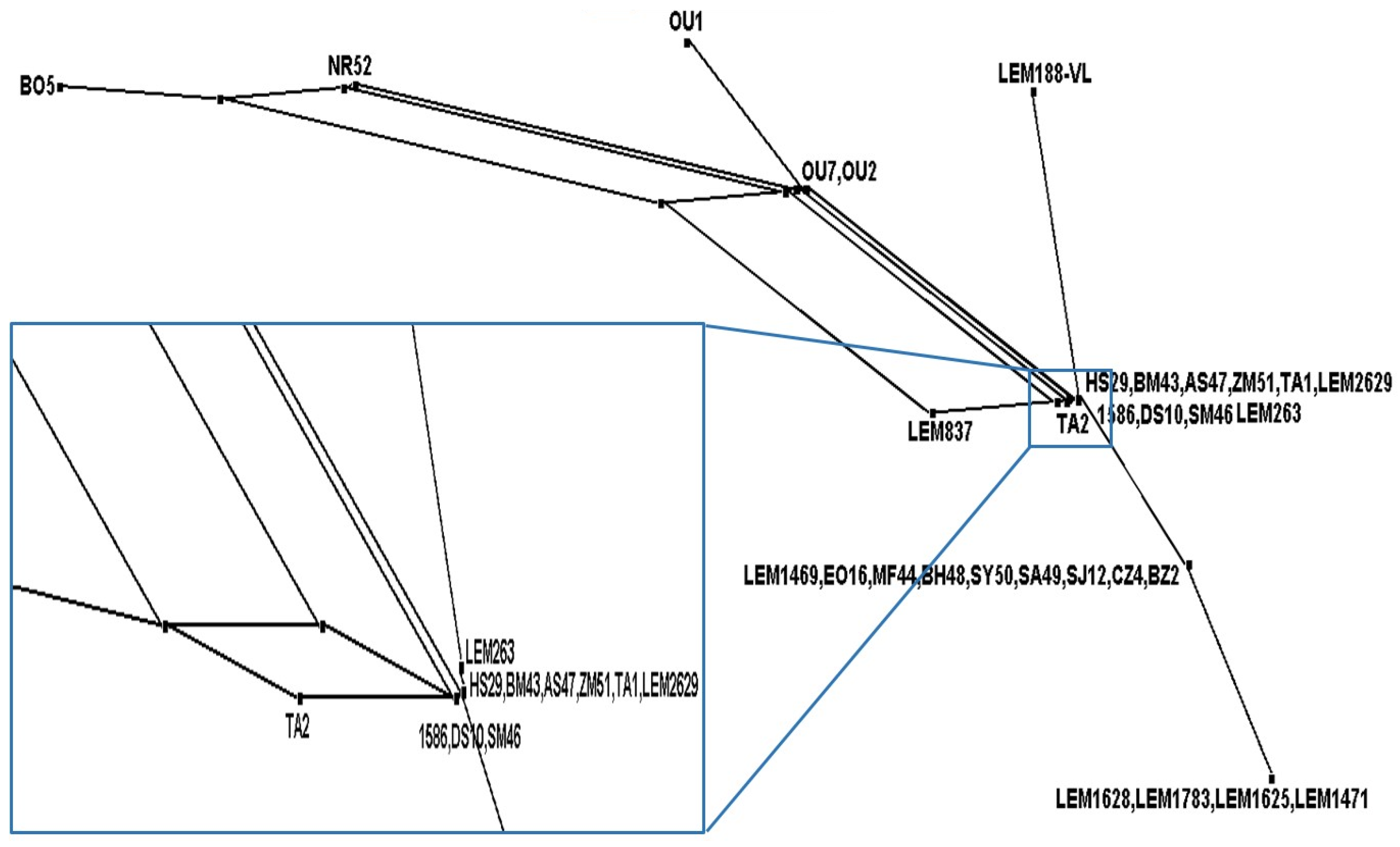
3.4. Genotypic Variation in L. infantum

From the 31 concatenated sequences, a total of 14 sequence types (STs) were deduced. The number of allele types at each locus were six at gpi, five at alat, g6pd, me and pgd, three at pgm and finally two at fh. This latter locus had the lowest discriminative power (0.065), while the locus pgd had the highest discriminative power with a value of 0.634 (Table 10).

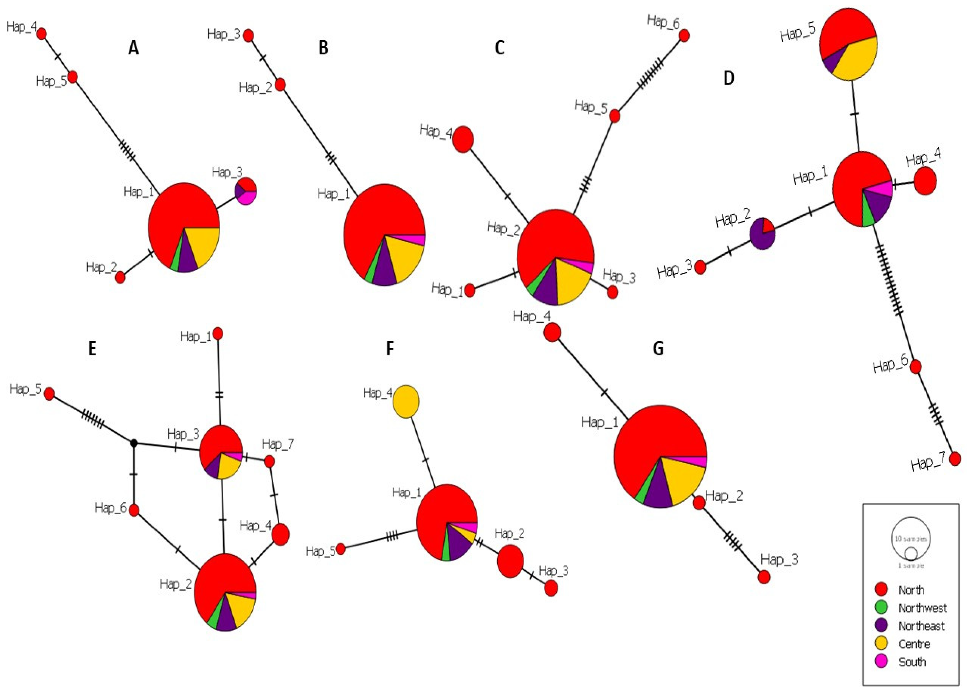
3.5. Haplotype Diversity of L. infantum

The number of haplotypes generated for each locus ranged between three in fh as the least polymorphic locus and seven in the markers gpi and pgd as the most polymorphic ones. In all the seven markers, the most common haplotype had strains from at least four geographic areas, and most of them had strains from different forms: CL, VL and CanL. In every network, at least two haplotypes were singletons including one strain each (Table 1).
We created the haplotype network of each gene separately; the seven haplotype networks had different patterns. The loci fh and pgm had the simplest networks with a straight-line pattern, and the loci g6pd, pgd and gpi were the most variable and displayed the most complex network patterns. The number of mutational events ranged from one mutation as the most frequent event and sixteen mutations that appeared once in pgd network (Figure 2).

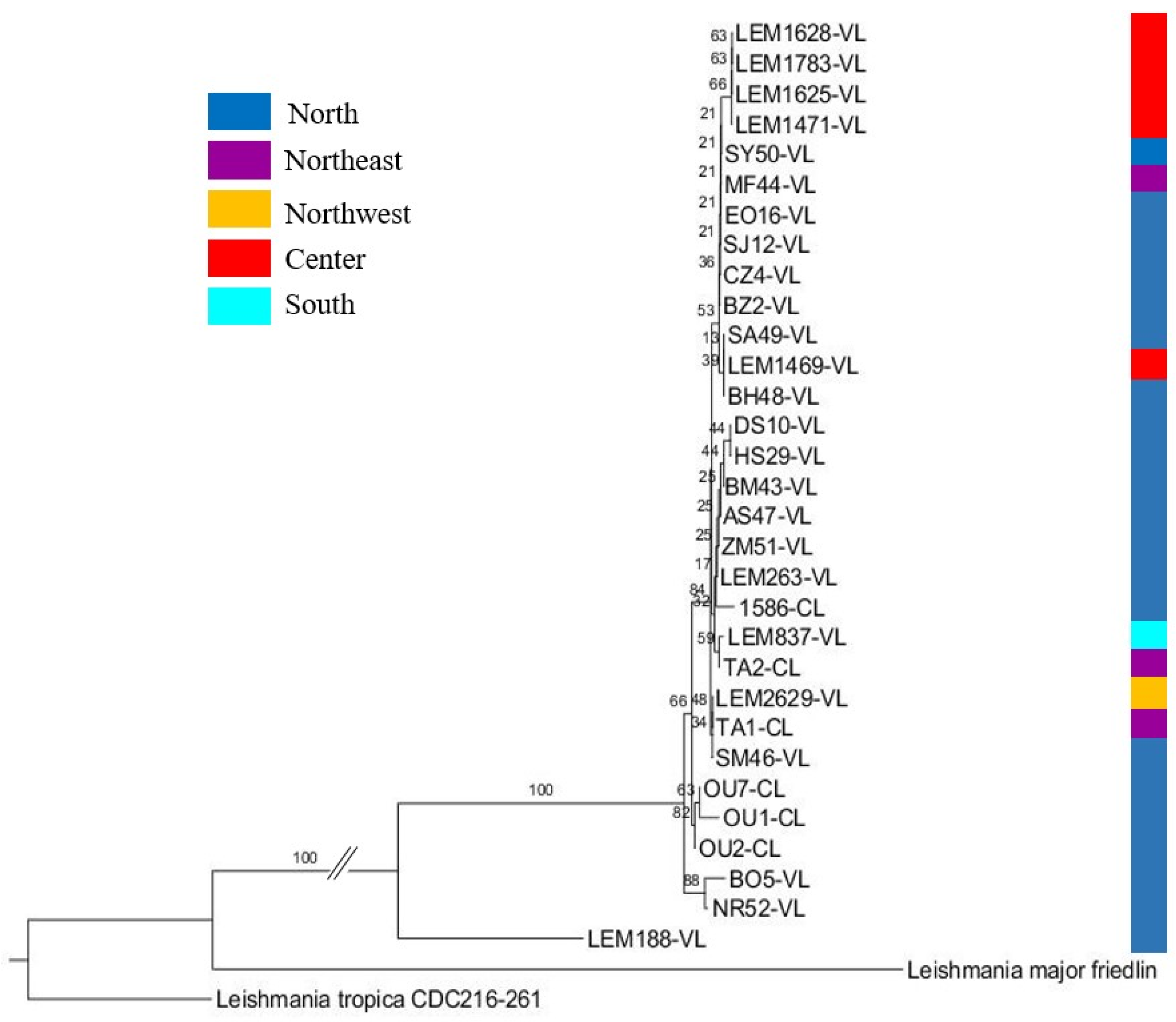
3.6. Phylogenetic Analysis of L. infantum

The phylogenetic tree was constructed to display the evolutionary biology of L. infantum using the seven polymorphic loci. L. major Friedlin and L. tropica CDC216-162 strains were used as outgroups.
All the L. infantum strains clustered together except for one strain, LEM188, which showed a prominent differentiation from the rest of the strains consistently with the haplotype network, where it was represented by Hap_14 and Hap_15, which was far from the closest haplotype Hap_13 (Figure 3).
The samples from the same geographic area clustered together (north and center), suggesting a correlation between the geographic area and the clustering. Three strains from the northern macrofocus (the northeast, the north and the northwest) clustered together (Figure 3).
Based on the disease form, almost all of the VL strains and canine strains clustered together into different groups; half of the CL strains formed a group on their own, while the other CL strains clustered with VL and CanL strains in different groups. However, the CL/HIV+ strain differentiated on its own (Figure 3).

3.7. Recombination among L. infantum Strains

The estimate of the recombination parameter (R) per gene was 0.001, and the minimum number of recombination events (Rm) recorded four events; these recombination events were detected between the following sites (11, 1045)-(1393, 1492)-(1492, 1783) and (2028, 2575).
The splits network based on the neighbor net method using the alignment of the concatenated sequences for seven loci of all the L. infantum strains suggested through the presence of parallelograms and the network-like structure the occurrence of recombination among the L. infantum strains (Figure 4).

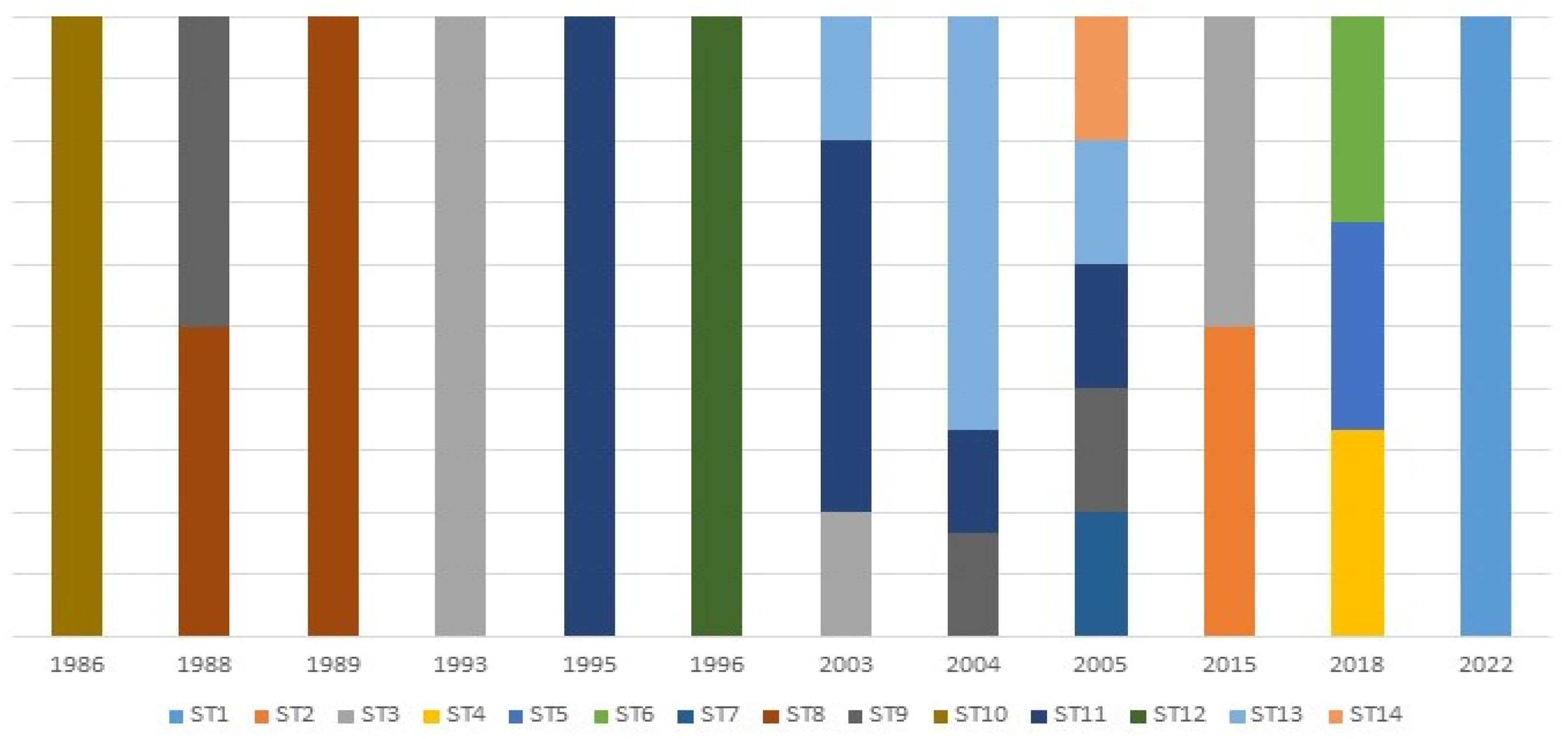
3.8. Temporal Variation Analysis

The temporal variation in the different sequence types for all the L. infantum strains was assessed. An association between the STs and the year of isolation was observed; out of the fourteen detected sequence types, nine were unique as they appeared only once; ST1 in 2022, ST2 in 2015, ST4 in 2018, ST5 in 2018, ST6 in 2018, ST7 in 2005, ST10 in 1986, ST12 in 1996 and ST14 in 2005. The other sequence types were relatively constant over the years; ST13 included strains from 2003–2005, ST11 included strains isolated between 1995 and 2003–2005, ST9 included strains from 1988, 2004–2005, ST8 appeared in 1988 and 1989 and ST3 included strains from 1993, 2003 and 2015 (Figure 5).

3.9. Analysis of the Genetic Exchange between L. infantum and L. tropica

The possibility of gene flow between the two endemic Leishmania species L. infantum and L. tropica in Morocco in two sympatric foci in the north and center was assessed. An additional set of the same seven markers for the fourteen L. tropica strains were used, twelve from the center and two from the north. The sequences of five markers (me, pgd, fh, g6pd and gpi) for the twelve strains of L. tropica were produced in our laboratory and already deposited in GenBank (Table 3), and the two missing markers, pgm and alat, were amplified and sequenced for these twelve L. tropica strains. In addition, the seven markers were amplified for the two other L. tropica strains from the north, whose sequences were submitted to GenBank and are accessible using the following accession numbers: OQ627481 to OQ627482 (for alat), OQ627545 to OQ627546 (for fh), OQ627578 to OQ627579 (for g6pd), OQ627611 to OQ627612 (for gpi), OQ627644 to OQ627645 (for me), OQ627677 to OQ627678 (for pgd) and OQ627710 to OQ627711 (for pgm).

3.9.1. Genotypic Variation Analysis of L. infantum and L. tropica

Twenty-two sequence types (STs) were deduced from forty-five concatenated sequences of L. infantum and L. tropica. The highest number of allele types was nine at fh and pgd and the lowest was at alat. The discrimination power and the typing efficiency were calculated for each locus (Table 11).

3.9.2. Haplotypes Diversity of L. infantum and L. tropica

The number of haplotypes for each marker ranged between six for the marker alat and eleven for the marker pgd. None of the haplotypes in all the markers had L. infantum and L. tropica strains together. Some haplotypes were private as they included strains of L. tropica from the same focal population, and others had L. tropica strains from two different population, namely the center and the north. Singleton haplotypes were present in all the markers for having one L. tropica strain from the north, the center or both (Table 1).

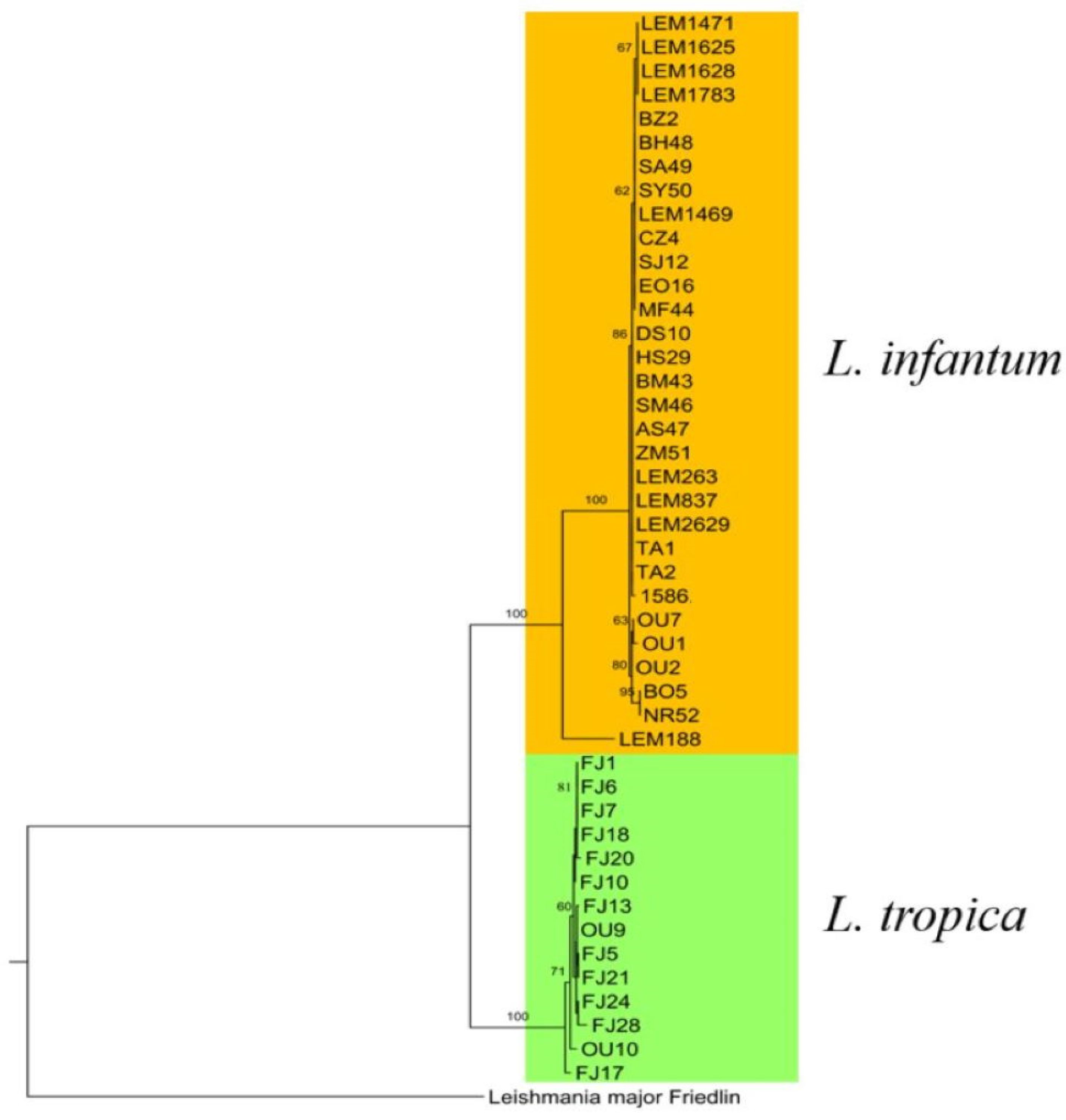
3.9.3. Phylogenetic Analysis of L. infantum and L. tropica

The phylogenetic tree was constructed to display the evolutionary biology of L. infantum and L. tropica using the seven polymorphic loci. L. major Friedlin was used as an out group.
In the phylogenetic tree, each species formed a cluster on its own. Inside L. tropica’s cluster, all of the center samples formed a group including (OU9) from the north, with the exception of two samples, one from the center (FJ17) and one from the north (OU10), as each one of them clustered on its own. L. tropica’s strains (OU9 and OU10) were isolated from the same focus as L. infantum’s strains (OU1, OU2 and OU7), and all the (FJ) strains were also isolated from the same focus as L. infantum’s strains (LEM1469, LEM1471, LEM1625, LEM1628 and LEM1783). However, these strains clustered with their respective species (Figure 6).

4. Discussion

Among the typing techniques of Leishmania species and strains, multilocus enzyme electrophoresis (MLEE) is still considered to be the gold standard [20]. However, it has many drawbacks; it is slow, laborious, costly, bulk cultures of live parasites are necessary and it has a low discriminatory power due to its inability to detect nucleotide substitutions that do not change the amino acid composition [21]. For Leishmania infantum, the discriminatory power of MLEE for studying genetic variability among strains is limited because most L. infantum strains in the Mediterranean countries belong to the zymodeme MON-1 regardless of the disease form, host and immune status [41]; it represents approximately 70% of all identified strains [42].
In Morocco, the L. infantum zymodeme MON-1 is responsible for the visceral form, which is lethal if left untreated in over 95% of cases [43], while zymodeme MON-24 is responsible for the cutaneous form that has been increasing in terms of its geographic spread and number of cases [7]. Few molecular studies have been conducted on the population genetics of this clinically important species; thus, a better understanding of the dynamics of leishmaniasis due to L. infantum and the population structure of this species in Morocco through genetic techniques with high discriminatory power is needed.
Housekeeping genes with high and variable rates of sequence diversity within and between species or genera are the ideal targets for characterization of microorganisms including Leishmania [24,44,45,46]. The MLST approach is based on the sequence of a set of seven to ten housekeeping genes that result in each strain having a distinct numerical allelic profile, which is abbreviated to a unique identifier, i.e., the sequence type (ST) [47]. This approach has proved its usefulness in being an accurate and highly discriminative portable typing system [47]. This method is considered as a potentially powerful phylogenetic approach that is poised to become the new reference method for Leishmania species characterization in the near future [23].
In our study, the 31 concatenated L. infantum sequences showed a high level of heterogeneity with 82 polymorphic/heterozygous sites (S), whereas the PCR-RFLP-KDNA polymorphisms of 188 L. infantum strains from Morocco and Portugal were less significant with values of S = 19 and S = 30, respectively [41]. The same technique applied to 44 L. infantum sequences from southeastern Spain revealed a value of S = 11 [48].
Fourteen sequence types were deduced, showing the high intraspecific diversity of L. infantum. In Morocco, other studies and through different techniques have proven the diversity of this species; using the multilocus microsatellite typing (MLMT) method on thirty-three Moroccan strains showed thirty-three genotypes [49], and kDNA-PCR-RFLP on seventy-five samples generated six genotypes [41]. The PCR-RFLP analysis of a single-copy gene encoding N-acetylglucosamine-1-phosphate transferase (nagt) was employed on a population of forty Moroccan L. infantum strains showed the genetic heterogeneity with six different genotypes [25]. In Spain, four RFLP-kDNA genotypes were deduced in a population of forty-four L. infantum sequences [48]. In another study in the south of France, the MLMT method was employed to assess 270 L. infantum isolates using 12 microsatellites, and the results revealed a total of 121 different genotypes [50].
The MLST scheme used in our study revealed a significant level of heterogeneity in L. infantum in Morocco. Moreover, it showed a higher number of heterozygous sites and genotypes, confirming the high resolution and discriminatory power of this technique. This conclusion is supported by the results of Banu et al., (2019), where seven L. infantum isolates, four from the Old World and three from the New World, clustered into seven sequence types using MLST. In the same work, it was reported that MLST possesses sufficient discriminatory power to detect genetic heterogeneity within and between species of the L. donovani complex [51]. Moreover, the MLST scheme we used in this work had a discrimination power of 0.9, and, according to Hunter and Gaston (1988), a discrimination power of 0.9 implies that the results can be interpreted with confidence [52].
According to Lauthier et al. (2020), phylogeny based on each marker separately might be associated with some inconsistency among different genes. Therefore, concatenation is a powerful approach as it is based on a complete set of genes, as previously shown in Leishmania parasites [23]. In the phylogenetic tree constructed based on the concatenated sequences from the seven genes, most of the strains from the same geographic areas clustered together, which was in agreement with other studies that demonstrated a geographical clustering of L. infantum strains isolated in different regions [53,54].
For the two canine strains, the one from the center clustered with two VL strains from the north and the one from the south clustered with a strain from the northeast. Ferroglio et al. (2006), through a study conducted in northern Italy using kDNA, showed that certain RFLP patterns were associated with specific geographic regions while others were more widely distributed [55]. In addition, Alam et al. (2014), reported that two Chinese populations did not correlate exactly with their geographical origin clusters [56].
The phylogenetic tree showed clusters including human or canine strains only and groups with both sources. Moreover, most of the canine strains clustered with human (CL and VL) strains, which was in accordance with previous works in the country that proved the zoonotic character of the transmission cycle of L. infantum [15,18,49]. Other studies have also reported the clustering of human and canine isolates together [48,57].
According to Seridi et al. (2008), there is no strong confirmation of an association be-tween L. infantum clinical forms and geography in Algeria [58], partly because the in-dividual genetic susceptibility and host immune response influence the tropism for the skin or for internal organs [59,60]. However, in the phylogenetic tree, almost all of the VL human strains coming from the same geographic region (north) clustered together into different groups, and half of the CL strains from the north clustered together. This result was in agreement with the findings of Chargui et al. (2009), who found that two MON-24 populations could be differentiated in Tunisia [54]. One included strains isolated from CL mainly from the north, and the other included VL strains from the center of Tunisia. Nevertheless, due to the small sample size, this should be further studied to confirm whether it is a general trait or only caused by sampling bias [54].
Two CL strains from the northeast clustered with VL strains in two groups. This could be due to the genetic similarity of parasites causing both forms. Indeed, the two CL strains came from Taza, which has been known to be a hypoendemic focus of VL due to L. infantum since 1995 [61]. Alam et al. (2014) reported the genetic similarity between the strains causing VL or CL [56]. Kuhls et al. (2011) also reported that CL strains from Honduras formed a single cluster with one VL strain from the same origin [62]. Wang et al. (2010) reported that a strain isolated from a VL patient grouped with isolates from CL patients from a different province [63]. Noyes et al. (1997) and Belli et al. (1999) confirmed that the two clinical forms in Honduras and Nicaragua were caused by genetically similar parasites [64,65]. Moreover, the tropism of L. infantum zymodemes is not clear cut; they are known to cause both VL and CL, and the reasons leading to their respective clinical pictures seem to be very complex [62].
The CL/HIV+ strain had a unique sequence type (ST1), and it clustered on its own in the phylogenetic tree, which was in accordance with Cruz et al. (2002), who reported, in the case of L. infantum, that most of the newly discovered zymodemes were heterozygous [66]. In addition, the results of Alvar et al. (1997) reported that the enzymatic characterization of Leishmania isolates from HIV-positive patients from the Mediterranean basin showed an extreme variability in L. infantum in such patients; a total of 17 zymodemes were described in 150 Leishmania isolates [67]. Eight of the seventeen zymodemes, MON-136, MON-183, MON-185, MON-188, MON-190, MON-198, MON-199 and MON-201, were new zymodemes that have been encountered only in HIV-positive individuals [67]. Moreover, the zymodeme MON-183, previously described in two cases of VL/HIV-positive individuals [68], was confirmed as a new zymodeme responsible not only for VL but also for CL in coinfected patients [69].
Three of the northern CL strains clustered on their own in both the phylogenetic tree and the haplotype network indicating their homogeneity. This result could be explained by the recent emergence of CL due to L. infantum in Morocco, as the first case was reported in 1996, while VL was first notified in 1920 [7]. Momen et al. (1993) reported that a low diversity supports the hypothesis of a recent import of selected strains of L. infantum from the Old World to the New World [70]. This is in accordance with Marlow et al. (2014), who showed that the high homogeneity observed among L. braziliensis in the state of Santa Catarina, Brazil, is due to the recent emergence of this species in the latter state [71].
The human L. infantum strains showed less diversity and more similarity than the ca-nine strains. The geographic origin of the strains could explain this difference in diversity, as all the human strains belonged to the northern focus, i.e., the same ecosystem, while the canine strains came from different geographic areas (north, northwest, center and south) and therefore different ecosystems. These results were consistent with Ghatee et al. (2018), as they reported that the similarity of human sequence haplotypes of L. infantum from central areas in Iran is due, among other reasons, to the similarity of the ecosystems in these foci [72].
According to Mauricio et al. (2006), heterozygosity at several sites within a gene and at several loci can be a consequence of genetic exchange; mutation is a less likely explanation [26]. Many authors have reported that heterozygosity might suggest sexual re-production in the natural Leishmania population, even if asexual reproduction via clonal propagation is considered the main reproductive mechanism in the Leishmania genus [73,74,75]. Our results showed evidence of recombination between L. infantum strains through the presence of the network-like structure and the four recorded recombination events, which was consistent with Rogers et al. (2014), who reported through the analysis of 11 L. infantum entire genomes isolated from the vector evidence of a cross between two diverse strains with subsequent recombination [76]. Other studies have also reported the presence of recombination amongst L. infantum isolates [51,54,58].
In recent years, many L. infantum CL cases were reported in different parts of the country, mostly in endemic foci of L. tropica, indicating the invasion of L. infantum CL to the former species distribution areas [7]. In the phylogenetic tree, the L. infantum and L. tropica strains formed two clearly divergent clusters. In L. tropica’s cluster, one of the two northern strains clustered with the strains from the center, which could be due to infected human behavior, as they are considered to be the main reservoir of L. tropica; it could have been spread through the movement of human populations [77]. In the phylogenetic tree, there was no gene flow between L. infantum and L. tropica from the two studied foci. This could be explained by the fact that the appearance of L. infantum in L. tropica’s focus in central Morocco is a recent event, as this focus is the oldest L. tropica focus (Azilal province) [78]. The same goes for the northern focus (Ouazzane province), which is known to be endemic for VL, while CL appeared until 1997 [79].
Through the temporal variation analysis using the sequence types, an association between the STs and the year of isolation was observed, as most of the STs were unique. Conversely, Herrera et al. (2017) observed no association between the STs and the year of isolation in L. panamensis and L. braziliensis [80], and Gouzelou et al. (2013) also reported that no correlation to the year of strain isolation was identified [81]. On the other hand, Pomares et al. reported that some genotypes were detected during a limited period and are no longer detected, while other genotypes were still detected [50].

5. Conclusions

To the best of our knowledge, this study is the first to apply the MLST approach on L. infantum in Morocco. This technique revealed the high genetic diversity of this species and showed its zoonotic character. Moreover, this approach disclosed the existence of recombination between L. infantum strains. The co-existence of L. infantum and L. tropica in the two endemic foci is a recent event; therefore, no gene flow was detected. In future work, increasing the number of samples from different geographic areas and different sources, especially from the vector throughout the country and in foci where different species are sympatric, could be of great help to understand the dynamics of the species circulating in the country.

Supplementary Materials

The supporting information can be downloaded at: https://www.mdpi.com/article/10.3390/pathogens12060785/s1. Figure S1. Agarose gel profiles of MLST genes: (A) alat: 589 bp; (B) pgm: 529 bp; (C) me: 687 bp; (D) gpi: 500 bp; (E) pgd: 697 bp; (F) g6pd: 684 bp; (G) fh 604 bp. NC: negative control; MW: 100 bp molecular weight marker.

Author Contributions

Conceptualization, M.L.; Software, S.E.M.; Validation, M.L. and K.B.; Formal Analysis, S.E.M.; Investigation, I.M., O.D., M.A.K., S.E.K., I.E.i.S. and M.R.; Resources, M.L.; Data Curation, S.E.M.; Writing—Original Draft Preparation, S.E.M.; Writing—Review and Editing, S.E.M., M.L., K.B., I.G. and M.B.; Visualization, S.E.M.; Supervision, M.L. and K.B.; Project Administration, M.L.; Funding Acquisition, M.L. All authors have read and agreed to the published version of the manuscript.

Funding

This work was funded by the Research and Innovation Staff Exchange (RISE) Program, Marie Skłodowska-Curie Actions, Horizon 2020 EU (grant agreement N°778298). The funders had no role in the study design, data collection or analysis, the decision to publish or the preparation of the manuscript.

Institutional Review Board Statement

This study was conducted according to the principles specified in the Declaration of Helsinki and under the local ethical guidelines of the Ethics Committee for Biomedical Research (CERB) of the Faculty of Medicine and Pharmacy, University Mohammed V of Rabat, Morocco (IORG 0006594, approved 16 April 2019) for studies involving humans. Ethical review and approval were waived for this study because in Morocco we do not have an ethics committee for animals.

Informed Consent Statement

Informed consent was obtained from all the participants involved in this study.

Data Availability Statement

The reported sequences in the present paper were deposited in the GenBank database under the following accession numbers: OQ627483 to OQ627513 (for alat), OQ627514 to OQ627544 (for fh), OQ627547 to OQ627577 (for g6pd), OQ627580 to OQ627610 (for gpi), OQ627613 to OQ627643 (for me), OQ627646 to OQ627676 (for pgd), OQ627679 to OQ627709 (for pgm) for L. infantum and: OQ627481 to OQ627482 (for alat), OQ627545 to OQ627546 (for fh), OQ627578 to OQ627579 (for g6pd), OQ627611 to OQ627612 (for gpi), OQ627644 to OQ627645 (for me), OQ627677 to OQ627678 (for pgd) and OQ627710 to OQ627711 (for pgm) for L. tropica.

Acknowledgments

The authors extend their appreciation to the medical teams of physicians and nurses for their efforts of the different provinces and Hicham Charoute, bioinformatician at Institut Pasteur of Morocco, for his assistance.

Conflicts of Interest

The authors declare no conflict of interest.

References

  1. Ashford, R.W. The leishmaniases as emerging and reemerging zoonoses. Int. J. Parasitol. 2000, 30, 1269–1281. [Google Scholar] [CrossRef]
  2. de Vries, H.J.; Reedijk, S.H.; Schallig, H.D. Cutaneous leishmaniasis: Recent developments in diagnosis and management. Am. J. Clin. Dermatol. 2015, 16, 99–109. [Google Scholar] [CrossRef] [PubMed]
  3. Remlinger, P. Un cas de Kala-azar infantile observé au Maroc. Arch. Inst. Pasteur Afr. Noi 1921, 1, 240–241. [Google Scholar]
  4. Rhajaoui, M. Human leishmaniases in Morocco: A nosogeographical diversity. Pathol. Biol. 2011, 59, 226–229. [Google Scholar] [CrossRef] [PubMed]
  5. Murray, H.W.; Berman, J.D.; Davies, C.R.; Saravia, N.G. Advances in leishmaniasis. Lancet 2005, 366, 1561–1577. [Google Scholar] [CrossRef] [PubMed]
  6. van Griensven, J.; Diro, E. Visceral leishmaniasis. Infect. Dis. Clin. N. Am. 2012, 26, 309–322. [Google Scholar] [CrossRef] [PubMed]
  7. El Mazini, S.; Ejghal, R.; Bekhti, K.; Lemrani, M. The Sporadic cutaneous leishmaniasis due to Leishmania infantum in Morocco: A presumably trend towards endemicity. Acta Trop. 2022, 227, 106288. [Google Scholar] [CrossRef] [PubMed]
  8. Hmamouch, A.; Amarir, F.; Fellah, H.; Karzaz, M.; Bekhti, K.; Rhajaoui, M.; Sebti, F. Coexistence of Leishmania tropica and Leishmania infantum in Sefrou province, Morocco. Acta Trop. 2013, 130, 94–99. [Google Scholar]
  9. Mhaidi, I.; El Kacem, S.; Ait Kbaich, M.; El Hamouchi, A.; Sarih, M.; Akarid, K.; Lemrani, M. Molecular identification of Leishmania infection in the most relevant sand fly species and in patient skin samples from a cutaneous leishmaniasis focus, in Morocco. PLoS Negl. Trop. Dis. 2018, 12, e0006315. [Google Scholar] [CrossRef]
  10. Chaffai, M.; Ben Rachid, M.S.; Ben-Ismail, R.; Ben Osman, A.; Makni, N. Clinico-epidemiologic forms of cutaneous leishmaniasis in Tunisia. Ann. Dermatol. Venereol. 1988, 115, 1255–1260. [Google Scholar]
  11. Rioux, J.A.; Mahjoub, J.; Gallego, M.; Dereure, J.; Perieres, J.; Lahmrani, A.J.B.S.F.P. Human cutaneous leishmaniasis due to Leishmania infantum zymodeme MON-24 in Morocco. Bull. Soc. Fr. Parasitol. 1996, 14, 179–183. [Google Scholar]
  12. Bachi, F. The epidemiological and clinical aspects of leishmaniasis in Algeria. La Lett. L’infectiologue 2006, 21, 9–15. [Google Scholar]
  13. Rhajaoui, M.; Nasereddin, A.; Fellah, H.; Azmi, K.; Amarir, F.; Al-Jawabreh, A.; Ereqat, S.; Planer, J.; Abdeen, Z. New clinico-epidemiologic profile of cutaneous leishmaniasis, Morocco. Emerg. Infect Dis. 2007, 13, 1358–1360. [Google Scholar] [CrossRef] [PubMed]
  14. Mouttaki, T.; Maksouri, H.; El Mabrouki, J.; Merino-Espinosa, G.; Fellah, H.; Itri, M.; Martin-Sanchez, J.; Soussi-Abdallaoui, M.; Chiheb, S.; Riyad, M. Concomitant visceral and localized cutaneous leishmaniasis in two Moroccan infants. Infect. Dis. Poverty 2018, 7, 32. [Google Scholar] [CrossRef] [PubMed]
  15. Nejjar, R.; Lemrani, M.; Malki, A.; Ibrahimy, S.; Amarouch, H.; Benslimane, A. Canine leishmaniasis due to Leishmania infantum MON-1 in northern Morocco. Parasite 1998, 5, 325–330. [Google Scholar] [CrossRef]
  16. Haralambous, C.; Dakkak, A.; Pratlong, F.; Dedet, J.P.; Soteriadou, K. First detection and genetic typing of Leishmania infantum MON-24 in a dog from the Moroccan Mediterranean coast: Genetic diversity of MON-24. Acta Trop. 2007, 103, 69–79. [Google Scholar] [CrossRef] [PubMed]
  17. Echchakery, M.; Chicharro, C.; Boussaa, S.; Nieto, J.; Carrillo, E.; Sheila, O.; Moreno, J.; Boumezzough, A. Molecular detection of Leishmania infantum and Leishmania tropica in rodent species from endemic cutaneous leishmaniasis areas in Morocco. Parasit Vectors 2017, 10, 454. [Google Scholar] [CrossRef] [PubMed]
  18. Guessous-Idrissi, N.; Hamdani, A.; Rhalem, A.; Riyad, M.; Sahibi, H.; Dehbi, F.; Bichichi, M.; Essari, A.; Berrag, B. Epidemiology of human visceral leishmaniasis in Taounate, a northern province of Morocco. Parasite 1997, 4, 181–185. [Google Scholar] [CrossRef] [PubMed]
  19. Es-Sette, N.; Ajaoud, M.; Laamrani-Idrissi, A.; Mellouki, F.; Lemrani, M. Molecular detection and identification of Leishmania infection in naturally infected sand flies in a focus of cutaneous leishmaniasis in northern Morocco. Parasit Vectors 2014, 7, 305. [Google Scholar] [CrossRef]
  20. Schönian, G.; Mauricio, I.; Gramiccia, M.; Cañavate, C.; Boelaert, M.; Dujardin, J.C. Leishmaniases in the Mediterranean in the era of molecular epidemiology. Trends Parasitol. 2008, 24, 135–142. [Google Scholar] [CrossRef]
  21. Ochsenreither, S.; Kuhls, K.; Schaar, M.; Presber, W.; Schönian, G. Multilocus microsatellite typing as a new tool for discrimination of Leishmania infantum MON-1 strains. J. Clin. Microbiol. 2006, 44, 495–503. [Google Scholar] [CrossRef] [PubMed]
  22. Schönian, G.; Kuhls, K.; Mauricio, I.L. Molecular approaches for a better understanding of the epidemiology and population genetics of Leishmania. Parasitology 2011, 138, 405–425. [Google Scholar] [CrossRef]
  23. Lauthier, J.J.; Ruybal, P.; Barroso, P.A.; Hashiguchi, Y.; Marco, J.D.; Korenaga, M. Development of a Multilocus sequence typing (MLST) scheme for Pan-Leishmania. Acta Trop. 2020, 201, 105189. [Google Scholar] [CrossRef] [PubMed]
  24. Marco, J.D.; Barroso, P.A.; Locatelli, F.M.; Cajal, S.P.; Hoyos, C.L.; Nevot, M.C.; Lauthier, J.J.; Tomasini, N.; Juarez, M.; Estévez, J.O.; et al. Multilocus sequence typing approach for a broader range of species of Leishmania genus: Describing parasite diversity in Argentina. Infect. Genet. Evol. J. Mol. Epidemiol. Evol. Genet. Infect. Dis. 2015, 30, 308–317. [Google Scholar] [CrossRef] [PubMed]
  25. El Hamouchi, A.; El Kacem, S.; Ejghal, R.; Lemrani, M. Genetic polymorphism in Leishmania infantum isolates from human and animals determined by nagt PCR-RFLP. Infect. Dis. Poverty 2018, 7, 54. [Google Scholar] [CrossRef] [PubMed]
  26. Mauricio, I.L.; Yeo, M.; Baghaei, M.; Doto, D.; Pratlong, F.; Zemanova, E.; Dedet, J.P.; Lukes, J.; Miles, M.A. Towards multilocus sequence typing of the Leishmania donovani complex: Resolving genotypes and haplotypes for five polymorphic metabolic enzymes (ASAT, GPI, NH1, NH2, PGD). Int. J. Parasitol. 2006, 36, 757–769. [Google Scholar] [CrossRef] [PubMed]
  27. Zemanová, E.; Jirků, M.; Mauricio, I.L.; Horák, A.; Miles, M.A.; Lukes, J. The Leishmania donovani complex: Genotypes of five metabolic enzymes (ICD, ME, MPI, G6PDH, and FH), new targets for multilocus sequence typing. Int. J. Parasitol. 2007, 37, 149–160. [Google Scholar] [CrossRef]
  28. El Kacem, S.; Kbaich, M.A.; Daoui, O.; Charoute, H.; Mhaidi, I.; Ejghal, R.; Barhoumim, M.; Guizani, I.; Bennani, H.; Lemrani, M. Multilocus sequence analysis provides new insight into population structure and genetic diversity of Leishmania tropica in Morocco. Infect. Genet. Evol. 2021, 93, 104932. [Google Scholar] [CrossRef]
  29. Schönian, G.; Schweynoch, C.; Zlateva, K.; Oskam, L.; Kroon, N.; Gräser, Y.; Presber, W. Identification and determination of the relationships of species and strains within the genus Leishmania using single primers in the polymerase chain reaction. Mol. Biochem. Parasitol. 1996, 77, 19–29. [Google Scholar] [CrossRef]
  30. Schönian, G.; Nasereddin, A.; Dinse, N.; Schweynoch, C.; Schallig, H.D.; Presber, W.; Jaffe, C.L. PCR diagnosis and characterization of Leishmania in local and imported clinical samples. Diagn. Microbiol. Infect. Dis. 2003, 47, 349–358. [Google Scholar] [CrossRef]
  31. Tamura, K.; Stecher, G.; Kumar, S. MEGA11: Molecular Evolutionary Genetics Analysis Version 11. Mol. Biol. Evol. 2021, 38, 3022–3027. [Google Scholar] [CrossRef] [PubMed]
  32. Rozas, J.; Ferrer-Mata, A.; Sánchez-DelBarrio, J.C.; Guirao-Rico, S.; Librado, P.; Ramos-Onsins, S.E.; Sánchez-Gracia, A. DnaSP 6: DNA Sequence Polymorphism Analysis of Large Data Sets. Mol. Biol. Evol. 2017, 34, 3299–3302. [Google Scholar] [CrossRef] [PubMed]
  33. Excoffier, L.; Lischer, H.E. Arlequin suite ver 3.5: A new series of programs to perform population genetics analyses under Linux and Windows. Mol. Ecol. Resour. 2010, 10, 564–567. [Google Scholar] [CrossRef] [PubMed]
  34. Tajima, F. Statistical method for testing the neutral mutation hypothesis by DNA polymorphism. Genetics 1989, 123, 585–595. [Google Scholar] [CrossRef] [PubMed]
  35. Tomasini, N.; Lauthier, J.J.; Llewellyn, M.S.; Diosque, P. MLSTest: Novel software for multi-locus sequence data analysis in eukaryotic organisms. Infect. Genet. Evol. 2013, 20, 188–196. [Google Scholar] [CrossRef]
  36. Stöver, B.C.; Müller, K.F. TreeGraph 2: Combining and visualizing evidence from different phylogenetic analyses. BMC Bioinform. 2010, 11, 7. [Google Scholar] [CrossRef]
  37. Leigh, J.; Bryant, D. PopART: Full-feature software for haplotype network construction. Methods Ecol. Evol 2015, 6, 1110–1116. [Google Scholar] [CrossRef]
  38. Huson, D.H.; Bryant, D. Application of phylogenetic networks in evolutionary studies. Mol. Biol. Evol. 2006, 23, 254–267. [Google Scholar] [CrossRef]
  39. Hudson, R.R.; Kaplan, N.L. Statistical properties of the number of recombination events in the history of a sample of DNA sequences. Genetics 1985, 111, 147–164. [Google Scholar] [CrossRef]
  40. Hudson, R.R. Estimating the recombination parameter of a finite population model without selection. Genet. Res. 1987, 50, 245–250. [Google Scholar] [CrossRef]
  41. El Hamouchi, A.; Ejghal, R.; Hida, M.; Lemrani, M. Intraspecific genetic variability in a population of Moroccan Leishmania infantum revealed by PCR-RFLP of kDNA minicircles. Acta Trop. 2017, 169, 142–149. [Google Scholar] [CrossRef]
  42. Gállego, M.; Pratlong, F.; Fisa, R.; Riera, C.; Rioux, J.A.; Dedet, J.P.; Portús, M. The life-cycle of Leishmania infantum MON-77 in the Priorat (Catalonia, Spain) involves humans, dogs and sandflies; also literature review of distribution and hosts of L. infantum zymodemes in the Old World. Trans. R. Soc. Trop. Med. Hyg. 2001, 95, 269–271. [Google Scholar] [CrossRef] [PubMed]
  43. WHO. Leishmaniasis 2023. Available online: https://www.who.int/news-room/fact-sheets/detail/leishmaniasis (accessed on 23 June 2022).
  44. Bennasar, A.; Mulet, M.; Lalucat, J.; García-Valdés, E. PseudoMLSA: A database for multigenic sequence analysis of Pseudomonas species. BMC Microbiol. 2010, 10, 118. [Google Scholar] [CrossRef] [PubMed]
  45. Weirather, J.L.; Jeronimo, S.M.; Gautam, S.; Sundar, S.; Kang, M.; Kurtz, M.A.; Haque, R.; Schriefer, A.; Talhari, S.; Carvalho, E.M.; et al. Serial quantitative PCR assay for detection, species discrimination, and quantification of Leishmania spp. in human samples. J. Clin. Microbiol. 2011, 49, 3892–3904. [Google Scholar] [CrossRef] [PubMed]
  46. Boité, M.C.; Mauricio, I.L.; Miles, M.A.; Cupolillo, E. New insights on taxonomy, phylogeny and population genetics of Leishmania (Viannia) parasites based on multilocus sequence analysis. PLoS Negl. Trop. Dis. 2012, 6, e1888. [Google Scholar] [CrossRef]
  47. Francisco, A.P.; Bugalho, M.; Ramirez, M.; Carriço, J.A. Global optimal eBURST analysis of multilocus typing data using a graphic matroid approach. BMC Bioinform. 2009, 10, 152. [Google Scholar] [CrossRef]
  48. Ortuño, M.; Latrofa, M.S.; Iborra, M.A.; Pérez-Cutillas, P.; Bernal, L.J.; Risueño, J.; Muñoz, C.; Bernal, A.; Sánchez-Lopez, P.F.; Segovia, M.; et al. Genetic diversity and phylogenetic relationships between Leishmania infantum from dogs, humans and wildlife in south-east Spain. Zoonoses Public Health 2019, 66, 961–973. [Google Scholar] [CrossRef]
  49. Amro, A.; Hamdi, S.; Lemrani, M.; Mouna, I.; Mohammed, H.; Mostafa, S.; Rhajaoui, M.; Hamarsheh, O.; Schönian, G. Moroccan Leishmania infantum: Genetic diversity and population structure as revealed by multi-locus microsatellite typing. PLoS ONE 2013, 8, e77778. [Google Scholar] [CrossRef]
  50. Pomares, C.; Marty, P.; Bañuls, A.L.; Lemichez, E.; Pratlong, F.; Faucher, B.; Jeddi, F.; Moore, S.; Michel, G.; Aluru, S.; et al. Genetic Diversity and Population Structure of Leishmania infantum from Southeastern France: Evaluation Using Multi-Locus Microsatellite Typing. PLoS Negl. Trop. Dis. 2016, 10, e0004303. [Google Scholar] [CrossRef]
  51. Banu, S.S.; Meyer, W.; Ferreira-Paim, K.; Wang, Q.; Kuhls, K.; Cupolillo, E.; Schönian, G.; Lee, R. A novel multilocus sequence typing scheme identifying genetic diversity amongst Leishmania donovani isolates from a genetically homogeneous population in the Indian subcontinent. Int. J. Parasitol. 2019, 49, 555–567. [Google Scholar] [CrossRef]
  52. Hunter, P.R.; Gaston, M.A. Numerical index of the discriminatory ability of typing systems: An application of Simpson’s index of diversity. J. Clin. Microbiol. 1988, 26, 2465–2466. [Google Scholar] [CrossRef] [PubMed]
  53. Botilde, Y.; Laurent, T.; Quispe Tintaya, W.; Chicharro, C.; Cañavate, C.; Cruz, I.; Kuhls, K.; Schönian, G.; Dujardin, J.C. Comparison of molecular markers for strain typing of Leishmania infantum. Infect. Genet. Evol. J. Mol. Epidemiol. Evol. Genet. Infect. Dis. 2006, 6, 440–446. [Google Scholar] [CrossRef]
  54. Chargui, N.; Amro, A.; Haouas, N.; Schönian, G.; Babba, H.; Schmidt, S.; Ravel, C.; Lefebvre, M.; Bastien, P.; Chaker, E.; et al. Population structure of Tunisian Leishmania infantum and evidence for the existence of hybrids and gene flow between genetically different populations. Int. J. Parasitol. 2009, 39, 801–811. [Google Scholar] [CrossRef]
  55. Ferroglio, E.; Romano, A.; Trisciuoglio, A.; Poggi, M.; Ghiggi, E.; Sacchi, P.; Biglino, A. Characterization of Leishmania infantum strains in blood samples from infected dogs and humans by PCR-RFLP. Trans. R. Soc. Trop. Med. Hyg. 2006, 100, 636–641. [Google Scholar] [CrossRef] [PubMed]
  56. Alam, M.Z.; Nakao, R.; Sakurai, T.; Kato, H.; Qu, J.Q.; Chai, J.J.; Chang, K.P.; Schönian, G.; Katakura, K. Genetic diversity of Leishmania donovani/infantum complex in China through microsatellite analysis. Infect. Genet. Evol. J. Mol. Epidemiol. Evol. Genet. Infect. Dis. 2014, 22, 112–119. [Google Scholar] [CrossRef] [PubMed]
  57. Kuhls, K.; Chicharro, C.; Cañavate, C.; Cortes, S.; Campino, L.; Haralambous, C.; Soteriadou, K.; Pratlong, F.; Dedet, J.P.; Mauricio, I.; et al. Differentiation and gene flow among European populations of Leishmania infantum MON-1. PLoS Negl. Trop. Dis. 2008, 2, e261. [Google Scholar] [CrossRef]
  58. Seridi, N.; Amro, A.; Kuhls, K.; Belkaid, M.; Zidane, C.; Al-Jawabreh, A.; Schönian, G. Genetic polymorphism of Algerian Leishmania infantum strains revealed by multilocus microsatellite analysis. Microbes Infect. 2008, 10, 1309–1315. [Google Scholar] [CrossRef]
  59. Alvar, J.; Jiménez, M. Could infected drug-users be potential Leishmania infantum reservoirs? AIDS 1994, 8, 854. [Google Scholar] [CrossRef]
  60. Rossi, M.; Fasel, N. How to master the host immune system? Leishmania parasites have the solutions! Int. Immunol. 2018, 30, 103–111. [Google Scholar] [CrossRef]
  61. Hakkour, M.; Hmamouch, A.; El Alem, M.M.; Rhalem, A.; Amarir, F.; Touzani, M.; Sadak, A.; Fellah, H.; Sebti, F. New epidemiological aspects of visceral and cutaneous leishmaniasis in Taza, Morocco. Parasites Vectors 2016, 9, 612. [Google Scholar] [CrossRef]
  62. Kuhls, K.; Alam, M.Z.; Cupolillo, E.; Ferreira, G.E.; Mauricio, I.L.; Oddone, R.; Feliciangeli, M.D.; Wirth, T.; Miles, M.A.; Schönian, G. Comparative microsatellite typing of new world Leishmania infantum reveals low heterogeneity among populations and its recent old world origin. PLoS Negl. Trop. Dis. 2011, 5, e1155. [Google Scholar] [CrossRef] [PubMed]
  63. Wang, Y.; Yang, Y.; Wang, J.; Bao, Y.; Guan, L.; Gao, C.; Shi, F. Molecular characterization of Leishamania isolates from China by inter-simple sequence repeat polymerase chain reaction. Parasitol. Res. 2010, 106, 1385–1394. [Google Scholar] [CrossRef] [PubMed]
  64. Belli, A.; García, D.; Palacios, X.; Rodriguez, B.; Valle, S.; Videa, E.; Tinoco, E.; Marín, F.; Harris, E. Widespread atypical cutaneous Leishmaniasis caused by Leishmania (L.) chagasi in Nicaragua. Am. J. Trop. Med. Hyg. 1999, 61, 380–385. [Google Scholar] [CrossRef] [PubMed]
  65. Noyes, H.; Chance, M.; Ponce, C.; Ponce, E.; Maingon, R. Leishmania chagasi: Genotypically similar parasites from Honduras cause both visceral and cutaneous leishmaniasis in humans. Exp. Parasitol. 1997, 85, 264–273. [Google Scholar] [CrossRef]
  66. Cruz, I.; Cañavate, C.; Rubio, J.M.; Morales, M.A.; Chicharro, C.; Laguna, F.; Jiménez-Mejías, M.; Sirera, G.; Videla, S.; Alvar, J. A nested polymerase chain reaction (Ln-PCR) for diagnosing and monitoring Leishmania infantum infection in patients co-infected with human immunodeficiency virus. Trans. R. Soc. Trop. Med. Hyg. 2002, 96 (Suppl. S1), S185–S189. [Google Scholar] [CrossRef]
  67. Alvar, J.; Cañavate, C.; Gutiérrez-Solar, B.; Jiménez, M.; Laguna, F.; López-Vélez, R.; Molina, R.; Moreno, J. Leishmania and human immunodeficiency virus coinfection: The first 10 years. Clin. Microbiol. Rev. 1997, 10, 298–319. [Google Scholar] [CrossRef]
  68. Pratlong, F.; Reynes, J.; Dereure, J.; Baixench, M.T.; Marchou, B.; Lefebvre, M.; Janbon, F. Characterization of Leishmania isolates from two AIDS patients originating from Valencia, Spain. Trans. R. Soc. Trop. Med. Hyg. 1993, 87, 705–706. [Google Scholar] [CrossRef]
  69. Jiménez, M.I.; Laguna, F.; de la Torré, F.; Solís, F.; Pratlong, F.; Alvar, J. New Leishmania (Leishmania) infantum zymodemes responsible for visceral leishmaniasis in patients co-infected with HIV in Spain. Trans. R. Soc. Trop. Med. Hyg. 1995, 89, 33. [Google Scholar] [CrossRef]
  70. Momen, H.; Pacheco, R.S.; Cupolillo, E.; Grimaldi Júnior, G. Molecular evidence for the importation of Old World Leishmania into the Americas. Biol. Res. 1993, 26, 249–255. [Google Scholar]
  71. Marlow, M.A.; Boité, M.C.; Ferreira, G.E.; Steindel, M.; Cupolillo, E. Multilocus sequence analysis for Leishmania braziliensis outbreak investigation. PLoS Negl. Trop. Dis. 2014, 8, e2695. [Google Scholar] [CrossRef]
  72. Ghatee, M.A.; Mirhendi, H.; Karamian, M.; Taylor, W.R.; Sharifi, I.; Hosseinzadeh, M.; Kanannejad, Z. Population structures of Leishmania infantum and Leishmania tropica the causative agents of kala-azar in Southwest Iran. Parasitol. Res. 2018, 117, 3447–3458. [Google Scholar] [CrossRef]
  73. Kelly, J.M.; Law, J.M.; Chapman, C.J.; Van Eys, G.J.; Evans, D.A. Evidence of genetic recombination in Leishmania. Mol. Biochem. Parasitol. 1991, 46, 253–263. [Google Scholar] [CrossRef] [PubMed]
  74. Nolder, D.; Roncal, N.; Davies, C.R.; Llanos-Cuentas, A.; Miles, M.A. Multiple hybrid genotypes of Leishmania (Viannia) in a focus of mucocutaneous Leishmaniasis. Am. J. Trop. Med. Hyg. 2007, 76, 573–578. [Google Scholar] [CrossRef] [PubMed]
  75. Odiwuor, S.; De Doncker, S.; Maes, I.; Dujardin, J.C.; Van der Auwera, G. Natural Leishmania donovani/Leishmania aethiopica hybrids identified from Ethiopia. Infect. Genet. Evol. J. Mol. Epidemiol. Evol. Genet. Infect. Dis. 2011, 11, 2113–2118. [Google Scholar] [CrossRef] [PubMed]
  76. Rogers, M.B.; Downing, T.; Smith, B.A.; Imamura, H.; Sanders, M.; Svobodova, M.; Volf, P. Genomic confirmation of hybridisation and recent inbreeding in a vector-isolated Leishmania population. PLoS Genet. 2014, 10, e1004092. [Google Scholar] [CrossRef] [PubMed]
  77. El Baidouri, F.; Diancourt, L.; Berry, V.; Chevenet, F.; Pratlong, F.; Marty, P.; Ravel, C. Genetic structure and evolution of the Leishmania genus in Africa and Eurasia: What does MLSA tell us. PLoS Negl. Trop. Dis. 2013, 7, e2255. [Google Scholar] [CrossRef]
  78. Marty, P.; Le Fichoux, Y.; Pratlong, F.; Rioux, J.A.; Rostain, G.; Lacour, J.P. Cutaneous leishmaniasis due to Leishmania tropica in a young Moroccan child observed in Nice, France. Trans. R. Soc. Trop. Med. Hyg. 1989, 83, 510. [Google Scholar] [CrossRef]
  79. El Miri, H.; Faraj, C.; Himmi, O.; Hmamouch, A.; Maniar, S.; Laaroussi, T.; Rhajaoui, M.; Sebti, F.; Benhoussa, A. Cutaneous leishmaniasis in Ouazzane and Sidi Kacem provinces, Morocco (1997–2012). Bull. Soc. Pathol. Exot. 2016, 109, 376–380. [Google Scholar] [CrossRef] [PubMed]
  80. Herrera, G.; Hernández, C.; Ayala, M.S.; Flórez, C.; Teherán, A.A.; Ramírez, J.D. Evaluation of a Multilocus Sequence Typing (MLST) scheme for Leishmania (Viannia) braziliensis and Leishmania (Viannia) panamensis in Colombia. Parasites Vectors 2017, 10, 236. [Google Scholar] [CrossRef]
  81. Gouzelou, E.; Haralambous, C.; Antoniou, M.; Christodoulou, V.; Martinković, F.; Živičnjak, T.; Smirlis, D.; Pratlong, F.; Dedet, J.P.; Özbel, Y.; et al. Genetic diversity and structure in Leishmania infantum populations from southeastern Europe revealed by microsatellite analysis. Parasites Vectors 2013, 6, 342. [Google Scholar] [CrossRef]
Figure 1. Map showing the sampling areas.
Pathogens 12 00785 g001
Figure 2. The haplotype networks of each gene: (A) alat; (B) fh; (C) g6pd; (D) pgd; (E) gpi; (F) me; (G) pgm. Circle size reflects the frequency of haplotypes and color represents the geographical origin. The black bars represent nucleotide changes.
Pathogens 12 00785 g002
Figure 3. Phylogenetic tree of L. infantum based on the concatenated sequences of the seven loci used for the MLST analysis with the disease form indicated.
Pathogens 12 00785 g003
Figure 4. Splits decomposition network of the concatenated DNA sequences of the seven loci for thirty-one L. infantum strains based on the neighbor net method.
Pathogens 12 00785 g004
Figure 5. Temporal variation in sequence types (STs) based on the seven markers MLST scheme for L. infantum.
Pathogens 12 00785 g005
Figure 6. Phylogenetic tree of the concatenated sequences of L. infantum and L. tropica of the seven loci used for the MLST analysis.
Pathogens 12 00785 g006
Table 1. Geographical origin, clinical form, strain codes, sequence types and haplotypes of the studied samples of both species.
Leishmania
Species
Disease FormStrain CodeGeographic OriginSequence TypesHaplotypes
alatfhg6pdgpimepgdpgm
L. infantumHIV+/CL1586NorthST11–211–21–2111
CLTA2NortheastST21–3122111
CLTA1NortheastST31122–3111
CLOU1NorthST411222–311
CLOU2NorthST51122211
CLOU7NorthST6112–322–32–31
VLNR52NorthST71144241
CanineLEM2629NorthwestST31122111
CanineLEM1783CenterST81122–3451
CanineLEM1628CenterST81122–3451
CanineLEM1625CenterST81122–3451
CanineLEM1471CenterST81122–3451
CanineLEM1469CenterST91122151
CanineLEM837SouthST103122–3111
CanineLEM263NorthST111122–3111
CanineLEM188NorthST124–52–35–65–61–56–72–3
VLZM51NorthST111122–3111
VLSY50NorthST131122–3151
VLSA49NorthST91122151
VLBH48NorthST91122151
VLAS47NorthST111122–3111
VLSM46NorthST31122111
VLMF44NortheastST131122–3151
VLBM43NorthST111122–3111
VLHS29NorthST111122–3111
VLEO16NorthST131122–3151
VLSJ12NorthST131122–3151
VLDS10NorthST111122–3111
VLBO5NorthST143144–7244
VLCZ4NorthST131122–3151
VLBZ2NorthST131122–3151
L. tropicaCLOU9NorthST1054–566685
CLOU10NorthST11567–86686
CLFJ1Center16546668–95
CLFJ5Center1454–866685
CLFJ6Center16546668–95
CLFJ7Center16546668–95
CLFJ10Center185–64–79–66685
CLFJ13Center19546669–115
CLFJ17Center175466–76–78–109
CLFJ18Center16546668–95
CLFJ20Center1554–96668–95
CLFJ21Center1454–86668–95
CLFJ24Center135466686
CLFJ28Center1255–766687–8
VL: visceral leismaniasis, CL: cutaneous leishmaniasis.
Table 2. Primer sequences and expected size of the amplified DNA markers used for the MLST scheme.
DNA MarkerChromosomePrimers Sequences (5′ to 3′)Amplicon Size (bp)
alat12FW: GTGTGCATCAACCCMGGGAA
RE: CGTTCAGCTCCTCGTTCCGC
589
fh29FW: GTCATCGACGAYRATGAACG
RE: CAACAAGARCGGCATYTACA
604
gpi12FW: TCCAAGTCRCAYATCAACGA
RE: GCATTCGTCAACGTTTCTTG
500
g6pd34FW: GATYCGMGAGAAGGAGAATG
RE: CGGTCGTTGTTGATGTTGAG
684
me24FW: AYCAGGTGGARCGGTACTGG
RE: TGTGGAARCCAGCAGCKATG
687
pgd35FW: TGTGAGCHTGGCRAGAATCT
RE: GTATCACAACGCTGGGGAGT
697
Pgm21FW: CAGAGAAGCTGACGTCCCG
RE: GACGGGTTCACGAAGAAGG
529
CytbKinetoplast MaxicircleFW: AGCGGAGAGRARAGAAAG
RE: GYTCRCAATAAAATGCAAC
618
Table 3. Leishmania tropica sequences retrieved from GenBank.
MarkerAccession Numbers
fhMW411684.1,MW411686.1-MW411692.1,MW411694.1-MW411696.1,MW411703.1
gpiMW411726.1,MW411728.1-MW411734.1,MW411736.1-MW411738.1
g6pdMW411705.1,MW411707.1-MW411713.1,MW411715.1-MW411717.1,MW411723.1
meMW411746.1,MW411748.1-MW411754.1,MW411756.1-MW411758.1,MW411765.1
pgdMW411768.1,MW411770.1-MW411776.1,MW411778.1-MW411780.1MW411787.1
Table 4. The genetic diversity indices of all L. infantum concatenated strains.
MarkerFragment SizeSHHdPiKdN/dS
alat412950.237970.001450.5960.5
fh483430.063990.000460.2231
g6pd5941660.240080.001230.7310.333
gpi4471270.565310.002080.9320.333
me485850.492330.0020.970.333
pgd5342570.625070.003541.890.4705
pgm431840.125330.000820.3520.1428
Concatenated336882180.892120.001685.694-
S: number of polymorphic/heterozygous sites; H: number of haplotypes; Hd: haplotype diversity; Pi: nucleotide diversity; K: average number of nucleotide differences; dN/dS: the ratio of synonymous and non-synonymous substitutions.
Table 5. The genetic diversity indices of the canine and human groups.
Markeralatfhg6pdgpimepgdpgmConcatenated
Canine strainsS84139522768
H43343439
Hd0.3980.2160.2150.6080.5820.6080.2160.895
Pi0.003820.001530.003090.003330.001990.008290.002260.00355
K1.5750.7381.8361.490.9674.4250.97412.006
Human strainsS201434117
H312535213
Hd0.1723000.169130.55630.376320.622620.088790.86364
Pi0.0004300.000350.001570.001670.001450.000210.00083
K0.1754800.169130.704020.807610.776960.088792.8129
S: number of polymorphic/heterozygous sites; H: number of haplotypes; Hd: haplotype diversity; Pi: nucleotide diversity; K: average number of nucleotide differences.
Table 6. The genetic diversity indices of the human VL and CL sub-groups.
Markeralatfhg6pdgpimepgdpgmConcatenated
VL human strainsS10122219
H21242327
Hd0.12100.2260.6080.2990.6130.1210.814
Pi0.0002900.000380.001590.001230.001390.000280.00070
K0.12100.2260.7090.5980.7420.1212.371
CL Human strainsS202332012
H31333317
Hd0.31800.3180.3180.6660.31800.864
Pi0.0008100.000560.001420.002870.0008800.00094
K0.33300.3330.6361.3940.46903.167
S: number of polymorphic/heterozygous sites; H: number of haplotypes; Hd: haplotype diversity; Pi: nucleotide diversity; K: average number of nucleotide differences.
Table 7. Tajima’s D statistic of the different groups.
All StrainsCanine StrainsHuman Strains
−0.89377
CL strainsVL strains
Tajima’s D statistic−2.31766 **−1.63404−0.853150.18667
**: p-value < 0.01.
Table 8. AMOVA among L. infantum from VL and CL forms and CanL and the differentiation test (Fst).
Among (VL, CL and CanL)
Among PopulationsWithin Populations
d.f.*259
Sum of Squares15.012104.472
Variance Components0.302361.77072
Percentage Variation14.58%85.41%
Fst0.14585
p-value<0.0001
* d.f.: degrees of freedom.
Table 9. Comparative Fst between L. infantum populations from different geographical origins.
NorthNortheastCenter
North-0.01170.226
Northeast--0.455
Table 10. Number of alleles, number of polymorphisms, typing efficiency and discrimination power.
MarkerNumber of AllelesNumber of
Polymorphisms
Typing EfficiencyDiscrimination Power (95% CI) *
alat590.5560.299 (0.071–0.527)
fh240.50.065 (0–0.194)
g6pd5160.3120.299 (0.071–0.527)
gpi6120.50.594 (0.439–0.748)
me580.6250.527 (0.315–0.739)
pgd5250.20.634 (0.532–0.737)
pgm380.3750.127 (0–0.3)
Total14820.3780.91 (0.858–0.962)
* 95% confidence interval.
Table 11. Number of alleles, number of polymorphisms, typing efficiency and discrimination power.
Number of
Alleles
Number of
Polymorphisms
Typing
Efficiency
Discrimination Power (95% CI) *
alat5240.2080.511 (0.382–0.64)
fh9160.5620.538 (0.364–0.712)
g6pd8290.2760.598 (0.45–0.746)
gpi6180.3330.566 (0.439–0.693)
me7160.4380.699 (0.596–0.802)
pgd9410.220.797 (0.731–0.862)
pgm7180.3890.543 (0.39–0.697)
Total221620.3150.907 (0.849–0.966)
* 95% Confidence Interval.
Disclaimer/Publisher’s Note: The statements, opinions and data contained in all publications are solely those of the individual author(s) and contributor(s) and not of MDPI and/or the editor(s). MDPI and/or the editor(s) disclaim responsibility for any injury to people or property resulting from any ideas, methods, instructions or products referred to in the content.

