Article

Molecular and Serological Identification of Anaplasma marginale and Borrelia burgdorferi in Cattle and Ticks from Nuevo Leon, Northern Mexico

by
José Ángel Ortiz-Ramírez
1,
Jorge Jesús Rodríguez-Rojas
2,
Jesús Jaime Hernández-Escareño
1,
Kame-A Galan-Huerta
3,
Eduardo Alfonso Rebollar-Téllez
4,
Gustavo Moreno-Degollado
1,
Carlos E. Medina-De la Garza
2,
Rosa María Sánchez-Casas
1,2,* and
Ildefonso Fernández-Salas
2,4,*
1
Facultad de Medicina Veterinaria y Zootecnia, Universidad Autónoma de Nuevo León, Escobedo 66054, NL, Mexico
2
Unidad de Patógenos y Vectores, Centro de Investigación y Desarrollo en Ciencias de la Salud, Universidad Autónoma de Nuevo León, Monterrey 66460, NL, Mexico
3
Departamento de Bioquímicay Medicina Molecular, Facultad de Medicina, Universidad Autónoma de Nuevo León, Monterrey 66460, NL, Mexico
4
Facultad de Ciencias Biológicas Laboratorio de Entomología Médica, Universidad Autónoma de Nuevo León, San Nicolás de los Garza 66455, NL, Mexico
*
Authors to whom correspondence should be addressed.
Pathogens 2023, 12(6), 784; https://doi.org/10.3390/pathogens12060784
Submission received: 1 April 2023 / Revised: 27 May 2023 / Accepted: 29 May 2023 / Published: 31 May 2023
(This article belongs to the Special Issue Climate Change and Ticks and Tick-Borne Diseases)

Abstract

:
Ticks and tick-borne diseases affect livestock productivity and cause significant economic losses. Therefore, surveillance of these pathogens and vectors is paramount to reducing these effects in livestock. This study aimed to identify Anaplasma marginale and Borrelia burgdorferi sensu lato in ticks collected from cattle. Molecular biology techniques were utilized to identify A. marginale for both types of samples, i.e., ticks and bovine blood. Serology of cattle using indirect immunofluorescence assay (IFA) was conducted to determine antibodies to B. burgdorferi s.l. from seven locations in Nuevo Leon, Mexico, between 2015 and 2017. From 404 bovines, 2880 ticks were collected: Rhipicephalus microplus (2391 females and 395 males), Amblyomma spp. (51 females and 42 males) and Dermacentor variabilis (1 female). Rhipicephalus microplus represented the largest specimens captured, with 96.7% within the seven study sites. PCR processed only 15% (442) of tick samples to identify A. marginale. Field genera proportions were followed to select testing tick numbers. Results showed that 9.9% (44/442) of A. maginale infected the pooled tick species, whereas the highest percent corresponded to 9.4% (38/404) in R. microplus. Regarding the molecular analysis of blood samples, 214 of 337 (63.5%) were positive for A. maginale. In each of the seven locations, at least one bovine sample tested positive for A. maginale. Borrelia burgdorferi s.l. was not found either in the ticks or serum samples. Two A.marginale DNA nucleotide sequences obtained in this study were deposited in the GenBank with the following accession numbers OR050501 cattle, and OR050500 R.microplus tick. Results of this work point to current distribution of bovine anaplasmosis in northern Mexico.

1. Introduction

Anaplasma marginale Theiler 1910 (Rickettsiales: Anaplasmataceae) is an obligate intracellular Gram-negative bacterium that parasites eukaryotic cells of only ruminants and causes bovine anaplasmosis. The most characteristic symptoms are fever, severe anemia, hemolytic anemia pyrexia, depression, abortion, decreased milk production, and weight loss. Bovine anaplasmosis is a disease that occurs in tropical and subtropical areas that impacts cattle production in many countries [1,2]. Anaplasma marginale is transmitted mainly by Rhipicephalus ticks but can also be transmitted mechanically by biting flies, blood-contaminated needles, contaminated farm equipment, and transplacental transmission [1]. At the same time, Borrelia burgdorferi Johnson, Schmid, Hyde, Steigerwalt, and Brenner 1984 (Spirocheatales: Spirochaetaceae) is a Gram-negative spirochete bacterium, a motile, microaerophilic, and helical-shaped microorganism that is the causative agent of borreliosis or Lyme disease [3]. This disease can affect domestic and wild animals and infect humans. In cattle, acute Lyme borreliosis often will show a swollen joint, stiffness, fever, decreased milk production, and to a lesser extent, laminitis, chronic weight loss, and abortion [4]. This species of bacteria is transmitted mainly by ticks of the Ixodes genus. Moreover, Dermacentor variabilis (Say 1821) or Amblyomma americanum (Linnaeus 1758) play an essential role in the transmission to livestock [5].
Bovine anaplasmosis is present in several southern, central, and northern Mexico states, including Nuevo Leon. The prevalence of A. marginale in cattle from different regions of Mexico ranges from 11 to 70% [6,7]. Moreover, A. marginale has been found in R. microplus (Canestrini 1888) from Tamaulipas [8]. While the information on B. burgdorferi in cattle is almost nil, only one report found Amblyomma cajennense sensu lato (Fabricius 1787) nymphs captured in three cattle, of which two ticks were positive for B. burgdorferi sensu stricto in Mexico [9]. There is more evidence in Mexico of the natural infection of B. burgdorferi in ticks since it has been reported in about thirteen species [5].
At the national level, it is estimated that 1,097,930 production units are dedicated to the breeding and exploitation of cattle, according to the National Agricultural Survey of Mexico [10]. All age groups of cattle populations are susceptible to bovine anaplasmosis and borreliosis. And this risk increases because Mexico is an endemic region for these diseases. Therefore, current and specific studies are needed on monitoring A. marginale and B. burgdorferi in cattle and their associated ticks. Thus, the present study aims to identify these two bacteria in cattle blood samples and ticks by molecular biology. Only B. burgdorfei antibodies testing was conducted by the IFA technique from seven locations in Nuevo Leon, Mexico.

2. Materials and Methods
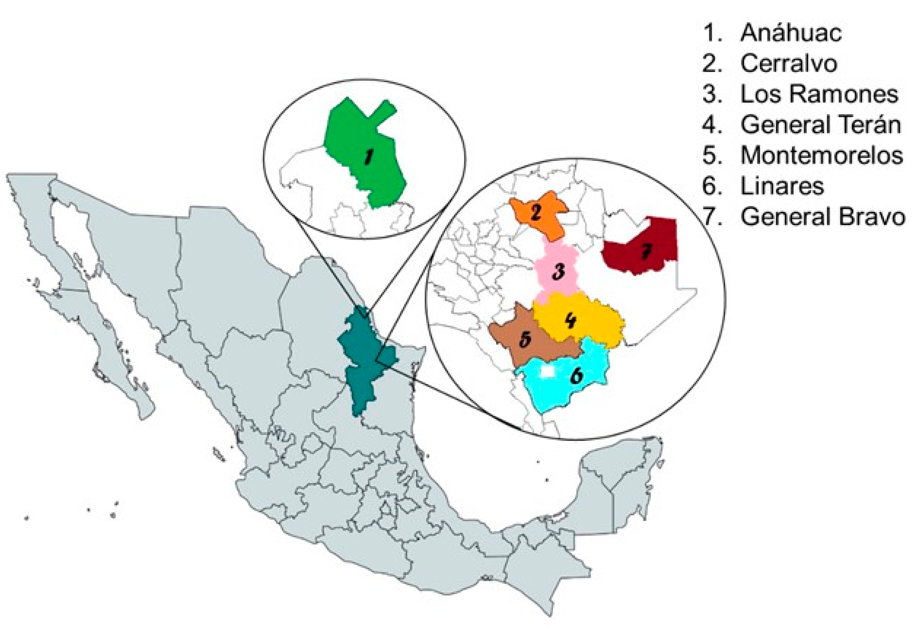
2.1. Study Areas

The study was conducted in seven municipalities of Nuevo Leon, Mexico: 1. Anahuac (27°14′27.5″ N, 100°08′01.5″ W; altitude 335 m), 2: General Teran (25°15′34.0″ N, 99°40′50.7″ W; altitude 230 m), 3. Los Ramones (25°41′47.0″ N, 99°37′27.9″ W; altitude 226 m), 4. General Teran (25°15′34.0″ N, 99°40′50.7″ W; altitude 230 m), 5. Montemorelos (25°11′13.0″ N, 99°50′05.0″ W; altitude 342 m), 6. Linares (25°51′45.2″ N, 99°33′52.0″ W; altitude 350 m), and 7. General Bravo (25°47′44.2″ N, 99°10′44.3″ W; altitude 150 m) (Figure 1). These study areas belong to the physiographic region of the North Gulf Coastal Plain, and the Tamaulipas Spiny Scrub ecoregion. The altitude of these sites ranges between 150 and 350 m. The climate is dry and semidry. The average annual temperature is around 20 °C, while the average yearly rainfall is 650 mm, with summer rains in August and September [11]. The sampling was conducted from 2015 to 2017, in April, June, July, September, October, and November.

2.2. Ticks and Bovine Blood Collections

Every herd of each locality was privately owned, and it was worked with the owner’s approval. Ethics committee approval for our study was number CEICANCL21012022- MOLSUNL-06. Cattle were manually restrained in a Powder River manual squeeze chute (crush). Ticks were captured directly from cattle (Simbrah and Simmental breed) and placed in vials with 90% alcohol.
Each tick was carefully observed under 8 to 32× magnifications using a stereoscopic microscope (Carl Zeiss Light Microscopy, Göttingen, Germany) to examine the external morphological characteristics of genera and species; in this way, the life stage and sex of each specimen were also determined. The identification was based on the taxonomic keys of [12,13,14,15]. Voucher tick specimens are deposited in the Walter Reed Biosystematics Unit, Smithsonian Institution, Suitland, MD, USA.
In contrast, the whole blood samples for molecular assay were taken from the caudal vein of the bovine in sterile tubes with EDTA, one sampling per animal. The tubes were immediately transported to the laboratory on ice (4 °C). Separate blood samples were allowed to settle and centrifuged at 2000× g for 10 min. The serum was recovered and stored at −20 °C until B. burgoderferi serological analyses.

2.3. DNA Extraction and PCR Amplification

DNA extraction from both ticks and whole blood was performed using the method of Ferrer et al. [16]. From the material obtained after crushing the tick in a mortar, the entire content was added to a new Eppendorf tube, and 500 μL of lysis buffer and 40 μL of proteinase K were placed and left to incubate in an AccuBlock (Labnet International Inc., Edison, NJ, USA) dry bath at 65 °C for one hour, shaking the sample in a vortex (VX-100 Lab Vortex Mixer Labnet International Inc.) every 15 min. After one hour, 500 μL of phenol–chloroform–isoamyl alcohol (49.5:49.5:1) was added to the sample and placed in a centrifuge (Centrifuge 5430 Eppendorf) at 14,000 rpm for 15 min. The supernatant was placed in a new tube, and two washes were performed with 500 μL of isoamyl alcohol (27:1), centrifuging for 10 min at 14,000 rpm, and collecting the supernatant. Subsequently, 65 μL of NaOAc was added, and 75 μL of NaCl was gently shaken using inversion. It was left at −20 °C for 24 h. The sample was centrifuged for 10 min at 14,000 rpm. The supernatant was collected and placed in a new tube to which 270 μL of isopropanol was added and left at −20 °C for 10 min. The sample was centrifuged for 10 min at 14,000 rpm, and the supernatant was removed. The pellet was resuspended in 500 μL of 80% molecular-grade ethanol and centrifuged for 5 min at 14,000 rpm. Once centrifuged, the supernatant was removed and placed in the 37 °C incubator to dry the ethanol completely. The DNA was resuspended in 20 μL of milli-Q water or TE buffer, and the DNA was extracted at −20 °C to continue with PCR.
For B. burgdorferi, the 234 bp PB directed at the flagellin gene sequence SC1 (5′-AAC ACA CCA GCA TCA CTT TCA GG-3′) and SC2 (5′-GAG AAT TAA CTC CGC CTT GAG AAG-3′) [17] was used. With the denaturation cycle at 94 °C for 1 min, 40 denaturation cycles at 93 °C for 30 s, primer binding at 57 °C for 30 s, DNA chain elongation at 72 °C for 30 s, and finally 72 °C for 1 min of elongation. Once the reaction is complete, the fraction is observed on an agarose gel.
For A. marginale, the 265 bp (Msp1 β), which is directed to the surface protein sequence SC1 (5′-GCT CTA GCA GGT TAT GCG TC-3′) and SC2 (5′-CTG CTT GGG AGA ATG CAC CT-3′ [18], was used with the denaturation cycle at 94 °C for 1 min, 30 denaturation cycles at 95 °C for 1 min, primer binding at 53 °C for 1 min, DNA chain elongation at 72 °C for 1 min, and finally at 72 °C for 10 min of elongation. Once the reaction was complete, the fraction was observed; all PCR products of ticks and bovine blood were carried out by electrophoresis on 1% agarose gels. They were visualized with ethidium bromide staining under ultraviolet light. Amplicons from blood cattle and the vector R. microplus were used for A. marginale sequencing, and further deposited to the GenBank.

2.4. Serological Analysis

Considering the almost unknown Lyme borreliosis in cattle in the area, we focused our study on only evaluating the presence of antibodies against B. burgdorferi using IFA. The substrate and bovine-specific anti-immunoglobulin to perform the tests were purchased commercially (Lyme (B. burgdorferi) IFA Substrate Slide from VMRD® laboratory; bovine anti-immunoglobulin). Of the serum, ten μL with titers of 1:64 was used, with an incubation time of one hour at 37 °C in a humid chamber. After the anti-immunoglobulin was conjugated with fluorescein isothiocyanate (FITC), ten μL was used, incubating at 37 °C for 45 min.

2.5. Statistical Analysis

The sample size of ticks for molecular analysis was calculated with a formula for a finite population with a 95% confidence level and a margin of error of 5%. Statistical analyses for variable associations were performed using the chi-square test. Infection rates were compared by study site and positive molecular samples. A value of p < 0.05 was considered significant. All analyses were conducted using the Epi InfoTM program ver. 7.2 from Centers for Disease Control and Prevention, Atlanta GA USA [19].

3. Results

3.1. Ticks Collected from Cattle

The group of 404 bovines yielded 2880 ticks distributed in three genera: R. microplus (2391 females and 395 males), Amblyomma spp. (51 females and 42 males), and D. variabilis (1 female). Rhipicephalus microplus represented the most significant number of specimens captured, with 96.7%. Within the seven study sites, Linares captured the highest number of ticks (29.9%), followed by General Teran (22.9%) (Table 1).

3.2. Ticks Sample Size from Cattle for PCR Analysis

Several 442-ticks, representing 15% of the total population of 2880 collected were calculated for molecular PCR analysis. The number of individuals per tick species described the same proportions as in the field frequency: R. microplus (n = 404), Amblyomma mixtum Koch 1844 (n = 10), A. tenellum (n = 27), and D. variabilis (n = 1).

3.3. Ticks Analyzed Using PCR to Identify A. marginale

Anaplasma marginale infection rates varied by tick species and study sites. Our results showed that, from 442 assayed individuals, only 44 (9.9%) ticks were positive for A. marginale. The number of positive specimens per species was R. microplus (n = 38; 9.4%), A. tenellum (n = 5; 18.5%), and D. variabilis (n = 1; 100%). Likewise, the localities with the highest infection rates of A. marginale in ticks were Linares (19.8%) and Montemorelos (18.7%) (Table 2). There was a significant (χ2 (4, n = 428) = 28.83, p < 0.00) association between study sites (Linares, General Teran, General Bravo, Cerralvo, and Montemorelos) and infection rate of A. marginale in ticks. Moreover, there was a significant (χ2 (4, n = 392) = 23.71, p = 0.00) result between localities (Linares, General Teran, General Bravo, Cerralvo, and Montemorelos) and the infection rate of A. marginale in R. microplus. One amplicon of R. microplus infected with A. marginale was sequenced and uploaded to the GenBank as a molecular genetic reference; the accession number is OR05500.

3.4. Bovine Blood Samples Analyzed Using PCR to Identify A. maginale

Of the 404 bovines, 337 blood samples were PCR-processed and yielded 214 positives for the presence of A. maginale (65.50%) infection rate (Table 3). Rates of cattle infected with A. marginale showed variation among localities. Cerralvo showed the highest infection rate of 92.45%, followed by Montemorelos (81.82%). A chi-square test examined the association between study sites (Linares, General Teran, General Bravo, Cerralvo, and Montemorelos) and infection-positive A. marginale in cattle. The relation between these variables was significant, χ2 (4, n = 327) = 20.02, p = 0.000. There was a significant association between study sites and the A. marginale infection rate. A host or cattle sample showing PCR infection to A. marginale was sequenced and GenBank accessed, number OR050501.

3.5. Ticks and Blood Bovine Samples Negative to B. burgdorferi s.l. by PCR and IFA

Negative results were determined using IFA antibody serology to identify B. burgdorferi s.l.; there was no identification in blood samples (n = 367) from cattle or PCR testing on ticks (n = 442). The same result was consistent throughout all study sites.

4. Discussion

In this work, the endpoint PCR identified A. marginale in cattle blood and tick tissue from several Nuevo Leon, Mexico localities. The amplification of the Msp1β gene delivered a product of the expected molecular size of 265 bp, confirming the presence of this hemoparasite in cattle and ticks. These data represent the first report in Nuevo Leon.
Bovine anaplasmosis is reported to be endemic in cattle in Mexico [7]. The infection rates of A. marginale in cattle in Mexico range from 10 to 78.9% [20,21,22]. The present study found an average infection rate of 63.50% in bovines in all seven sampled localities of Nuevo Leon. Percent rates ranged from 20% at Anahuac and Los Ramones to 81.82% in Montemorelos and 92.45% in Cerralvo. In other studies, a variation in the infection rates was found, for example, in the neighboring state of Tamaulipas by Lopez-Sanchez et al. [23], reporting 11.1% using serology, and by Almazán et al. [8] 36.7% using blood smear. Moreover, other percentages of infection of A. marginale in cattle from some southern states of Mexico have been documented. Pacific Ocean coastal states such as Guerrero (78.9% serology) [24], Veracruz (56% serology and 69.2% PCR) [25,26], and Yucatan (69.8% serology) [21] showed the highest percentages of infection. These differences between the data may be due to different ecological conditions; the breed and age of cattle; the difference in sample size and sampling effort of each locality; and the high infestation of susceptible tick populations.
Molecular identifications exhibited 9.9% of infection found in all the tick species and study localities (Table 2). Five of the seven localities resulted in favorable rates, whereby localities such as Montemorelos and Linares showed the highest rates: 18.7% and 19.8%, respectively. Although A. marginale was found in Anahuac and Los Ramones cattle, their tick populations were PCR-negative. The number of ticks captured in Anahuac (n = 10) and Los Ramones (n = 3) was very low compared to the other localities. Thus, the probability decreases in the detection of the bacteria. Conversely, Montemorelos and Cerralvo were the opposite. Particularly in Montemorelos, where both reservoir and vector yielded (81.82%) and (18.7%) infected with the bacteria, respectively.
In Mexico, it has been documented that two tick species, R. microplus and Rhipicephalus annulatus (Say, 1821), are proven vectors of A. marginale [2,27]. Only one study from Tamaulipas State has demonstrated 40% of the natural infection of A. marginale in R. microplus [8]. Here, we were first to reveal infection of A. marginale in R. microplus collected from cattle from Nuevo Leon. Similarly, we reported the presence of A. marginale in the ticks A. tenellum and D. variabilis. Our results showed that A. marginale in R. microplus and A. tenellum played a variable role as vectors from locality to locality. This result suggests that R. microplus may act as a vector of A. marginale in Linares, General Teran, General Bravo, Cerralvo, and Montemorelos. Undoubtedly, our findings point out that Rhipicephalus microplus, known as cattle tick, is a species that has a wide distribution in the American region and causes severe damage to cattle due to its direct parasitic action and because it is a vector of important pathogens [28].
We have shown here the natural infection of A. marginale in cattle and its R. microplus, A. tenellum, and D. variabilis ticks from some Nuevo Leon, Mexico localities. Surveillance is the first step to controlling tick-borne diseases; it is necessary to monitor ectoparasites and their causal agents [29]. This is important to avoid economic losses in Mexico, with a cattle population of 35,224,960 heads at risk [6]. Therefore, this work offers a clearer perspective on bovine anaplasmosis in Nuevo Leon for the first time.
In addition to surveillance, accurate diagnostic methods are the next step to confirming specific pathogen presence. Several traditional and modern approaches have been developed: microscopic observation, cELISA, card agglutination test, and, more recently, PCR [1]. However, the PCR method used in the present study has gained more popularity due to its specificity. Thus, it can be used in epidemiological studies as an early and accurate diagnostic method [30].
Although this study showed negative results for B. burgdorferi in cattle and ticks from Nuevo Leon, in a systematic review, a total of 1,347 records of Borrelia were found in Mexico [5]. Additionally, Nuevo Leon was one of the three states with the highest number of reports (253 records; 18.5%): humans [31,32,33,34], domestic dogs [35,36], wild rodents [37], and ticks [9,38]. However, there are no reports of this spirochete in cattle in Nuevo Leon.
For perspective and future studies of these diseases, it will be necessary to sequence the positive samples according to collection localities, tick species, and disease incidence, to carry out the respective phylogenetic and clinical analyses. Since it is known that there is a genetic diversity of A. marginale in Mexico [8,39,40], this way establishes superinfections [20] in places with a high rate of infection such as Montemorelos and Cerralvo, Nuevo Leon, Mexico. Therefore, molecular surveillance of these two diseases must be continued.

5. Conclusions

In the current study, we made the first presentation of A. marginale natural infection in the following tick species: R. microplus, A. tenellum, and D. variabilis. These species were collected from domestic cattle from Nuevo Leon, northeastern Mexico. Molecular PCR techniques confirmed A. marginale in bovine blood samples. Conversely, we did not find B. burgdorferi in the same ticks and bovine blood samples.

Author Contributions

Conceptualization, J.Á.O.-R. and G.M.-D.; methodology J.Á.O.-R., K.-A.G.-H., E.A.R.-T., J.J.H.-E., G.M.-D., R.M.S.-C. and I.F.-S.; validation, J.Á.O.-R., K.-A.G.-H., J.J.R.-R., E.A.R.-T., J.J.H.-E., G.M.-D., R.M.S.-C., C.E.M.-D.l.G. and I.F.-S.; formal analysis, J.J.R.-R.; investigation, J.Á.O.-R. and J.J.H.-E.; resources, G.M.-D., R.M.S.-C. and I.F.-S.; data curation, J.Á.O.-R. and J.J.R.-R.; writing—original draft preparation, J.Á.O.-R. and J.J.R.-R.; writing—review and editing, J.Á.O.-R., J.J.R.-R., R.M.S.-C. and I.F.-S.; visualization, J.J.R.-R., C.E.M.-D.l.G. and R.M.S.-C.; supervision, J.J.H.-E., G.M.-D., R.M.S.-C. and I.F.-S.; project administration, G.M.-D., R.M.S.-C. and I.F.-S.; funding acquisition, J.J.H.-E., G.M.-D., R.M.S.-C. and I.F.-S. All authors have read and agreed to the published version of the manuscript.

Funding

This research was funded by Universidad Autónoma de Nuevo León- Facultad de Medicina Veterinaria y Zootecnia, “Programa de Apoyo al Fortalecimiento de la investigación,” CONACYT. Grant number 12-2019.

Institutional Review Board Statement

Not applicable.

Informed Consent Statement

CEICANCL21012022-MOLSUNL-06.

Data Availability Statement

Data is private property of cattle farm owners.

Acknowledgments

The authors acknowledge the support of the research project by the CONACYT. Moreover, to Centro Nacional de Servicios de Constatación de Salud Animal (CENAPA). We thank the ranchers who kindly allowed us to work with their animals for this research. Additionally, we thank María D. Esteve Gassent from the Department of Veterinary Pathology at Texas A&M University, USA, and Sergio D. Rodriguez, Coordinator of the Anaplasmosis Unit of the National Institute of Forest, Agricultural and Livestock Research, who provided the positive controls.

Conflicts of Interest

The authors declare no conflict of interest.

References

  1. Atif, F.A. Anaplasma marginale and Anaplasma phagocytophilum: Rickettsiales pathogens of veterinary and public health significance. Parasitol. Res. 2015, 114, 3941–3957. [Google Scholar] [CrossRef]
  2. Kocan, K.M.; de la Fuente, J.; Blouin, E.F.; Coetzee, J.F.; Ewing, S.A. The natural history of Anaplasma marginale. Vet. Parasitol. 2010, 167, 95–107. [Google Scholar] [CrossRef] [PubMed]
  3. Johnson, R.C.; Schmid, G.P.; Hyde, F.W.; Steingerwalt, A.G.; Brenner, D.J. Borrelia burgdorferi sp. nov.: Etiological agent of Lyme disease. Int. J. Syst. Bacterio. 1984, 34, 496–497. [Google Scholar] [CrossRef]
  4. Parker, J.L.; White, K.K. Lyme borreliosis in cattle and horses: A review of the literature. Cornell. Vet. 1992, 82, 253–274. [Google Scholar] [PubMed]
  5. Colunga-Salas, P.; Sánchez-Montes, S.; Volkow, P.; Ruíz-Remigio, A.; Becker, I. Lyme disease and relapsing fever in Mexico: An overview of human and wildlife infections. PLoS ONE 2020, 15, e0238496. [Google Scholar] [CrossRef]
  6. Ferreira, G.C.M.; Canozzi, M.E.A.; Peripolli, V.; de Paula Moura, G.; Sánchez, J.; Martins, C.E.N. Prevalence of bovine Babesia spp., Anaplasma marginale, and their co-infections in Latin America: Systematic review-meta-analysis. Ticks Tick Borne Dis. 2022, 13, 101967. [Google Scholar] [CrossRef]
  7. Rodríguez, S.D.; Ortiz, M.A.G.; Ocampo, R.J.; Murguía, C.A.V. Molecular epidemiology of bovine anaplasmosis with a particular focus in Mexico. Infect Genet Evol 2009, 9, 1092–1101. [Google Scholar] [CrossRef]
  8. Almazán, C.; Medrano, C.; Ortiz, M.; de la Fuente, J. Genetic diversity of Anaplasma marginale strains from an outbreak of bovine anaplasmosis in an endemic area. Vet. Parasitol. 2008, 158, 103–109. [Google Scholar] [CrossRef] [PubMed]
  9. Gordillo-Pérez, G.; Vargas, M.; Solórzano-Santos, F.; Rivera, A.; Polaco, O.J.; Alvarado, L.; Muñoz, O.; Torres, J. Demonstration of Borrelia burgdorferi sensu stricto infection in ticks from the northeast of Mexico. Clin. Microbiol. Infect. 2009, 15, 496–498. [Google Scholar] [CrossRef] [PubMed]
  10. Instituto Nacional de Estadística, Geografía e Informática. Encuesta Nacional Agropecuaria. Available online: https://www.inegi.org.mx/programas/ena/2019/ (accessed on 5 December 2022).
  11. Instituto Nacional de Estadística y Geografía. Available online: https://cuentame.inegi.org.mx/monografias/informacion/nl/territorio/clima.aspx?tema=me&e=19 (accessed on 5 December 2022).
  12. Guzmán-Cornejo, C.; Robbins, R.G.; Guglielmone, A.A.; Montiel-Parra, G.; Pérez, T.M. The Amblyomma (Acari: Ixodida: Ixodidae) of Mexico: Identification keys, distribution and hosts. Zootaxa 2011, 2998, 16–38. [Google Scholar] [CrossRef]
  13. Guzmán-Cornejo, C.; Robbins, R.G.; Guglielmone, A.A.; Montiel-Parra, G.; Rivas, G.; Pérez, T.M. The Dermacentor (Acari, Ixodida, Ixodidae) of Mexico: Hosts, geographical distribution and new records. Zookeys 2016, 569, 1–22. [Google Scholar] [CrossRef]
  14. Guglielmone, A.A.; Robbins, R.G.; Apanaskevich, D.A.; Petney, T.N.; Estrada-Peña, A.; Horak, I.G.; Shao, R.; Barker, S.C. The Argasidae, Ixodidae and Nuttalliellidae (Acari: Ixodida) of the world: A list of valid species names. Zootaxa 2010, 2528, 1–28. Available online: http://www.mapress.com/zootaxa/2010/f/z02528p028f.pdf (accessed on 8 August 2022). [CrossRef]
  15. Walker, J.B.; Keirans, J.E.; Horak, I.G. Genus Rhipicephalus (Acari, Ixodidae). In A Guide to the Brown Ticks of the World; Cambridge University Press: Cambridge, UK, 2000. [Google Scholar] [CrossRef]
  16. Ferrer, C.; Colom, F.; Frasés, S.; Mulet, E.; Abad, J.L.; Alió, J.L. Detection and identification of fungal pathogens by PCR and ITS2 and 5.8 S ribosomal DNA typing in ocular infections. J. Clin. Microbiol. 2001, 39, 2873–2879. [Google Scholar] [CrossRef] [PubMed]
  17. Jaulhac, B.; Heller, R.; Limbach, F.X.; Hansmann, Y.; Lipsker, D.; Monteil, H.; Sibilia, J.; Piémont, Y. Direct molecular typing of Borrelia burgdorferi sensu lato species in synovial samples from patients with Lyme arthritis. J. Clin. Microbiol. 2000, 38, 1895–1900. [Google Scholar] [CrossRef]
  18. Bilgiç, H.; Karagenç, T.; Simuunza, M.; Shiels, B.; Tait, A.; Eren, H.; Weir, W. Development of a multiplex PCR assay for simultaneous detection of Theileria annulata, Babesia bovis and Anaplasma marginale in cattle. Exp. Parasitol. 2013, 133, 222–229. [Google Scholar] [CrossRef]
  19. Centers for Disease Control and Prevention. Division of Health Informatics & Surveillance (DHIS), Center for Surveillance, Epidemiology & Laboratory Services (CSELS). Available online: http://www.cdc.gov/epiinfo (accessed on 9 January 2023).
  20. Castañeda-Ortiz, E.J.; Ueti, M.W.; Camacho-Nuez, M.; Mosqueda, J.J.; Mousel, M.R.; Johnson, W.C.; Palmer, G.H. Association of Anaplasma marginale strain superinfection with infection prevalence within tropical regions. PLoS ONE 2015, 10, e0120748. [Google Scholar] [CrossRef]
  21. Rodríguez-Vivas, R.I.; Mata-Mendez, Y.; Pérez-Gutierrez, E.; Wagner, G. The effect of management factors on the seroprevalence of Anaplasma marginale in Bos indicus cattle in the Mexican tropics. Trop. Anim. Health Prod. 2004, 36, 135–143. [Google Scholar] [CrossRef]
  22. Vega, L.E.O.; Rodríguez, S.D.; Alarcón, G.J.C.; Flores, R.L.; Ocampo, R.J.; Ortiz, M.Á.G.; Preciado de la Torre, J.F.; Ramírez, E.E.R. Anaplasma marginale field challenge: Protection by an inactivated immunogen that shares partial sequence of msp1α variable region with the challenge strain. Vaccine 2007, 25, 519–525. [Google Scholar] [CrossRef]
  23. López-Sánchez, F.; Canto-Alarcón, G.J.; Falcón-Neri, A.; Aboytes-Torres, R. Anaplasmosis and babesiosis prevalence in the Experimental Station of Aldama, Tamaulipas. Técnica Pecu. México 1984, 46, 88–92. [Google Scholar]
  24. Fragoso, S.G.; Suazo, F.M. Prevalencia de anticuerpos contra Anaplasma marginale y Babesia spp. en la zona centro del estado de Guerrero. Rev. Mex Cienc. Pecu 1984, 47, 133–136. Available online: https://cienciaspecuarias.inifap.gob.mx/index.php/Pecuarias/article/viewFile/3313/2733 (accessed on 8 August 2022).
  25. Cossio-Bayugar, R.; Rodriguez, S.D.; Garcia-Ortiz, M.A.; Garcia-Tapia, D.; Aboytes-Torres, R. Bovine anaplasmosis prevalence in northern Veracruz state, Mexico. Prev. Vet. Med. 1997, 32, 165–170. [Google Scholar] [CrossRef] [PubMed]
  26. García-Tapia, D.G.; Rojas, M.L.; Bayugar, R.C.; Vazquez, Z.G.; Ortiz, M.A.G.; Jalil, P.D.; Torres, R.A. Seroprevalencia de anaplasmosis en explotaciones bovinas de 18 municipios de la zona norte de Veracruz. Rev. Mex Cienc. Pecu 1996, 34, 38–45. Available online: https://cienciaspecuarias.inifap.gob.mx/index.php/Pecuarias/article/viewFile/698/696 (accessed on 10 August 2022).
  27. Osorno, M.B.; Ristic, M. Anaplasmosis bovina con énfasis en control, diagnostico, distribución de la enfermedad en México y uso de una vacuna atenuada de Anaplasma marginale. Vet. México 1977, 8, 85–98. [Google Scholar]
  28. Estrada-Peña, A.; García, Z.; Sánchez, H.F. The distribution and ecological preferences of Boophilus microplus (Acari: Ixodidae) in Mexico. Exp. Appl. Acarol. 2006, 38, 307–316. [Google Scholar] [CrossRef] [PubMed]
  29. Carvajal-de la Fuente, V.; Merino-Charrez, O.; Tovar-Carman, E.; Rodríguez-Camarillo, S.D.; Lagunes-Quintanilla, R.E.; Muñoz-Tenería, F.A.; Contreras, M.; de la Fuente, J. Differential expression analysis for subolesin in Rhipicephalus microplus infected with Anaplasma marginale. Exp. Appl. Acarol. 2018, 76, 229–241. [Google Scholar] [CrossRef] [PubMed]
  30. Estrada-Peña, A.; Szabó, M.; Labruna, M.; Mosqueda, J.; Merino, O.; Tarragona, E.; Venzal, J.M.; de la Fuente, J. Towards an effective, rational and sustainable approach for the control of cattle ticks in the Neotropics. Vaccines 2020, 8, 9. [Google Scholar] [CrossRef]
  31. Arroyave, C.M.; Tamez-González, R. Enfermedad de Lyme. Informe de dos casos. Bol. Med. Hosp. Infant. Mex 1994, 51, 117–121. [Google Scholar]
  32. Gordillo-Pérez, G.; Torres, J.; Solórzano-Santos, F.; Garduño-Bautista, V.; Tapia-Conyer, R.; Muñoz, O. Estudio seroepidemiológico de borreliosis de Lyme en la Ciudad de México y el noreste de la República Mexicana. Salud. Publ. Mex 2003, 45, 351–355. [Google Scholar] [CrossRef]
  33. Skinner-Taylor, C.M.; Flores-González, M.S.; Esquivel-Valerio, J.A.; Salinas-Meléndez, J.A.; Salinas-Palacios, C.K.; Rodríguez-Amado, J.; Garza, E.M.A. Evidencia de la enfermedad de Lyme en una población de alto riesgo del noreste de México. Med. Univer. 2007, 9, 105–111. Available online: https://www.medigraphic.com/pdfs/meduni/mu-2007/mu073c.pdf (accessed on 8 August 2022).
  34. Skinner-Taylor, C.M.; Flores, M.S.; Salinas, J.A.; Arevalo-Nińo, K.; Galán-Wong, L.J.; Maldonado, G.; Garza-Elizondo, M.A. Antibody profile to Borrelia burgdorferi in veterinarians from Nuevo León, Mexico, a non-endemic area of this zoonosis. Rheumatology 2016, 54, 97–102. [Google Scholar] [CrossRef]
  35. Salinas-Meléndez, J.A.; Tamez-González, R.; Welsh-Lozano, O.; Barrera-Saldaña, H.A. Detection of Borrelia burgdorferi DNA in human skin biopsies and dog synovial fluid by the polymerase chain reaction. Rev. Latinoam. Microbiol. 1995, 37, 7–10. [Google Scholar] [PubMed]
  36. Salinas-Meléndez, J.A.; Avalos-Ramírez, R.; Riojas-Valdez, V.M.; Martínez-Muñoz, A. Serological survey of canine borreliosis. Rev. Latinoam. Microbiol. 1999, 41, 1–4. [Google Scholar]
  37. Rodríguez-Rojas, J.J.; Rodríguez-Moreno, Á.; Sánchez-Casas, R.M.; Hernández-Escareño, J.J. Molecular detection of Leptospira interrogans and Borrelia burgdorferi in wild rodents from Mexico. Vector Borne Zoonotic Dis. 2020, 20, 860–863. [Google Scholar] [CrossRef] [PubMed]
  38. Galaviz-Silva, L.; Pérez-Treviño, K.C.; Molina-Garza, Z.J. Distribution of ixodid ticks on dogs in Nuevo León, Mexico, and their association with Borrelia burgdorferi sensu lato. Exp. Appl. Acarol. 2013, 61, 491–501. [Google Scholar] [CrossRef] [PubMed]
  39. Jiménez-Ocampo, R.; Vega y Murguía, C.A.; Oviedo-Ortega, N.; Rojas-Ramírez, E.E.; García-Ortiz, M.A.; Preciado de la Torre, J.F.; Rodríguez-Camarillo, S.D. Diversidad genética de la región variable de los genes msp1a y msp4 en cepas de Anaplasma marginale de México. Rev. Mex Cienc. Pecu 2012, 3, 373386. Available online: https://www.scielo.org.mx/scielo.php?pid=S2007-11242012000300008&script=sci_arttext (accessed on 8 August 2022).
  40. Martínez-Ocampo, F.; Quiroz-Castañeda, R.E.; Amaro-Estrada, I.; Dantán-González, E.; de la Torre, J.F.; Rodríguez-Camarillo, S. Whole-genome sequencing of mexican strains of Anaplasma marginale: An approach to the causal agent of bovine anaplasmosis. Int. J. Genom. 2020, 2020, 5902029. [Google Scholar] [CrossRef]
Figure 1. Geographic location of the seven study sites of molecular surveillance of Anaplasma marginale and Borrelia burgdorferi in cattle and ticks from Nuevo Leon, Mexico, between 2015 and 2017.
Pathogens 12 00784 g001
Table 1. Species of ticks captured from bovines in seven localities of Nuevo Leon, Mexico, from 2015 to 2017.
LocalitiesRhipicephalus
microplus
(F:M)
Amblyomma
spp. (F:M)
Dermacentor
variabilis
(F:M)
Total (F:M)
Linares860
(734:126)
1
(0:1)
1
(1:0)
862
(735:127)
General Teran653
(584:69)
7
(7:0)
0660
(591:69)
General Bravo256
(182:74)
21
(3:18)
0277
(185:92)
Cerralvo523
(469:54)
10
(8:2)
0533
(477:56)
Montemorelos355
(297:58)
53
(32:21)
0408
(329:79)
Anahuac134
(120:14)
00134
(120:14)
Los Ramones5
(5:0)
1
(1:0)
06
(6:0)
Total (F:M)2786
(2391:395)
93
(51:42)
1
(1:0)
2880
(2443:437)
Note. F: female; M: male
Table 2. Anaplasma marginale infection rates (%) in ticks collected from cattle at study sites in Nuevo Leon, Mexico, from 2015 to 2017.
LocalitiesRhipicephalus microplus (%)Amblyomma mixtum (%)Amblyomma tenellum (%)Dermacentor variabilis (%)Total (%)
Linares20/105 (9.5)001/1 (100)21/106 (19.8)
General Teran5/110 (4.5)00/4 (0)05/114 (4.4)
General Bravo3/60 (5.0)0/4 (0)003/64 (4.7)
Cerralvo1/65 (1.5)0/5 (0)001/70 (1.4)
Montemorelos9/52 (17.3)05/23 (21.7)014/75 (18.7)
Anahuac0/10 (0)0000/10 (0)
Los Ramones0/2 (0)0/1 (0)000/3 (0)
Total (%)38/404 (9.4)0/10 (0)5/27 (18.5)1/1 (100)44/442 (9.9)
Table 3. Positive PCR blood samples of Anaplasma marginale from cattle at study sites in Nuevo Leon, Mexico from 2015 to 2017.
LocalitiesTotal Samples + Samples Total (%)
Linares956164.21
General Teran903437.78
General Bravo453271.11
Cerralvo534992.45
Montemorelos443681.82
Anahuac5120.00
Los Ramones5120.00
Total33721463.50
Disclaimer/Publisher’s Note: The statements, opinions and data contained in all publications are solely those of the individual author(s) and contributor(s) and not of MDPI and/or the editor(s). MDPI and/or the editor(s) disclaim responsibility for any injury to people or property resulting from any ideas, methods, instructions or products referred to in the content.

