Article

Characterization of Collagen Binding Activity of Clostridium perfringens Strains Isolated from Broiler Chickens

1
Animal Bioscience and Biotechnology Laboratory, Beltsville Agricultural Research Center, Agricultural Research Service, US Department of Agriculture, Beltsville, MD 20705, USA
2
Environment Microbial and Food Safety Laboratory, Beltsville Agricultural Research Center, Agricultural Research Service, US Department of Agriculture, Beltsville, MD 20705, USA
*
Author to whom correspondence should be addressed.
Pathogens 2023, 12(6), 778; https://doi.org/10.3390/pathogens12060778
Submission received: 23 March 2023 / Revised: 13 May 2023 / Accepted: 23 May 2023 / Published: 30 May 2023

Abstract

:
Clostridium perfringens is the etiological agent for necrotic enteritis (NE) in broiler chickens, which causes a substantial economic loss of an estimated USD 6 billion annually in the global poultry industry. Collagen adhesion is involved in the NE pathogenesis in poultry. In this study, the binding capabilities of chicken C. perfringens isolates of various genetic backgrounds (netBtpeL, netB+tpeL, netB+tpeL+) to collagen types I–V and gelatin were examined, and the putative adhesin protein cnaA gene was investigated at the genomic level. In total, 28 C. perfringens strains from healthy and NE-inflicted sick chickens were examined. The results on collagen adhesin-encoding gene cnaA by the quantitative-PCR results indicated that netBtpeL isolates had much lower copies of the detectable cnaA gene than netB+ isolates (10 netB+tpeL isolates, 5 netB+tpeL+ isolates). Most of the virulent C. perfringens isolates demonstrated collagen-binding abilities to types I–II and IV–V, while some strains showed weak or no binding to collagen type III and gelatin. However, the netB+tpeL+ isolates showed significantly higher binding capabilities to collagen III than netBtpeL and netB+tpeL isolates. The data in this study suggest that the collagen-binding capability of clinical C. perfringens isolates correlates well with their NE pathogenicity levels, especially for C. perfringens isolates carrying genes encoding crucial virulence factors and virulence-associated factors such as netB, cnaA, and tpeL. These results indicate that the presence of the cnaA gene may be correlated with C. perfringens virulence (particularly for netB+ isolates).

1. Introduction

Clostridium perfringens is an anaerobic Gram-positive rod-shaped bacterium, and its type A and G isolates are the key etiologic factor contributing to necrotic enteritis (NE) in broiler chickens [1]. NE is characterized by sudden death (clinical form) or underperformance in growth and feed conversion (subclinical form), which could inflict substantial economic damage with an estimated annual loss of USD 6 billion to the global poultry industry [2,3]. NE is a complex and multi-factorial disease [3]. The β-pore-forming toxin netB is found to be an essential contributing factor to the virulence of pathogenic C. perfringens isolates [4,5,6]. Disease severity can be enhanced by multiple factors, such as using highly pathogenic netB+tpeL+ isolates expressing the largest TpeL toxin (a size of ~205 kDa), predisposition to coccidiosis, and nutritional manipulation with the use of cereals rich in non-starch polysaccharides or animal proteins [3,7].
Bacterial colonization is the first step for infection and plays a critical role in the normal development, differentiation, function, and regulation of the intestinal mucosal immune system [8]. Bacteria have evolved various mechanisms to adhere to the surface of organs in contact with the intestine, such as adhesin, pili, fimbriae, biofilm, and lipopolysaccharides [9]. It is suggested that the strong capability of C. perfringens adherence to extracellular matrix molecules promotes virulence in the induction of NE [10]. Furthermore, the adherent capabilities of C. perfringens isolates to collagens may be positively associated with the pathogenesis of avian NE [11,12]. The putative collagen gene cnaA is located within pathogenicity locus VR-10B mainly in netB-positive strains [13], and the mutation of cnaA has abolished NE pathogenesis in an animal model and reduced the capability of bacterial colonization in the chicken intestinal mucosa [11]. In this study, we characterized the capability of 28 C. perfringens isolates of laboratory collections in adherence to several collagens (I, II, III, IV, and V) and gelatins and screened the cnaA gene in bacteria with a polymerase chain reaction (PCR) and quantitative-PCR (qPCR).

2. Materials and Methods

2.1. Bacterial Strains and Growth Conditions

Twenty-eight isolates of C. perfringens were used in this study. N10 and N11 were isolated from healthy chickens, while most others came from collections of NE-afflicted chickens [14]: nine netBtpeL isolates, fourteen netB+tpeL isolates, and five netB+tpeL+ isolates. The isolates carrying the tpeL gene may induce enhanced NE disease severity. All strains were characterized for cnaA gene by PCR and qPCR. Of all 28 C. perfringens isolates, some have been characterized for NE induction in our NE disease model (See Table 1).
All C. perfringens strain collections were maintained as stock cultures in 25% glycerol and stored in a –80 °C freezer in the laboratory [15,16,17]. C. perfringens strains were grown anaerobically at 37 °C in chopped meat glucose (CMG) medium (Anaerobe Systems, Morgan Hill, CA, USA), and then in BYC medium containing 3.7% brain heart infusion medium (BHI Broth, RPI, Research Products International, Mt. Prospect, IL, USA), 0.5% yeast extract (Fisher Scientific, Hampton, NH, USA), 0.05% L-cysteine (Sigma-Aldrich, St. Louis, MO, USA), and 0.1% sodium thioglycolate (Sigma-Aldrich). The bacteria were incubated at 37 °C under anaerobic conditions (AnaerPack® System, MGC, Mitsubishi Gas Chemical Co., Inc., Tokyo, Japan) overnight.
Table 1. Sources and background information of Clostridium perfringens collections.
StrainSourcenetBtpeLReference
13 SoilThis study
CP1Field NE[16]
CP2Field NE[16]
CP15Field NE[16]
CP23Field NE[16]
JGSField NE[16]
LLY_N10Healthy chickens[16]
LLY_N11Healthy chickens[16]
SM101Food poisoning[17]
C11Field NE+±[15]
tpeL15Field NE++[15]
tpeL17Field NE++[15]
tpeL18Field NE++[15]
tpeL19Field NE++[15]
C1Field NE+[15]
C2Field NE+[15]
C3Field NE+[15]
C5Field NE+[15]
C6Field NE+[15]
C7Field NE+[15]
C8Field NE+[15]
C9Field NE+[15]
C10Field NE+[15]
C12Field NE+[15]
C13Field NE+This study
C14Field NE+[15]
C16Field NE+[15]
Del1Field NE+[16]
Note: “−” = negative in PCR, “+” = postive in PCR, “±” = weakly positive in PCR.

2.2. Preparation of Genomic DNA from C. perfringens

Five mL of BHI Broth was inoculated with each strain of C. perfringens and incubated at 37 °C under anaerobic conditions overnight. An amount of 1.5 mL of the culture was centrifuged at 8000× g for 2 min in a microcentrifuge, and the pellet was collected. Then, 100 μL of 200 mg/mL lysozyme was added to each tube, mixed thoroughly, and incubated at 37 °C for 30 min. Samples were vortexed well, and then incubated at 37 °C for an additional 15 min; 467 µL of TE buffer was added, and the pellet was resuspended by repeated pipetting. Then, 30 µL of 10% SDS and 3 µL of 20 mg/mL proteinase K (Sigma-Aldrich) were added to give a final concentration of 100 µg/mL proteinase K in 0.5% SDS. Samples were mixed thoroughly and incubated at 37 °C for 1 h. Then, 100 µL of 5 M NaCl was added and mixed thoroughly, followed by adding 80 µL of cetyltrimethylammonium bromide (CTAB, Sigma-Aldrich) /NaCl solution for a 10 min incubation at 65 °C. An approximately equal volume (0.7 to 0.8 mL) of chloroform/isoamyl alcohol (Sigma-Aldrich) was added and mixed thoroughly, and samples were centrifuged for 5 min. Aqueous and viscous supernatants were transferred to new tubes. An equal volume of phenol/chloroform/isoamyl alcohol (Sigma-Aldrich) was then added, extracted thoroughly, and centrifuged for 5 min. The supernatant was transferred to a fresh tube, and a volume of 0.6 of isopropanol was added to precipitate the nucleic acids. DNA was washed with 70% ethanol to remove residual CTAB, and DNA was centrifuged for 5 min at room temperature. The supernatant was carefully removed, and the pellet was briefly air-dried. The pellet was redissolved in 100 µL of TE buffer.

2.3. PCR for Detection of CnaA

All C. perfringens strains were screened for the presence of cnaA using PCR. Primers were designed using the cnaA gene of strain EHE-NE 18 (Accession number KT749987.1) (Table 2). Primer sequences were as follows: forward primer 5′-GGTGGATGGGCAACATTTAC-3′, reverse primer 5′-CCTTGCTTGGATTCACCAGT-3′, with an expected product size of 220 bp. PCR was performed using GoTaq green master mix (Promega, Madison, WI, USA). PCR conditions were used as follows: 95 °C for 3 min; followed by 35 repeats of 95 °C for 30 s, 55 °C for 30 s, and 72 °C for 3 min; followed by 72 °C for 5 min.

2.4. qPCR for Detection of cnaA

Genomic DNA (gDNA) was assessed using a NanoDrop spectrophotometer (NanoDrop One; Thermo Fisher Scientific, Waltham, MA, USA) at 260/280 and 260/230 nm to measure its concentrations and to check its purity. Each sample of gDNA was diluted to a concentration of 50 ng/µL in RNase-free water (Thermo Fisher Scientific), and followed by qPCR analysis with 5 μL of cDNA using SYBR Green qPCR Master Mix (PowerTrack, Applied Biosystems, Waltham, MA, USA) in triplicates using Applied Biosystems QuantStudio 3 Real-Time PCR Systems (Life Technologies, Carlsbad, CA, USA). The gDNA was analyzed with the following PCR conditions: denaturation at 95 °C for 10 min, followed by amplification at 56 °C for 1 min for 40 cycles. The oligonucleotide primer sequences used for 16S rRNA and cnaA genes are listed in Table 2. The relative expression level of each gDNA was normalized to the control gene 16S rRNA using the 2−ΔΔCt method [15].

2.5. Adhesion Assay

Bacterial adherence to five types of collagens and one gelatin was conducted as previously described and repeated in triplicates [12]. Collagens (types I, II, III, IV, and V) were all purchased from Sigma: collagen I from rat tail; collagen II from bovine nasal septum; collagen III from human placenta and Bornstein and Traub type III (Sigma Type X); collagens IV and V both from human placenta and gelatin (gelatin from porcine skin, gel strength 300, type A). Nunclon Delta-Treated, Flat-Bottom Microplates (Thermo Fisher Scientific) were coated with collagens (1 µg/well for collagens III and V; 2 µg/well for collagens I, II, and gelatin; and 0.031 µg/well for collagen IV) in 50 µL of phosphate-buffered saline (PBS, Quality Biological Inc., Gaithersburg, MD, USA) overnight at 4 °C and blocked using 200 μL of PBS plus 0.5% (w/v) bovine serum albumin (BSA) (Sigma-Aldrich) (PBSA) for 2 h at 4 °C and rinsed three times with 200 μL of PBSA.
Single C. perfringens colonies were suspended in 100 μL of sterile PBSA, spread onto BHI-supplemented C. perfringens-specific nutrient agar plates, and incubated overnight to produce a confluent layer. Bacteria were harvested from the agar surface in PBSA. Cells were pelleted by centrifugation at 3000× g for 2 min and washed three times with 5 mL of PBSA. The suspension was adjusted to an optical density (OD) of around 0.8 at 600 nm. Cells were added to wells in 50 μL aliquots and incubated at room temperature for 2 h with agitation, followed by rinsing three times with 200 μL of PBSA. C. perfringens were stained with 0.5% (w/v) crystal violet for 5 min. After rinsing three times with 200 μL of PBSA, 50 μL of solution of ethanol:acetone (1/1, v/v) was added to each well to destain adherent C. perfringens, and the OD value at 562 nm was measured. Wells incubated with bacteria but no collagen, and collagen-coated wells to which no bacteria were added, were used as blank and negative controls, respectively. Values from the adhesion assay were background-corrected by subtracting the blank value from the test sample absorbance values for that particular strain. Any values that were less than zero were adjusted to zero. All assays were repeated in biological triplicates.

2.6. Statistical Analysis

Statistical analysis was undertaken using GraphPad Prism 5 (GraphPad, San Diego, CA, USA). Mean treatment group values were compared using one-way analysis of variance followed by a Dunnett post hoc test. Differences were considered statistically significant at p ≤ 0.05. All the data were expressed as mean ± standard error of the mean.

3. Results

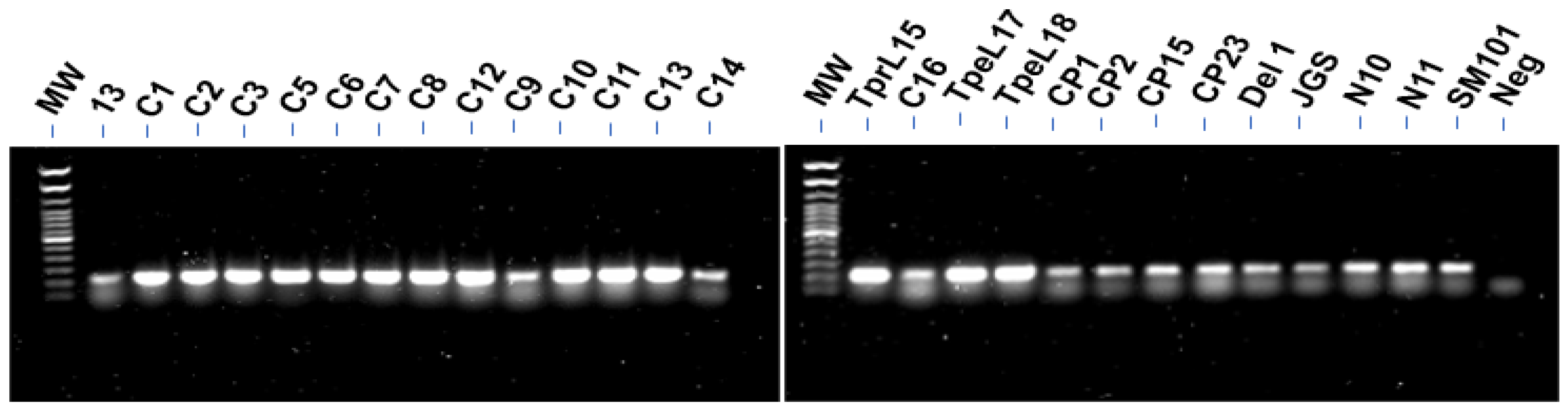
3.1. PCR for Detection of CnaA

Fifteen of 28 C. perfringens strains tested showed thick bands for the cnaA gene (isolate tpeL19 not shown in Figure 1), among which all were netB+ isolates, including five netB+tpeL+ isolates (C11, tpeL15, tpeL17-19), while thirteen isolates had relatively thin bands for cnaA by PCR (Figure 1): strains 13, C9, C14, C16, CP1, CP2, CP15, CP23, Del1, JGS, N10, N11, and SM101, among which most were netBtpeL, and four netB+tpeL isolates (C9, C14, C16, Del1). The PCR screening results for the cnaA gene within the genomes of these C. perfringens strains are summarized in Table 1.

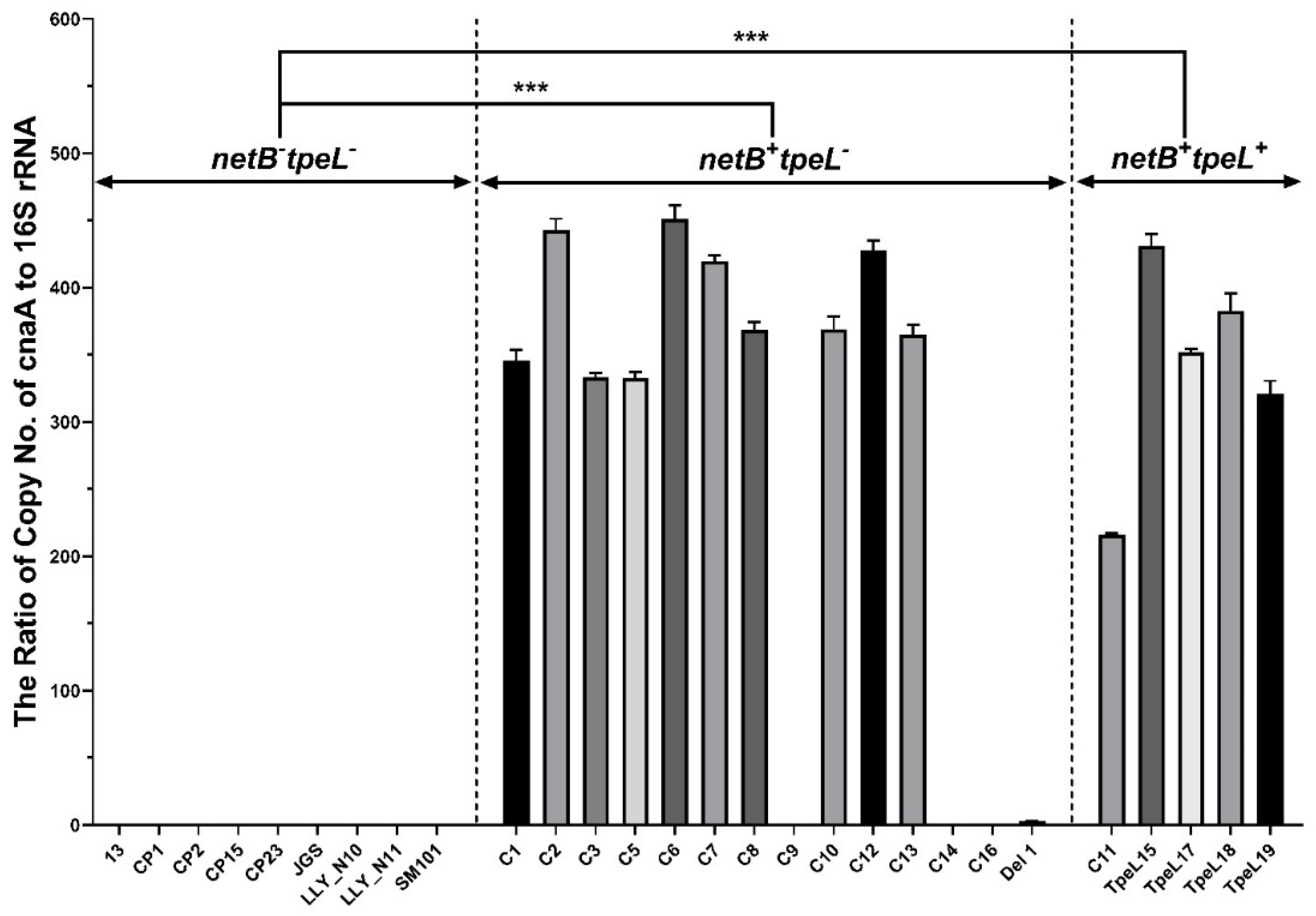
3.2. Quantitative Polymerase Chain Reaction (qPCR) for cnaA Gene Detection
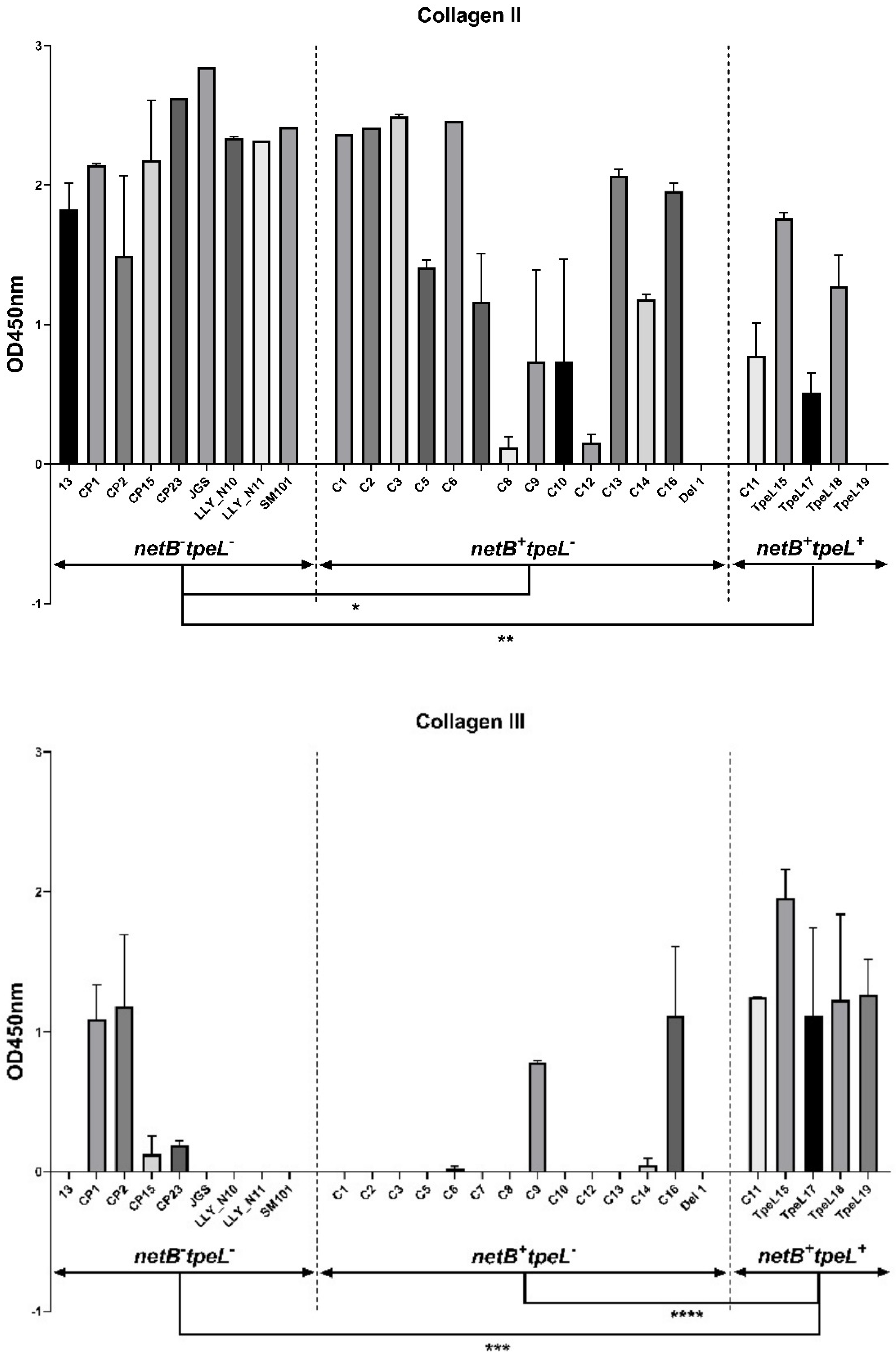
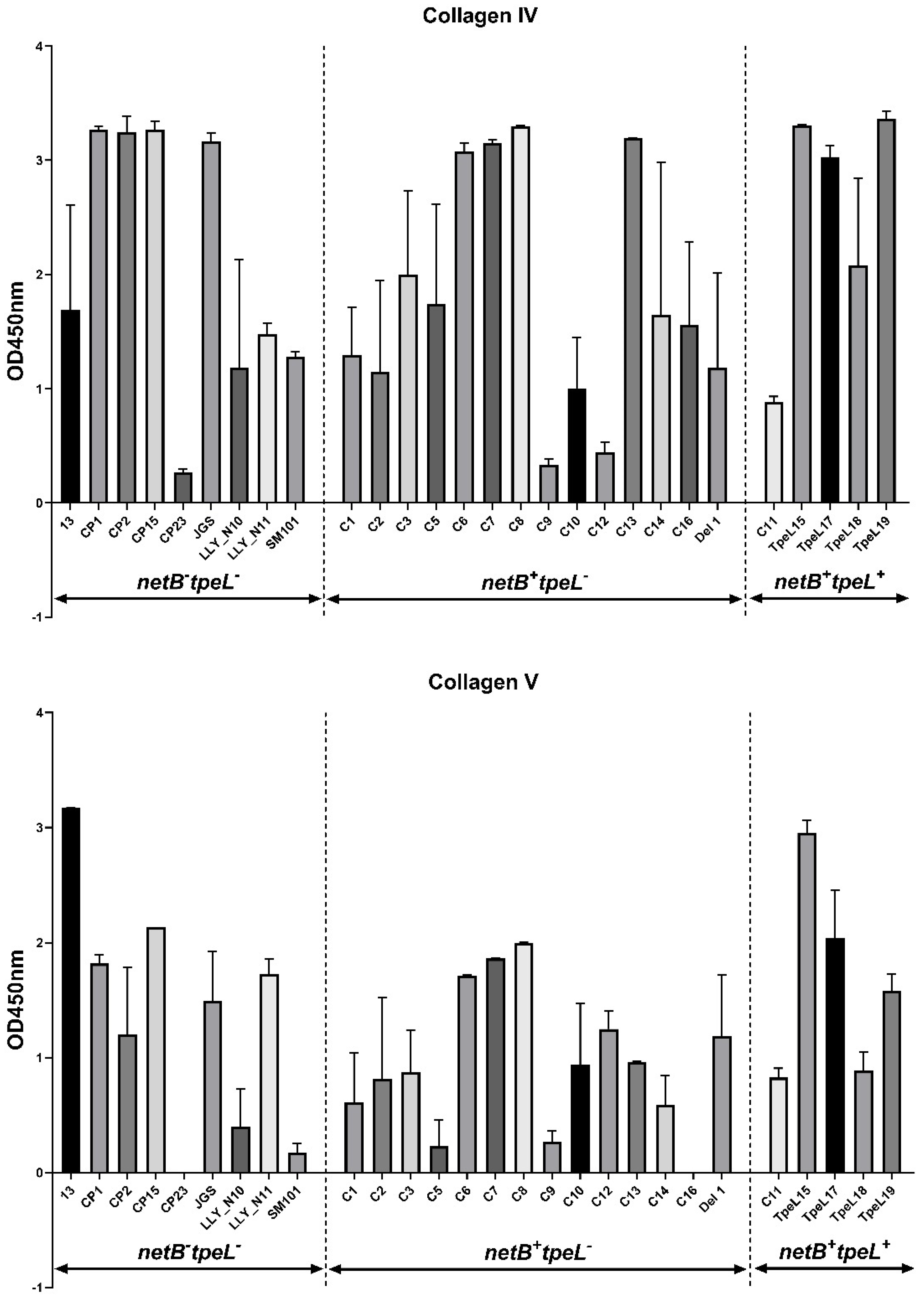
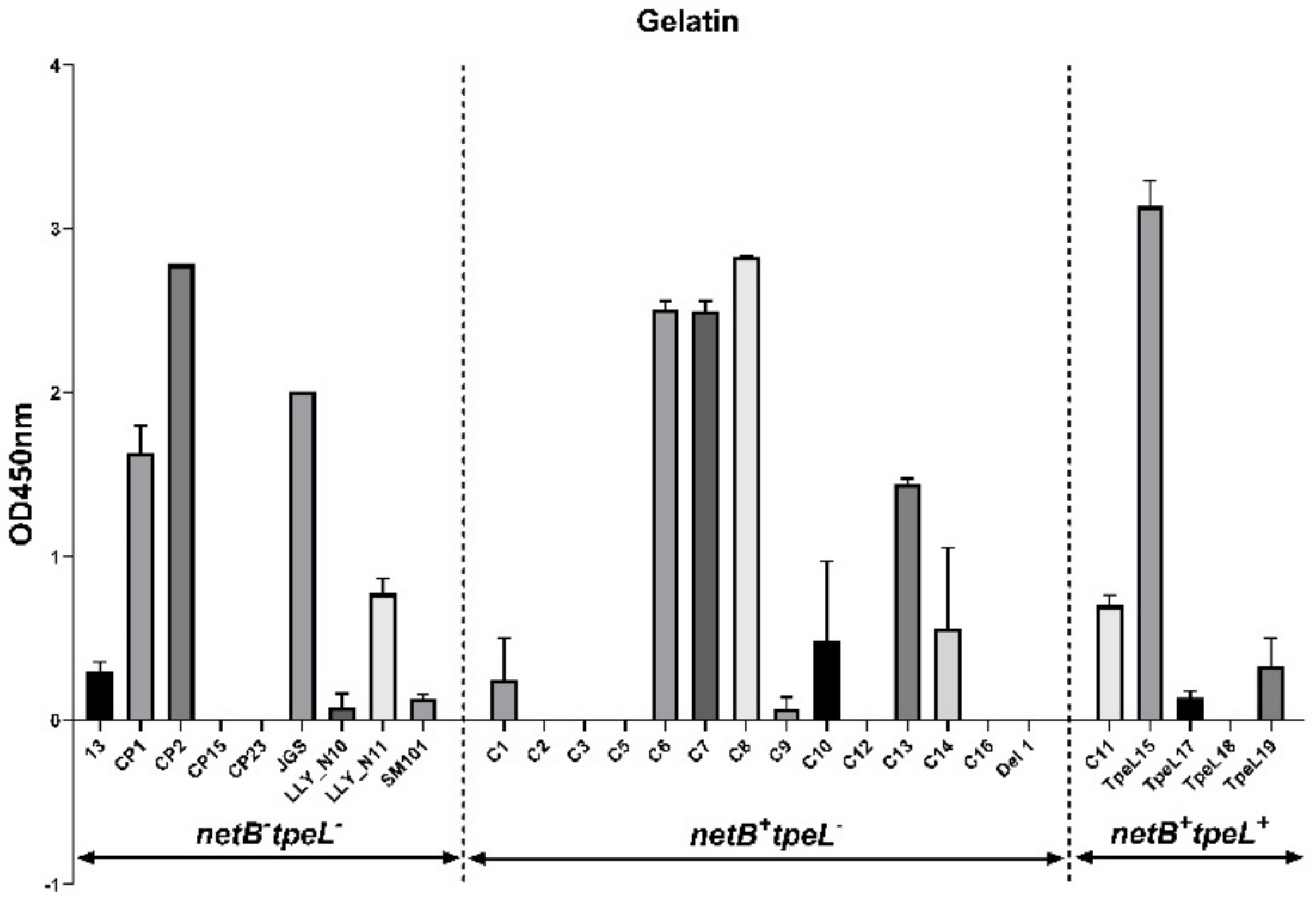
Twenty-eight C. perfringens isolates were tested for cnaA presence by qPCR, among which 15 of them showed higher signals (high copy numbers of cnaA), and the remaining 13 of the isolates were negative (Figure 2), which is correlated well with the regular PCR result (Figure 1 and Table 3). No significant differences were found in the copy numbers of the cnaA gene between netB+tpeL+ and netB+tpeL (p > 0.05). However, significant differences were observed either between netBtpeL and netB+tpeL, or between netBtpeL and netB+tpeL+ (p < 0.001). Generally, there is a positive association between cnaA gene and netB gene expression except for a few samples (C5C8), which agrees with our previously published data on netB and tpeL gene expression [15].

3.3. Adhesion Assay

The 28 C. perfringens isolates were tested for their abilities to bind to collagens, and results show that C. perfringens isolates showed variable levels of binding to collagens. The majority of the virulent isolates were capable of binding well to collagen types I, II, IV, and V, while most of the isolates bound poorly to collagen type III and gelatin (Figure 3). The netBtpeL isolates showed the significantly stronger capabilities of binding to collagen type II than the netB+tpeL (p < 0.05) and the netB+tpeL+ isolates (p < 0.01). Interestingly, out of eight isolates binding potently to collagen type III, five isolates came from netB+tpeL+ isolates (C11, tpeL15, tpeL17-19) (OD562 ≥ 1.0). The netB+tpeL+ isolates demonstrated the significantly superior binding capabilities to collagen type III than either the netBtpeL (p < 0.001) or the netB+tpeL isolates (p < 0.0001). The netB+tpeL+ isolate tpeL15 bound firmly to the collagen types I~V and gelatin.

4. Discussion

C. perfringens is a complex pathogen, causing histotoxic infections, enteritis/enterocolitis, and enterotoxemias with this virulence versatility attributable mostly to its ability to produce various potent toxins [18], which results in the intestinal tissue damage associated with NE, exposure of extracellular matrix molecules (ECMM, such as collagen types I–III and V), and basement membrane or substances in the lamina propria (collagen type IV being the major component) [10]. Other than toxins, other C. perfringens virulence factors and their contribution to NE need to be better investigated. Furthermore, C. perfringens pathogenicity depends upon the colonization to persistently proliferate/survive in vivo, and this colonization process normally consists of nutrient acquisition for growth/survival (for example, by the production of sialidases NanI) and adherence [18]. In addition to sialidases, adhesins play important roles in the C. perfringens colonization stage for the pathogenesis of C. perfringens prior to secreting a panoply of toxins in the tissues.
It has been reported that the levels of virulence of C. perfringens strains are related to their binding abilities to collagen types II, IV, and V [10,12]. In the initial colonization stage, C. perfringens readily adheres to the damaged tissue where many extracellular matrix proteins, including abundant collagens and fibronectin (Fn) proteins, are presented in the damaged tissue [12]. Eimeria pre-exposure, which damages gut tissues and enhances the development of NE, acts as an essential predisposing factor in chickens [3,19].
Interaction between C. perfringens, tissue collagens, and cnaA as a pilus component of C. perfringens contributes to enteritis in chickens and pigs by promoting the adhesion to damaged intestinal tissue [10,12,20]. Inactivation of the pilus genes resulted in the inhibition of pilus production, highly reducing the capability of C perfringens to bind collagen and initiate disease [11,12,21]. It is also implied that fibronectin is a possible extracellular matrix glycoprotein that facilitates host–pathogen contact for C. perfringens to invade and colonize the gut [18,22,23]. In the present study, the majority of C. perfringens isolates came from cases of avian NE, and some of them, including the netBtpeL isolates, were capable of inducing NE when chickens were pre-exposed to coccidiosis. Within the virulent netB+ isolates of C. perfringens, their abilities to adhere to collagen types I, II, IV, and V may be correlated with the presence of a putative collagen adhesin cnaA gene. Therefore, this study demonstrated a direct association between the virulence of C. perfringens strains and their abilities to adhere to immobilized collagens and between the virulence and the cnaA gene copies. Our results were in line with the observations obtained from other researchers [10,12]. Interestingly, most C. perfringens strains did not directly bind well to collagen type III or gelatin, although they bound to collagen types I, II, IV, or V. Possibly, the loss or reduced expression of fibronectin proteins on these isolates may impact the collagen binding ability. It has been reported that C. perfringens may firmly bind to collagens in the presence of fibronectin, therefore enhancing adherence and colonization [18]. This hypothesis merits further investigation. Mutations of three pilin structure subunits (cnaA, fimA, and fimB) have been reported to lead to the loss of filamentous structures; reduce the capabilities of C. perfringens’s binding to collagen types I, II, and IV; and result in attenuation of their pathogenicity in a chicken NE challenge model [21]. Understanding the roles of these virulence-associated factors in NE pathogenesis may help scientists to design optimal strategies to efficiently disrupt the colonization of C. perfringens during the early infection stage. Chickens immunized subcutaneously three times with either CnaA or FimB (another pilin structure subunit) showed significantly reduced NE lesions against pathogenic C. perfringens challenge [24]. In our laboratory, immunization with CnaA alone or in combination with other toxoids/virulence factors conferred partial protection against virulent C. perfringens challenge [25].
The present study investigated the capability of collagen binding and cnaA gene level among the virulent and less virulent clinical C. perfringens isolates and evaluated any association between the capability to bind to collagen types I–V/gelatin and NE pathogenicity in chickens. For simplicity, three C. perfringens groups were assigned: netBtpeL, netB+tpeL, and netB+tpeL+. The netBtpeL group also contained the avirulent isolates (such as N10 and SM101, demonstrated by Eimeria maxima/C. perfringens challenge studies) and clinical NE strains. Our results showed that the avirulent isolates (N10 and SM101, netB) demonstrated a weak binding activity to collagen types III, V, and gelatin compared to the other virulent isolates in the same group or other group. There were variable capabilities of binding to these collagens among different groups. Interestingly, the netBtpeL group had statistically significant capabilities of binding to collagen II than the netB+tpeL and netB+tpeL+ groups. The reason why the netB C. perfringens isolates bind more potently to collagen II than netB+ isolates (netB+tpeL and netB+tpeL+) is not completely understood. The lower binding activities to collagenase type II in C. perfringens type G isolates may be associated with a high frequency of collagenase variant type II gene fragments that encode a truncated collagenase with the missing N-terminal part of the collagenase unit [26]. The absence of the NetB toxin in netB isolates may lead to the upregulation of the production of the collagen II binding protein that compensates for the loss of NetB or less interference with their binding to collagen II. Interestingly, among the eight isolates that showed the potent binding capability to collagen III (OD562 ≥ 1.0), five out of eight isolates came from netB+tpeL+ isolates. All five netB+tpeL+ isolates showed statistically significant potent capabilities of binding to collagen III than the tpeL (netB+tpeL and netBtpeL) groups. This unique characteristic may be related to the presence of the tpeL+ gene. The TpeL protein, a member of the large clostridial toxin (LCT) family [27,28], is reported to promote the disease severity of NE in chickens [3,15], while NetB is a pore-forming toxin that has been shown to disrupt the integrity of cell membranes [4]. The presence of both TpeL and NetB toxins may cause more severe tissue damage and superior binding to collagen type III. The isolate netB+tpeL+ tpeL15 was the outstanding isolate that firmly bound to all collagen types and gelatin. In our previous genomic study [17], netB+tpeL+ isolate tpeL17 shared the same fibronectin-binding protein with other strains (Del1, N11, SM101, Strain 13), and shared the same fimbrial adhesion protein with other virulent isolates (Del1 and N11 isolates). The reason why the netB+tpeL+ C. perfringens isolates potently bound to collagen type III merits further investigation.
Adhesin CnaA protein may play an important role in bacterial interaction with collagens and colonization, and our PCR and qPCR results demonstrated that more copies of cnaA gene were readily detected in a majority of the netB+ virulent isolates (netB+tpeL and netB+ tpeL+), but much less in netBtpeL isolates. The association of higher copies of the cnaA gene with netB+ strains suggested that the product encoded by this cnaA gene may have a synergistic effect with NetB toxin in the NE pathogenesis. Furthermore, the sandwich ELISA assay in another pilot study using CnaA-specific monoclonal antibodies enabled us to detect cnaA gene expression at the protein level in various C. perfringens overnight culture supernatants [29], especially in few clinical virulent isolates, including the well-characterized pathogenic N11 (28.5 ng/mL), Del1 (20.1 ng/mL), and tpeL17 (6.6 ng/mL) isolates. N11, a netBtpeL strain isolated from healthy chicken gut from our previous study, induced necro-hemorrhagic enteritis lesions in combination with Eimeria maxima pre-exposure [16,30,31]. It appears that the concentrations of CnaA proteins in the supernatant of these pathogenic C. perfringens isolates may be associated with the bacterial growth rate (N11 > Del1 > tpeL17) as well, and the generation of the higher levels of toxins (for example, NetB and TpeL secreted by tpeL 17 isolate) may slow down the growth rate, therefore affecting the CnaA production in the supernatant (unpublished data). Further investigations may be warranted to better define the complex association among growth rate, toxin productions, genotypes, and pathogenicity. The collagen adhesin-like proteins, as an important virulence factor also developed by many other pathogenic Gram-positive bacteria to promote bacteria–host interaction [32,33], may be useful targets utilized to control multiple bacterial infections at the critical steps of initiation and persistence.

5. Conclusions

We have demonstrated that clinical isolates of C. perfringens are capable of binding to collagen types I and III-V, compared to avirulent isolates. The netB+tpeL+ isolates potently bound to collagen type III. All the collection isolates carrying the cnaA gene with higher copy numbers were mainly netB+ isolates. The collagen adhesin cnaA gene plays an important role in the adherence of C. perfringens to collagen types during the bacterial colonization stage in poultry. Our results suggest that the collagen binding capability correlates positively with their NE pathogenicity levels, especially for C. perfringens isolates carrying crucial virulence factors and virulence-associate factors such as NetB, CnaA, and TpeL.

Author Contributions

Conceptualization: C.L.; Data curation: Z.S., M.L., Y.L., D.G., B.Y. and C.L.; Formal analysis: Z.S., M.L., X.Y., Y.L., D.G., X.Y. and C.L.; Software: Z.S. and M.L.; Funding acquisition: H.L. and C.L.; Supervised the project, Project administration: C.L.; Writing—original draft: Z.S., M.L. and C.L.; Writing—review and editing: Z.S., M.L., H.L. and C.L. All authors have read and agreed to the published version of the manuscript.

Funding

This research was funded by ARS Animal Health project number 8042-32000-115-00D.

Institutional Review Board Statement

The animal study protocols were approved by the local Institutional Animal Care and Use Committee at Beltsville Agricultural Research Center (Animal Use Protocol No 17-027).

Informed Consent Statement

Not applicable.

Data Availability Statement

The data presented in this study are available within the article.

Acknowledgments

This work was supported by the funds from Agricultural Research Service, USDA. The sponsors had no role in the writing process of the article. Some isolates of Clostridium strains used in this study were obtained from the Poultry Microbiological Safety Research Unit, ARS, USDA, Athens, GA.

Conflicts of Interest

There is no conflict of interest to the scientific integrity of this article.

Abbreviations

CMG = chopped meat glucose; CP = Clostridium perfringens; NE = necrotic enteritis; netB = necrotic enteritis toxin B-like gene; tpeL = Clostridium perfringens lethal toxin gene; cnaA = adhesin protein can gene; OD = optical density; qPCR = quantitative polymerase chain reaction.

References

  1. Rood, J.I.; Adams, V.; Lacey, J.; Lyras, D.; McClane, B.A.; Melville, S.B.; Moore, R.J.; Popoff, M.R.; Sarker, M.R.; Songer, J.G.; et al. Expansion of the Clostridium perfringens toxin-based typing scheme. Anaerobe 2018, 53, 5–10. [Google Scholar] [CrossRef] [PubMed]
  2. Tsiouris, V. Poultry management: A useful tool for the control of necrotic enteritis in poultry. Avian Pathol. 2016, 45, 323–325. [Google Scholar] [CrossRef]
  3. Prescott, J.F.; Smyth, J.A.; Shojadoost, B.; Vince, A. Experimental reproduction of necrotic enteritis in chickens: A review. Avian Pathol. 2016, 45, 317–322. [Google Scholar] [CrossRef] [PubMed]
  4. Keyburn, A.L.; Yan, X.X.; Bannam, T.L.; Van Immerseel, F.; Rood, J.I.; Moore, R.J. Association between avian necrotic enteritis and Clostridium perfringens strains expressing NetB toxin. Vet. Res. 2010, 41, 21. [Google Scholar] [CrossRef]
  5. Keyburn, A.L.; Boyce, J.D.; Vaz, P.; Bannam, T.L.; Ford, M.E.; Parker, D.; Di Rubbo, A.; Rood, J.I.; Moore, R.J. NetB, a new toxin that is associated with avian necrotic enteritis caused by Clostridium perfringens. PLoS Pathog. 2008, 4, e26. [Google Scholar] [CrossRef] [PubMed]
  6. Rood, J.I.; Keyburn, A.L.; Moore, R.J. NetB and necrotic enteritis: The hole movable story. Avian Pathol. 2016, 45, 295–301. [Google Scholar] [CrossRef]
  7. Shojadoost, B.; Vince, A.R.; Prescott, J.F. The successful experimental induction of necrotic enteritis in chickens by Clostridium perfringens: A critical review. Vet. Res. 2012, 43, 74. [Google Scholar] [CrossRef]
  8. Shi, H.N.; Walker, A. Bacterial colonization and the development of intestinal defences. Can. J. Gastroenterol. 2004, 18, 493–500. [Google Scholar] [CrossRef]
  9. Ribet, D.; Cossart, P. How bacterial pathogens colonize their hosts and invade deeper tissues. Microbes Infect. 2015, 17, 173–183. [Google Scholar] [CrossRef]
  10. Martin, T.G.; Smyth, J.A. The ability of disease and non-disease producing strains of Clostridium perfringens from chickens to adhere to extracellular matrix molecules and Caco-2 cells. Anaerobe 2010, 16, 533–539. [Google Scholar] [CrossRef]
  11. Wade, B.; Keyburn, A.L.; Haring, V.; Ford, M.; Rood, J.I.; Moore, R.J. The adherent abilities of Clostridium perfringens strains are critical for the pathogenesis of avian necrotic enteritis. Vet. Microbiol. 2016, 197, 53–61. [Google Scholar] [CrossRef] [PubMed]
  12. Wade, B.; Keyburn, A.L.; Seemann, T.; Rood, J.I.; Moore, R.J. Binding of Clostridium perfringens to collagen correlates with the ability to cause necrotic enteritis in chickens. Vet. Microbiol. 2015, 180, 299–303. [Google Scholar] [CrossRef]
  13. Lepp, D.; Gong, J.; Songer, J.G.; Boerlin, P.; Parreira, V.R.; Prescott, J.F. Identification of accessory genome regions in poultry Clostridium perfringens isolates carrying the netB plasmid. J. Bacteriol. 2013, 195, 1152–1166. [Google Scholar] [CrossRef] [PubMed]
  14. Li, C.; Yan, X.; Lillehoj, H.S. Complete genome sequences of Clostridium perfringens Del1 strain isolated from chickens affected by necrotic enteritis. Gut Pathog. 2017, 9, 69. [Google Scholar] [CrossRef] [PubMed]
  15. Gu, C.; Lillehoj, H.S.; Sun, Z.; Lee, Y.; Zhao, H.; Xianyu, Z.; Yan, X.; Wang, Y.; Lin, S.; Liu, L.; et al. Characterization of virulent netB(+)/tpeL(+) Clostridium perfringens strains from necrotic enteritis-affected broiler chicken farms. Avian Dis. 2019, 63, 461–467. [Google Scholar] [CrossRef]
  16. Li, C.; Lillehoj, H.S.; Gadde, U.D.; Ritter, D.; Oh, S. Characterization of Clostridium perfringens strains isolated from healthy and necrotic enteritis-afflicted broiler chickens. Avian Dis. 2017, 61, 178–185. [Google Scholar] [CrossRef]
  17. Liu, L.; Yan, X.; Lillehoj, H.; Sun, Z.; Zhao, H.; Xianyu, Z.; Lee, Y.; Melville, S.; Gu, C.; Wang, Y.; et al. Comparison of the pathogenicity of five Clostridium perfringens isolates using an Eimeria maxima coinfection necrotic enteritis disease model in commercial broiler chickens. Avian Dis. 2020, 64, 386–392. [Google Scholar] [CrossRef]
  18. Mehdizadeh Gohari, I.; Navarro, M.A.; Li, J.; Shrestha, A.; Uzal, F.; McClane, B.A. Pathogenicity and virulence of Clostridium perfringens. Virulence 2021, 12, 723–753. [Google Scholar] [CrossRef]
  19. Van Immerseel, F.; Lyhs, U.; Pedersen, K.; Prescott, J.F. Recent breakthroughs have unveiled the many knowledge gaps in Clostridium perfringens-associated necrotic enteritis in chickens: The first International Conference on Necrotic Enteritis in Poultry. Avian Pathol. 2016, 45, 269–270. [Google Scholar] [CrossRef]
  20. Jost, B.H.; Billington, S.J.; Trinh, H.T.; Songer, J.G. Association of genes encoding beta2 toxin and a collagen binding protein in Clostridium perfringens isolates of porcine origin. Vet. Microbiol. 2006, 115, 173–182. [Google Scholar] [CrossRef]
  21. Lepp, D.; Zhou, Y.; Ojha, S.; Mehdizadeh Gohari, I.; Carere, J.; Yang, C.; Prescott, J.F.; Gong, J. Clostridium perfringens produces an adhesive pilus required for the pathogenesis of necrotic enteritis in poultry. J. Bacteriol. 2021, 203, e00578-20. [Google Scholar] [CrossRef]
  22. Hitsumoto, Y.; Morita, N.; Yamazoe, R.; Tagomori, M.; Yamasaki, T.; Katayama, S. Adhesive properties of Clostridium perfringens to extracellular matrix proteins collagens and fibronectin. Anaerobe 2014, 25, 67–71. [Google Scholar] [CrossRef]
  23. Katayama, S.; Nozu, N.; Okuda, M.; Hirota, S.; Yamasaki, T.; Hitsumoto, Y. Characterization of two putative fibronectin-binding proteins of Clostridium perfringens. Anaerobe 2009, 15, 155–159. [Google Scholar] [CrossRef]
  24. Lepp, D.; Ojha, S.; Mehdizadeh Gohari, I.; Chakravarty, B.; Prescott, J.F.; Gong, J. Immunization with subunits of a novel pilus produced by virulent Clostridium perfringens strains confers partial protection against necrotic enteritis in chickens. Vet. Microbiol. 2019, 230, 7–13. [Google Scholar] [CrossRef] [PubMed]
  25. Yuan, B.; Sun, Z.; Lu, M.; Lillehoj, H.; Lee, Y.; Liu, L.; Yan, X.; Yang, D.A.; Li, C. Immunization with pooled antigens for Clostridium perfringens conferred partial protection against experimental necrotic enteritis in broiler chickens. Vaccines 2022, 10, 979. [Google Scholar] [CrossRef]
  26. Van Damme, L.; Cox, N.; Callens, C.; Dargatz, M.; Flügel, M.; Hark, S.; Thiemann, F.; Pelzer, S.; Haesebrouck, F.; Ducatelle, R.; et al. Protein truncating avriants of colA in Clostridium perfringens Type G strains. Front. Cell. Infect. Microbiol. 2021, 11, 645248. [Google Scholar] [CrossRef] [PubMed]
  27. Amimoto, K.; Noro, T.; Oishi, E.; Shimizu, M. A novel toxin homologous to large clostridial cytotoxins found in culture supernatant of Clostridium perfringens type C. Microbiology 2007, 153, 1198–1206. [Google Scholar] [CrossRef] [PubMed]
  28. Chen, J.; McClane, B.A. Characterization of Clostridium perfringens TpeL toxin gene carriage, production, cytotoxic contributions, and trypsin sensitivity. Infect. Immun. 2015, 83, 2369–2381. [Google Scholar] [CrossRef]
  29. Goo, D.; Park, I.; Nam, H.; Lee, Y.; Sawall, J.; Smith, A.; Rehberger, T.; Li, C.; Lillehoj, H. Collagen adhesin protein and necrotic enteritis B-like toxin as biomarkers for early diagnosis of necrotic enteritis in commercial broiler chickens. Poult. Sci. 2023, 102, 102647. [Google Scholar] [CrossRef]
  30. Li, C.; Yan, X.; Lillehoj, H.S. Complete egnome sequence of Clostridium perfringens LLY_N11, a necrotic enteritis-inducing strain isolated from a healthy chicken intestine. Genome Announc. 2017, 5, e01225-17. [Google Scholar] [CrossRef]
  31. Goossens, E.; Dierick, E.; Ducatelle, R.; Van Immerseel, F. Spotlight on avian pathology: Untangling contradictory disease descriptions of necrotic enteritis and necro-haemorrhagic enteritis in broilers. Avian Pathol. 2020, 49, 423–427. [Google Scholar] [CrossRef] [PubMed]
  32. Madani, A.; Garakani, K.; Mofrad, M.R. Molecular mechanics of Staphylococcus aureus adhesin, CNA, and the inhibition of bacterial adhesion by stretching collagen. PLoS ONE 2017, 12, e0179601. [Google Scholar] [CrossRef] [PubMed]
  33. De Gaetano, G.V.; Coppolino, F.; Lentini, G.; Famà, A.; Cullotta, C.; Raffaele, I.; Motta, C.; Teti, G.; Speziale, P.; Pietrocola, G.; et al. Streptococcus pneumoniae binds collagens and C1q via the SSURE repeats of the PfbB adhesin. Mol. Microbiol. 2022, 117, 1479–1492. [Google Scholar] [CrossRef] [PubMed]
Figure 1. Detection of cnaA gene from C. perfringens isolates by PCR. Extracted genomic DNAs were applied as templates, and a pair of cnaA primers were used to amplify the cnaA gene. The PCR products were run on 1% agarose gels and imaged with the Chemi-Touch image analysis system. Neg = negative buffer control.
Pathogens 12 00778 g001
Figure 2. Detection of cnaA gene copies from C. perfringens isolates shown as the relative gene copies by qPCR. The gDNA was extracted, and the relative gene copy numbers of cnaA in real-time PCR were estimated by qPCR and normalized based on relative 16S rDNA levels. The data were analyzed by one-way ANOVA, and means were calculated using Duncan’s multiple range test. Each bar represents the mean ± SEM (n = 3). *** represents p ≤ 0.001.
Pathogens 12 00778 g002
Figure 3. Adhesion assay of C. perfringens isolates to collagen types I, II, III, IV, and V, and gelatin. Collagens (types I, II, III, IV, and V) and gelatin were coated on Nunclon Delta-Treated, Flat-Bottom Microplates overnight at 4 °C and blocked using PBS plus 0.5% (w/v) bovine serum albumin. Bacterial cells were added to wells and incubated at room temperature for 2 h with agitation, followed by rinses. C. perfringens were stained with 0.5% (w/v) crystal violet for 5 min. After rinsing, solution of ethanol:acetone was added to each well to destain adherent C. perfringens, and the OD value at 562 nm was measured. All assays were repeated in triplicates. *, **, ***, **** marks represent statistically p ≤ 0.05, 0.01, 0.001, and 0.0001, respectively.
Pathogens 12 00778 g003aPathogens 12 00778 g003bPathogens 12 00778 g003cPathogens 12 00778 g003d
Table 2. Primer sequences and target genes used in the qRT–PCR analysis.
Target GenePrimer Sequences (5’-3’)
16S rRNAF-GGGGGTTTCAACACCTCC
R-GCAAGGGATATCAAGTGT
cnaAF-GGTGGATGGGCAACATTTAC
R-CCTTGCTTGGATTCACCAGT
Table 3. Results of comparison of C. perfringens isolates for cnaA gene detection by PCR and qPCR.
GENOTYPEnetBtpeLnetBtpeL+
ISOLATE ID13CP1CP10CP15CP23JGSN10N11SM101C11tpeL15tpeL17tpeL18tpeL19
cnaA PCR+++++++++++++++++++
cnaA qPCR+++++++++++++++
GENOTYPEnetB+tpeL+
ISOLATE IDC1C2C3C5C6C7C8C9CP10CP12CP13CP14CP16Del 1
cnaA PCR++++++++++++++++++++++++
cnaA qPCR+++++++++++++++++++++++++++++++/−
Note: +++ (strong expression), ++ (intermediate expression), + (weak expression), − (very weak or no expression).
Disclaimer/Publisher’s Note: The statements, opinions and data contained in all publications are solely those of the individual author(s) and contributor(s) and not of MDPI and/or the editor(s). MDPI and/or the editor(s) disclaim responsibility for any injury to people or property resulting from any ideas, methods, instructions or products referred to in the content.

