Article

Production of Escovopsis weberi (Ascomycota: Hypocreales) Mycelial Pellets and Their Effects on Leaf-Cutting Ant Fungal Gardens

by
Thais Berçot Pontes Teodoro
,
Aline Teixeira Carolino
,
Raymyson Rhuryo de Sousa Queiroz
,
Patrícia Batista de Oliveira
,
Denise Dolores Oliveira Moreira
,
Gerson Adriano Silva
and
Richard Ian Samuels
*
Laboratório de Entomologia e Fitopatologia, Centro de Ciências e Tecnologias Agropecuárias, Universidade Estadual do Norte Fluminense Darcy Ribeiro, Campos dos Goytacazes 28013-602, RJ, Brazil
*
Author to whom correspondence should be addressed.
Pathogens 2023, 12(2), 330; https://doi.org/10.3390/pathogens12020330
Submission received: 19 December 2022 / Revised: 7 February 2023 / Accepted: 10 February 2023 / Published: 15 February 2023
(This article belongs to the Special Issue Entomopathogenic Fungi and Nematodes in Modern Agriculture)

Abstract

:
The maintenance of the symbiosis between leaf-cutting ants and their mutualistic fungus Leucoagaricus gongylophorus Singer (Moller) is vital for the survival of both species. The specialist fungal parasite Escovopsis weberi Muchovej & Della Lucia is a threat to this symbiosis, causing severe damage to the fungal garden. Mycelial pellets are resistant fungal structures that can be produced under laboratory conditions. These structures were studied for use in biological pest control, but the production of mycelial pellets has not previously been documented in Escovopsis. One of the aims of this study was to induce Escovopsis weberi to produce mycelial pellets and investigate the potential of these pellets for the control of leaf-cutting ants. We compared the pathogenicity of Escovopsis weberi mycelial pellets and conidia against mini-colonies of Acromyrmex subterraneus subterraneus Forel when applied in the form of baits. Worker ants were able to distinguish mycelial pellets from conidia, as baits with mycelial pellets were more attractive to workers than those with conidia, causing a greater negative impact on colony health. All types of baits containing Escovopsis weberi influenced the foraging activity but only treatments with viable fungal propagules resulted in an increase in the quantity of waste material, with a significant negative impact on the fungal garden biomass. The results provided novel information regarding Escovopsis recognition by worker ants and differences between conidia and mycelial pellet dynamics in leaf-cutting ant colonies, with new perspectives for the biological control of these important pests.

1. Introduction

Leaf-cutting ants (Attini: Formicidae) are amongst the most important agricultural and forestry pests throughout the Neotropics [1,2]. They cause severe damage due to the large amount of fresh leaves they cut and carry to their nests, which they use to cultivate the symbiotic fungus Leucoagaricus gongylophorus (Möller) Singer (Agaricales: Basidiomycota) [3,4,5]. In this symbiosis, the ants provide the fungus shelter, fresh leaves, and protection against parasites; in exchange, L. gongylophorus provides the ants with easily assimilable nutrients via specialized hyphal structures called gongylidia, which are the main food resource for the queens, brood, and workers [6,7]. Additionally, gongylidia provide the ants with enzymes that are useful for degrading plant material [7,8].
Brazil is one of the most important wood producers in the world. Wood production represents the fourth largest crop when considered by area, consisting of mainly fast-growing monocultures, such as eucalyptus, pine, acacia, and teak [9,10]. However, leaf-cutting ants can cause serious damage to these plantations, especially at the sapling stage [11]. The defoliation caused by ants significantly reduces wood production, causing a decrease in tree growth and also making the trees more vulnerable to attack by other pests [12,13].
Despite being ecologically important in natural environments, where leaf-cutting ants improve soil penetrability, nutrient cycling, and seed dispersal [14,15,16,17], these insects are a major concern in agricultural and forestry systems. The genus Acromyrmex includes 35 species; however, only 6 species are important as pests, i.e., A. balzani, A. octospinosus, A. rugosus, A. subterraneus brunneus, A. subterraneus molestans, and A. subterraneus subterraneus [18,19]. Acromyrmex subterraneus subterraneus, which was studied here, is mainly found in Brazil, Argentina, and Paraguay.
Synthetic chemical insecticide baits continue to be the most common control method used against these ants [20]. Currently, sulfluramid, which is a persistent organic pollutant, is the most efficient insecticide for controlling leaf-cutting ants and it is widely used in Brazil [13], even though the Stockholm Convention banned its use. However, chemical control methods cause negative impacts on human health and the environment [21]. This insecticide was found to contaminate soil, eucalyptus leaves, and water (underground, river, and coastal waters), which is of great concern, mainly in relation to human exposure to this compound [22,23]. In addition, the use of chemical insecticides promotes pest resistance and negatively affects non-target organisms [24,25,26,27]. The search for alternative control methods that are not detrimental to the environment or human health will benefit agriculture, animal health, and the ecosystem.
The use of biological agents could be an environmentally safe alternative for the control of leaf-cutting ants [2,28]. Escovopsis weberi (Ascomycota: Hypocreales) is a virulent specialist fungus that parasitizes L. gongylophorus and has the potential to be employed in biocontrol programs [29,30,31]. In this parasitism, L. gongylophorus is degraded by direct hyphal contact between the two fungi, aided by hook-like protuberances that consume nutrients from L. gongylophorus living cells, leading to host death and consequent colony collapse if the worker ants cannot control this parasite [31,32,33,34].
Despite the capacity of Escovopsis to rapidly over-run L. gongylophorus fungus gardens, the ants have developed strategies to prevent or deal with eventual infections. The ants possess metapleural glands that produce antimicrobial secretions [35,36], and they constantly weed and groom the gardens to remove any contaminants [37,38]. The presence of Pseudonocardia, which is a symbiotic bacterium, on the bodies of some species of worker ants produces antibiotics that reduce the chances of Escovopsis infections [30,39,40]. Even so, E. weberi is still frequently found attacking leaf-cutting ant colonies [41].
A diverse range of fungi can produce structures called sclerotia to survive challenging conditions [42]. Sclerotia are dense hyphal aggregations that are highly resistant to adverse conditions, such as the absence of the host or desiccation. These structures are viable for a longer period compared with conidia and can germinate whenever the environmental conditions become favorable [43]. In nature, sclerotia are commonly found, especially in the soil [44]. However, when cultured in liquid media under constant agitation, it is possible to induce certain fungi to produce structures similar to sclerotia. In many cases, these so-called microsclerotia [45,46,47] are probably better described as mycelial pellets (hereafter referred to as MPs).
Specialist mycoparasites, such as Escovopsis, have not previously been documented to produce such structures [48], and the application of Escovopsis MPs could be an interesting and novel approach to control leaf-cutting ants. Interestingly, Escovopsis mycelium, unlike conidia, are not susceptible to ants’ metapleural gland secretions, which are responsible for antimicrobial activity against a variety of pathogens [35]. MPs store large amounts of nutrients in their structures [43], which may promote faster development of the parasite when attacking leaf-cutting ant fungus gardens, and have an increased shelf life [44,49].
The aim of this study was to develop an alternative strategy for leaf-cutting ant control. The four main objectives were as follows: (1) to induce E. weberi to produce MPs; (2) to assess the reproductive capacity of MPs; (3) to assess the effect of E. weberi MP baits against A. subterraneus subterraneus mini-colonies when compared with baits containing E. weberi conidia; and (4) to study the acceptance of both propagules, viable or dead, when offered to the ants in the form of baits. The results showed the potential of E. weberi in the form of MPs for leaf-cutting ant control with specific advantages over the application of conidia.

2. Materials and Methods

2.1. Field Collection of Colonies, Maintenance, and Mini-Colony Establishment

Acromyrmex subterraneus subterraneus colonies were collected in Bom Jardim, Rio de Janeiro State, Brazil (22°13′16.37″ S, 42°15′14.74″ W; 709 m a.s.l.). This region is a tropical montane forest, located near urban and agricultural areas. We collected whole colonies after gently excavating the soil around each colony. Fungus gardens were transferred to clean containers with as many ants as possible, including the queens. The colonies were maintained in the Myrmecology Unit at the Universidade Estadual do Norte Fluminense Darcy Ribeiro (UENF). Acromyrmex species identification followed the keys provided by Fowler et al. [50]. The colonies were offered fresh Acalypha wilkesiana leaves on a daily basis and kept at a controlled room temperature (26 °C) and 70% relative humidity. For the bioassays, queenless mini-colonies were established randomly from the collected colonies. Each mini-colony was composed of ants of all 4 castes in a 350 mL of fungus garden inside a clear plastic pot that was placed in a white plastic tray (40 cm × 30 cm × 10 cm) that was maintained in the dark at room temperature and 70% RH.

2.2. Escovopsis Isolation, Identification, and Conidial Production

Escovopsis weberi (AT-02; NCBI accession numbers: OQ345555 and OQ345556) was isolated from an Atta sexdens rubropilosa colony collected in a eucalyptus plantation in São Francisco do Itabapoana (21°26′57″ S, 41°11′51″ W; 50 m a.s.l.), Rio de Janeiro State, Brazil, and maintained in the Entomology and Phytopathology Department (UENF) Pathogenic Fungi collection. In order to establish a pure culture of this isolate, single spores were cultured as described by Choi et al. [51].
After single spore culturing, AT-02 was inoculated on potato dextrose agar (PDA) culture medium in Petri dishes (9 cm diameter) and maintained in the dark at 27 °C and 70% RH. After 5 days, spores were harvested with the aid of a spatula and suspended in Tween 80 (0.03% in sterile distilled water). Conidial concentrations were estimated using a Neubauer hemocytometer and a suspension of 1 × 10⁷ conidia mL−1 was used after serial dilution. This concentration was either used for MP production or for making baits containing conidia that were used in the subsequent bioassays.

2.3. Mycelial Pellet Production in Liquid Culture

Escovopsis weberi AT-02 MP was grown in liquid culture following a protocol developed by Carolino et al. [52]. The medium consisted of chickpea flour (3%), yeast extract (4%), and dextrose (4%) in distilled water. The procedure was as follows: First, the chickpea flour and distilled water were autoclaved for 20 min at 121 °C and then filtered using Miracloth (Sigma-Aldrich, Barueri, São Paulo, Brazil). Then, yeast extract and dextrose were added to this infusion. The culture medium was then autoclaved again.
Forty-nine milliliters of sterile liquid culture medium were added to 250 mL sterile Erlenmeyer flasks with 1 mL of conidial suspension (1 × 10⁷ conidia mL−1). The flasks were placed in an orbital shaker (Solab®, Piracicaba, São Paulo, Brazil, model SL-223/F) in the dark at 27 °C and 230 rpm for 4 days. At 24 h, 48 h, 72 h, and 96 h post-inoculation, 1 mL aliquots were taken from the culture medium to evaluate MP development and morphology. After 4 days, MPs were washed 5 times with sterile distilled water and centrifuged at 3000 rpm for 5 min (Nova Técnica®, Piracicaba, São Paulo, Brazil, model NT 810). Fresh MPs were stored at 4 °C. To determine the MP diameter, measurements were taken (20 repetitions) using a Nikon Eclipse 80i microscope with NIS Elements v. 3.22 software.

2.4. Biomass Production

To assess the mean MP dry weight of stock suspensions, 1 mL of fresh biomass was taken from storage (4 °C), placed in Petri dishes, and dried in an incubator (Nova Ética®, model 411d, Vargem Grande Paulista, São Paulo, Brazil) at 25 °C for 2 days. This assay was repeated 6 times. The dry weight was calculated in mg mL−1.
The MP reproductive capacity was then evaluated monthly (10 repetitions for each month) for one year by plating out 500 µL of MP biomass stock onto the potato dextrose agar culture medium in Petri dishes, spread uniformly with the aid of a Drigalski spatula, and incubated at 27 °C. The conidia produced were harvested with the aid of a spatula, and the plates were rinsed with 10 mL sterile 0.03% Tween 80 (v/v) to remove all remaining conidia from the plates. The conidial concentration was determined using a hemocytometer.

2.5. Bait Production

Baits were produced using powdered orange peel as a base. Oranges were peeled and the peel was dried in a forced air-drying chamber (Nova Ética® model 411d) at 28 °C for 48 h and then ground using an electric blender. A stainless-steel sieve with a 1 mm aperture was used to establish a homogenous powder. Baits were composed of powdered orange peel (90%), carboxymethylcellulose (5%), and soy oil (5%). All material was autoclaved for 20 min at 121 °C. After sterilization, 500 µL of fresh MPs in 10 mL sterile 0.03% Tween 80 or a 10 mL suspension of 1 × 10⁷ conidia mL−1 in 0.03% Tween 80 was added to each bait portion to be offered to the mini-colonies. After blending all components, bait pellets were produced with the aid of a sterile disposable syringe and then dried for 4 h at 27 °C in an air-drying chamber to remove all remaining water. Control baits were produced likewise, but without fungi.

2.6. Mini-Colony Bioassays

Baits were offered to mini-colonies to assess the effect of exposure of the fungus gardens to MPs in comparison to conidia and to verify the influence of viable and dead propagules on bait acceptance by worker ants. Five treatments were established, with five repetitions each: (1) viable MPs, (2) viable conidia, (3) dead MPs, (4) dead conidia, and (5) controls. In treatments 3 and 4, the MPs and conidia were autoclaved at 121 °C for 20 min prior to the bait production. The control treatment consisted of sterile distilled water + Tween 80 (0.03%).
During the bioassay period, fresh A. wilkesiana leaves were offered daily for 7 days to mini-colonies, except on the 8th day, when 3 g of bait pellets were placed in the foraging area of each mini-colony, and during that time, no leaves were offered to the ants. Twenty-four hours later (day 9), all remaining bait was removed and quantified in order to establish the acceptance rate. After the baits had been offered, the bioassays continued for the next 15 days with fresh leaves offered every day. The foraging activity was recorded daily as the fresh leaf area (cm2) cut by workers, established from the difference between the initial leaf area and the remaining leaf area on the following day, which was estimated using a CI-202 portable leaf area meter (CID Bio-Science Inc., Camas, WA, USA).
The waste pile was removed daily from the plastic trays with the aid of a spatula to estimate the waste disposal rate. For the waste disposal analysis, bioassays were divided into 3 periods: from day 1 to day 7 (time 1), from day 8 to day 15 (time 2), and from day 16 to day 23 (time 3). The fungus garden weight was determined every 3 days. To do this, the plastic pots containing the fungus gardens were temporarily sealed to prevent workers from escaping and weighed with the aid of an analytical balance before returning the pots to the trays.

2.7. Statistical Analyses

The experimental design was completely randomized. Experimental units were mini-colonies that were randomly established from field colonies maintained under laboratory conditions. The variables analyzed were the (1) MP reproductive capacity, (2) bait acceptance rate, (3) foraging activity, (4) waste disposal, and (5) fungus garden weight. The treatments established were (1) viable MPs, (2) viable conidia, (3) dead MP, (4) dead conidia, and (5) control baits. For variable 1, ten repetitions (120 experimental samples) were performed and five repetitions of variables 2, 3, 4, and 5, with a total of 20 experimental samples each. A normality test (Shapiro–Wilk) was used to evaluate all variables.
To compare the waste disposal rate (g) between periods and between treatments, values for the mean waste disposal were subjected to a two-way ANOVA followed by Tukey’s post hoc test (α = 0.05). To compare the mean bait acceptance rate, fungus garden weight, and foraging activity, data were subjected to a one-way ANOVA followed by Tukey’s post hoc test (α = 0.05). The MP reproductive capacity was subjected to Kruskal–Wallis analysis, followed by Tukey’s post hoc test (α = 0.05), as it did not pass the normality test. All statistical analyses were carried out using Sigma-Plot (v. 12.5) software.

3. Results

3.1. MP Production in Liquid Culture

After four days, E. weberi conidia (Figure 1A) that had been inoculated in liquid medium produced MPs with variable sizes (57.47 to 685.86 µm in diameter), with a mean value of 280.82 µm (SEM ± 35.69). Hyphal development could be observed 48 h after conidial inoculation (Figure 1B,C). The liquid culture with chickpea flour, yeast extract, and dextrose induced the production of MPs with a compact structure (Figure 1D), and no melanization was observed at any time. Each milliliter of MP suspension had a mean dry weight of 0.411 mg (SEM ± 0.05). The MPs had a high capacity to produce conidia. Productivity assays showed that for each 500 µL of fresh MP biomass placed in Petri dishes and incubated for 5 days, 1 × 10⁹ conidia mL−1 (SEM ± 8 × 10⁷) was produced.
The viability of the MPs was evaluated monthly and statistical differences were observed over time (H11 = 64.995, p < 0.001) (Table 1). However, a statistical difference was only observed after six months. Conidia production remained stable during the first five months, with a range of 1.4 × 10⁹ to 1.1 × 10⁹ conidia mL−1 (Table 1). However, during months 10, 11, and 12 of the bioassay, the productivity dropped to 1.3 × 10⁸, 3.3 × 10⁸, and 4.6 × 10⁸ conidia mL−1, respectively.

3.2. Mini-Colony Bioassays

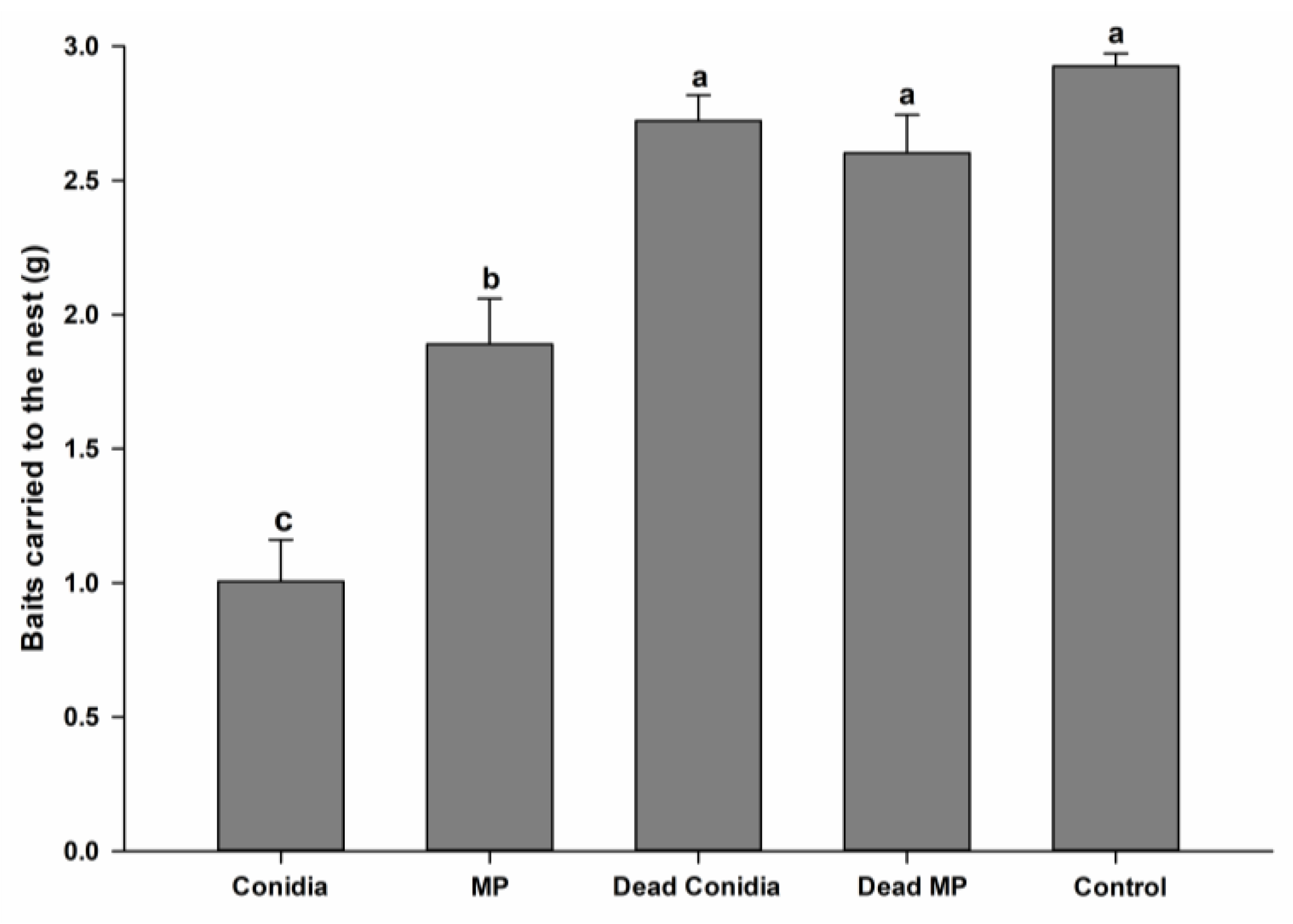
The baits containing E. weberi offered to A. subterraneus subterraneus mini-colonies on the eighth day of the bioassay showed different acceptance patterns between the treatments. Baits containing viable conidia were the least accepted, with a mean of 1.006 g carried to the nest (F4,20 = 36.964, p < 0.001, normality test p = 0.708) (Figure 2). Viable MP baits were more acceptable to worker ants (1.890 g carried), and the weight of the baits carried to the nest was statistically different (p < 0.001) from that of viable conidia baits carried by the ants. Dead conidia (2.722 g), dead MP (2.602 g), and control (2.926 g) baits were highly acceptable and readily carried to the nest, with no statistical differences observed between these treatments (Figure 2).

3.3. Leaf Area Cut following Bait Exposure

Daily foraging activity (leaf area cut) showed slight differences between treatments that contained fungi (F4,20 = 14.622, p < 0.001, normality test p = 0.174) (Figure 3). The leaf areas cut following exposure to baits with viable MP were significantly different from those of mini-colonies exposed to dead conidia, but the viable conidia and dead MP treatments were not statistically different. Control treatments differed significantly from all other treatments (p < 0.001), with the highest foraging activity observed here (Figure 3).

3.4. Waste Disposal

Before offering the baits (time 1), all treatments presented similar amounts of waste disposed of in the rubbish tip (F4,60 = 57.467, p < 0.001, normality test p = 0.141) (Table 2). After offering dead conidia, dead MP, and control baits (times 2 and 3), the waste disposal quantities remained similar to those prior to offering baits (time 1). However, for viable conidia and viable MP treatments, there were significant increases in the waste disposal rates during time 2 and time 3 (F2,60 = 17.302, p < 0.001; Table 2).

3.5. Fungal Garden Biomass

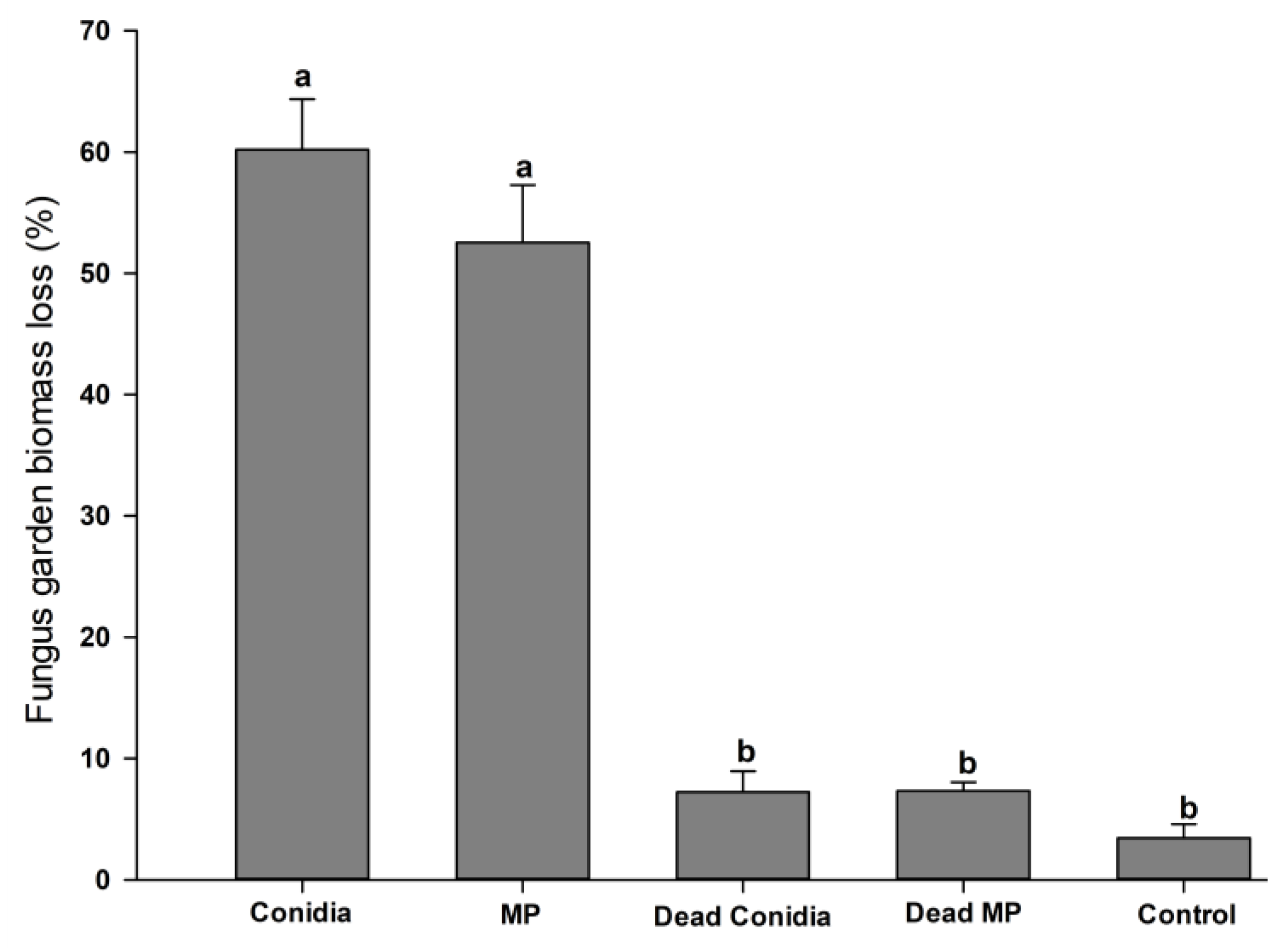
Loss in fungal garden biomass could also be observed for colonies treated with viable conidia or viable MPs. From day one until the end of the bioassay, viable-conidia- and viable-MP-treated colonies lost 60.2% and 52.53% of their fungal garden biomass, respectively (F4,20 = 86.615, p < 0.001, normality test p = 0.479) (Figure 4). Conversely, loss of fungal garden biomass when colonies were exposed to dead conidia, dead MP, or control treatments was very low and did not present statistical differences when comparing these three groups. The biomass losses were 7.23%, 7.33%, and 3.44% (p > 0.005) in these three treatments, respectively (Figure 4).

4. Discussion

Here, it is reported for the first time that E. weberi, when cultured in a liquid medium, can produce hyphal aggregations regarded to be mycelial pellets (MPs). Under laboratory conditions, it is possible to induce certain species of fungi to produce these types of propagules [53,54,55]. The medium used here was originally developed to stimulate the production of blastospores by Metarhizium anisopliae [52]. However, in the current study, the protocol was adapted to induce the production of MPs by E. weberi.
Escovopsis weberi grown in a liquid medium produced MPs of variable sizes, similar to those seen in previous studies of entomopathogenic fungi [45,47,56]. Escovopsis weberi MP presented faster germination than conidia when plated onto a solid culture medium (unpublished data). It is normal for MPs to germinate rapidly in favorable conditions due to the high numbers of mitochondria and high levels of polysaccharides and lipids in the cytoplasm of the hyphal cells [57]. As observed in this study, when using a PDA culture medium, the MPs germinated and formed conidiphores, which subsequently resulted in high concentrations of conidia. The MPs maintained a high level of conidial productivity over a 12-month storage period, with only the last three months presenting statistical differences from the previous months, but the results still demonstrated high levels of conidial production. This result is important when considering the shelf life and stability of this type of propagule, increasing its potential as a biological control agent.
Sclerotia found in soils and plant material generally present as dark brown or black due to the presence of melanin [44], different from mycelial pellets produced in liquid culture under constant agitation, which are mostly light brown [49,58]. Likewise, the E. weberi MPs obtained in this study were light brown, with no melanization observed during any of the developmental stages.
In A. subterraneus subterraneus mini-colonies, bait acceptance rates of worker ants offered viable and dead E. weberi conidia or MPs were assessed. We observed that the ants could distinguish between the type of propagule (conidia or MP) due to the significant differences in acceptance rates. Workers carried a lower percentage of conidial baits to the fungal garden in comparison to the others treatments. Some studies showed that Escovopsis can enter the colony as conidia present in the environment attached to arthropod bodies [41,59,60]. Augustin et al. [60] showed that Escovopsis conidia enter the ant colonies through accidental contact of workers with spores when spores are present in foraging areas around the nest. They also observed that workers were not able to immediately recognize such small quantities of conidia on their bodies and, therefore, did not carry out self-grooming. Thus, the high conidial concentration employed in the present study may explain its detection by worker ants, even though the orange-based baits were highly attractive, which may disguise the pathogen [61]. These results were similar to those seen by Augustin et al. [60].
Fungal hyphae and conidia have certain similarities and certain differences in chemical composition. While both propagules have polysaccharides, such as β-1,3 glucan and chitin, in their cell walls, conidia are covered by a protective layer containing I-hydrophobin RodA and DHN-melanin [62]. These two compounds are critical in providing hydrophobicity and tolerance to environmental conditions [63,64]. It is possible that the difference in acceptance patterns observed in this study between conidia and MPs was influenced by differences in the cell wall composition of these propagules, i.e., ants may recognize the compounds in conidia as a greater threat to the colony. More studies on the differences between conidia, hyphae, MP cell walls, and their recognition by ants are necessary.
Leaf-cutting ants can forage a wide variety of plant material at high speed. They carry the plant material into their nest and incorporate it into the fungal garden. This capacity to cut large quantities of fresh leaves makes leaf-cutting ants one of the most important herbivores in the Neotropics, causing damage and economic losses in agricultural systems [1,3,11,12,13,64]. Leaf-cutting ants are very selective regarding the plant material they carry to the nest and have the capacity to recognize organisms or substances that are harmful either to individuals or to their symbiont fungus [3,65]. This factor is one of the main challenges when using baits to control these insects, as the ants may refuse the baits containing biological control agents. Thus, it is important to develop strategies that can effectively deliver these agents to the ants.
Important differences were observed in the acceptance rates of baits incorporating dead or viable propagules. Almost 100% of the bait containing both types of dead propagules was carried to the nest by workers. In contrast, baits with viable propagules were significantly less likely to be accepted. Despite the differences in the cell walls of conidia and hyphae mentioned above, it is possible that viable fungi may produce volatile organic compounds that are detected by worker ants. Such volatiles may be absent in dead fungal propagules. Fungi naturally produce volatile compounds for different purposes, such as promoting conidiation, recognition, or behavioral changes [66,67,68]. Kandasamy et al. [66] observed that Ips typographus, which is a bark beetle that attacks pine trees in coniferous forests, can distinguish its symbiotic fungus from other fungi through volatile organic compounds emitted by the former. Thus, the beetle is correctly attracted to food sources colonized by the symbiont. Future studies concerning insect–fungus recognition in leaf-cutting ants and their associated fungus may clarify the observations found in this study.
Although fungal viability had a marked influence on bait acceptance, it did not have the same effect on the foraging activity. In all treatments containing fungi, viable or not, the A. wilkesiana accumulated leaf area (cm2) offered daily to mini-colonies that was cut by workers was not statistically different. However, in the control treatments, the total leaf area cut was higher than in all fungal treatments. This observation indicated that the presence of Escovopsis, viable or not, modified the foraging behavior of the ants. This behavior strategy may be an attempt to prevent a possible Escovopsis reintroduction into the colony [65].
The presence of Escovopsis baits in the nest influenced the amount of waste disposed of by the ants. After worker ants tended the fungal garden into which foragers had carried baits containing both types of viable propagules, the amount of waste disposed of by the ants increased notably in comparison to assays with both types of dead propagules and control treatments. This may be associated with the presence of Escovopsis in the colonies. Behaviors such as fungus grooming and weeding, which are done to prevent colony collapse, are commonly observed in colonies infected by Escovopsis [38]. Once the colony has been contaminated with Escovopsis, the ants attempt to free the fungus garden from infection by gathering Escovopsis spores and sterilizing them in their infrabuccal pockets [37,69,70,71]. Workers also detach and dispose of portions of the fungal garden contaminated with the parasite. In both cases, workers deposit the alien fungus or the contaminated garden fragments in the rubbish tips [38].
Despite the effort of fungus grooming and weeding, Escovopsis can still dominate the colony. It was shown that only Escovopsis conidia are inhibited by metapleural glands [35]. However, after conidial germination, Escovopsis hyphae are no longer affected by the compounds produced by this gland. In addition, once Escovopsis hyphae begin to develop and grow inside the colony, Escovopsis produces substances such as melinacidin IV and shearinine D, both of which are lethal to Pseudonocardia [72]. The non-susceptibility of Escovopsis hyphae to metapleural gland secretions and the inhibition of Pseudonocardia indicate that this parasite employs these strategies to successfully colonize its host. As a means to control Escovopsis infection, worker ants increase weeding and fungus grooming activities, which, in turn, increases the waste disposal rate and decreases the fungal garden biomass, as observed in the current study. There was a clear influence of these two factors during the bioassays using viable conidia and MPs. This pattern of increased waste disposal and decreased fungus garden biomass was not recorded for dead propagules or control treatments.
Previously, there were no reports of MP production by myco-parasitic fungi, such as Escovopsis. Escovopsis weberi is a specialist parasite of Leucoagaricus gongylophorus and is a threat to leaf-cutting ant colonies [59]. The use of entomopathogenic, phytopathogenic, and nematophagous fungal MPs for biological control was studied and shown as a potential alternative to chemical control [45,46,47,55,58,73,74]. The application of Escovopsis MPs could also have the potential for use in biological pest control programs due to its capacity to affect colony integrity and remain viable for long periods.

5. Conclusions

This is the first report of mycelial pellet production by Escovopsis weberi. Here, we observed that fresh and stored MPs were able to rapidly germinate and produce conidia. Baits containing MPs were more attractive to leaf-cutting ants than baits incorporating conidia, indicating the potential of MPs for use in biocontrol strategies with a lower risk of rejection. MPs had a negative effect on the fungus garden, resulting in a deterioration in colony health as measured by the sharp decline in biomass.

Author Contributions

Conceptualization, T.B.P.T. and A.T.C.; methodology, T.B.P.T. and A.T.C.; validation, T.B.P.T. and A.T.C.; formal analysis, T.B.P.T., R.R.d.S.Q. and G.A.S.; investigation, T.B.P.T., A.T.C., R.R.d.S.Q., P.B.d.O. and D.D.O.M.; data curation, T.B.P.T.; writing—original draft preparation, T.B.P.T. and A.T.C.; writing—review and editing, T.B.P.T., A.T.C. and R.I.S.; visualization, T.B.P.T. and G.A.S.; project administration, R.I.S.; supervision, R.I.S.; funding acquisition, R.I.S. All authors have read and agreed to the published version of the manuscript.

Funding

This research was funded by the Fundação Carlos Chagas Filho de Amparo à Pesquisa do Estado do Rio de Janeiro (FAPERJ E-26/210.120/2022). R.I.S. is a Conselho Nacional de Desenvolvimento Científico e Tecnológico (CNPq) research fellow.

Institutional Review Board Statement

Not applicable.

Informed Consent Statement

Not applicable.

Data Availability Statement

The sequence data is available at NCBI under accession numbers OQ345555 and OQ345556.

Conflicts of Interest

The authors declare no conflict of interest.

References

  1. Montoya-Lerma, J.; Giraldo-Echeverri, C.; Armbrecht, I.; Farji-Brener, A.; Calle, Z. Leaf-cutting ants revisited: Towards rational management and control. Int. J. Pest Manag. 2012, 58, 225–247. [Google Scholar] [CrossRef]
  2. Murguia-Gonzalez, J.; Presa-Parra, E.; Serna-Lagunes, R.; Andres-Meza, P.; Rosas-Mejia, M.; Garcia-Martinez, M.A. Low Concentration of Azadirachtin Has the Same Toxic Effect as Imidacloprid + Lambda-Cyhalothrin in Workers of Two Species of Leaf-Cutter Ants. Southwest. Entomol. 2022, 47, 313–324. [Google Scholar] [CrossRef]
  3. Della Lucia, T.M.C.; Gandra, L.C.; Guedes, R.N.C. Managing leaf-cutting ants: Peculiarities, trends and challenges. Pest Manag. Sci. 2013, 70, 14–23. [Google Scholar] [CrossRef] [PubMed] [Green Version]
  4. Espinoza, C.; Zavala, I.I.; Couttolenc, A.; Landa-Cadena, G.; Valenzuela, J.; Trigos, Á. In vitro isolation and identification of Leucoagaricus gongylophorus from Atta mexicana (Hymenoptera: Formicidae) fungal garden. Rev. Mex. Micol. 2017, 46, 3–8. [Google Scholar]
  5. Bustamante, S.; Amarillo-Suárez, A. Foraging plasticity of Atta cephalotes (Hymenoptera: Formicidae) in response to temperature differences between forest and pasture. Rev. Biol. Trop. 2019, 67, 963–974. [Google Scholar] [CrossRef]
  6. Quinlan, R.J.; Cherrett, J.M. The role of the fungus in the diet of the leafcutting ant Atta cephalotes. Ecol. Entomol. 1979, 4, 151–160. [Google Scholar] [CrossRef]
  7. Shik, J.Z.; Rytter, W.; Arnan, X.; Michelsen, A. Disentangling nutritional pathways linking leafcutter ants and their co-evolved fungal symbionts using stable isotopes. Ecology 2018, 99, 1999–2009. [Google Scholar] [CrossRef]
  8. Vigueras, G.; Paredes-Hernandez, D.; Revah, S.; Valenzuela, J.; Olivares-Hernandez, R.; Le Borgne, S. Growth and enzymatic activity of Leucoagaricus gongylophorus, a mutualistic fungus isolated from the leaf-cutting ant Atta mexicana, on cellulose and lignocellulosic biomass. Lett. Appl. Microbiol. 2017, 65, 173–181. [Google Scholar] [CrossRef]
  9. Della Lucia, T.M.C.; Amaral, K.D. Past and Current Strategies for the Control of Leaf-Cutting Ants in Brazil. In Forest Pest and Disease Management in Latin America; Estay, S.E., Ed.; Springer Nature: Cham, Switzerland, 2020; pp. 31–43. [Google Scholar] [CrossRef]
  10. Afonso, R.; Miller, D.C. Forest plantations and local economic development: Evidence from Minas Gerais, Brazil. For. Policy Econ. 2021, 133, 102618. [Google Scholar] [CrossRef]
  11. Souza, A.; Zanetti, R.; Calegario, N. Economic damage level for leaf-cutting ants in function of the productivity index of eucalyptus plantations in an Atlantic Forest region. Neotrop. Entomol. 2011, 40, 483–488. [Google Scholar]
  12. Giesel, A.; Boff, P.; Boff, M.I.C.; Fernandes, P. Ocorrência de formigas cortadeiras em campos de altitude no sul do Brasil. Res. Soc. Dev. 2020, 9, e839986365. [Google Scholar] [CrossRef]
  13. Vinha, G.L.; De La Cruz, R.A.; Della Lucia, T.M.C.; Wilcken, C.F.; Silva, E.D.; Lemes, P.G.; Zanuncio, J.C. Leaf-cutting ants in commercial forest plantations of Brazil: Biological aspects and control methods. South. For. 2020, 82, 95–103. [Google Scholar] [CrossRef]
  14. Hudson, T.M.; Turner, B.L.; Herz, H.; Robinson, J.S. Temporal patterns of nutrients availability around nests of leaf-cutting ants (Atta colombica) in secondary moist tropical forest. Soil Biol. Biochem. 2009, 41, 1088–1093. [Google Scholar] [CrossRef]
  15. Pikart, T.G.; Souza, G.K.; Zanuncio, T.V.; Zanetti, R.; Polanczyk, R.A.; Serrão, J.E.; Zanuncio, J.C. Dispersion of seeds of tree species by the leaf-cutting ant Acromyrmex subterraneus molestans (Hymenoptera: Formicidae) in Viçosa, Minas Gerais State, Brazil. Sociobiology 2010, 52, 645–652. [Google Scholar]
  16. Farji-Brener, A.; Tadey, M. Consequences of leaf-cutting ants on plant fitness: Integrating negative effects of herbivory and positive effects from soil improvement. Insectes Soc. 2016, 64, 45–54. [Google Scholar] [CrossRef]
  17. García-Cárdenas, D.R.; Armbrecht, I.; Torres, W.; Baena, M.L.; Montoya-Lerma, J. Functional significance of the leaf-cutting ant Atta cephalotes (Formicidae) in coffee plantations: An enemy or an ally? Pedobiologia 2022, 93, 150825. [Google Scholar] [CrossRef]
  18. Cherrett, J.M. The economic importance and control of leaf-cutting ants. In Economic Impact and Control of Social Insects, 2nd ed.; Vinson, S.B., Ed.; Praeger: New York, NY, USA, 1986; pp. 165–192. [Google Scholar]
  19. Della Lucia, T.M.C. Hormigas de importancia económica en la region neotropical. In Introducción a las Hormigas de la Region Neotropical, 1st ed.; Fernandez, F., Ed.; Instituto de Investigación de Recursos Biologicos Alexander Von Humboldt: Bogotá, Colombia, 2003; pp. 337–349. [Google Scholar]
  20. Perri, D.V.; Gorosito, N.B.; Schilman, P.E.; Casaubón, E.A.; Dávila, C.; Fernández, P.C. Push-pull to manage leaf-cutting ants: An effective strategy in forestry plantations. Pest Manag. Sci. 2021, 77, 432–439. [Google Scholar] [CrossRef]
  21. Soares, W.L.; Porto, M.F.S. Estimating the social cost of pesticide use: An assessment from acute poisoning in Brazil. Ecol. Econ. 2009, 68, 2721–2728. [Google Scholar] [CrossRef]
  22. Britto, J.S.; Forti, L.C.; Oliveira, M.A.; Zanetti, R.; Wilcken, C.F.; Zanuncio, J.C.; Loeck, A.E.; Caldato, N.; Nagamoto, N.S.; Lemes, P.G.; et al. Use of alternatives to PFOS, its salts and PFOSF for the control of leaf-cutting ants Atta and Acromyrmex. Int. J. Res. Environ. Stud. 2016, 3, 11–92. [Google Scholar]
  23. Nascimento, M.M.; Rocha, G.O.; Andrade, J.B. Pesticides in the atmospheric environment: An overview on their determination methodologies. Anal. Methods 2018, 10, 4484–4504. [Google Scholar] [CrossRef]
  24. Flury, M. Experimental evidence of transport of pesticides through field soils—A review. J. Environ. Qual. 1996, 25, 25–45. [Google Scholar] [CrossRef]
  25. Cocco, P. On the rumors about the silent spring: Review of the scientific evidence linking occupacitional and enviromental pesticide exposure to endocrine disruption health effects. Cad. Saude Publica 2002, 18, 379–402. [Google Scholar] [CrossRef]
  26. Laabs, V.; Amelung, W.; Pinto, A.; Zech, W. Fate of pesticides in tropical soils of Brazil under field conditions. J. Environ. Qual. 2002, 31, 256–268. [Google Scholar] [CrossRef] [PubMed]
  27. Bass, C.; Jones, C.M. Editorial overview: Pests and resistance: Resistance to pesticides in arthropod crop pests and disease vectors: Mechanisms, models and tools. Curr. Opin. Insect Sci. 2018, 27, 4–7. [Google Scholar] [CrossRef] [PubMed]
  28. Travaglini, R.V.; Vieira, A.S.; Arnosti, A.A.; Camargo, R.S.; Stefanelli, L.E.P.; Forti, L.C.; Camargo-Mathias, M.I. Leaf-cutter ants and microbial control. In The Complex World of Ants, 2nd ed.; Shields, V., Ed.; IntechOpen: London, UK, 2018. [Google Scholar] [CrossRef] [Green Version]
  29. Currie, C.R.; Mueller, U.G.; Malloch, D. The agricultural pathology of ant fungus gardens. Proc. Natl. Acad. Sci. USA 1999, 96, 7998–8002. [Google Scholar] [CrossRef] [Green Version]
  30. Folgarait, P.; Gorosito, N.; Poulsen, M.; Currie, C.R. Preliminary in vitro insights into the use of natural fungal pathogens of leaf-cutting ants as biocontrol agents. Curr. Microbiol. 2011, 63, 250–258. [Google Scholar] [CrossRef]
  31. Varanda-Haifig, S.S.; Albarici, T.R.; Nunes, P.H.; Haifig, I.; Vieira, P.C.; Rodrigues, A. Nature of the interactions between hypocrealean fungi and the mutualistic fungus of leaf-cutter ants. Antonie Leeuwenhoek 2017, 110, 593–605. [Google Scholar] [CrossRef] [Green Version]
  32. Silva, A.; Rodrigues, A.; Pagnocca, F.C.; Bueno, O.C. Susceptibility of the ant-cultivated fungus Leucoagaricus gongylophorus (Agaricales: Basidiomycota) towards microfungi. Mycopathologia 2006, 162, 115–119. [Google Scholar] [CrossRef]
  33. Taerum, S.J.; Cafaro, M.J.; Little, A.E.; Schultz, T.R.; Currie, C.R. Low host-pathogen specificity in the leaf-cutting ants microbe symbiosis. Proc. R. Soc. 2007, 274, 1971–1978. [Google Scholar] [CrossRef] [Green Version]
  34. Marfetan, J.R.; Romero, A.I.; Folgarait, P.J. Pathogenic interaction between Escovopsis weberi and Leucoagaricus sp.: Mechanisms involved and virulence levels. Fungal Ecol. 2015, 17, 52–61. [Google Scholar] [CrossRef]
  35. Bot, A.N.M.; Ortius-Lechner, D.; Finster, K.; Maile, R.; Boomsma, J.J. Variable sensitivity of fungi and bacteria to compounds produced by the metapleural glands of leaf-cutting ants. Insectes Soc. 2002, 49, 363–370. [Google Scholar] [CrossRef]
  36. Fernandez-Marın, H.; Zimmerman, J.K.; Rehner, S.A.; Wcislo, W.T. Active use of the metapleural glands by ants in controlling fungal infection. Proc. R. Soc. B 2006, 273, 1689–1695. [Google Scholar] [CrossRef] [Green Version]
  37. Bot, A.N.M.; Currie, C.R.; Hart, A.G.; Boomsma, J.J. Waste management in leaf-cutting ants. Ethol. Ecol. Evol. 2001, 13, 225–237. [Google Scholar] [CrossRef]
  38. Currie, C.R.; Stuart, A.E. Weeding and grooming of pathogen in agriculture by ants. Proc. R. Soc. B 2001, 268, 1033–1039. [Google Scholar] [CrossRef] [Green Version]
  39. Mattoso, T.C.; Moreira, D.D.O.; Samuels, R.I. Symbiotic bacteria on the cuticle of the leaf-cutting ant Acromyrmex subterraneus subterraneus protect workers from attack by entomopathogenic fungi. Biol. Lett. 2011, 8, 461–464. [Google Scholar] [CrossRef] [Green Version]
  40. Samuels, R.I.; Mattoso, T.C.; Moreira, D.D.O. Chemical warfare: Leaf-cutting ants defend themselves and their gardens against parasite attack by deploying antibiotic secreting bacteria. Commun. Integr. Biol. 2013, 6, e23095. [Google Scholar] [CrossRef] [Green Version]
  41. Currie, C.R. Prevalence and impact of a virulent parasite on a tripartite mutualism. Oecologia 2001, 128, 99–106. [Google Scholar] [CrossRef]
  42. Smith, M.E.; Henkel, T.W.; Rollins, J.A. How many fungi make sclerotia? Fungal Ecol. 2014, 13, 211–220. [Google Scholar] [CrossRef]
  43. Coley-Smith, J.R.; Cooke, R.C. Survival and germination of fungal sclerotia. Annu. Rev. Phytopathol. 1971, 9, 65–92. [Google Scholar] [CrossRef]
  44. Song, Z. Fungal microsclerotia development: Essential prerequisites, influencing factors and molecular mechanism. Appl. Microbiol. Biotechnol. 2018, 102, 9873–9880. [Google Scholar] [CrossRef]
  45. Jackson, M.A.; Jaronski, S.T. Production of microsclerotia of the fungal entomopathogen Metarhizium anisopliae and their potential for use as a biocontrol agent for soil-inhabiting insects. Mycol. Res. 2009, 113, 842–850. [Google Scholar] [CrossRef] [PubMed]
  46. Kobori, N.N.; Mascarin, G.M.; Jackson, M.A.; Schisler, D.A. Liquid culture production of microsclerotia and submerged conidia by Trichoderma harzianum active against damping-off disease caused by Rhizoctonia solani. Fungal Biol. 2015, 119, 179–190. [Google Scholar] [CrossRef] [PubMed] [Green Version]
  47. Huarte-Bonnet, C.; Paixão, F.R.S.; Mascarin, G.M.; Santana, M.; Fernandes, E.K.K.; Pedrini, N. The entomopathogenic fungus Beauveria bassiana produces microsclerotia-like pellets mediated by oxidative stress and peroxisome biogenesis. Environ. Microbiol. Rep. 2019, 11, 518–524. [Google Scholar] [CrossRef] [PubMed]
  48. Castaño-Quintana, K. Revisión de la biología y métodos de control de la hormiga arriera Atta cephalotes (Hymenoptera: Myrmicinae). In Manejo Integrado de Insectos Herbívoros en Sistemas Ganaderos Sostenibles; Castaño-Quintana, K., Chará-Orozco, J., Giraldo, C., Calle, Z., Eds.; Fundación Centro para la Investigación en Sistemas Sostenibles de Producción Agropecuaria: Cali, Colombia, 2019; pp. 108–149. [Google Scholar]
  49. Song, Z.Y.; Yin, Y.P.; Jiang, S.S.; Liu, J.J.; Wang, Z.K. Optimization of culture medium for microsclerotia production by Nomuraea rileyi and analysis of their viability for use as a mycoinsecticide. BioControl 2014, 59, 597–605. [Google Scholar] [CrossRef]
  50. Fowler, H.G.; Della Lucia, T.M.C.; Moreira, D.D.O. Posição taxonomica das formigas cortadeiras. In As Formigas Cortadeiras, 1st ed.; Della Lucia, T.M.C., Ed.; Folha de Viçosa: Viçosa, Brazil, 1993; pp. 4–25. [Google Scholar]
  51. Choi, Y.W.; Hyde, K.D.; Ho, W.H. Single spore isolation of fungi. Fungal Divers. 1999, 3, 29–38. [Google Scholar]
  52. Carolino, A.T.; Teodoro, T.B.P.; Gomes, S.A.; Samuels, R.I. Using Chick Pea flour for the production of Metarhizium anisopliae blastospores for control of Aedes albopictus larvae. 2023; manuscript in preparation. [Google Scholar]
  53. Jackson, M.A.; Schisler, D.A. Liquid culture production of microsclerotia of Colletotrichum truncatum for use as bioherbicidal propagules. Mycol. Res. 1995, 99, 879–884. [Google Scholar] [CrossRef]
  54. Shearer, J.F.; Jackson, M.A. Liquid culturing of microsclerotia of Mycoleptodiscus terrestris, a potential biological control agent for the management of Hydrilla. Biol. Control 2006, 38, 298–306. [Google Scholar] [CrossRef]
  55. Boyette, C.D.; Abbas, H.K.; Johnson, B.; Hoagland, R.E.; Weaver, M.A. Biological control of the weed Sesbania exaltata using a microsclerotia formulation of the bioherbicide Colletotrichum truncatum. Am. J. Plant Sci. 2014, 5, 2672–2685. [Google Scholar] [CrossRef] [Green Version]
  56. Villamizar, L.F.; Nelson, T.L.; Jones, S.A.; Jackson, T.A.; Hurst, M.R.H.; Marshall, S.D.G. Formation of microsclerotia in three species of Beauveria and storage stability of a prototype granular formulation. Biocontrol. Sci. Technol. 2018, 28, 1097–1113. [Google Scholar] [CrossRef]
  57. Chet, I.; Henis, Y.; Kislev, N. Ultrastructure of sclerotia and hyphae of Sclerotium rolfsii Sacc. J. Gen. Microbiol. 1969, 57, 143–147. [Google Scholar] [CrossRef] [Green Version]
  58. Song, Z.Y.; Shen, L.; Zhong, Q.; Yin, Y.P.; Wang, Z.K. Liquid culture production of microsclerotia of Purpureocillium lilacinum for use as bionematicide. Nematology 2016, 18, 719–726. [Google Scholar] [CrossRef]
  59. Currie, C.R. A community of ants, fungi, and bacteria: A multilateral approach to studying symbiosis. Annu. Rev. Microbiol. 2001, 55, 357–380. [Google Scholar] [CrossRef] [Green Version]
  60. Augustin, J.O.; Simões, T.G.; Dijksterhuis, J.; Elliot, S.L.; Evans, H.C. Putting the waste out: A proposed mechanism for transmission of the mycoparasite Escovopsis between leafcutter ant colonies. R. Soc. Open Sci. 2017, 4, 161013. [Google Scholar] [CrossRef] [Green Version]
  61. Boaretto, M.A.C.; Forti, L.C. Perspectivas no controle de formigas cortadeiras. Sér. Téc. IPEF 1997, 11, 31–46. [Google Scholar]
  62. Beauvais, A.; Latgé, J.P. Special issue: Fungal cell wall. J. Fungi 2018, 4, 91. [Google Scholar] [CrossRef] [Green Version]
  63. Riquelme, M.; Aguirre, J.; Bartnicki-García, S.; Braus, G.H.; Feldbrügge, M.; Fleig, U.; Hansberg, W.; Herrera-Estrella, A.; Kämper, J.; Kück, U.; et al. Fungal morphogenesis, from the polarized growth of hyphae to complex reproduction and infection structures. Microbiol. Mol. Biol. Rev. 2018, 79, 243–262. [Google Scholar] [CrossRef] [Green Version]
  64. Zanuncio, J.C.; Lemes, P.G.; Antunes, L.R.; Maia, J.L.S.; Mendes, J.E.P.; Tanganelli, K.M.; Salva, J.F.; Serrão, J.E. The impact of the Forest Stewardship Council (FSC) pesticide policy in the management of leaf-cutting ants and termites in certified forests in Brazil. Ann. For. Sci. 2016, 73, 205–214. [Google Scholar] [CrossRef] [Green Version]
  65. Goes, A.C.; Barcoto, M.O.; Kooij, P.W.; Bueno, O.C.; Rodrigues, A. How do leaf-cutting ants recognize antagonistic microbes in their fungal crops? Front. Ecol. Evol. 2020, 8, 95. [Google Scholar] [CrossRef]
  66. Kandasamy, D.; Gershenzon, J.; Andersson, M.N.; Hammerbacher, A. Volatile organic compounds influence the interaction of the Eurasian spruce bark beetle (Ips typographus) with its fungal symbionts. ISME J. 2019, 13, 1788–1800. [Google Scholar] [CrossRef] [Green Version]
  67. Moisan, K.; Cordovez, V.; Zande, E.M.V.; Raaijmakers, J.M.; Dicke, M.; Barbosa, D.L. Volatiles of pathogenic and non-pathogenic soil-borne fungi affect plant development and resistance to insects. Oecologia 2019, 190, 589–604. [Google Scholar] [CrossRef] [Green Version]
  68. Roncal, T.; Cordobés, S.; Sterner, O.; Ugalde, U. Conidiation in Penicillium cyclopium is induced by conidiogenone, an endogenous diterpene. Eukaryot. Cell 2002, 1, 823–829. [Google Scholar] [CrossRef] [PubMed] [Green Version]
  69. Hart, A.G.; Bot, A.N.M.; Brown, M.J.F. A colony-level response to disease control in a leaf-cutting ant. Sci. Nat. 2002, 89, 275–277. [Google Scholar] [CrossRef] [PubMed]
  70. Little, A.E.F.; Murakami, T.; Mueller, U.G.; Currie, C.R. Defending against parasites: Fungus-growing ants combine specialized behaviours and microbial symbionts to protect their fungus gardens. Biol. Lett. 2006, 2, 12–16. [Google Scholar] [CrossRef] [PubMed] [Green Version]
  71. Bonadies, E.; Wcislo, W.T.; Gálvez, D.; Hughes, W.H.O.; Fernández-Marín, H. Hygiene defense behaviors used by a fungus-growing ant depend on the fungal pathogen stages. Insects 2019, 10, 130. [Google Scholar] [CrossRef] [Green Version]
  72. Heine, D.; Holmes, N.A.; Worsley, S.F.; Santos, A.C.A.; Innocent, T.M.; Scherlach, K.; Patrick, E.H.; Yu, D.W.; Murrell, J.C.; Vieria, P.C.; et al. Chemical warfare between leafcutter ant symbionts and a co-evolved pathogen. Nat. Commun. 2018, 9, 2208. [Google Scholar] [CrossRef]
  73. Mascarin, G.M.; Kobori, N.N.; Vital, R.C.J.; Jackson, M.A.; Quintela, E.D. Production of microsclerotia by brazilian strains of Metarhizium spp. using submerged liquid culture fermentation. World J. Microbiol. Biotechnol. 2013, 30, 1583–1590. [Google Scholar] [CrossRef]
  74. Goble, T.A.; Hajek, A.E.; Jackson, M.A.; Gardescu, S. Microsclerotia of Metarhizium brunneum F52 applied in hydromulch for control of asian longhorned beetles (Coleoptera: Cerambycidae). Econ. Entomol. 2015, 108, 433–443. [Google Scholar] [CrossRef]
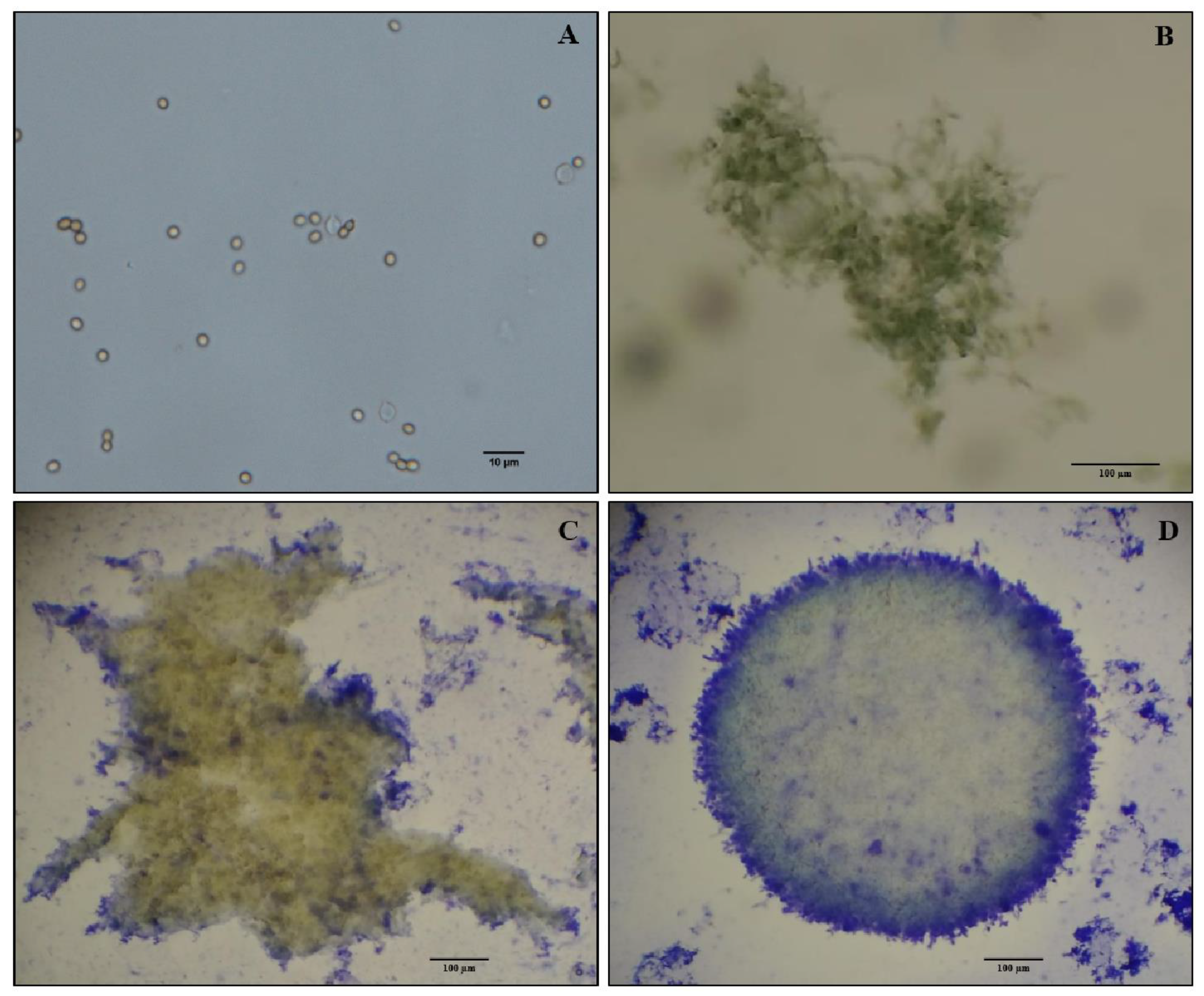
Figure 1. Escovopsis weberi MP development in liquid culture using an orbital shaker at 27 °C and 230 rpm: (A) conidial inoculation in liquid culture; (B) hyphae aggregating at 48 h; (C) early MP formation at 72 h; (D) complete MP formation at 96 h. (A): scale bar is 10 µm; (BD): scale bar is 100 µm.
Pathogens 12 00330 g001
Figure 2. Mean weights of the baits transported by ant workers during a 24 h period. Different letters (a–c) indicated significant differences between treatments according to an ANOVA and Tukey’s test (α = 0.05).
Pathogens 12 00330 g002
Figure 3. Cumulative daily foraging activity (leaf area cut (cm2)) performed by A. subterraneus subterraneus workers when offered A. wilkesiana during the bioassay period. Different letters (a–c) indicated significant differences between treatments according to Tukey’s test (α = 0.05).
Pathogens 12 00330 g003
Figure 4. Fungal garden biomass loss (%) during the 23 days of the bioassay. Different letters (a,b) indicated significant differences between treatments according to Tukey’s test (α = 0.05).
Pathogens 12 00330 g004
Table 1. Monthly mycelial pellet reproductive capacity (conidia mL−1) during a 12-month storage period.
Evaluation Time (Months)Median (×108)
111.68 a
212.40 a
311.72 a
414.54 a
512.64 a
69.67 abc
710.23 ab
89.44 abc
910.05 abc
101.3 d
113.35 cd
124.68 bcd
Medians followed by different letters (a–d) were statistically different to each other according to the Kruskal–Wallis test, followed by Tukey’s test (α = 0.05).
Table 2. Waste weight in grams (mean ± SEM) dumped during 23 days of the bioassay. From day 1 to day 7 (time 1), day 8 to day 15 (time 2), day 16 to day 23 (time 3).
TreatmentsTimes
123
Conidia0.20 ± 0.05 Ba0.58 ± 0.05 Aa0.51 ± 0.03 Aa
MP0.21 ± 0.05 Ba0.56 ± 0.05 Aa0.47 ± 0.02 Aa
Dead Conidia0.15 ± 0.16 Aa0.17 ± 0.02 Ab0.16 ± 0.01 Ab
Dead MP0.21 ± 0.02 Aa0.20 ± 0.01 Ab0.20 ± 0.02 Ab
Control0.16 ± 0.02 Aa0.19 ± 0.02 Ab0.17 ± 0.02 Ab
Uppercase letters (A,B): comparisons between periods; lowercase letters (a,b): comparisons among treatments. Means followed by different letters indicated statistical differences according to Tukey’s test (α = 0.05).
Disclaimer/Publisher’s Note: The statements, opinions and data contained in all publications are solely those of the individual author(s) and contributor(s) and not of MDPI and/or the editor(s). MDPI and/or the editor(s) disclaim responsibility for any injury to people or property resulting from any ideas, methods, instructions or products referred to in the content.