Article

Independent Circulation of Leishmania major and Leishmania tropica in Their Respective Sandfly Vectors for Transmission of Zoonotic and Chronic Cutaneous Leishmaniasis Co-Existing in a Mixed Focus of Central Tunisia

1
Pasteur Institute of Tunis, Unit of Vector Ecology, Tunis 1002, Tunisia
2
Pasteur Institute of Tunis, Laboratory of Veterinary Microbiology, Tunis 1002, Tunisia
3
Pasteur Institute of Tunis, Laboratory of Bioinformatics, Mathematic, and Biostatistics, Tunis 1002, Tunisia
4
Mediterranean Institute of Technology, South Mediterranean University, Les Berges du Lac 2, Tunis 1053, Tunisia
*
Author to whom correspondence should be addressed.
Pathogens 2022, 11(8), 855; https://doi.org/10.3390/pathogens11080855
Submission received: 16 June 2022 / Revised: 15 July 2022 / Accepted: 19 July 2022 / Published: 29 July 2022
(This article belongs to the Special Issue Leishmania & Leishmaniasis)

Abstract

:
Zoonotic cutaneous leishmaniasis (ZCL) and chronic cutaneous leishmaniasis (CCL) are known to overlap in Central Tunisia. Sandflies were collected using sticky traps and CDC light-traps set in rodent burrows at the ecotones surrounding the village, in houses, and in animal shelters during July–October 2017, 2018, and 2019. A total of 17,175 sandflies were collected during the three sandfly seasons and identified morphologically to species level. Of a total of 18 sandfly species reported in Tunisia, 16 were identified in this mixed focus of ZCL and CCL. Except for the rocky mountainous areas, Phlebotomus papatasi was the most abundant sandfly species in all biotopes. In the mountainous areas, Phlebotomus sergenti is the most abundant sandfly species belonging to the genus Phlebotomus. Female sandflies were tested for the presence of Leishmania species by PCR. The overall infection prevalence of sandflies with Leishmania major and Leishmania tropica was 0.42% and 0.065%, respectively. The sequencing of PCR-amplified ITS1 products showed that L. major is the predominant species in all biotopes and transmitted mostly by P. papaptasi followed by Phlebotomus longicuspis and Sergentomyia species. Leishmania tropica was detected in Phlebotomus sergenti and in Phlebotomus longicuspis collected in bedrooms and in the ecotone of rocky mountainous areas. Our results provided strong evidence that the proximity of human settlements to biotopes of rodent reservoir hosts of L. major and of L. tropica resulted into the cocirculation of both Leishmania species leading to a mixed focus of ZCL and CCL. The epidemiology of leishmaniases in North Africa is highly complex by the high diversity of sandfly vectors and their associated Leishmania species, leading to a mixed form of cutaneous leishmaniasis. It is of major epidemiological importance to point to the risk of spillover from rural to urban areas leading to the anthroponization of cutaneous leishmaniasis. Therefore, efficient control to reduce the indoor abundance of sandfly vectors in order to reduce the incidence of leishmaniases is urgently needed.

1. Introduction

Leishmaniases are vector-borne diseases caused by Leishmania protozoan parasites and they are transmitted to humans by the bite of infected female sandflies. Leishmaniases are widespread across 98 countries and 3 territories on 5 continents, with more than 58,000 visceral leishmaniasis cases (VL) and 220,000 cutaneous leishmaniasis cases (CL) per year [1]. The two forms of leishmaniasis coexist in Tunisia with a higher prevalence of CL compared to VL [1]. In Tunisia, CL is caused by three different Leishmania species: Leishmania major, Leishmania infantum, and Leishmania tropica (synonym, Killicki), etiologic agent of zoonotic cutaneous leishmaniasis (ZCL), sporadic cutaneous leishmaniasis (SCL), and chronic cutaneous leishmaniasis (CCL), respectively [2,3,4].
Sporadic cutaneous leishmaniasis caused by L. infantum is distributed in the humid, subhumid and semiarid biogeographical areas of Northern Tunisia and occurs sporadically with 50–150 cases per year [5]. To date, the life cycle of SCL has not yet been elucidated. Zoonotic cutaneous leishmaniasis (ZCL) caused by L. major is transmitted by the sandfly vector Phlebotomus papatasi [6,7]. Psammomys obesus, Meriones shawi, and Meriones libycus are the principal reservoir hosts of L. major [8,9,10,11,12]. Mustela nivalis, Paraechinus aethiopicus, Atelerix algirus, Ctenodactylus gundi, and Psammomys vexillaris are considered as potential reservoirs for L. major [13,14,15,16]. Annually, thousands of ZCL cases are reported from Central and Southern Tunisia [17,18], with the governorate of Sidi Bouzid alone having an estimated annual ZCL incidence rate of 669.7 per 100,000 inhabitants [19]. The population estimated to be at risk in the governorates of Kairouan, Sidi Bouzid, and Gafsa, the most endemic for ZCL, represents 87% of the total population at risk [20]. Chronic cutaneous leishmaniasis (CCL) occurs in hypoendemic microfoci located in the arid, rocky, mountainous areas of Southeastern Tunisia [21,22]. CCL is less prevalent than ZCL with 50–150 cases per year [5]. In Southeastern Tunisia, CCL is caused by L. tropica and transmitted by Phlebotomus sergenti [23]. The North African gundi (Ctenodactylus gundi) is considered as a putative reservoir host of L. tropica [24]. CCL is spreading towards the center and the southwest of Tunisia, leading to mixed foci in predominantly endemic ZCL areas [2,25,26,27]. The overlap of human cutaneous leishmaniasis due to L. major and to L. tropica is an increasingly important public health problem, as reported in the southeast [22], in the southwest [25], and in the center [25,26,27]. In the present study, an entomological investigation was carried out aiming to identify sandfly species and circulating Leishmania species in a mixed focus of ZCL with sporadic cases of CCL in Central Tunisia.

2. Material and Methods
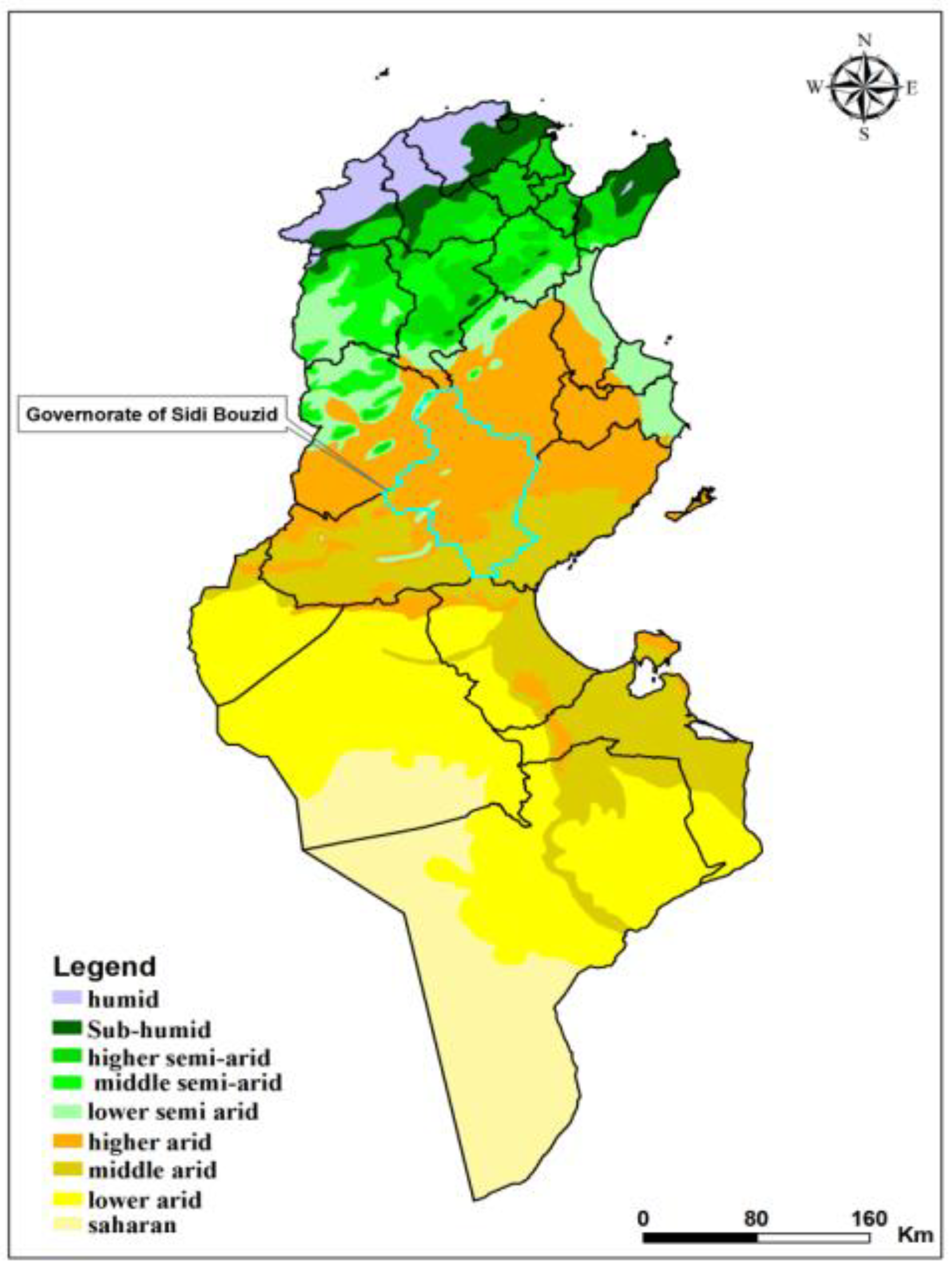
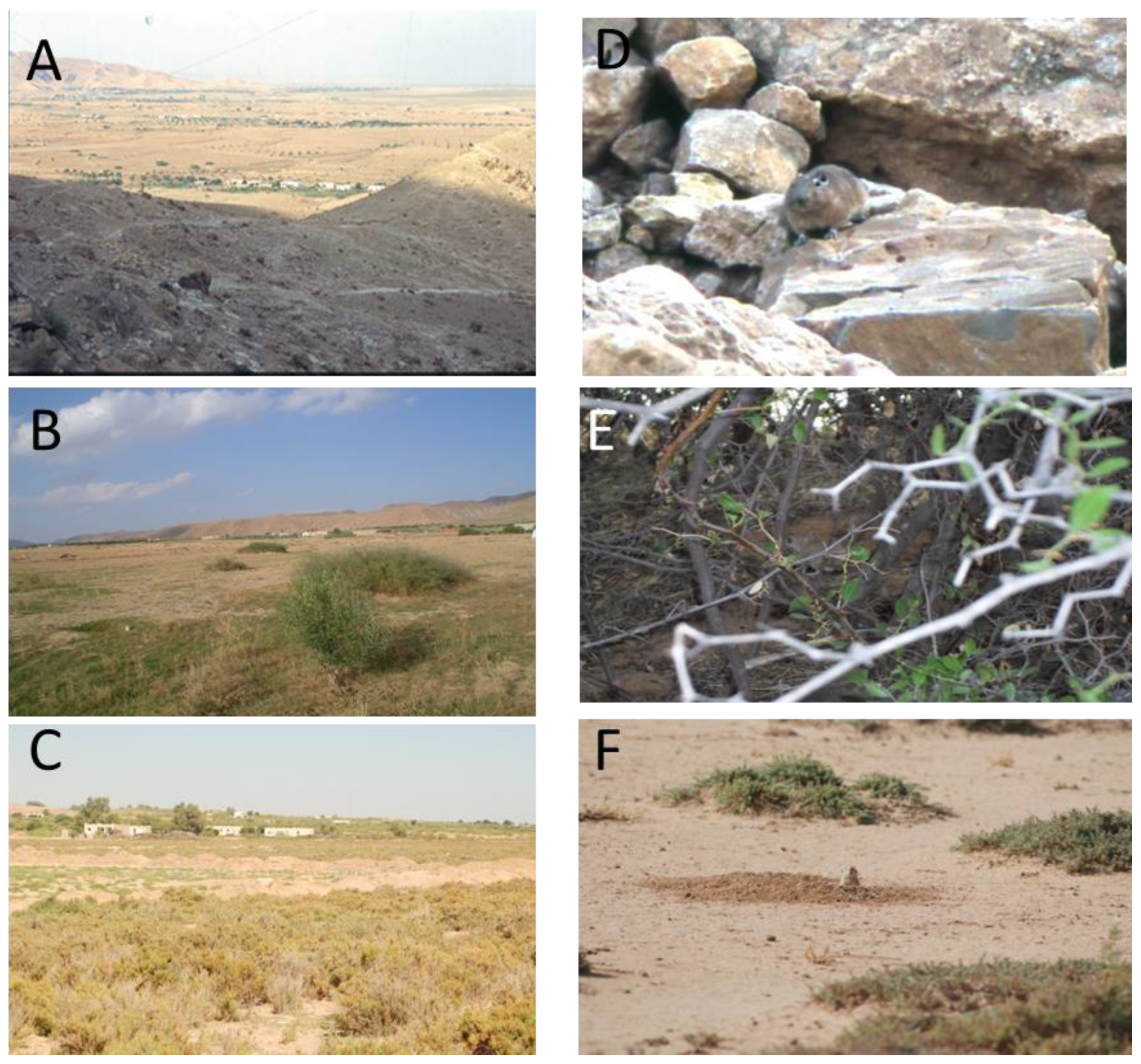
2.1. Study Site

The study was carried out in the governorate of Sidi Bouzid situated in an arid bioclimatic zone [28] (Figure 1), a highly endemic area with multiple foci of ZCL located in Central Tunisia [19]. The average annual incidence rate of ZCL was 666.7/100,000 inhabitants in the governorate of Sidi Bouzid and the dynamics of ZCL incidence are significantly heterogenic, occurring in outbreaks and clustering in space and time [19].
The study was performed in the village of Gouleb (9° 36′ E, 34° 48′ N), located in the delegation of Souk Jedid, within the governorate of Sidi Bouzid, a highly endemic focus of ZCL with an annual incidence of 3822.8 cases per 100,000 inhabitants [29], and it is considered as an emerging focus of CCL [30]. A hypoendemic focus of CCL has been reported in the delegation of Meknassy, which is adjacent to the delegation of Souk Jedid where the village of Gouleb is located [4,26]. The village is situated at the flank of the rocky mountainous areas which is the natural habitat of C. gundi [15] (Figure 2A,D). On the other side, the village is surrounded by agricultural fields harboring jujube trees (Figure 2B,E) and by nonagricultural fields made of chenopods, which are the natural habitat of M. shawi and P. obesus (Figure 2C,F), respectively. Thus, in the study sites, three ecotones which are the interface between human settlements and natural ecosystems were considered [31].

2.2. Sandfly Trapping and Identification

Our entomological investigation was carried out in the village of Gouleb during three consecutive years (2017–2019). In Tunisia, the phenology of P. papatasi is characterized by two main peaks of activity: one in June–July and a second, larger peak in September–October [18]. Each season, sandflies were collected during July–October by using CDC light traps (LT) and by sticky traps (ST) placed inside houses, animal shelters located in peridomestic areas, between rocks in the mountainous areas, in burrows of M. shawi and of P. obesus. Traps were placed from dusk to dawn and brought back the following morning to the laboratory. The collected sandflies were dissected to remove the head and the genitalia for species identification and the rest of the body was placed in a 1.5 mL microfuge tube to be examined for the presence of Leishmania DNA. Sandflies were identified at the species level by using the identification keys of Croset et al. [32] with special attention given to the atypical form of female P. perniciosus that could be confused with P. longicuspis [33,34]. Following identification, unfed female sandflies were pooled with up to a maximum of 30 specimens per pool based on the date of collection, species, and biotype and then stored in phosphate-buffered saline (PBS) for molecular analysis.

2.3. Detection of Leishmania DNA in Female Sandflies

Because this study deals with the circulation of two forms of cutaneous leishmaniasis in one focus, we used two different methods to detect Leishmania infection. Firstly, we used a nested-PCR-based schizodeme method targeting the partially conserved region of the kinetoplast minicircle DNA, enabling Leishmania species discrimination on the basis of PCR amplicon size, where L. tropica generated a 750 bp product, L. infantum produced a 680 bp product, whereas the product size of L. major was 560 bps [35]. Secondly, we used a nested PCR of a partial region of ITS-rDNA gene allowing the sequencing of detected Leishmania DNA as described by Parvizi et al. [36,37,38].
Female sandflies in pools were homogenized in 200 μL of PBS through high-speed shaking using the automated Tissue Lyser LT (Qiagen, Hilden, Germany) with glass beads. The mixture was clarified by centrifugation at 6000× g for 2 min for use in DNA extraction with a Qiagen DNA Mini Kit (Qiagen, Hilden, Germany) according to the manufacturer’s instructions. Extracted DNA from female sandflies was screened for infections with Leishmania species by a nested PCR based on the schizodeme method targeting the partial conserved region of the kinetoplast minicircle DNA as previously described [35]. This method allows the discrimination of Leishmania species originating from North Africa based on the PCR amplicon size. The nested PCR product sizes for L. tropica, L. major and L. infantum were 750, 560 bp and 680 bp, respectively [35]. The first PCR was performed using the Taq DNA recombinant polymerase kit (Invitrogen, Waltham, MA, USA) in 25 μL containing: 2.5 μL 10X buffer, 1.5 μL MgCl2 (50 mM), 1 μL dNTP mix (10 mM), 1 μL of each reverse and forward primers CSB2xF/CSB1xR (10 μM), 0.25 μL Taq DNA polymerase (5 U/μL), 12.75 μL nuclease-free water and 5 μL of extracted DNA. The nested PCR was carried out in 50 μL containing 3 μL of the first PCR step DNA product and 47 μL of a mixture containing: 5 μL 10X buffer, 3 μL MgCl2 (50 mM), 2 μL dNTP mix (10 mM), 2 μL of each reverse and forward internal primers 13Z/LIR (10 μM), 0.5 μL of Taq DNA polymerase (5 U/μL) (Invitrogen, Waltham, MA, USA), and 32.5 μL of RNase and DNase-free H2O. Optimized cycling conditions for the first and second PCR steps were performed as follows: 94 °C for 5 min followed by 35 cycles, repeating denaturation at 94 °C for 30 s, annealing at 55 °C for 60 s and elongation at 72 °C for 90 s, and an extension step at 72 °C for 10 min. Previously extracted L. tropica (MHOM/TN/88/TAT3) and L. major (MHOM/TN/97/LPN162) DNA were used as a positive control for Leishmania detection. Cross-contamination was monitored by negative controls for sample extraction and PCR assay. Amplification products of the nested PCR were then visualized by electrophoresis in 1.5% agarose gel supplemented with ethidium bromide under UV-light transillumination. Positive PCR product sizes were estimated according to 100 bp molecular weight (Invitrogen, Waltham, MA, USA) to identify sandfly-associated Leishmania species.
In this study, the infection of sandfly species by Leishmania species is reported using the minimum infection rate (MIR) which is calculated by: ([number of positive pools/total number of tested sandflies] × 100) [39].

2.4. Detection of Leishmania DNA, DNA Sequencing and Phylogenetic Analysis

Extracted DNA was screened for infections of Leishmania species by a nested PCR of a partial region of the ITS-rDNA gene as previously described [36,37]. The first amplification steps were performed using the Taq DNA recombinant polymerase kit (Invitrogen, Waltham, MA, USA) in 50 μL reaction containing: 5 μL 10X buffer, 3 μL MgCl2 (50 mM), 2 μL dNTP mix (10 mM), 1 μL of each reverse and forward primers IR1/IR2 (10 μM), 0.5 μL Taq DNA polymerase enzyme and 10 μL of total extracted DNA. The nested PCR was carried out in 50 μL containing 2 μL of the first PCR step DNA product and 48 μL of mixture containing: 5 μL 10X buffer, 3 μL MgCl2 (50 mM), 2 μL dNTP mix (10 mM), 1 μL of each reverse and forward internal primers ITS1F/ITS2R4 (10 μM) and 0.5 μL of Taq DNA polymerase (Invitrogen, Waltham, MA, USA). Optimized cycling conditions for the first and second PCR step were performed as follows: (i) 94 °C for 3 min followed by 40 cycles of 94 °C for 60 s, 58 °C for 60 s and 72 °C for 90 s, followed by a final extension step (72 °C) for 10 min; (ii) nested PCR with 94 °C for 3 min followed by 5 cycles of 94 °C for 60 s, 55 °C for 60 s and 72 °C for 60 s, and 35 cycles of incubation at 94 °C, 59 °C and 72 °C for 60 s each. The extension step was continued for 10 min at 72 °C. Cross-contamination was monitored by negative controls for sample extraction and PCR solutions for the PCR test. Amplification products of the nested PCR were separated in 2% agarose gel stained with ethidium bromide and visualized under UV-light illumination. Positive PCR products were directly sequenced to identify sandfly-associated Leishmania species.
The 462bp nested PCR products were purified by the ExoSAP-IT method using the Exonuclease-I and the Shrimp Alkaline Phosphatase were sequenced in both directions using a Big Dye Terminator ready reaction cycle sequencing v3.1 kit (Applied Biosystems, Waltham, MA, USA) with forward and reverse nested PCR primers (ITS1F/ITS2R4) [38]. The resulting consensus sequences were deduced by aligning the respective forward and reverse sequences using CLUSTAL_W 1.4 implemented in MEGA v.5.22 [40]. In addition to the studied sequences, several Leishmania species sequences, including 1 L. brazeliensis, 13 L. tropica, and 33 L. major, were selected from the GenBank database. Phylogenetic analysis was performed using the maximum likelihood analysis method and the Tamura-3 parameter model. The tree topology was supported by 1000 bootstrap replicates.

2.5. Data Analysis

Sandfly Species Diversity

Biological diversity was quantified by measuring richness and evenness for a better understanding of community structure [41]. To assess the sandfly fauna structure, the following ecological parameters and diversity indexes were calculated:
Relative abundance (ni): Number of sandfly species/Total number of sandflies in the sample.
Specific richness (S): number of species in the sample [41].
Specific diversity was also measured by the Shannon–Wiener index (H′) that takes into consideration the probability of encountering a specific species in a stand. To better discuss this Shannon index, it is often accompanied by the Piélou equitability index (J), or equirepartition index (E). Its formula corresponds to the ratio between H′ and Hmax: E = H′/Hmax. This index varies between 0 and 1. If it tends towards E = 1, then the species present in the stand have identical abundances. If it tends towards E = 0, then we are in the presence of an imbalance where a single species dominates the entire stand.
H   =   i = 1   S p i   l o g p i
  • pi = the proportional abundance or percent abundance of a species present (pi = ni/N).
  • ni = the number of individuals counted for the species.
  • N = the total number of individuals counted, all species combined.
  • S = the total or cardinal number of the list of present species.
Statistical analysis was performed using python Jupiter notebook. Fisher’s exact test was applied to a 2 × 2 contingency table to compare the relative abundance of sandfly species in different habitats and their distribution within infected sandflies. The significance level was set at 5%.

3. Results

3.1. Sandfly Fauna

In the focus on Gouleb, 16 sandfly species were identified, belonging to two genera, Phlebotomus (ten species) and Sergentomyia (six species). Among sandfly species of the genus Phlebotomus, Phlebotomus (Phlebotomus) papatasi was the most abundant species followed by P. (Larroussius) longicuspis and P. (Paraphlebotomus) sergenti. The remaining sandfly species of the genus Phlebotomus were less prevalent including: P. (Larroussius) perniciosus, P. (Larroussius) perfiliewi, P. (Larroussius) ariasi, P. (Larroussius) langeroni, P. (Paraphlebotomus) alexandri, P. (Paraphlebotomus) riouxi, and P. (Paraphlebotomus) chabaudi. Among sandfly species of the genus Sergentomyia, Sergentomyia (Sergentomyia) fallax was the most prevalent followed by S. (Sergentomyia) minuta. The remaining sandfly species of the genus Sergentomyia were less prevalent including: S. (Sergentomyia) antennata, S. (Grassomyia) dreyfussi, S. (Sintonius) christophersi, and S. (Sintonius) clydei (Table 1).
Of the total collected sandflies in all biotopes during the three sandfly seasons, P. (Phlebotomus) papatasi, S. (Sergentomyia) fallax, S. (Sergentomyia) minuta and P. (Larroussius) longicuspis corresponded to 93% of all specimens. The 12 remaining species were less prevalent and represented 7% of the total sandfly fauna. Since the Piélou’s equitability index E = H′/Hmax was 0.55, the sandfly fauna was imbalanced with four dominant species in the entire stand.
The relative abundance of predominant sandfly species varied significantly among biotopes (Table 2). Among sandfly species of the genus Phlebotomus, P. sergenti was the predominant sandfly in the mountainous areas, the natural habitat of the gundi, compared to other biotopes (p < 0.05). Phlebotomus papatasi was less prevalent in gundi’s biotope, predominant in bedrooms, animal shelters located in the peridomestic areas, and rabbit holes, and highly abundant in burrows of M. shawi and P. obesus (p < 0.05). Phlebotomus longicuspis was abundant in animal shelters located in the peridomestic areas, bedrooms, and to a lesser extent, in gundi’s biotope. Sergentomyia fallax and S. minuta had the same patterns of distribution, being the most abundant in gundi’s biotope followed by bedrooms, and animal shelters located in the peridomestic areas (Table 2).

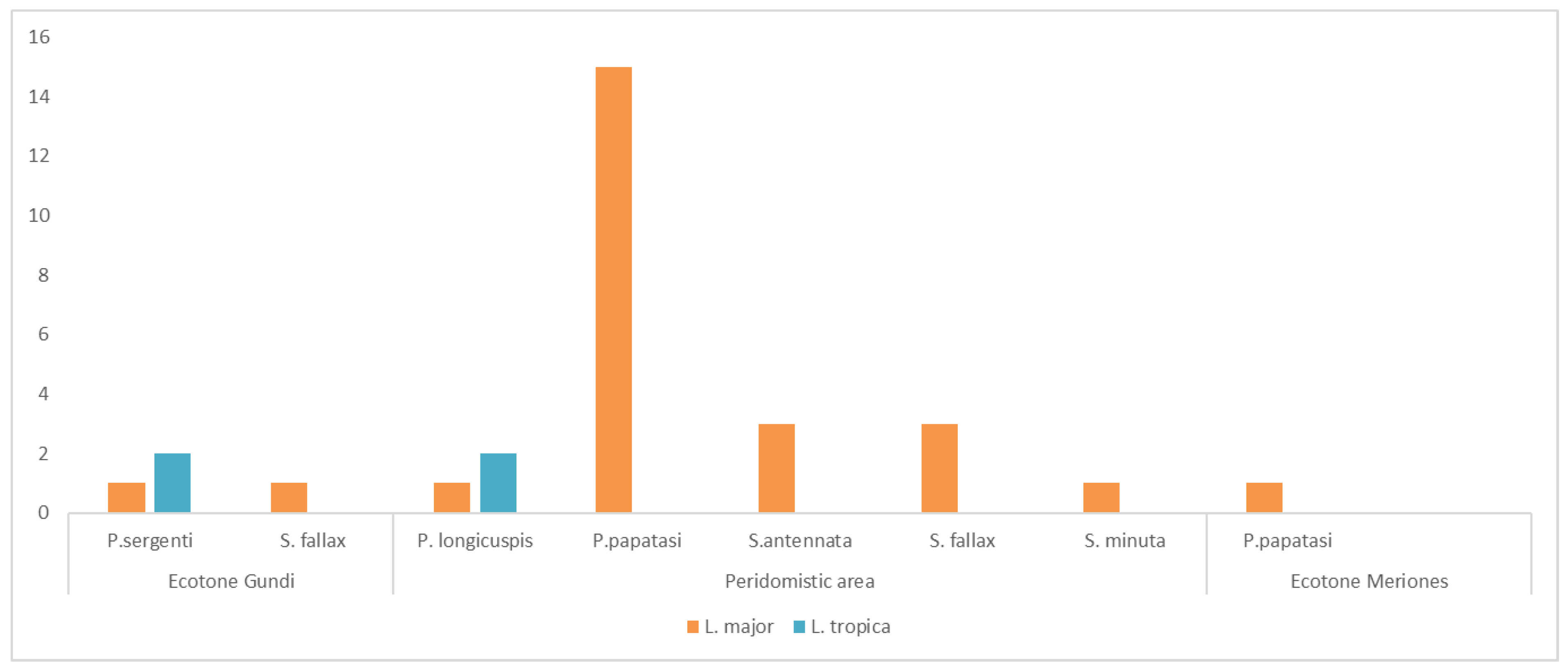
3.2. Leishmania Detection

A total of 957 pools of unfed female sandflies were screened for Leishmania infection by the nested-PCR-based schizodeme method targeting the partially conserved region of the kinetoplast minicircle DNA. Thirty pools were found to be positive for Leishmania DNA. Hence, the overall minimum infection rate of sandflies with Leishmania DNA was 0.5% (30/6187). Sandflies forming positive pools were collected from bedrooms, animal shelters located in the peridomestic areas, gundi’s biotope, and rodents’ burrows (Table 3).
The overall infection prevalence of sandflies with L. major and L. tropica was 0.42% (26/6187), and 0.065% (4/6187), respectively. Among the 30 positive pools, 26 were positive for L. major DNA (86.6%), and 4 positive for L. tropica DNA (13.3%) (Table 3). Of a total of 26 positive pools for L. major DNA, 16 (61.5%) were detected in pools of P. papatasi, 4 in pools of S. fallax (15.4%), 3 in pools of S. antennata (11.5%), 1 in a pool of S. minuta (3.8%), 1 in a pool of P. longicuspis (3.8%), and 1 in a pool of P. sergenti (3.8%) (Table 3). Among positive pools for L. tropica DNA, two were detected in two pools of P. sergenti and two were detected in two pools of P. longicuspis (Table 3).
All positive pools of sandflies collected from bedrooms (N = 7) were infected only with L. major (five pools of P. papatasi (71.4%), one pool of S. fallax (14.2%), and one pool of S. antennata (14.2%)) (Figure 3). Among the 18 positive pools of sandflies collected from the peridomestic areas including rabbits’ holes and animal shelters, 10 (55.5%) were L. major-DNA-infected pools of P. papatasi, 5 L. major-DNA-infected pools of Sergentomyia sp. (27.7%), 1 L. major-DNA-infected pool of P. longicuspis (5.5%), and 2 L. tropica-DNA-infected pools of P. longicuspis (11.1%) (Figure 3). One pool of P. papatasi collected from burrows of M. shawi and/or P. obesus was positive for L. major DNA (Figure 3). Despite the fact that L. major was detected in Sergentomyia species, P. sergenti, and P. longicuspis, it remained highly associated with P. papatasi (p < 0.05). In the rocky mountainous areas, which are the natural biotope of C. gundi, in addition to two pools of P. sergenti positive for L. tropica DNA (50%), two pools (one S. fallax and one P. sergenti) were positive for L. major DNA. Statistically, an association was shown between P. sergenti and P. longicuspis towards L. tropica (p < 0.05).

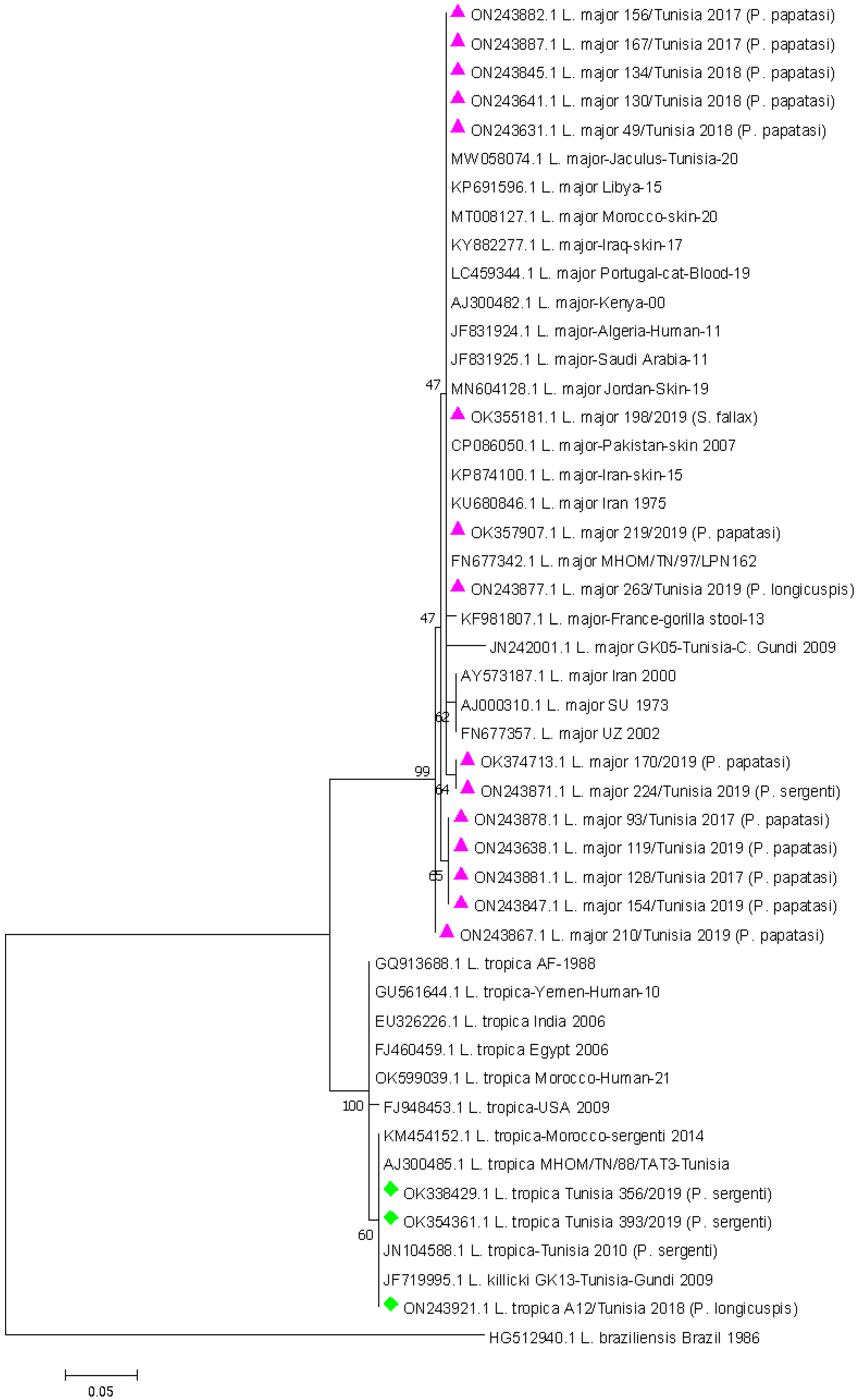
3.3. Leishmania DNA Sequencing and Phylogenetic Analysis

From the 30 PCR-positive products for Leishmania DNA which were sequenced, only 18 were readable. The alignment of ITS sequences obtained confirmed that samples corresponded to 3 L. tropica and 15 L. major. Three sequences corresponding to three L. tropica (from two P. sergenti and one P. longicuspis) were selected and deposited in GenBank under accession numbers OK338429, OK354361, and ON243921. Fifteen sequences corresponding to L. major (12 from P. papatasi, 1 from P. longicuspis, 1 from P. sergenti, and 1 from S. fallax) were deposited in GenBank under accession numbers OK355181, OK357907, OK374713, ON243631, ON243638, ON243641, ON243845, ON243847, ON243867, ON243871, ON243877, ON243878, ON243881, ON243882, and ON243887.
A phylogenetic analysis was performed to observe the phylogenetic relationships of ITS among species. The identified Leishmania DNA sequences are closely related to the reference sequence of L. major and L. tropica with (96.8–99%) and (99–100%) of identity, respectively.
The sequence analyses showed that Tunisian L. major sequences found in this study (OK355181, OK357907, OK374713, ON243631, ON243638, ON243641, ON243845, ON243847, ON243867, ON243871, ON243877, ON243878, ON243881, ON243882 and ON243887) were closely related to a Tunisian sequence (MHOM/TN/97/LPN162 accession number FN677342) isolated in 1997 with (98 to 100%) identity at the nucleotide level. Regarding sequences of L. tropica (OK338429, OK354361 and ON243921), they showed 100% identity to Tunisian sequences (MHOM/TN/88//TAT3 accession number AJ300485) isolated in 1988. Phylogenetic reconstructions revealed clustering of obtained Leishmania sequences within L. major and L. tropica genetic clades. The phylogenetic branch was supported by a high bootstrap value of 99% and 100% for L. major and L. tropica, respectively (Figure 4).
The evolutionary history was inferred by using the maximum likelihood method based on the Tamura three-parameter model. The 1000 bootstrap pseudo-replication values were reported at nodes. The scale bar represents 0.05% divergence with branch lengths measured in the number of substitutions per site. The analysis involved 47 nucleotide sequences aligned using the CLUSTAL algorithm. All positions containing gaps and missing data were eliminated. There was a total of 155 positions in the final dataset. Evolutionary analysis was conducted in MEGA7. The sequences obtained are marked with a pink triangle and a green diamond. Leishmania braziliensis was used as the outgroup.

4. Discussion

Despite the high annual prevalence of ZCL in the delegation of Souk Jedid where the village of Gouleb is located [18,19,20,29], little is known regarding the epidemiological aspects of the coexistence of ZCL and CCL in this area. Species identification of the sandfly vectors and etiological-agent typing in these vectors represent steps forward in understanding the epidemiology of leishmaniasis, which should lead to the implementation of improved disease control programs. Taking into account the coexistence of ZCL and CCL, in a nearby focus of CCL [26], the focus of Gouleb is of particular epidemiological importance. The present study aimed: (1) to clarify the diversity and the abundance of sandflies, and (2) to identify the Leishmania species infecting sandfly species in this emerging CCL within a focus of a predominantly ZCL.
Following this biological approach, we quantified the species diversity of sandfly populations in and around houses of CL cases due to L. major and in biotopes of the reservoir host Meriones. Species diversity was low in all habitats. As previously reported in ZCL foci, the proven vector of L. major in Tunisia, P. papatasi, was the dominant species [31]. Other species were probably insufficiently abundant in the Meriones habitat to be easily captured in and around the houses. In these recently established settlements, P. papatasi is probably the first species, or the only species, to invade new houses, because of its specific behavior.
Of a total of 18 sandfly species reported from Tunisia [42,43], 16 species were collected from the site of Gouleb, representing 88.8% of the sandfly fauna reported from Tunisia. Except in the mountainous areas, P. papatasi was the predominant sandfly species in all biotopes including bedrooms, peridomestic areas, and agricultural fields surrounding the village harboring burrows of rodents which are reservoir hosts of L. major.
In addition, P. papatasi was the most prevalent L. major-infected sandfly species in all biotopes including rodents’ burrows. Recently, it was shown that ZCL incidence is significantly higher in the ecotones of M. shawi compared to ecotones of P. obesus [31]. This finding could be explained by the high infection prevalence of M. shawi with L. major reaching 53% in autumn compared to the infection prevalence of P. obesus (41%) [11], and by its migratory behavior leading to the dispersal of ZCL [44]. Considering that the flight range of P. papatasi is around 0.75 km [45], increases in densities of L. major-infected P. papatasi in the ecotone of M. shawi expand the overlap of the infected ZCL vector with human habitations and communities contributing to the emergence of epidemics among naïve human populations [46]. Taking into account that P. papatasi (i) is highly associated with burrows of M. shawi at the ecotone level [31] and (ii) is the most abundant sandfly species indoors and is highly endophilic with a trophic preference for humans and rodents [46], consequently, ZCL is the predominant form of CL in the focus of Gouleb [19,29].
Phlebotomus sergenti is the predominant sandfly species belonging to the genus Phlebotomus in the mountainous areas of the village of Gouleb, a natural biotope of the gundi [15]. Indoors, P. sergenti is endophilic but it may take longer for this sandfly species to invade houses compared to the most abundant species P. papaptasi, as has been reported by Jaouadi et al. [47] in the same area and by Tabbabi et al. [23] in a mixed focus of ZCL and CCL in Southeastern Tunisia. Previous studies reported the detection of L. tropica in P. sergenti collected from bedrooms [47,48]. In the present work, we reported for the first time the detection of L. tropica in P. sergenti collected from the rocky mountainous areas of the village, a natural biotope of the gundi. In addition, L. tropica was detected in gundi [49], and also in P. sergenti trapped from a bedroom in a site near to the village of Gouleb [47], and isolated from a human in the same region [27]. The aforementioned findings provide strong evidence to incriminate P. sergenti and C. gundi as the vector and the reservoir of L. tropica, respectively, in Central Tunisia, as has been reported from the Southeast of Tunisia [24,48].
In the present study, we showed that P. sergenti is totally absent from burrows of M. shawi and P. obesus; therefore, this sandfly species is not cavernicolous compared to P. papatasi. Thus, it is expected that P. sergenti will not be involved in the transmission of L. major between M. shawi and/or P. obesus. However, we reported for the first time the natural infection of unfed female P. sergenti collected from the rocky mountainous areas with L. major. Concomitantly, a high infection prevalence of gundi with L. major (30%, N = 23) trapped in a nearby site named Khabina was reported [15]. The detection of L. major DNA in P. sergenti does not mean necessarily that this sandfly species is permissive for L. major. Phlebotomus sergenti was described as specific vector only for L. tropica and not for L. major [50,51]. Kamhawi et al. [50] highlighted the role of sandfly midgut lipophosphoglycan (LPG) receptors in Leishmania attachment and the impact of LPG on the vectorial competence of P. sergenti for only L. tropica and, consequently, this sandfly species is not permissive for other Leishmania species such as L. major and/or L. donovani. Since P. sergenti may not have the genetic characteristics of a single species [52], it is of major epidemiological importance to study the vectorial competence and the vectorial capacity of the North Africa strain of P. sergenti in the transmission of L. major.
In Central Tunisia, ZCL is the predominant form of CL caused by L. major due to the predominance of the sandfly vector P. papatasi in the peridomestic areas and its high infection prevalence with the parasite. However, the occurrence of sporadic human cases of CCL in Central Tunisia located at the flank of rocky, arid, mountainous areas is most probably due to the involvement of P. sergenti and the gundi in the transmission of L. tropica, and, subsequently, leading to the coexistence of ZCL and CCL. In Morocco, several studies conducted in historical foci of CL, due to L. major being close to mountainous areas where P. sergenti is the most commonly collected sandfly species, have shown the emergence of L. tropica in these foci [53,54,55]. The North African form of L. tropica tends to be zoonotic and endemic in rural areas compared to the Middle East form which is anthroponotic and endemic in urban cities [56]. Recently, several epidemics with hundreds of cases of L. tropica CL have been reported in several Moroccan cities [57,58]. It is of major epidemiological importance to point to the risk of spillover of CCL from rural settlements to urban areas in Tunisia [59], added to the risk of anthroponization which may lead to major outbreaks of cutaneous leishmaniasis due to L. tropica in major cities located in Central and Southern Tunisia. Further studies are needed to assess the risk of urbanization of CCL in Central Tunisia.
Among sandfly species belonging to the genus Phlebotomus, P. longicuspis is the second most dominant species in bedrooms and in animal shelters located in the peridomestic areas and are rare in the rocky mountainous areas as well as in burrows of M. shawi and P. obesus. The infection of P. longicuspis collected from animal shelters with L. tropica point to the potential role of this sandfly species in the transmission of CCL in Central Tunisia. Similar results were reported by Remadi et al. [60] from arid Central Tunisia. Phlebotomus longicuspis is abundant in the Saharan and arid bioclimatic zones with a relative abundance of 60% and 40%, respectively [61]. Recently, we showed the involvement of P. longicuspis in the transmission of L. infantum in highly irrigated areas of Central Tunisia [39]. Similar results were reported by Remadi et al. [60], and it is suspected to be the main vector of L. infantum in Southern Tunisia [61]. In Morocco, P. longicuspis is considered a vector of L. infantum in a CL focus where P. sergenti is confirmed as the main vector of L. tropica [62,63]. In Northern Algeria, P. longicuspis is suspected to be a competent vector of zoonotic visceral leishmaniasis in these areas [64]. As for P. perniciosus, P. longicuspis appears to be a permissive vector for Leishmania species.
Sandflies of the genus Sergentomyia, mainly S. fallax, S. antennata, and S. minuta, are predominant mainly in the rocky mountainous areas, indoors, animal shelters, and rodents’ burrows. Indoors, L. major was detected in S. fallax and S. antennata. In the rocky mountainous areas, S. fallax was shown to be infected with L. major. In animal shelters, L. major was detected in S. fallax, S. antennata, and S. minuta. It is important to point out that no L. tropica was detected in S. fallax, S. antennata, and S. minuta trapped in the rocky mountainous areas, the natural biotope of gundi. In an old emerging ZCL focus of Southern Tunisia (Gafsa), L. major was detected in S. minuta trapped in the peridomestic areas [65]. Here, we reported for the first time the detection of L. major in S. fallax and S. antennata. Our findings strongly suggest that S. fallax, S. antennata, and S. minuta are suspected vectors of L. major and the gundi is considered a potential reservoir host in the rocky mountainous areas close to communities, and may play a significant role in the transmission of ZCL.
In conclusion, the epidemiology of leishmaniases in North Africa is highly complex due to the high diversity of sandfly vectors and their associated Leishmania species, leading to a mixed form of CL caused by different pathogens, with the risk of spillover from rural to urban areas. Therefore, efficient control to reduce the indoor abundance of sandfly vectors to reduce the incidence of leishmaniases is urgently needed.

Author Contributions

Conceptualization, E.Z.; Data curation, M.A.S.A., J.L., D.L. and S.B.M.; Formal analysis, M.A.S.A., J.L., I.C., K.D. and E.Z.; Investigation, M.A.S.A., J.L., D.L. and E.Z.; Methodology, M.A.S.A., J.L., I.C., D.L., K.D., S.B.M. and E.Z.; Resources, E.Z.; Supervision, E.Z.; Validation, J.L.; Visualization, M.A.S.A. and E.Z.; Writing—original draft, E.Z.; Writing—review & editing, J.L. All authors have read and agreed to the published version of the manuscript.

Funding

This research received no external funding.

Institutional Review Board Statement

Not applicable.

Informed Consent Statement

Not applicable.

Data Availability Statement

This study did not report any data.

Conflicts of Interest

The authors declare no conflict of interest.

References

  1. Alvar, J.; Vélez, I.D.; Bern, C.; Herrero, M.; Desjeux, P.; Cano, J.; Jannin, J.; de Boer, M. Leishmaniasis worldwide and global estimates of its incidence. PLoS ONE 2012, 7, e35671. [Google Scholar] [CrossRef] [PubMed]
  2. Kallel, K.; Pratlong, F.; Belhadj, S.; Cherif, F.; Hammami, M.; Dedet, J.P.; Chaker, E. Cutaneous leishmaniasis in Tunisia: Results of the iso-enzymatic characterization of 71 Strains. Ann. Trop. Med. Parasitol. 2005, 99, 11–19. [Google Scholar] [CrossRef] [PubMed]
  3. Aoun, K.; Amri, F.; Chouihi, E.; Haouas, N.; Bedoui, K.; Benikhlef, R.; Ghrab, J.; Babba, H.; Chahed, M.K.; Harrat, Z.; et al. Épidémiologie de Leishmania (L.) infantum, L. major et L. killicki en Tunisie: Résultats et analyse de l’identification de 226 isolats humains et canins et revue de la littérature. Bull. Soc. Pathol. Exot. 2008, 101, 323–328. [Google Scholar] [CrossRef]
  4. Haouas, N.; Chaker, E.; Chargui, N.; Gorcii, M.; Belhadj, S.; Kallel, K.; Aoun, K.; Akrout, F.M.; Ben Said, M.; Pratlong, F.; et al. Geographical distribution updating of Tunisian leishmaniasis foci: About the isoenzymatic analysis of 694 Strains. Acta Trop. 2012, 124, 221–228. [Google Scholar] [CrossRef]
  5. Aoun, K.; Bouratbine, A. Cutaneous leishmaniasis in North Africa: A Review. Parasite 2014, 21, 11820. [Google Scholar] [CrossRef] [Green Version]
  6. Derbali, M.; Chelbi, I.; Ben Hadj Ahmed, S.; Zhioua, E. Leishmania major Yakimoff et Schokhor, 1914 (Kinetoplastida: Trypanosomatidae) chez Meriones shawi Duvernoy, 1842 (Rodentia: Gerbillidae): Persistance de l’infection du mérion et de son infectivité pour le phlébotome vecteur Phlebotomus (Phlebotomus) papatasi. Bull. Soc. Pathol. Exot. 2012, 105, 399–402. [Google Scholar] [CrossRef] [PubMed]
  7. Ismail, R.B.; Gramiccia, M.; Gradoni, L.; Helal, H.; Rachid, M.S.B.; Ben Ismail, R.; Gramiccia, M.; Gradoni, L.; Helal, H.; Ben Rachid, M.S. Isolation of Leishmania major from Phlebotomus papatasi in Tunisia. Trans. R. Soc. Trop. Med. Hyg. 1987, 81, 749. [Google Scholar] [CrossRef]
  8. Rioux, J.A.; Petter, F.; Zahaf, A.; Lanotte, G.; Houin, R.; Jarry, D.; Perieres, J.; Martini, A.; Sarhani, S. Isolation of Leishmania major Yakimoff and Shokhor, 1914 (Kinetoplastida-Trypanosomatidae) in Meriones shawi-shawi (Duvernoy, 1842) (Rodentia-Gerbillidae) in Tunisia. Ann. Parasitol. Hum. Comp. 1986, 61, 139–145. [Google Scholar] [CrossRef] [Green Version]
  9. Ben-Ismail, R.; Ben Rachid, M.S.; Gradoni, L.; Gramiccia, M.; Helal, H.; Bach-Hamba, D. Zoonotic cutaneous leishmaniasis in Tunisia: Study of the disease reservoir in the Douara area. Ann. Soc. Belg. Med. Trop. 1987, 67, 335–343. [Google Scholar]
  10. Fichet-Calvet, E.; Jomâa, I.; Ben Ismail, R.; Ashford, R.W. Leishmania major infection in the fat sand rat Psammomys obesus in Tunisia: Interaction of host and parasite populations. Ann. Trop. Med. Parasitol. 2003, 97, 593–603. [Google Scholar] [CrossRef] [PubMed]
  11. Ghawar, W.; Toumi, A.; Snoussi, M.A.; Chlif, S.; Zâatour, A.; Boukthir, A.; Bel Haj Hamida, N.; Chemkhi, J.; Diouani, M.F.; Ben Salah, A. Leishmania major infection among Psammomys obesus and Meriones shawi: Reservoirs of zoonotic cutaneous leishmaniasis in Sidi Bouzid (Central Tunisia). Vector Borne Zoonotic. Dis. 2011, 11, 1561–1568. [Google Scholar] [CrossRef] [PubMed] [Green Version]
  12. Derbali, M.; Polyakova, L.; Boujaama, A.; Burruss, D.; Cherni, S.; Barhoumi, W.; Chelbi, I.; Poché, R.; Zhioua, E. Laboratory and field evaluation of rodent bait treated with fipronil for feed through and systemic control of Phlebotomus papatasi. Acta Trop. 2014, 135, 27–32. [Google Scholar] [CrossRef] [PubMed]
  13. Ghawar, W.; Snoussi, M.A.; Bel Hadj Hamida, N.; Boukthir, A.; Yazidi, R.; Chaâbane, S.; Chemkhi, J.; Zâatour, A.; Ben Salah, A. First report of natural infection of Least Weasel (Mustela Nivalis Linnaeus, 1776) with Leishmania major in Tunisia. Vector Borne Zoonotic. Dis. 2011, 11, 1507–1509. [Google Scholar] [CrossRef]
  14. Chemkhi, J.; Souguir, H.; Ali, I.B.; Driss, M.; Guizani, I.; Guerbouj, S. Natural infection of Algerian hedgehog, Atelerix Algirus (Lereboullet 1842) with Leishmania parasites in Tunisia. Acta Trop. 2015, 150, 42–51. [Google Scholar] [CrossRef] [PubMed]
  15. Ghawar, W.; Bettaieb, J.; Salem, S.; Snoussi, M.A.; Jaouadi, K.; Yazidi, R.; Ben-Salah, A. Natural infection of Ctenodactylus gundi by Leishmania major in Tunisia. Acta Trop. 2018, 177, 89–93. [Google Scholar] [CrossRef] [PubMed]
  16. Ben Othman, S.; Ghawar, W.; Chaouch, M.; Ayari, C.; Chemkhi, J.; Cancino-Faure, B.; Tomás-Pérez, M.; Alcover, M.M.; Riera, C.; Ben Salah, A.; et al. First detection of Leishmania DNA in Psammomys obesus and Psammomys vexillaris: Their potential involvement in the epidemiology of leishmaniasis in Tunisia. Infect. Genet. Evol. 2018, 59, 7–15. [Google Scholar] [CrossRef] [PubMed]
  17. Chelbi, I.; Kaabi, B.; Béjaoui, M.; Derbali, M.; Zhioua, E.; BéJaoui, M.; Zhioua, E. Spatial correlation between Phlebotomus papatasi Scopoli (Diptera: Psychodidae) and incidence of zoonotic cutaneous leishmaniasis in Tunisia. J. Med. Entomol. 2009, 46, 400–402. [Google Scholar] [CrossRef] [PubMed] [Green Version]
  18. Chelbi, I.; Derbali, M.; Al-Ahmadi, Z.; Zaafouri, B.; El Fahem, A.; Zhioua, E. Phenology of Phlebotomus papatasi (Diptera: Psychodidae) relative to the seasonal prevalence of zoonotic cutaneous leishmaniasis in Central Tunisia. J. Med. Entomol. 2007, 44, 385–388. [Google Scholar] [CrossRef] [PubMed] [Green Version]
  19. Ben Salah, A.; Kamarianakis, Y.; Chlif, S.; Ben Alaya, N.; Prastacos, P. Zoonotic cutaneous leishmaniasis in Central Tunisia: Spatio-temporal dynamics. Int. J. Epidemiol. 2007, 36, 991–1000. [Google Scholar] [CrossRef] [PubMed] [Green Version]
  20. Chalghaf, B.; Chlif, S.; Mayala, B.; Ghawar, W.; Bettaieb, J.; Harrabi, M.; Benie, G.B.; Michael, E.; Ben Salah, A. Ecological niche modeling for the prediction of the geographic distribution of lutaneous leishmaniasis in Tunisia. Am. J. Trop. Med. Hyg. 2016, 94, 844–851. [Google Scholar] [CrossRef] [PubMed] [Green Version]
  21. Rioux, J.-A.; Lanotte, G.; Pratlong, F. Leishmania killicki n. sp. (Kinetoplasitda, Trypanosomatidae). In Leishmania: Taxonomie et Phylogenèse: Applications Eco-Epidémiologiques; IMEEE: Montpellier, France, 1986; pp. 139–142. [Google Scholar]
  22. Bousslimi, N.; Aoun, K.; Ben-Abda, I.; Ben-Alaya-Bouafif, N.; Raouane, M.; Bouratbine, A. Epidemiologic and clinical features of cutaneous leishmaniasis in Southeastern Tunisia. Am. J. Trop. Med. Hyg. 2010, 83, 1034–1039. [Google Scholar] [CrossRef] [PubMed] [Green Version]
  23. Tabbabi, A.; Bousslimi, N.; Rhim, A.; Aoun, K.; Bouratbine, A. Short Report: First report on natural infection of Phlebotomus sergenti with Leishmania promastigotes in the cutaneous leishmaniasis focus in Southeastern Tunisia. Am. J. Trop. Med. Hyg. 2011, 85, 646–647. [Google Scholar] [CrossRef] [PubMed] [Green Version]
  24. Bousslimi, N.; Ben-Ayed, S.; Ben-Abda, I.; Aoun, K.; Bouratbine, A. Natural infection of North African gundi (Ctenodactylus gundi) by Leishmania tropica in the focus of cutaneous leishmaniasis, Southeast Tunisia. Am. J. Trop. Med. Hyg. 2012, 86, 962–965. [Google Scholar] [CrossRef] [Green Version]
  25. Bouratbine, A.; Aoun, K.; Ghrab, J.; Harrat, Z.; Ezzedini, M.S.; Etlijani, S. Spread of Leishmania killicki to Central and South-West Tunisia. Parasite 2005, 12, 59–63. [Google Scholar] [CrossRef] [Green Version]
  26. Haouas, N.; Chargui, N.; Chaker, E.; Ben Said, M.; Babba, H.; Belhadj, S.; Kallel, K.; Pratlong, F.; Dedet, J.P.; Mezhoud, H.; et al. Anthroponotic cutaneous leishmaniasis in Tunisia: Presence of Leishmania killicki outside its original focus of Tataouine. Trans. R. Soc. Trop. Med. Hyg. 2005, 99, 499–501. [Google Scholar] [CrossRef] [PubMed]
  27. Haouas, N.; Gorcii, M.; Chargui, N.; Aoun, K.; Bouratbine, A.; Messaadi Akrout, F.; Masmoudi, A.; Zili, J.; Ben Said, M.; Pratlong, F.; et al. Leishmaniasis in Central and Southern Tunisia: Current geographical distribution of zymodemes. Parasite 2007, 14, 239–246. [Google Scholar] [CrossRef] [PubMed] [Green Version]
  28. Gounot, M.; Le Houerou, H.N. 1967. Essai de synthèse sur la végétation et la phyto-écologie tunisiennes. In Élément de Botanique et de Phyto-Ecologie Tunisiennes; Tome, I., Nabli, M.A., Eds.; Faculty of Sciences of Tunis and MAB: Tunis, Tunisia, 1989; 387p. [Google Scholar]
  29. Bellali, H.; Hchaichi, A.; Harizi, C.; Mrabet, A.; Chahed, M.K. Comparison between active surveillance and passive detection of zoonotic cutaneous leishmaniasis in endemic rural areas in Central Tunisia, 2009 to 2014. Asian Pacific J. Trop. Dis. 2015, 5, 515–519. [Google Scholar] [CrossRef]
  30. Ghrab, J.; Rhim, A.; Bach-Hamba, D.; Chahed, M.K.; Aoun, K.; Nouira, S.; Bouratbine, A. Phlebotominae (Diptera: Psychodidae) of human leishmaniosis sites in Tunisia. Parasite 2006, 13, 23–33. [Google Scholar] [CrossRef] [Green Version]
  31. Barhoumi, W.; Chelbi, I.; Fares, W.; Zhioua, S.; Abbas, M.; Derbali, M.; Ramalho-Ortigao, M.; Zhioua, E. Risk Assessment of the role of the ecotones in the transmission of zoonotic cutaneous leishmaniasis in Central Tunisia. Int. J. Environ. Res. Public Health 2021, 18, 9274. [Google Scholar] [CrossRef]
  32. Croset, H.; Rioux, J.; Maistre, M.; Bayar, N. Les phlébotomes de la Tunisie (Diptera, Phlebotominae). Mise au point systématique, chorologique et éthologique. Ann. Parasitol. Hum. Comp. 1978, 53, 711–749. [Google Scholar] [CrossRef] [Green Version]
  33. Pesson, B.; Ready, J.S.; Benabdennbi, I.; Martín-Sánchez, J.; Esseghir, S.; Cadi-Soussi, M.; Morillas-Marquez, F.; Ready, P.D. Sandflies of the Phlebotomus perniciosus complex: Mitochondrial introgression and a new sibling species of P. longicuspis in the Moroccan Rif. Med. Vet. Entomol. 2004, 18, 25–37. [Google Scholar] [CrossRef] [PubMed]
  34. Boussaa, S.; Boumezzough, A.; Remy, P.E.; Glasser, N.; Pesson, B. Morphological and isoenzymatic differentiation of Phlebotomus perniciosus and Phlebotomus longicuspis (Diptera: Psychodidae) in Southern Morocco. Acta Trop. 2008, 106, 184–189. [Google Scholar] [CrossRef] [PubMed]
  35. Noyes, H.A.; Reyburn, H.; Bailey, J.W.; Smith, D. A nested-PCR-based schizodeme method for identifying Leishmania kinetoplast minicircle classes directly from clinical samples and its application to the study of the epidemiology of Leishmania tropica in Pakistan. J. Clin. Microbiol. 1998, 36, 2877–2881. [Google Scholar] [CrossRef] [PubMed] [Green Version]
  36. Parvizi, P.; Ready, P.D. Nested PCRs and sequencing of nuclear ITS-rDNA fragments detect three Leishmania species of gerbils in sandflies from Iranian foci of zoonotic cutaneous leishmaniasis. Trop. Med. Int. Health 2008, 13, 1159–1171. [Google Scholar] [CrossRef] [PubMed]
  37. Parvizi, P.; Mauricio, I.; Aransay, A.M.; Miles, M.A.; Ready, P.D. First detection of Leishmania major in peridomestic Phlebotomus papatasi from Isfahan Province, Iran: Comparison of nested PCR of nuclear ITS ribosomal DNA and semi-Nested PCR of minicircle kinetoplast DNA. Acta Trop. 2005, 93, 75–83. [Google Scholar] [CrossRef]
  38. Parvizi, P.; Moradi, G.; Akbari, G.; Farahmand, M.; Ready, P.D.; Piazak, N.; Assmar, M.; Amirkhani, A. PCR detection and sequencing of parasite ITS-RDNA gene from reservoirs host of zoonotic cutaneous leishmaniasis in Central Iran. Parasitol. Res. 2008, 103, 1273–1278. [Google Scholar] [CrossRef] [PubMed]
  39. Barhoumi, W.; Fares, W.; Cherni, S.; Derbali, M.; Dachraoui, K.; Chelbi, I.; Ramalho-Ortigao, M.; Beier, J.C.; Zhioua, E. Changes of sand fly populations and Leishmania infantum infection rates in an irrigated village located in arid Central Tunisia. Int. J. Environ. Res. Public Health 2016, 13, 329. [Google Scholar] [CrossRef] [Green Version]
  40. Tamura, K.; Dudley, J.; Nei, M.; Kumar, S. MEGA4: Molecular evolutionary genetics analysis (MEGA) software version 4.0. Mol. Biol. Evol. 2007, 24, 1596–1599. [Google Scholar] [CrossRef] [PubMed]
  41. Spellerberg, I.A.N.F.; Fedor, P.J. Tribute to Claude Shannon (1916-2001) and a plea for more rigorous use of species diversity and the “Shannon-Wiener” Index. Glob. Ecol. Biogeogr. 2003, 12, 177–179. [Google Scholar] [CrossRef] [Green Version]
  42. Chelbi, I.; Zhioua, E. Confirmation de la présence en Tunisie de Sergentomyia (Sintonius) clydei (Sinton, 1928). Bull. Soc. Pathol. Exot. 2012, 105, 396–398. [Google Scholar] [CrossRef]
  43. Chemkhi, J.; Guerbouj, S.; Saadawi, W.; Shaibi, T.; Faris, S.; Ghawar, W.; Boukthir, A.; Guizani, I.; Annajar, B.B.; Ben Salah, A. Presence of Sergentomyia (Parrotomyia) lewisi (Diptera: Psychodidae) in Tunisia. J. Med. Entomol. 2019, 56, 560–564. [Google Scholar] [CrossRef]
  44. Zaatour, W.; Marilleau, N.; Giraudoux, P.; Martiny, N.; Amara, A.B.H.; Ben Miled, S. An agent-based model of a cutaneous leishmaniasis reservoir host, Meriones shawi. Ecol. Modell. 2021, 443, 109455. [Google Scholar] [CrossRef]
  45. Orshan, L.; Elbaz, S.; Ben-Ari, Y.; Akad, F.; Afik, O.; Ben-Avi, I.; Dias, D.; Ish-Shalom, D.; Studentsky, L.; Zonstein, I. Distribution and dispersal of Phlebotomus papatasi (Diptera: Psychodidae) in a zoonotic cutaneous leishmaniasis focus, the Northern Negev, Israel. PLoS Negl. Trop. Dis. 2016, 10, e0004819. [Google Scholar] [CrossRef] [PubMed]
  46. Chelbi, I.; Mathlouthi, O.; Zhioua, S.; Fares, W.; Boujaama, A.; Cherni, S.; Barhoumi, W.; Dachraoui, K.; Derbali, M.; Abbass, M.; et al. The Impact of illegal waste sites on the transmission of zoonotic cutaneous leishmaniasis in Central Tunisia. Int. J. Environ. Res. Public Health 2021, 18, 66. [Google Scholar] [CrossRef]
  47. Jaouadi, K.; Bettaieb, J.; Bennour, A.; Salem, S.; Rjeibi, M.R.; Chaabane, S.; Yazidi, R.; Khabouchi, N.; Gharbi, A.; Ben Salah, A. First report on natural infection of Phlebotomus sergenti with Leishmania tropica in a classical focus of Leishmania major in Tunisia. Am. J. Trop. Med. Hyg. 2017, 97, 291–294. [Google Scholar] [CrossRef]
  48. Tabbabi, A.; Ghrab, J.; Aoun, K.; Ready, P.D.; Bouratbine, A. Habitats of the sandfly vectors of Leishmania tropica and L. major in a mixed focus of cutaneous leishmaniasis in Southeast Tunisia. Acta Trop. 2011, 119, 131–137. [Google Scholar] [CrossRef] [PubMed]
  49. Jaouadi, K.; Haouas, N.; Chaara, D.; Gorcii, M.; Chargui, N.; Augot, D.; Pratlong, F.; Dedet, J.P.; Ettlijani, S.; Mezhoud, H.; et al. First Detection of Leishmania killicki (Kinetoplastida, Trypanosomatidae) in Ctenodactylus gundi (Rodentia, Ctenodactylidae), a possible reservoir of human cutaneous leishmaniasis in Tunisia. Parasite Vectors 2011, 4, 4–6. [Google Scholar] [CrossRef] [PubMed] [Green Version]
  50. Kamhawi, S.; Modi, G.B.; Pimenta, P.F.P.; Rowton, E.; Sacks, D.L. The vectorial competence of Phlebotomus sergenti is specific for Leishmania tropica and is controlled by species-specific, lipophosphoglycan-mediated midgut attachment. Parasitology 2000, 121, 25–33. [Google Scholar] [CrossRef]
  51. Volf, P.; Myskova, J. Sand flies and Leishmania: Specific versus permissive vectors. Trends Parasitol. 2007, 23, 91–92. [Google Scholar] [CrossRef] [Green Version]
  52. Moin-Vaziri, V.; Depaquit, J.; Yaghoobi-Ershadi, M.R.; Oshaghi, M.A.; Derakhshandeh-Peykar, P.; Ferté, H.; Kaltenbach, M.; Bargues, M.D.; Léger, N.; Nadim, A. Intraspecific variation within Phlebotomus sergenti Parrot (1917) (Diptera: Psychodidae) based on MtDNA sequences in Islamic Republic of Iran. Acta Trop. 2007, 102, 29–37. [Google Scholar] [CrossRef]
  53. Ait Kbaich, M.; Mhaidi, I.; Ezzahidi, A.; Dersi, N.; El Hamouchi, A.; Riyad, M.; Akarid, K.; Lemrani, M. New epidemiological pattern of cutaneous leishmaniasis in two pre-Saharan arid provinces, Southern Morocco. Acta Trop. 2017, 173, 11–16. [Google Scholar] [CrossRef]
  54. Hmamouch, A.; El Alem, M.M.; Hakkour, M.; Amarir, F.; Daghbach, H.; Habbari, K.; Fellah, H.; Bekhti, K.; Sebti, F. Circulating species of Leishmania at microclimate area of Boulemane Province, Morocco: Impact of environmental and human factors. Parasite Vectors 2017, 10, 100. [Google Scholar] [CrossRef] [PubMed] [Green Version]
  55. Mohamed Mahmoud, E.A.; Faiza, S.; Lemine, M.; Smaine, C.; Adlaoui, E.B.; Khalid, H.; Abderrahim, S.; Hajiba, F. Geographical distribution and new Situation of Leishmania species after the control of cutaneous Leishmaniasis foci in Errachidia Province, Morocco, in 2014. Biomed. Res. Int. 2016, 2016, 8642373. [Google Scholar] [CrossRef] [PubMed] [Green Version]
  56. Reithinger, R.; Dujardin, J.-C.; Louzir, H.; Pirmez, C.; Alexander, B.; Brooker, S. Cutaneous leishmaniasis. Lancet. Infect. Dis. 2007, 7, 581–596. [Google Scholar] [CrossRef] [Green Version]
  57. Rhajaoui, M.; Nasereddin, A.; Fellah, H.; Azmi, K.; Amarir, F.; Al-Jawabreh, A.; Ereqat, S.; Planer, J.; Abdeen, Z. New Clinico-epidemiologic profile of cutaneous leishmaniasis, Morocco. Emerg. Infect. Dis. 2007, 13, 1358–1360. [Google Scholar] [CrossRef] [PubMed]
  58. Baghad, B.; Razanapinaritra, R.; Maksouri, H.; El Bouri, H.; Outlioua, A.; Fellah, H.; Lemrani, M.; Akarid, K.; Martin-Sanchez, J.; Chiheb, S.; et al. Possible introduction of Leishmania Tropica to urban areas determined by pidemiological and clinical profiles of patients with cutaneous ceishmaniasis in Casablanca (Morocco). Parasite Epidemiol. Control 2020, 9, e00129. [Google Scholar] [CrossRef]
  59. Jaouadi, K.; Depaquit, J.; Haouas, N.; Chaara, D.; Gorcii, M.; Chargui, N.; Dedet, J.P.; Pratlong, F.; Boubabous, R.; Babba, H. Twenty-four new human cases of cutaneous leishmaniasis due to Leishmania killicki in Metlaoui, Southwestern Tunisia. Probable role of Phlebotomus sergenti in the transmission. Acta Trop. 2012, 122, 276–283. [Google Scholar] [CrossRef] [PubMed]
  60. Remadi, L.; Chargui, N.; Jiménez, M.; Molina, R.; Haouas, N.; González, E.; Chaabane-Banaouas, R.; Ben Salah, E.; Haddaji, M.; Chaabouni, Y.; et al. Molecular detection and identification of Leishmania DNA and blood meal analysis in Phlebotomus (Larroussius) species. PLoS Negl. Trop. Dis. 2020, 14, e0008077. [Google Scholar] [CrossRef]
  61. Zhioua, E.; Kaabi, B.; Chelbi, I. Entomological investigations following the spread of visceral leishmaniasis in Tunisia. J. Vector Ecol. 2007, 32, 371–374. [Google Scholar] [CrossRef]
  62. Es-Sette, N.; Ajaoud, M.; Laamrani-Idrissi, A.; Mellouki, F.; Lemrani, M. Molecular detection and identification of Leishmania infection in naturally infected sand flies in a focus of cutaneous leishmaniasis in Northern Morocco. Parasite Vectors 2014, 7, 305. [Google Scholar] [CrossRef] [Green Version]
  63. Mhaidi, I.; El Kacem, S.; Ait Kbaich, M.; El Hamouchi, A.; Sarih, M.; Akarid, K.; Lemrani, M. Molecular identification of Leishmania infection in the most relevant sand fly species and in patient skin samples from a cutaneous leishmaniasis focus, in Morocco. PLoS Negl. Trop. Dis. 2018, 12, e0006315. [Google Scholar] [CrossRef] [PubMed] [Green Version]
  64. Berdjane-Brouk, Z.; Charrel, R.N.; Hamrioui, B.; Izri, A. First detection of Leishmania infantum DNA in Phlebotomus longicuspis Nitzulescu, 1930 from visceral leishmaniasis endemic focus in Algeria. Parasitol. Res. 2012, 111, 419–422. [Google Scholar] [CrossRef]
  65. Jaouadi, K.; Ghawar, W.; Salem, S.; Gharbi, M.; Bettaieb, J.; Yazidi, R.; Harrabi, M.; Hamarsheh, O.; Ben Salah, A. First report of naturally infected Sergentomyia minuta with Leishmania major in Tunisia. Parasites Vectors 2015, 8, 649. [Google Scholar] [CrossRef] [Green Version]
Figure 1. Bioclimatic map of Tunisia showing in the governorate of Sidi Bouzid.
Pathogens 11 00855 g001
Figure 2. Biotopes of a mixed focus of zoonotic cutaneous leishmaniasis and chronic cutaneous leishmaniasis. The village is situated at the flank of the rocky mountainous areas which are the natural habitat of Ctenodactylus gundi (Figure 2A,D), and it is surrounded by agricultural fields harboring jujube trees which are the natural habitat of Meriones shawi (Figure 2B,E), and by nonagricultural fields made of chenopods, which are the natural habitat of Psammomys obesus (Figure 2C,F).
Pathogens 11 00855 g002
Figure 3. Infected sandflies according to Leishmania species, sandfly species, and biotopes.
Pathogens 11 00855 g003
Figure 4. Phylogenetic tree based on partial Leishmania ITS-rDNA 5.8 s sequences.
Pathogens 11 00855 g004
Table 1. Sandfly fauna in the focus on Gouleb (2017–2019).
Species (Subgenus)ST /SubtotalLT /SubtotalTotal (%)
P. (Phlebotomus) papatasi1874/47423483364/189752617609(44.30)
P. (Paraphlebotomus) sergenti68/198770/89159246(1.43)
P. (Paraphlebotomus) alexandri15/82367/44111134(0.78)
P. (Paraphlebotomus) chabaudi1/230/336(0.03)
P. (Paraphlebotomus) riouxi2/02 2(0.01)
P. (Larroussius) ariasi8/081/6715(0.09)
P. (Larroussius) longicuspis29/0291210/76919792008(11.69)
P. (Larroussius) perfiliewi3/1410/81822(0.13)
P. (Larroussius) perniciosus40/24214/173173(0.43)
P. (Larroussius) langeroni 0/222(0.01)
S. (Sergentomyia) fallax994/47114651506/119527014166(24.26)
S. (Sergentomyia) minuta767/3201087765/37611412228(12.97)
S. (Sergentomyia) antennata40/216182/247329390(2.27)
S. (Grassomyia) dreyfussi2/1353/191244247(1.44)
S. (Sintonius) christophersi1/122/192123(0.13)
S. (Sintonius) clydei0/220/224(0.02)
Total3844/132251667144/486512,00917,175
Table 2. Sandflies caught by biotype (2017–2019).
G.BBedA.SR.HBMsBPoTotal
SpeciesSu.T (%)Su.T (%)Su.T (%)Su.T (%)Su.T (%)Su.T (%)Total (%)
P. papatasi63 (2.1)1747 (33.13)1623 (39.13)1866 (88.31)1799 (88.36)511 (84.6)7609 (44.3)
P. sergenti100 (3.33)77 (1.46)55 (1.33)14 (0.66) 246 (1.43)
P. chabaudi5 (0.17) 1 (0.02) 6 (0.03)
P. alexandri26 (0.87)56 (1.06)47 (1.13)2 (0.99)2 (0.1)1 (0.17)134 (0.78)
P. riouxi2 (0.07) 2 (0.01)
S. fallax1487 (49.55)1895 (35.94)502 (12.1)105 (4.97)118 (5.8)59 (9.77)4166 (24.26)
S. minuta1145 (38.15)659 (12.5)277 (6.68)39 (1.85)90 (4.42)18 (2.98)2228 (12.97)
S. antennata63 (2.10)208 (3.94)85 (2.05)12 (0.57)16 (0.79)6 (0.99)390 (2.27)
S. dreyfussi24 (0.8)115 (2.18)103 (2.48)3 (0.14)1 (0.05)1 (0.17)247 (1.44)
S. clydei 1 (0.02) 2 (0.1) 4 (0.02)
S. christophersi1 (0.03)9 (0.17)13 (0.31) 1 (0.05) 23 (0.13)
P. longicuspis25 (0.83)487 (9.24)1416 (34.14)67 (3.17)6 (0.29)7 (1.16)2008 (11.69)
P. perniciosus47 (1.57)10 (0.19)12 (0.29)3 (0.14)1 (0.05) 73 (0.43)
P. perfiliewi3 (0.1)10 (0.19)7 (0.17)1 (0.05) 1 (0.17)22 (0.13)
P. langeroni 2 (0.05) 2 (0.01)
P. ariasi10 (0.33) 4 (0.1)1 (0.05) 15 (0.04)
Total3001527341482113203660417,175
Legend of abbreviations: G.B: gundi’s biotope, Bed: bedroom, A. S: animal shelter, R.H: rabbit hole, BMs: burrows of Meriones shawi, BPo: burrows of Psammomys obesus.
Table 3. Leishmania-infected sandflies according to biotypes.
DateBiotypeSandfly/Pool (Total)Sandfly SpeciesLeishmania Species
13 July 2017A. S1 (435)P. longicuspisL. tropica
Bed4 (494)P. papatasiL. major
A. S24 (435)P. papatasiL. major
Bed3 (494)P. papatasiL. major
A. S1 (435)S. antennataL. major
5 September 2017A. S1 (136)P. papatasiL. major
A. S1 (136)S. antennataL. major
R.H19 (1062)P. papatasiL. major
19 September 2017R.H2 (277)P. papatasiL. major
Bed9 (154)S. fallaxL. major
26 September 2017Bed3 (485)S. antennataL. major
A. S2 (215)S. fallaxL. major
23 August 2018A.S1 (39)P. longicuspisL. tropica
7 September 2018A. S3 (82)P. papatasiL. major
12 September 2018A. S6 (260)S. fallaxL. major
R.H8 (59)P. papatasiL. major
Bed2 (445)P. papatasiL. major
A. S1 (260)P. papatasiL. major
10 October 2018A. S1 (127)S. minutaL. major
25 September 2019G.B14 (210)S. fallaxL. major
2 October 2019R.B6 (24)P. papatasiL. major
2 October 2019A. S3 (39)P. papatasiL. major
25 September 2019Bed2 (170)P. papatasiL. major
3 October 2019A. S5 (276)P. longicuspisL. major
25 September 2019A. S30 (279)P. papatasiL. major
24 September 2019A. S30 (695)P. papatasiL. major
24 September 2019Bed1 (219)P. papatasiL. major
17 October 2019G.B2 (389)P. sergentiL. tropica
23 October 2019G.B5 (181)P. sergentiL. tropica
2 October 2019G.B2 (224)P. sergentiL. major
Legend of abbreviations: A. S: animal shelter, Bed: bedroom, R.H: rabbit hole, G.B: gundi’s biotope, R.B: rodents’ burrows near houses.
Publisher’s Note: MDPI stays neutral with regard to jurisdictional claims in published maps and institutional affiliations.

