The Efficiency of Commercial Immunodiagnostic Assays for the Field Detection of Schistosoma japonicum Human Infections: A Meta-Analysis
Abstract
1. Introduction
2. Methods
2.1. Literature Search
2.2. Inclusion and Exclusion Criteria
2.3. Data Extraction
2.4. Asymmetry Test
2.5. Meta-Analysis
3. Results
3.1. Study Characteristics
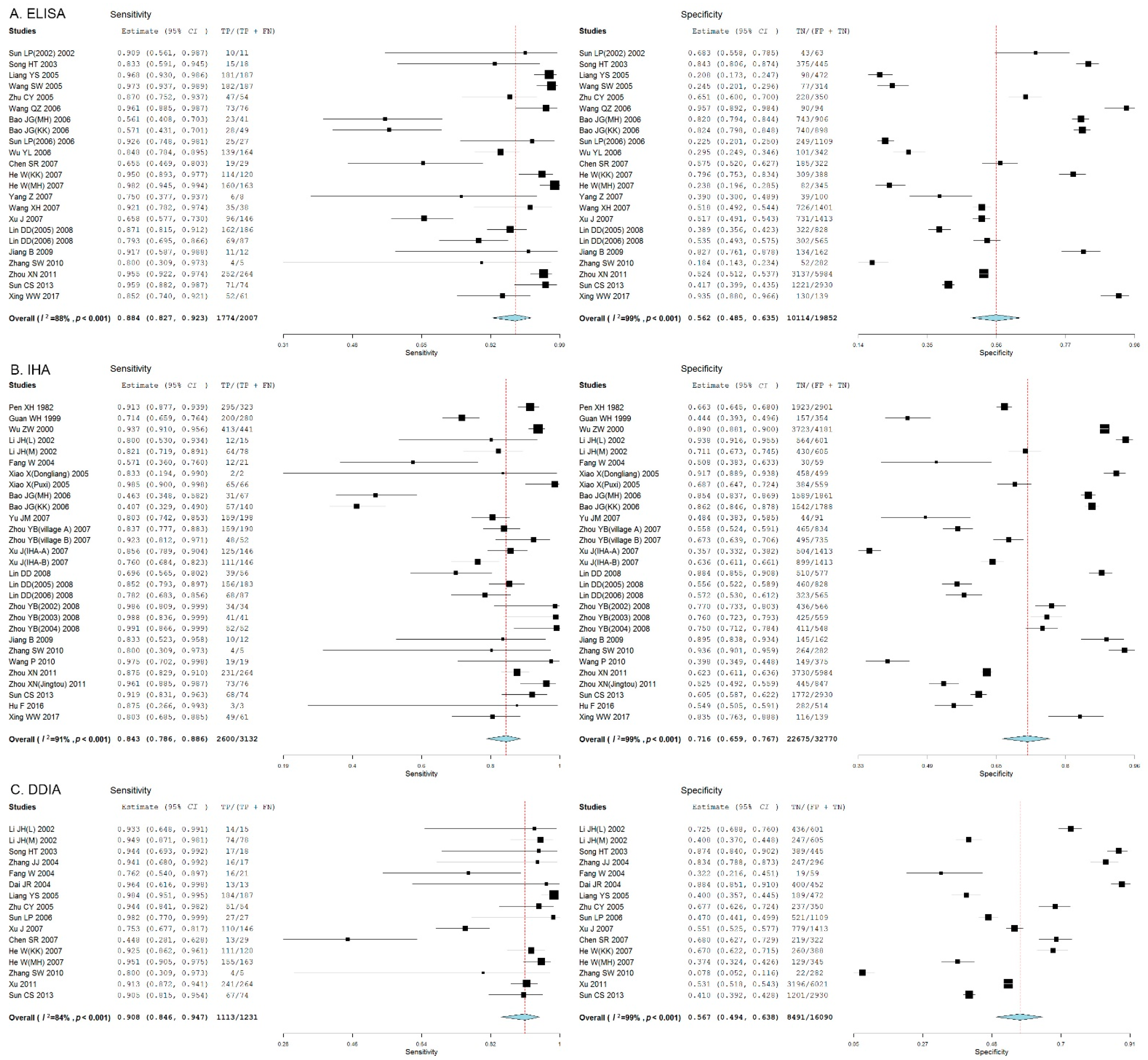
3.2. Meta-Analysis
3.3. Comparison of the Diagnostic Accuracy of Three Immunodiagnostic Assays
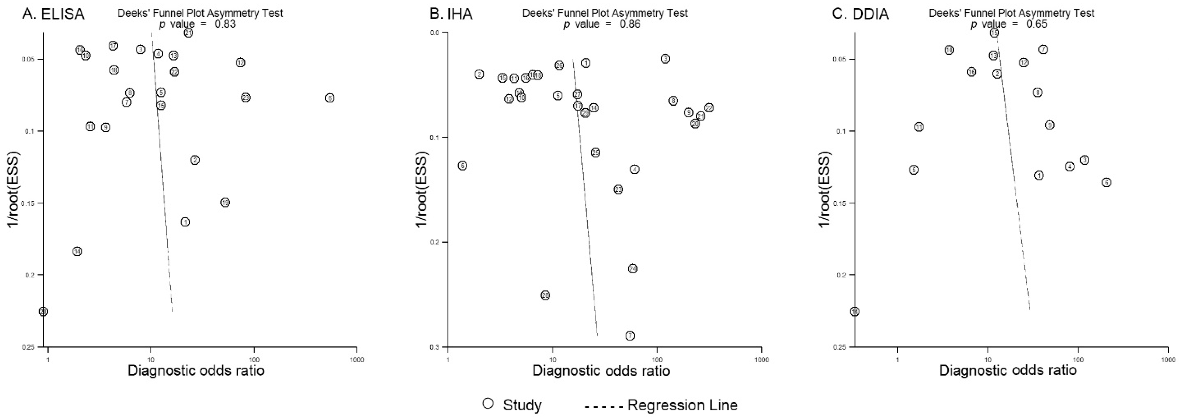
3.4. Publication Bias
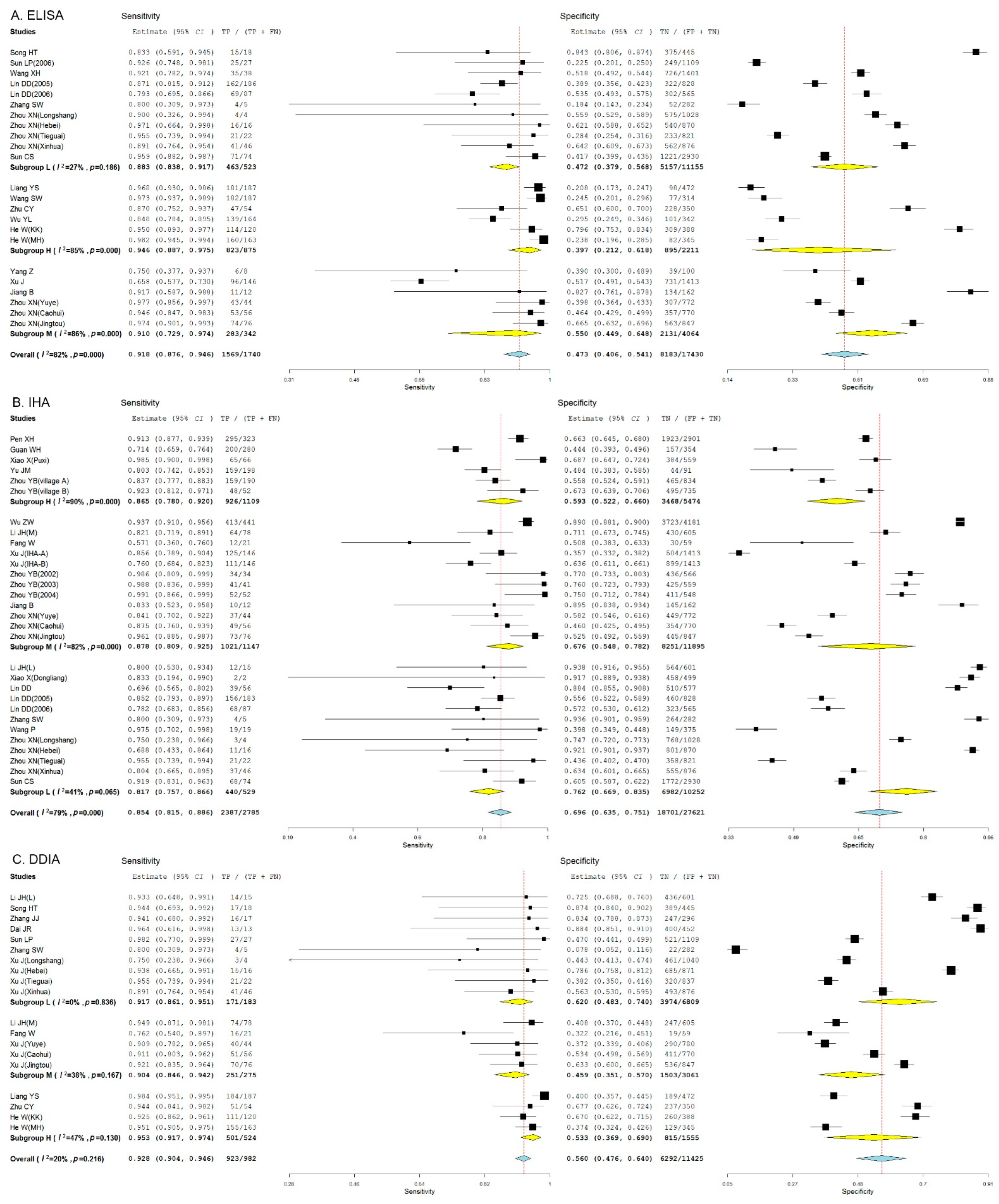
3.5. Subgroup Analysis
4. Discussion
Author Contributions
Funding
Institutional Review Board Statement
Informed Consent Statement
Data Availability Statement
Acknowledgments
Conflicts of Interest
References
- Verjee, M.A. Schistosomiasis: Still a Cause of significant morbidity and mortality. Res. Rep. Trop. Med. 2019, 10, 153–163. [Google Scholar] [CrossRef] [PubMed]
- Giboda, M.; Bergquist, R.; Utzinger, J. Schistosomiasis at the crossroad to elimination: Review of eclipsed research with emphasis on the post-transmission agenda. Trop. Med. Infect. Dis. 2022, 7, 55. [Google Scholar] [CrossRef] [PubMed]
- Deol, A.K.; Fleming, F.M.; Calvo-Urbano, B.; Walker, M.; Bucumi, V.; Gnandou, I.; Tukahebwa, E.M.; Jemu, S.; Mwingira, U.J.; Alkohlani, A.; et al. Schistosomiasis—Assessing progress toward the 2020 and 2025 global goals. N. Engl. J. Med. 2019, 381, 2519–2528. [Google Scholar] [CrossRef] [PubMed]
- Li, G.; Xu, D.; Hu, Y.; Xu, M.; Zhang, L.; Du, X.; Zhang, L.; Sun, C.; Xie, Y.; Tan, X. Impact of the coronavirus disease 2019 lockdown on Schistosoma host Oncomelania hupensis density in Wuhan. Acta Trop. 2022, 226, 106224. [Google Scholar] [CrossRef] [PubMed]
- Toor, J.; Adams, E.R.; Aliee, M.; Amoah, B.; Anderson, R.M.; Ayabina, D.; Bailey, R.; Basáñez, M.G.; Blok, D.J.; Blumberg, S.; et al. Predicted impact of COVID-19 on neglected tropical disease programs and the opportunity for innovation. Clin. Infect. Dis. 2021, 72, 1463–1466. [Google Scholar] [CrossRef]
- Mantica, G.; Martini, M.; Riccardi, N. The possible impact of SARS-CoV-2 on neglected tropical diseases in Europe: The out of spotlights emerging of schistosomiasis. J. Prev. Med. Hyg. 2021, 62, E3–E4. [Google Scholar]
- McManus, D.P.; Dunne, D.W.; Sacko, M.; Utzinger, J.; Vennervald, B.J.; Zhou, X.N. Schistosomiasis. Nat. Rev. Dis. Primers 2018, 4, 13. [Google Scholar] [CrossRef]
- Hong, Z.; Li, L.; Zhang, L.; Wang, Q.; Xu, J.; Li, S.; Zhou, X.N. Elimination of schistosomiasis japonica in China: From the One Health perspective. China CDC Wkly. 2022, 4, 130–134. [Google Scholar] [CrossRef]
- Xu, J.; Li, S.Z.; Zhang, L.J.; Bergquist, R.; Dang, H.; Wang, Q.; Lv, S.; Wang, T.P.; Lin, D.D.; Liu, J.B.; et al. Surveillance-based evidence: Elimination of schistosomiasis as a public health problem in the Peoples’ Republic of China. Infect. Dis. Poverty 2020, 9, 63. [Google Scholar] [CrossRef]
- Wang, W.; Bergquist, R.; King, C.H.; Yang, K. Elimination of schistosomiasis in China: Current status and future prospects. PLoS Negl. Trop. Dis. 2021, 15, e0009578. [Google Scholar] [CrossRef]
- Utzinger, J.; Becker, S.L.; van Lieshout, L.; van Dam, G.J.; Knopp, S. New diagnostic tools in schistosomiasis. Clin. Microbiol. Infect. 2015, 21, 529–542. [Google Scholar] [CrossRef] [PubMed]
- Lindholz, C.G.; Favero, V.; Verissimo, C.M.; Candido, R.R.F.; de Souza, R.P.; Dos Santos, R.R.; Morassutti, A.L.; Bittencourt, H.R.; Jones, M.K.; St Pierre, T.G.; et al. Study of diagnostic accuracy of Helmintex, Kato-Katz, and POC-CCA methods for diagnosing intestinal schistosomiasis in Candeal, a low intensity transmission area in northeastern Brazil. PLoS Negl. Trop. Dis. 2018, 12, e0006274. [Google Scholar] [CrossRef] [PubMed]
- Gray, D.J.; Ross, A.G.; Li, Y.S.; McManus, D.P. Diagnosis and management of schistosomiasis. BMJ 2011, 342, d2651. [Google Scholar] [CrossRef] [PubMed]
- LoVerde, P.T. Schistosomiasis. Adv. Exp. Med. Biol. 2019, 1154, 45–70. [Google Scholar]
- Lv, C.; Deng, W.; Wang, L.; Qin, Z.; Zhou, X.; Xu, J. Molecular techniques as alternatives of diagnostic tools in China as schistosomiasis moving towards elimination. Pathogens 2022, 11, 287. [Google Scholar] [CrossRef]
- Hinz, R.; Schwarz, N.G.; Hahn, A.; Frickmann, H. Serological approaches for the diagnosis of schistosomiasis—A review. Mol. Cell Probes 2017, 31, 2–21. [Google Scholar] [CrossRef]
- Chen, C.; Guo, Q.; Fu, Z.; Liu, J.; Lin, J.; Xiao, K.; Sun, P.; Cong, X.; Liu, R.; Hong, Y. Reviews and advances in diagnostic research on Schistosoma japonicum. Acta Trop. 2021, 213, 105743. [Google Scholar] [CrossRef]
- Zhang, J.F.; Xu, J.; Bergquist, R.; Yu, L.L.; Yan, X.L.; Zhu, H.Q.; Wen, L.Y. Development and application of diagnostics in the national schistosomiasis control programme in the People’s Republic of China. Adv. Parasitol. 2016, 92, 409–434. [Google Scholar]
- Zhou, Y.; Chen, Y.; Jiang, Q. History of human schistosomiasis (bilharziasis) in China: From discovery to elimination. Acta Parasitol. 2021, 66, 760–769. [Google Scholar] [CrossRef]
- Wang, X.Y.; Yang, K. Serological diagnosis methods of schistosomiasis japonica at different prevalence: A meta-analysis. Chin. J. Schisto. Control 2016, 28, 18–25. [Google Scholar]
- Zhu, H.; Yu, C.; Xia, X.; Dong, G.; Tang, J.; Fang, L.; Du, Y. Assessing the diagnostic accuracy of immunodiagnostic techniques in the diagnosis of schistosomiasis japonica: A meta-analysis. Parasitol. Res. 2010, 107, 1067–1073. [Google Scholar] [CrossRef] [PubMed]
- Wang, W.; Li, Y.; Li, H.; Xing, Y.; Qu, G.; Dai, J.; Liang, Y. Immunodiagnostic efficacy of detection of Schistosoma japonicum human infections in China: A meta analysis. Asian Pac. J. Trop. Med. 2012, 5, 15–23. [Google Scholar] [CrossRef]
- Liu, J. The role of the funnel plot in detecting publication and related biases in meta-analysis. Evid. Based Dent. 2011, 12, 121–122. [Google Scholar] [CrossRef] [PubMed]
- Hartzes, A.M.; Morgan, C.J. Meta-analysis for diagnostic tests. J. Nucl. Cardiol. 2019, 26, 68–71. [Google Scholar] [CrossRef] [PubMed]
- Zhou, X.N.; Guo, J.G.; Wu, X.H.; Jiang, Q.W.; Zheng, J.; Dang, H.; Wang, X.H.; Xu, J.; Zhu, H.Q.; Wu, G.L.; et al. Epidemiology of schistosomiasis in the People’s Republic of China, 2004. Emerg. Infect. Dis. 2007, 13, 1470–1476. [Google Scholar] [CrossRef] [PubMed]
- Cao, C.L.; Zhang, L.J.; Deng, W.P.; Li, Y.L.; Lv, C.; Dai, S.M.; Feng, T.; Qin, Z.Q.; Duan, L.P.; Zhang, H.B.; et al. Contributions and achievements on schistosomiasis control and elimination in China by NIPD-CTDR. Adv. Parasitol. 2020, 110, 1–62. [Google Scholar]
- Peng, X.H.; Hou, C.S.; Shi, C.C. Evaluating the value of indirect hemagglutination assay for the field diagnosis of schistosomiasis japonica. Sichuan Med. 1982, 3, 72–73. [Google Scholar]
- Guan, W.H.; Yuan, H.C.; Zhao, G.M.; Yu, J.M.; Yang, Q.J. Survey of schistosomiasis epidemic in dam-circle marsh region. Chin. J. Public Health 1999, 15, 686–688. [Google Scholar]
- Wu, Z.W.; Liu, Z.C.; Yang, G.F. Reliability of application of IHA method for determine chemotherapy targets of schistosomiasis in moderately endemic areas of lake region. Chin. J. Schist. Control 2000, 12, 21–23. [Google Scholar]
- Li, J.H.; Wang, T.P.; Xiao, X.; Wu, W.D.; Lv, D.B.; Fang, G.R.; Cai, W.; Zheng, J.; Xu, J.; Wang, R.R. Cost-effectiveness analysis on different schistosomiasis case screen methods in hypo-endemic area. J. Pract. Parasit. Dis. 2002, 10, 145–148. [Google Scholar]
- Li, J.H.; Wang, T.P.; Xiao, X.; Wu, W.D.; Lv, D.B.; Fang, G.R.; Cai, W.; Zheng, J.; Xu, J.; Wang, R.R. Cost-effectiveness analysis on different schistosomiasis case screen methods in hypo-endemic area. Chin. J Schist. Control. 2002, 14, 418–421. [Google Scholar]
- Sun, L.P.; Hong, Q.B.; Zhou, X.N.; Huang, Y.X.; Wu, F.; Zhang, Y.P.; Yang, G.J. Field evaluation of fraction antigen of SEA applied in screening of schistosomiasis. Chin. Parasit. Dis Control. 2002, 15, 42–44. [Google Scholar]
- Song, H.T.; Liang, Y.S.; Dai, J.R.; Li, H.J.; Ji, C.S.; Shen, X.H.; Li, L.G.; Yin, F. Cost-effectiveness of three immunoassays for diagnosis of schistosomiasis in lower endemic area. Chin. J. Schist. Control 2003, 15, 300–301. [Google Scholar]
- Zhang, J.J.; Xu, L.; Song, H.T. Application of DDIA for screening schistosomiasis in high-risk populations. J. Trop. Dis. Parasitol. 2004, 2, 50–51. [Google Scholar]
- Fang, W.; Gan, Z.M.; Dong, P.H.; Yang, T.L.; Chen, F.; Luo, B.R.; Qiu, Z.L. Field application of dipstick dye immunoassay in schistosomiasis-endemic areas in Yunnan Province. Chin. J. Schist. Control 2004, 16, 325–326. [Google Scholar]
- Dai, J.R.; Zhu, Y.C.; Liang, Y.S.; Zhao, S.; Li, H.J.; Xu, Y.L.; Hua, W.Q.; Cao, G.Q.; Xu, M. Study on scheme for screening schistosomiasis in low endemic areas. Chin. J. Schist. Control 2004, 16, 13–15. [Google Scholar]
- Zhu, Y.C.; He, W.; Dai, J.R.; Xu, M.; Liang, Y.S.; Tang, J.X.; Hua, W.Q.; Cao, G.Q.; Chen, H.G.; Lou, P.A. Application of dipstick dye immunoassay (DDIA) kit on detection of schistosomiasis japonica on large scale in endemic areas. Chin. J. Schist. Control 2004, 16, 13–15. [Google Scholar]
- Liang, Y.S.; Zhu, Y.C.; Dai, J.R.; He, W.; Xu, M.; Li, Y.L.; Wang, S.W.; Tang, J.X.; Hua, W.Q.; Li, H.J.; et al. Field application of dipstick dye immunoassay (DDIA) kit for detecting schistosomiasis in mountainous endemic regions in Yunnan Province. Chin. J. Schist. Control 2005, 17, 405–408. [Google Scholar]
- Wang, S.W.; Yang, Z.; Yin, G.L.; Li, Y.L.; Yang, J.; Zhao, J.B.; Luo, B.R.; Zuo, X.F.; Zou, H.M.; Zhang, J.P. Application of antibody-based ELISA for detection of schistosomiasis in highly endemic regions. Parasit. Infect. Dis. 2005, 3, 79–80. [Google Scholar]
- Xiao, X.; Wang, T.; Ye, H.; Qiang, G.; Wei, H.; Tian, Z. Field evaluation of a rapid, visually-read colloidal dye immunofiltration assay for Schistosoma japonicum for screening in areas of low transmission. Bull. World Health Organ. 2005, 83, 526–533. [Google Scholar]
- Wang, Q.Z.; Wang, F.F.; Yin, X.M.; Zhu, L.; Zhang, G.H.; Fang, G.R.; Wang, T.P.; Xiao, X.; Jiang, Q.W. Evaluation of screening effects of ELISA and IHA techniques in different epidemic areas of schistosomiasis. J. Trop. Dis. Parasitol. 2006, 4, 135–139. [Google Scholar]
- Wu, Y.L.; Cheng, M.; Meng, W.; Li, H.X. Comparison between effect of two schistosomiassi diagnostic kit application to the multimountain area in Yunnan Province. BMC J. 2006, 29, 168–169. [Google Scholar]
- Bao, J.G.; Qiang, G.X.; Deng, Y.J.; Zhang, R.; Chen, Y. Comparison of four diagnostic assays for the field detection of schistosomiasis. J. Trop. Dis. Parasitol. 2006, 4, 33–34. [Google Scholar]
- Sun, L.P.; Hong, Q.B.; Huang, Y.X.; Liang, Y.S.; Xu, M.; Zhang, L.H.; Gao, Y.; Zhou, M.; Yang, K.; Zhu, Y.C. Comparison of two immunoassays for schistosomiaisis diagnosis in the field. Chin. J. Schist. Control 2004, 16, 192–196. [Google Scholar]
- Chen, S.R.; Chen, F.; Zhou, X.N.; Li, H.J.; Stenmann, P.J.; Yang, Z.; Li, Y.L. Comparison of aetiological and serological diagnosis methods in schistosomiasis mountainous endemic area. Parasitol. Infect. Dis. 2007, 5, 1–4. [Google Scholar]
- He, W.; Zhu, Y.C.; Liang, Y.S.; Dai, J.R.; Xu, M.; Tang, J.X.; Cao, G.Q.; Hua, W.Q.; Li, Y.L.; Yang, Z. Comparison of stool examination and immunodiagnosis for schistosomiasis. Chin. J. Schist. Control 2007, 19, 107–109. [Google Scholar]
- Xu, J.; Chen, N.G.; Feng, T.; Wang, E.M.; Wu, X.H.; Chen, H.G.; Wang, T.P.; Zhou, X.N.; Zheng, J. Effectiveness of routinely used assays for the diagnosis of schistosomiasis japonica in the field. Chin. Parasitol. Parasit. Dis. 2007, 25, 175–179. [Google Scholar]
- Yang, Z.; Yin, G.L.; Fan, C.Z.; Luo, B.R.; Liu, Y.H.; Duan, Y.C.; Cui, Y.H.; Yang, Y.N.; Sun, H.Y.; Wang, S.W. Effect of gold labeling immunoassay for diagnosis of schistosomiasis. Parasitol. Infect. Dis. 2007, 5, 97–98. [Google Scholar]
- Zhou, Y.B.; Yang, M.X.; Wang, Q.Z.; Zhao, G.M.; Wei, J.G.; Peng, W.X.; Jiang, Q.W. Field comparison of immunodiagnostic and parasitological techniques for the detection of Schistosomiasis japonica in the People’s Republic of China. Am. J. Trop. Med. Hyg. 2007, 76, 1138–1143. [Google Scholar] [CrossRef]
- Yu, J.M.; de Vlas, S.J.; Jiang, Q.W.; Gryseels, B. Comparison of the Kato-Katz technique, hatching test and indirect hemagglutination assay (IHA) for the diagnosis of Schistosoma japonicum infection in China. Parasitol. Int. 2007, 56, 45–49. [Google Scholar] [CrossRef]
- Wang, X.H.; Zhou, X.N.; Li, Y.L.; Lv, S.; Li, L.H.; Jia, T.W.; Chen, S.R.; Yang, Z.; Fang, W.; Chen, F. Evaluation of two tests for detecting Schistosoma japonicum infection using a Bayesian approach. Chin. J. Health Stat. 2007, 24, 361–364. [Google Scholar]
- Lin, D.D.; Liu, Y.M.; Hu, F.; Tao, B.; Wang, X.M.; Zuo, X.X.; Li, J.Y.; Wu, G.L. Evaluation on application of common diagnosis methods for schistosomiasis japonica in endemic areas of China I Evaluation on estimation of prevalence of Schistosoma japonicum infection by IHA screening method. Chin. J. Schist. Control 2008, 20, 179–183. [Google Scholar]
- Zhou, Y.B.; Yang, M.X.; Tao, P.; Jiang, Q.L.; Zhao, G.M.; Wei, J.G.; Jiang, Q.W. A longitudinal study of comparison of the Kato-Katz technique and indirect hemagglutination assay (IHA) for the detection of schistosomiasis japonica in China, 2001–2006. Acta Trop. 2008, 107, 251–254. [Google Scholar] [CrossRef]
- Lin, D.D. Evaluation of the Performance of Commonly Used Diagnostic Assays for the Field Detection of Schistosomiasis Japonica in China; Nanjing Medical University: Nanjing, China, 2008. [Google Scholar]
- Lin, D.D.; Xu, J.M.; Zhang, Y.Y.; Liu, Y.M.; Hu, F.; Xu, X.L.; Li, J.Y.; Gao, Z.L.; Wu, H.W.; Kurtis, J.; et al. Evaluation of IgG-ELISA for the diagnosis of Schistosoma japonicum in a high prevalence, low intensity endemic area of China. Acta Trop. 2008, 107, 128–133. [Google Scholar] [CrossRef] [PubMed]
- Jiang, B.; Zhou, Y.D.; Meng, Q.Y.; Luo, Q.L.; Shen, J.L. Study on IgY immunoglobulin-based double antibody sandwich ELISA for schistosomiaisis diagnosis in the field. J. Trop. Med. Parasitol. 2009, 7, 138–140. [Google Scholar]
- Zhang, S.W.; Cheng, B.; Qu, H.J.; Chen, Z.M.; Zou, Q.; Chu, L.P.; Zhang, L.; He, H.R.; Tang, S.H.; Huang, X.P.; et al. Validity evaluation of dipstick dye immuno-assay (DDIA) for screening in low endemic areas of schistosomiasis. Chin. J. Schist. Control 2010, 22, 171–173. [Google Scholar]
- Wang, P.; Ren, C.P.; Wang, T.P.; Shen, J.J. Evaluation of recombinant 29 000 extra membranous protein for the ummunodiagnosis of schistosomiasis japonica. Chin. J. Parasitol. Parasit. Dis. 2010, 28, 284–286. [Google Scholar]
- Xu, J.; Feng, T.; Lin, D.D.; Wang, Q.Z.; Tang, L.; Wu, X.H.; Guo, J.G.; Peeling, R.W.; Zhou, X.N. Performance of a dipstick dye immunoassay for rapid screening of Schistosoma japonicum infection in areas of low endemicity. Parasit. Vectors 2011, 4, 87. [Google Scholar] [CrossRef]
- Zhou, X.N.; Xu, J.; Chen, H.G.; Wang, T.P.; Huang, X.B.; Lin, D.D.; Wang, Q.Z.; Tang, L.; Guo, J.G.; Wu, X.H.; et al. Tools to support policy decisions related to treatment strategies and surveillance of Schistosomiasis japonica towards elimination. PLoS Negl. Trop. Dis. 2011, 5, e1408. [Google Scholar] [CrossRef]
- Sun, C.S.; Wang, F.F.; Wang, Y.; Zhou, L.; Yin, X.M.; Wang, Q.Z.; Zhang, L.S.; Wang, E.M.; Zhang, S.Q. Effectiveness of an indirect hemagglutination assay kit at diagnosing schistosomiasis in the field. J. Parasit. Biol. 2013, 8, 982–985. [Google Scholar]
- Hu, F.; Li, Z.J.; Li, Y.F.; Yuan, M.; Xie, S.Y.; Liu, Y.M.; Li, J.Y.; Gao, Z.L.; Pu, Y.; Wang, J.M.; et al. Study on cut-off value of IHA method for schistosomiasis diagnosis in different endemic areas. Chin. J. Schist. Control 2016, 28, 644–647, 682. [Google Scholar]
- Xing, W.; Yu, X.; Feng, J.; Sun, K.; Fu, W.; Wang, Y.; Zou, M.; Xia, W.; Luo, Z.; He, H.; et al. Field evaluation of a recombinase polymerase amplification assay for the diagnosis of Schistosoma japonicum infection in Hunan province of China. BMC Infect. Dis. 2017, 17, 164. [Google Scholar] [CrossRef] [PubMed]
- Weerakoon, K.G.; Gobert, G.N.; Cai, P.; McManus, D.P. Advances in the diagnosis of human schistosomiasis. Clin. Microbiol. Rev. 2015, 28, 939–967. [Google Scholar] [CrossRef]
- Zhang, S.Q.; Sun, C.S.; Wang, M.; Lin, D.D.; Zhou, X.N.; Wang, T.P. Epidemiological features and effectiveness of schistosomiasis control programme in lake and marshland region in the People’s Republic of China. Adv. Parasitol. 2016, 92, 39–71. [Google Scholar] [PubMed]
- Shi, L.; Li, W.; Wu, F.; Zhang, J.F.; Yang, K.; Zhou, X.N. Epidemiological features and control progress of schistosomiasis in waterway-network region in the People’s Republic of China. Adv. Parasitol. 2016, 92, 97–116. [Google Scholar] [PubMed]
- Liu, Y.; Zhou, Y.B.; Li, R.Z.; Wan, J.J.; Yang, Y.; Qiu, D.C.; Zhong, B. Epidemiological features and effectiveness of schistosomiasis control programme in mountainous and hilly region of the People’s Republic of China. Adv. Parasitol. 2016, 92, 73–95. [Google Scholar] [PubMed]
- He, P.; Gordon, C.A.; Williams, G.M.; Li, Y.; Wang, Y.; Hu, J.; Gray, D.J.; Ross, A.G.; Harn, D.; McManus, D.P. Real-time PCR diagnosis of Schistosoma japonicum in low transmission areas of China. Infect. Dis. Poverty 2018, 7, 8. [Google Scholar] [CrossRef]
- Fung, M.S.; Xiao, N.; Wang, S.; Carlton, E.J. Field evaluation of a PCR test for Schistosoma japonicum egg detection in low-prevalence regions of China. Am. J. Trop. Med. Hyg. 2012, 87, 1053–1058. [Google Scholar] [CrossRef]
- Lier, T.; Johansen, M.V.; Hjelmevoll, S.O.; Vennervald, B.J.; Simonsen, G.S. Real-time PCR for detection of low intensity Schistosoma japonicum infections in a pig model. Acta Trop. 2008, 105, 74–80. [Google Scholar] [CrossRef]
- Xu, J.; Guan, Z.X.; Zhao, B.; Wang, Y.Y.; Cao, Y.; Zhang, H.Q.; Zhu, X.Q.; He, Y.K.; Xia, C.M. DNA detection of Schistosoma japonicum: Diagnostic validity of a LAMP assay for low-intensity infection and effects of chemotherapy in humans. PLoS Negl. Trop. Dis. 2015, 9, e0003668. [Google Scholar] [CrossRef]
- Xu, J.; Rong, R.; Zhang, H.Q.; Shi, C.J.; Zhu, X.Q.; Xia, C.M. Sensitive and rapid detection of Schistosoma japonicum DNA by loop-mediated isothermal amplification (LAMP). Int. J. Parasitol. 2010, 40, 327–331. [Google Scholar] [CrossRef] [PubMed]
- Wang, C.; Chen, L.; Yin, X.; Hua, W.; Hou, M.; Ji, M.; Yu, C.; Wu, G. Application of DNA-based diagnostics in detection of schistosomal DNA in early infection and after drug treatment. Parasit. Vectors 2011, 4, 164. [Google Scholar] [CrossRef] [PubMed]
- Song, Z.; Ting, L.; Kun, Y.; Wei, L.; Jian-Feng, Z.; Li-Chuan, G.; Yan-Hong, L.; Yang, D.; Qing-Jie, Y.; Hai-Tao, Y. Establishment of a recombinase-aided isothermal amplification technique to detect Schistosoma japonicum specific gene fragments. Chin. J. Schisto. Control 2018, 30, 273–277. [Google Scholar]
- Ye, Y.Y.; Zhao, S.; Liu, Y.H.; Zhang, J.F.; Xiong, C.R.; Ying, Q.J.; Yang, K. Establishment of a nucleic acid dipstick test for detection of Schistosoma japonicum specific gene fragments based on the recombinase-aided isothermal amplification assay. Chin. J. Schist. Control 2021, 33, 334–338. [Google Scholar]
- Zhao, S.; Liu, Y.H.; Li, T.; Li, W.; Zhang, J.F.; Guo, L.C.; Ying, Q.J.; Yang, H.T.; Yang, K. Rapid detection of Schistosoma japonicum specific gene fragment by recombinase aided isothermal amplification combined with fluorescent probe. Chin. J. Parasitol. Parasit. Dis 2019, 37, 23–27. [Google Scholar]
- Sun, K.; Xing, W.; Yu, X.; Fu, W.; Wang, Y.; Zou, M.; Luo, Z.; Xu, D. Recombinase polymerase amplification combined with a lateral flow dipstick for rapid and visual detection of Schistosoma japonicum. Parasit. Vectors 2016, 9, 476. [Google Scholar] [CrossRef][Green Version]
- Deng, W.; Wang, S.; Wang, L.; Lv, C.; Li, Y.; Feng, T.; Qin, Z.; Xu, J. Laboratory evaluation of a basic recombinase polymerase amplification (RPA) assay for early detection of Schistosoma japonicum. Pathogens 2022, 11, 319. [Google Scholar] [CrossRef]
- WHO. Ending the Neglect to Attain the Sustainable Development Goals: A Road Map for Neglected Tropical Diseases 2021–2030. Available online: https://www.who.int/publications/i/item/9789240010352 (accessed on 6 June 2022).
- WHO. WHO Guideline on Control and Elimination of Human Schistosomiasis. Available online: https://www.who.int/publications/i/item/9789240041608 (accessed on 6 June 2022).
- Chen, J.; Bergquist, R.; Zhou, X.N.; Xue, J.B.; Qian, M.B. Combating infectious disease epidemics through China’s Belt and Road Initiative. PLoS Negl. Trop. Dis. 2019, 13, e0007107. [Google Scholar] [CrossRef]
- Wang, X.Y.; He, J.; Juma, S.; Kabole, F.; Guo, J.G.; Dai, J.R.; Li, W.; Yang, K. Efficacy of China-made praziquantel for treatment of schistosomiasis haematobium in Africa: A randomized controlled trial. PLoS Negl. Trop. Dis. 2019, 13, e0007238. [Google Scholar] [CrossRef]
- Xing, Y.T.; Dai, J.R.; Yang, K.; Jiang, T.; Jiang, C.G.; Mohammed, S.J.; Kabole, F. Bulinus snails control by China-made niclosamide in Zanzibar: Experiences and lessons. In Sino-African Cooperation for Schistosomiasis Control in Zanzibar; Yang, K., Mehlhorn, H., Eds.; Springer Nature Switzerland AG: Cham, Switzerland, 2021; pp. 147–159. [Google Scholar]
- Zhang, L.J.; Mwanakasale, V.; Xu, J.; Sun, L.P.; Yin, X.M.; Zhang, J.F.; Hu, M.C.; Si, W.M.; Zhou, X.N. Diagnostic performance of two specific Schistosoma japonicum immunological tests for screening Schistosoma haematobium in school children in Zambia. Acta Trop. 2020, 202, 105285. [Google Scholar] [CrossRef]
- Zhu, Y.C.; Socheat, D.; Bounlu, K.; Liang, Y.S.; Sinuon, M.; Insisiengmay, S.; He, W.; Xu, M.; Shi, W.Z.; Bergquist, R. Application of dipstick dye immunoassay (DDIA) kit for the diagnosis of schistosomiasis mekongi. Acta Trop. 2005, 96, 137–141. [Google Scholar] [CrossRef] [PubMed]
- Hua, H.Y.; Wang, W.; Cao, G.Q.; Tang, F.; Liang, Y.S. Improving the management of imported schistosomiasis haematobia in China: Lessons from a case with multiple misdiagnoses. Parasit. Vectors 2013, 6, 260. [Google Scholar] [CrossRef] [PubMed][Green Version]
- Zhu, Y.C.; Hassen, S.; He, W.; Cao, G.Q. Preliminary study on detection of schistosomiasis mansoni with dipstick dye immunoassay (DDIA) kit. Chin. J. Schist. Control 2006, 18, 419–421. [Google Scholar]


| Publication Year | Subjects’ Age (Years) | Degree of Endemicity | Epidemic Types | Immunological Assay | Parasitological Technique | True Positives | False Negatives | True Negatives | False Positives | Reference |
|---|---|---|---|---|---|---|---|---|---|---|
| 1982 | >15 | High | Hilly and mountainous regions | IHA | Miracidium hatching test (three slides from three stool samples) | 295 | 28 | 1923 | 978 | [27] |
| 1999 | 3 to 70 | High | Plain regions with waterway networks | IHA | Kato-Katz (two slides from one stool sample) | 200 | 80 | 157 | 197 | [28] |
| 2000 | 6 to 60 | Medium | Marshland and lake regions | IHA | Kato-Katz | 413 | 28 | 3723 | 458 | [29] |
| 2002 | 5 to 56 | Low | Marshland and lake regions | IHA | Kato-Katz (three slides from one stool sample) | 12 | 3 | 564 | 37 | [30] |
| DDIA | 14 | 1 | 436 | 165 | ||||||
| 2002 | N/A | Medium | Marshland and lake regions | IHA | Kato-Katz (three slides from one stool sample) | 64 | 14 | 430 | 175 | [31] |
| DDIA | 74 | 4 | 247 | 358 | ||||||
| 2002 | 5 to 60 | N/A | Marshland and lake regions | ELISA | Miracidium hatching test | 10 | 1 | 43 | 20 | [32] |
| 2003 | 6 to 64 | Low | Marshland and lake regions | DDIA | Kato-Katz (three slides from one stool sample) and miracidium hatching test | 17 | 1 | 389 | 56 | [33] |
| ELISA | 15 | 3 | 375 | 70 | ||||||
| 2004 | N/A | Low | Marshland and lake regions | DDIA | Kato-Katz | 16 | 1 | 247 | 49 | [34] |
| 2004 | 6 to 60 | Medium | Hilly and mountainous regions | IHA | Miracidium hatching test | 12 | 9 | 30 | 29 | [35] |
| DDIA | 16 | 5 | 19 | 40 | ||||||
| 2004 | 60 to 65 | Low | Marshland and lake regions | DDIA | Miracidium hatching test (three slides from three stool samples) | 13 | 0 | 400 | 52 | [36] |
| 2005 | 15 to 70 | High | Marshland and lake regions | DDIA | Miracidium hatching test (three slides from one stool sample) | 51 | 3 | 237 | 113 | [37] |
| ELISA | 47 | 7 | 228 | 122 | ||||||
| 2005 | 10 to 70 | High | Hilly and mountainous regions | DDIA | Kato-Katz (three slides from one stool sample) | 184 | 3 | 189 | 283 | [38] |
| ELISA | 181 | 6 | 98 | 374 | ||||||
| 2005 | 6 to 65 | High | Hilly and mountainous regions | ELISA | Kato-Katz (three slides from one stool sample) and miracidium hatching test | 182 | 5 | 77 | 237 | [39] |
| 2005 | N/A | Low | Hilly and mountainous regions | IHA | Kato-Katz (three slides from one stool sample) | 2 | 0 | 458 | 41 | [40] |
| High | 65 | 1 | 384 | 175 | ||||||
| 2006 | 5 to 65 | N/A | Hilly and mountainous regions, and marshland and lake regions | ELISA | Kato-Katz (six slides from two stool samples) | 73 | 3 | 90 | 4 | [41] |
| IHA | 68 | 8 | 94 | 4 | ||||||
| 2006 | N/A | High | Hilly and mountainous regions | ELISA | Miracidium hatching test | 139 | 25 | 101 | 241 | [42] |
| 2006 | >5 | High, medium, and low | Hilly and mountainous regions | IHA | Miracidium hatching test (three slides from three stool samples) | 31 | 36 | 1589 | 272 | [43] |
| Kato-Katz (two slides from one stool sample) | 57 | 83 | 1542 | 246 | ||||||
| ELISA | Miracidium hatching test (three slides from three stool samples) | 23 | 18 | 743 | 163 | |||||
| Kato-Katz (two slides from one stool sample) | 28 | 21 | 740 | 158 | ||||||
| 2006 | 6 to 65 | Low | Marshland and lake regions, plain regions with waterway networks, and hilly and mountainous regions | ELISA | Miracidium hatching test (one slide from one stool sample) | 25 | 2 | 249 | 860 | [44] |
| DDIA | 27 | 0 | 521 | 588 | ||||||
| 2007 | >5 | High, medium, and low | Hilly and mountainous regions | ELISA | Miracidium hatching test (one slide from one stool sample) and Kato-Katz (four slides from one stool sample) | 19 | 10 | 185 | 137 | [45] |
| DDIA | 13 | 16 | 219 | 103 | ||||||
| 2007 | 10 to 70 | High | Hilly and mountainous regions | DDIA | Kato-Katz (three slides from one stool sample) | 111 | 9 | 260 | 128 | [46] |
| ELISA | 114 | 6 | 309 | 79 | ||||||
| DDIA | Miracidium hatching test (one slide from one stool sample) | 155 | 8 | 129 | 216 | |||||
| ELISA | 160 | 3 | 82 | 263 | ||||||
| 2007 | 6 to 65 | Medium | Marshland and lake regions | DDIA | Miracidium hatching test (three slides from one stool sample) and Kato-Katz (three slides from one stool sample) | 110 | 36 | 779 | 634 | [47] |
| ELISA | 96 | 50 | 731 | 682 | ||||||
| IHA-A | 125 | 21 | 504 | 909 | ||||||
| IHA-B | 111 | 35 | 899 | 514 | ||||||
| 2007 | 6 to 65 | Medium | Hilly and mountainous regions | ELISA | Miracidium hatching test (three slides from three stool samples) | 6 | 2 | 39 | 61 | [48] |
| 2007 | 5 to 75 | Village A: high | Marshland and lake regions | IHA | Kato-Katz (three slides from one stool sample) | 159 | 31 | 465 | 369 | [49] |
| Village B: medium | IHA | 48 | 4 | 495 | 240 | |||||
| 2007 | N/A | High | Marshland and lake regions | IHA | Kato-Katz (seven slides from one stool sample) and miracidium hatching test | 159 | 39 | 44 | 47 | [50] |
| 2007 | 6 to 65 | Low | Hilly and mountainous regions | ELISA | Kato-Katz (four slides from one stool sample) | 35 | 3 | 726 | 675 | [51] |
| 2008 | N/A | N/A | Marshland and lake regions | IHA | Kato-Katz (twelve slides from two stool samples) | 39 | 17 | 510 | 67 | [52] |
| 2008 | 6 to 65 | Medium | Hilly and mountainous regions | IHA | Kato-Katz (three slides from one stool sample) | 34 | 0 | 436 | 130 | [53] |
| IHA | 41 | 0 | 425 | 134 | ||||||
| IHA | 52 | 0 | 411 | 137 | ||||||
| 2008 | >5 | High | Marshland and lake regions | IHA | Kato-Katz (six slides from two stool samples) | 156 | 27 | 460 | 368 | [54] |
| IHA | 68 | 19 | 323 | 242 | ||||||
| 2008 | >5 | High | Marshland and lake regions | ELISA | Kato-Katz (six slides from two stool samples) | 162 | 24 | 322 | 506 | [55] |
| ELISA | 69 | 18 | 302 | 263 | ||||||
| 2009 | 11 to 46 | Medium | Marshland and lake regions | IHA | Kato-Katz (three slides from one stool sample) | 10 | 2 | 145 | 17 | [56] |
| ELISA | 11 | 1 | 134 | 28 | ||||||
| 2010 | 6 to 65 | Low | Marshland and lake regions | IHA | Miracidium hatching test (three slides from one stool sample) | 4 | 1 | 264 | 18 | [57] |
| DDIA | 4 | 1 | 22 | 260 | ||||||
| ELISA | 4 | 1 | 52 | 230 | ||||||
| 2010 | 5 to 80 | Low | Hilly and mountainous regions | IHA | Kato-Katz (nine slides from three stool samples) and miracidium hatching test | 19 | 0 | 149 | 226 | [58] |
| 2011 | 6 to 65 | Medium and low | Marshland and lake regions, and hilly and mountainous regions | DDIA | Kato-Katz (three slides from one stool sample) and miracidium hatching test | 241 | 23 | 3196 | 2825 | [59] |
| 2011 | 6 to 65 | Medium and low | Marshland and lake regions, and hilly and mountainous regions | IHA | Kato-Katz (three slides from one stool sample) and miracidium hatching test | 231 | 33 | 3370 | 2254 | [60] |
| ELISA | 252 | 12 | 3137 | 2847 | ||||||
| 2013 | 6 to 65 | Low | Marshland and lake regions, and hilly and mountainous regions | IHA | Kato-Katz (three slides from one stool sample) and miracidium hatching test | 68 | 6 | 1772 | 1158 | [61] |
| DDIA | 67 | 7 | 1201 | 1729 | ||||||
| ELISA | 71 | 3 | 1221 | 1709 | ||||||
| 2016 | >5 | N/A | Marshland and lake regions | IHA | Kato-Katz (twenty seven slides from three stool samples) | 3 | 0 | 282 | 232 | [62] |
| 2017 | N/A | N/A | Marshland and lake regions | IHA | Kato-Katz (three slides from one stool sample) and miracidium hatching test | 49 | 12 | 116 | 23 | [63] |
| ELISA | 52 | 9 | 130 | 9 |
Publisher’s Note: MDPI stays neutral with regard to jurisdictional claims in published maps and institutional affiliations. |
© 2022 by the authors. Licensee MDPI, Basel, Switzerland. This article is an open access article distributed under the terms and conditions of the Creative Commons Attribution (CC BY) license (https://creativecommons.org/licenses/by/4.0/).
Share and Cite
Mei, Z.; Lv, S.; Tian, L.; Wang, W.; Jia, T. The Efficiency of Commercial Immunodiagnostic Assays for the Field Detection of Schistosoma japonicum Human Infections: A Meta-Analysis. Pathogens 2022, 11, 791. https://doi.org/10.3390/pathogens11070791
Mei Z, Lv S, Tian L, Wang W, Jia T. The Efficiency of Commercial Immunodiagnostic Assays for the Field Detection of Schistosoma japonicum Human Infections: A Meta-Analysis. Pathogens. 2022; 11(7):791. https://doi.org/10.3390/pathogens11070791
Chicago/Turabian StyleMei, Zhongqiu, Shan Lv, Liguang Tian, Wei Wang, and Tiewu Jia. 2022. "The Efficiency of Commercial Immunodiagnostic Assays for the Field Detection of Schistosoma japonicum Human Infections: A Meta-Analysis" Pathogens 11, no. 7: 791. https://doi.org/10.3390/pathogens11070791
APA StyleMei, Z., Lv, S., Tian, L., Wang, W., & Jia, T. (2022). The Efficiency of Commercial Immunodiagnostic Assays for the Field Detection of Schistosoma japonicum Human Infections: A Meta-Analysis. Pathogens, 11(7), 791. https://doi.org/10.3390/pathogens11070791


