Article

Pet Reptiles in Poland as a Potential Source of Transmission of Salmonella

1
Department of Veterinary Prevention and Avian Diseases, Faculty of Veterinary Medicine, University of Life Sciences in Lublin, 20-033 Lublin, Poland
2
Department of Microbiology, National Veterinary Research Institute, 24-100 Puławy, Poland
3
Department of Parasitology and Fish Diseases, Faculty of Veterinary Medicine, University of Life Sciences in Lublin, 20-033 Lublin, Poland
*
Authors to whom correspondence should be addressed.
Pathogens 2022, 11(10), 1125; https://doi.org/10.3390/pathogens11101125
Submission received: 10 September 2022 / Revised: 27 September 2022 / Accepted: 27 September 2022 / Published: 29 September 2022
(This article belongs to the Special Issue Bacterial, Fungal and Parasitic Zoonoses)

Abstract

:
Reptiles are considered a potential source of Salmonella transmission to humans. The aim of this research was to determine the incidence of Salmonella in pet reptiles in Poland and to examine Salmonella isolates with regard to their biochemical characteristics, serotype, antimicrobial susceptibility, and pathogenic and zoonotic potential. The research material consisted of 67 reptile faeces samples. The taxonomic affiliation of the Salmonella isolates was determined by MALDI-TOF mass spectrometry, biochemical analyses, and serotyping; whole genome sequencing (WGS) analysis was performed on three isolates whose serotype could not be determined by agglutination. The antimicrobial susceptibility of the Salmonella isolates was determined by the broth dilution method, and in the case of some antimicrobials by the disk diffusion method. The pathogenic and zoonotic potential of the identified serotypes was estimated based on available reports and case studies. The presence of Salmonella was confirmed in 71.6% of faecal samples, with the highest incidence (87.1%) recorded for snakes, followed by lizards (77.8%) and turtles (38.9%). All isolates (n = 51) belonged to the species S. enterica, predominantly to subspecies I (66.7%) and IIIb (25.5%). Among these, 25 serotypes were identified, including 10 that had previously been confirmed to cause reptile-associated salmonellosis (RAS). Salmonella isolates were susceptible to all antimicrobial substances used except streptomycin, to which 9.8% of the strains showed resistance. None of the strains contained corresponding resistance genes. The study demonstrates that pet reptiles kept in Poland are a significant reservoir of Salmonella and contribute to knowledge of the characteristics of reptilian Salmonella strains. Due to the risk of salmonellosis, contact with these animals requires special hygiene rules.

1. Introduction

Salmonellosis is the second most frequently reported zoonotic disease in the EU, after campylobacteriosis. In 2020, 52,702 cases (13.7 per 100,000 population) were reported in the EU. Nearly 30% of patients required hospitalization, and the mortality rate was 0.19% [1]. In the United States, approximately 1.35 million human infections, 26,500 hospitalizations, and 420 deaths occur every year [2]. In Poland, the number of salmonellosis cases has remained stable in the last few years, ranging from about 9000 to 10,000 cases per year (26 per 100,000 in 2017). The disease is noted mainly in children aged 0–4 (~34%) and 5–9 years (~19%) and in the elderly 60+ (~17%). In 2017, hospitalization was required for over 60% of patients, and 10 patients died [3,4]. Food poisoning (97.1% of cases) concerned mainly children, while extraintestinal forms (2.9%) most often concerned people aged over 60. The most frequently isolated serotype was S. Enteritidis (75.5%) [3].
The genus Salmonella includes two species, S. enterica and S. bongori, with S. enterica further subdivided into six subspecies: enterica (I), salamae (II), arizonae (IIIa), diarizonae (IIIb), houtenae (IV), and indica (VI). According to the last published supplement (no. 48–2014) of the White–Kauffmann–Le Minor scheme, 2659 Salmonella serovars had been identified [5]. The vast majority of human salmonellosis is caused by strains of S. enterica subsp. enterica, within which as many as 1586 serotypes are distinguished [6].
Although the most common source of Salmonella infection in humans is contaminated food of animal origin, such as eggs and meat, about 6% of salmonellosis cases (11% of infections in persons <21 years old) are acquired through direct or indirect contact with reptiles [7]. These animals are well-known asymptomatic carriers of a wide variety of Salmonella serovars, which can cause non-typhoidal salmonellosis (NTS) in humans. Despite many reports of the presence of Salmonella in reptiles, awareness of the dangers of exposure to these animals is still limited. Reptiles are popular as pets in many countries. Across Europe, the population of captive reptiles is over 9 million, with the most living in the United Kingdom (1,450,000), Italy (1,365,000), Spain (~1,240,000), and France (1,090,000). The estimated number of captive reptiles in Polish households in 2020 was 215,000 [8]; however, the frequency of RAS in Poland is not known as there is no obligation to conduct an epidemiological interview with patients. In addition to Salmonella transmission via direct human–animal contact, indirect transmission may also play an important role in human infections. Reptiles are not always kept in terrariums; they can often move freely indoors and come into contact with furniture and other objects, thus contaminating the owner’s environment. People can also come into contact with reptiles at reptile exhibitions or at resorts, where street traders encourage tourists to pick up and photograph themselves with snakes or other exotic reptiles [9]. Reptile-related salmonellosis (RAS) mainly affects infants and young children, often resulting in self-limiting 4–7-day gastroenteritis, known as non-typhoidal non-invasive salmonellosis. In some cases, however, Salmonella, which normally causes diarrhoea, enters the bloodstream and spreads through the body, leading to sepsis, septic arthritis, meningitis, myocarditis, and even death. Such invasive non-typhoidal Salmonella infections are most common in persons in high-risk groups, e.g., young children, the immunocompromised, and the elderly, and they require antibiotic therapy [6,10,11].
Given the growing interest in reptile breeding and the associated potential risk of salmonellosis in humans, the aim of our research was to determine the prevalence of Salmonella in pet reptiles in Poland and to examine Salmonella isolates for their biochemical characteristics, serotype, and antimicrobial susceptibility. The identification of reservoirs of human-threatening microbes, such as Salmonella, is important for assessing the risks associated with their spread and potential infection. An additional aim was to use literature data to estimate the pathogenic and zoonotic potential of the Salmonella serotypes identified in this study.

2. Materials and Methods

2.1. Collection of Faecal Samples

The research material consisted of faecal samples collected between 2017 and 2020 from 67 reptiles kept as pets: snakes (n = 31), lizards (n = 18), and tortoises (n = 18). The animals did not show any symptoms of disease and came from private owners (n = 53) and pet shops (n = 14) in Lubelskie Province, Poland. One or two samples were taken from households, and seven samples from different terrariums were taken from each of the two pet shops. The stool samples were collected in sterile cups or collected with a swab while cleaning terrariums.

2.2. Salmonella sp. Isolation

Stool specimens collected with a swab were suspended in peptone water (Buffered Peptone Water, Oxoid, UK) and incubated for approximately 18 h; then, 1 mL of inoculum was transferred into 9 mL of Rappaport Vassiliadis Soya (RSV) Peptone Broth (BTL, PL) and incubated at 41.5 °C for 24 h under aerobic conditions according to the PN-EN ISO 6579:2003/A1:2007 standard. One loopful (10 µL) of the incubated broth was streaked onto each of the triple plates: xylose–lysine–deoxycholate (XLD) agar (Oxoid, UK), Salmonella–Shigella (SS) agar (Oxoid, UK), and Brilliant Green Agar (BGA) (Oxoid, UK). Single colonies (1 or 2 from each plate) with typical Salmonella morphology were inoculated on TSB (Trypticase soy broth; Oxoid Ltd., Oxoid, UK), and pure cultures supplemented with 20% glycerol were stored at −80 °C for further analysis.

2.3. Identification of Salmonella by MALDI-TOF Mass Spectrometry

The collected isolates grown on TSB agar were identified by matrix-assisted laser desorption ionization-time of flight (MALDI-TOF) mass spectrometry (MS) using a standard ethanol/formic acid extraction method [12]. The mass spectra obtained from each isolate were processed with the MALDI Biotyper 3.1 software package (Bruker, Germany), and the results were shown as the top 10 identification matches along with confidence scores ranging from 0.000 to 3.000, according to the manufacturer’s criteria (www.bruker.com accessed on 1 March 2022).

2.4. Biochemical Profiles of Salmonella Strains

In order to assign Salmonella isolates to species and subspecies, the API 20E test (Biomerieux, PL) and the series of biochemical tests listed in the White-Kauffmann-Le Minor scheme were performed [13]. The ability to produce β-glucuronidase was determined by culturing the isolates on Tryptone Bile X-Glucuronide (TBX, BTL, PL) agar. Utilization of o-nitrophenyl-6-D-galactopyranoside (ONPG) and sorbitol was assessed using the API 20E test (bioMerieux, PL). Ewing’s malonate modified broth (Biomaxima, PL) was used to evaluate the ability of the strains to utilize sodium malonate. The utilization of galacturonate and mucate was determined using homemade broths [14]. To assess the ability of Salmonella strains to utilize dulcitol, lactose and salicin, the isolates were inoculated in peptone water containing 1% sugar and a pH indicator (a 2% solution of bromocresol purple was added to the broth in a volume of 100 µL per 5 mL). The results were read after 24 and 48 h of incubation at 36 °C. The change in the colour of the medium from red to yellow or yellow-brown indicated the utilization of carbohydrates.
To determine the ability of Salmonella isolates to utilize L(+)-tartrate (dT+ strains), an indirect method was used, consisting of detection of the sequence located in the region between the STM 3357 and STM 3356 genes in the genome of the strains. The STM 3356 gene encodes a protein responsible for the transport of cations in the L(+)-tartrate fermentation pathway. To discriminate between dT- and dT+ strains, multiplex PCR was performed using primers 166, 167, ST11 and ST15 and DreamTaq polymerase (Thermo Fisher Scientific, USA) [15]. ST11 and ST15 enable the detection of DNA regions characteristic of the genus Salmonella, and primers 166 and 167 were used to amplify regions associated with L(+)-tartrate utilization.

2.5. Serotyping

To determine Salmonella, antigenic structure strains were streaked on Nutrient Agar (NA) (Biomaxima, PL) and AKG plates [16] (home-made, PIWET, PL) and incubated at 37 °C for 24 h. Then, isolates were serotyped with specific O- and H-antisera (Si-fin, DE; SSI Diagnostica, DK) by using agglutination slide tests. The presence of granular agglutination after the application of isolate with specific antisera was classified as a positive result. Based on these results, the composition of O- and H- antigens was established, and serovar names were identified according to the White–Kauffmann–Le Minor scheme [13], including the latest supplement (no. 48) [5].

2.6. Whole Genome Sequencing (WGS) and Bioinformatics Tools

Whole genome sequencing was performed for the three strains (nos. 33B, 50, and 51) whose serovar could not be determined in the agglutination test, as well as for one strain of S. Muenchen (no. 33A). DNA was extracted using the Maxwell® RSC Cultured Cells DNA Kit—Automated DNA Purification from Mammalian and Bacterial Cultured Cells (AS1620 Promega, Madison, WI, USA) according to the manufacturer’s instructions with the Maxwell® RSC Instrument (Promega, Madison, WI, USA). For yield and purity checks, all samples were measured with NanoDropTM One following extraction (Thermo Scientific, Waltham, MA, USA). DNA libraries prepared with the NextSeq MidOutput Kit 2 × 300 (Illumina, Inc., San Diego, CA, USA) according to the manufacturer’s instructions were sequenced with the NextSeq platform (Illumina, Inc., San Diego, CA, USA). The FastQC 0.11.5 tool was used to check the quality of raw sequencing data, and Trimmomatic 0.36 [17] was used to remove the low-quality sequences at the ends of reads. Corrected reads were assembled de novo by SPAdes v.3.15.3 [18]. The SeqSero 1.2 tool was used to identify the Salmonella serovars based on curated databases of Salmonella serotype determinants (rfb gene cluster, fliC and fljB alleles) [19].
Resistance genes were identified using the ResFinder [20], available at the Center for Genomic Epidemiology web server (https://cge.cbs.dtu.dk/services, accessed on 4 July 2022), and the AMRFinderPlus tool (https://www.ncbi.nlm.nih.gov/pathogens/antimicrobial-resistance/AMRFinder/, accessed on 4 July 2022), with a 90% threshold for identity with the reference and minimum 80% coverage of the gene length. Detection of genes responsible for lactose uptake and utilization was performed in RAST v2.0 (https://rast.nmpdr.org/, accessed on 4 July 2022). The genomic sequences of Salmonella strains were deposited in the GenBank database (BioSample/Acc. No. SAMN25211031- SAMN25211033 and SAMN25046248).

2.7. Evaluation of Pathogenic and Zoonotic Potential of Identified Salmonella Serovars Based on Review of Cases

To assess the pathogenic and zoonotic potential of the Salmonella serotypes identified in this study, a review of literature from PubMed and other online resources was performed. Reports from the Center for Disease Control and Prevention (CDC), European Food Safety Authority (EFSA) and others were searched for information on the frequency of human infections with specific serotypes. A database search to determine zoonotic potential was performed using the search terms “Salmonella”, “infection”, “reptile”, and “zoonosis”, in addition to the names of specific serovars.
Salmonella serotypes that have been reported to cause illness with a frequency of at least 0.05 cases per 100,000 population annually (which corresponds to >150 cases in the USA and >220 cases in the EU annually) were considered to have high pathogenic potential. All serotypes that have been reported to induce RAS were considered to have high zoonotic potential.

2.8. Evaluation of Antimicrobial Susceptibility of Salmonella Isolates

The drug susceptibility of Salmonella isolates to 16 antimicrobial substances was determined using the broth dilution method [21] (for ampicillin (AMP), cefotaxime (CTX), ceftazidime (CFZ), meropenem (MEM), nalidixic acid (NA), ciprofloxacin (CIP), chloramphenicol (CHL), gentamicin (CN), trimethoprim (W), sulfamethoxazole (SMX), tetracycline (T), colistin (CT), and azithromycin (AZM)) or the disk diffusion method [22] (for streptomycin (S, 10 µg), amikacin (AK, 30 µg) and nitrofurantoin (F, 100 µg)). Ready-to-use Sensititre EU Surveillance Salmonella/E. coli EUVSEC microplates (ThermoFisher Scientific, Waltham, MA, USA) were used for the microdilution method. A bacterial inoculum was prepared by suspending the pure culture grown on NA agar (24 h, 37 °C) in 0.9% NaCl to obtain a density of 0.5 McFarland. A 10 µL volume of the inoculum was added to 11 mL of Muller-Hinton broth (ThermoFisher Scientific, USA). Microplates were inoculated with 50 µL of the bacterial suspension and incubated at 35 °C for 18 h. The E. coli ATCC 25922 reference strain was used as a quality control. Strains were considered resistant (non-wild-type, NWT) if the minimum inhibitory concentration (MIC) for a given antimicrobial substance was above the epidemiological cut-off value (ECOFF) established by European Committee on Antimicrobial Susceptibility Testing (EUCAST) [21]. Salmonella with MIC values equal or below the ECOFF were recognized as susceptible (wild type, WT; WT ≤ z mg/L, NWT >z mg/L). WHONET software (v.22.7.21) was used for MIC data management (https://whonet.org/, accessed on 25 July 2022). As EUCAST does not indicate epidemiological cut-offs for SMX and CT, the results for these antimicrobials were interpreted based on the clinical cut-offs according to the Clinical and Laboratory Standards Institute (CLSI) [22] (for SMX) and EUCAST [23] (for CT). For S and AK, the strains were classified as susceptible, intermediate or resistant according to CLSI guidelines [22], while EUCAST guidelines [23] were used for nitrofurantoin.

2.9. Detection of Aminoglycoside Resistance Genes

All of the Salmonella spp. strains that showed phenotypic resistance or intermediate susceptibility to streptomycin were tested for the presence of corresponding resistance genes, i.e., strA/strB, aadA, aac(3)-IV and aphA1, by uniplex PCR [24]. Resistance genes were detected using the primers and annealing temperatures shown in Table S1.

2.10. Statistical Analysis

The results of the biochemical tests on Salmonella isolates were subjected to statistical analysis, which in total included the results of 28 assays, i.e., lactose, dulcitol, salicin, malonate, mucate, galacturonate, β-glucuronidase, L(+)-tartrate and 20 reactions from the API 20E test, including ONPG (Table S2). Positive and negative results were coded as 1 and 0, respectively, in a data matrix (Excel, Microsoft Office 2019), and the unweighted pair group method with arithmetic averages (UPGMA) was used to generate a dendrogram of dissimilarity.
The relationships between the reptile type (snakes, lizards, or turtles) and the presence of Salmonella were determined using the chi-square test. Normal distribution of data was tested using the Shapiro–Wilk test. The level of significance was set at p < 0.05. All statistical analyses were performed using Statistica 14.0.0 (TIBCO Software Inc., USA).

3. Results

3.1. Identification and Prevalence of Salmonella Isolates

The identification of putative Salmonella isolates (n = 51) grown on XLD, SS or BGA plates was confirmed by MALDI-TOF mass spectrometry. All isolates were identified only up to the Salmonella genus. The discrimination power of MALDI-TOF MS was insufficient to determine the species or subspecies of Salmonella. For 14 strains (27.45%), the log (score) identification value was in the range of 2.300–3.000, for 32 isolates (62.74%) it was between 2.000 and 2.299, and for 5 strains (9.80%) it was in the range of 1.700–1.999.
The presence of Salmonella was confirmed in 48 of 67 faecal samples (71.6%). A total of 35 out of 53 samples from households (66,04%) and 13 out of 14 samples from pet stores (92,85%) were positive. The incidence of Salmonella was highest for snakes, i.e., 87.1% (n = 27/31), and slightly lower for lizards, i.e., 77.8% (n = 14/18), while only 38.9% (n = 7/18) of turtle samples contained these bacteria. The occurrence of Salmonella in snakes and lizards was significantly more frequent than in turtles (chi2 =13.483, df = 2, p = 0.001, C = 0.409) (Table 1).

3.2. Biochemical Profiles of Salmonella Strains

Detailed results of the biochemical assays of all Salmonella isolates as well as subspecies and serotypes corresponding to specific biochemical profiles are presented in the Supplementary Materials (Table S2). Summarized data on the biochemical analysis are presented in Table 2.
The results of the tests with dulcitol, salicine, sorbitol, galacturonate, β-glucuronidase, and malonate were largely consistent with the information on the biochemical characteristics of individual Salmonella subspecies provided in the WHO Collaborating Center for Reference and Research on Salmonella guidelines [13]. Surprisingly, none of the Salmonella isolates tested produced gelatinase. According to the above-mentioned guidelines [13], gelatinase production is characteristic of the subspecies salamae, arizoneae and diarizonae.
Activity of β-galactosidase (ONPG test) was demonstrated in all (n = 13) strains of S. enterica subsp. diarizonae and S. enterica subsp. arizonae (n = 2), 1 strain of S. enterica subsp. salamae (50%) and 10 strains of S. enterica subsp. enterica, including 8 strains representing the Lattenkamp serotype. The positive results of the ONPG test for enterica subspecies strains are surprising, as the vast majority of strains of subsp. I do not produce β-galactosidase and are lactose-negative [13]. Interestingly, 5 of 26 ONPG+ strains did not utilize lactose (Table S2, Table 2).
L(+)-tartrate utilization sequences located in the region between the STM 3357 and STM 3356 genes were detected in all S. enterica subsp. enterica strains, as well as in one (50%) salamae strain (No. 36a) (Table 2 and Table S2, Figure 1).
Based on the results of the biochemical tests, and in the case of several strains additionally by serotyping, all isolates (n = 51) were assigned to the species Salmonella enterica and to the following subspecies: enterica (66.7%, n = 34), diarizonae (25.5%, n = 13), salamae (3.9%, n = 2), and arizonae (3.9%, n = 2) (Table S1). The greatest variation in terms of Salmonella subspecies was noted in snakes; among 29 isolates derived from this type of reptile, 15 (52%) belonged to subspecies I, 11 (38%) to IIIb, 2 (7%) to IIIa, and one (3%) to subspecies II. Among 15 isolates isolated from lizards, as many as 13 (86.7%) belonged to subspecies I, and two (13.3%) represented subspecies IIIb. In strains from turtles (total n = 7), subspecies I was most often identified (n = 6; 86%); only one isolate (14%) belonged to subspecies II. The vast majority of isolates of the diarizonae (IIIb) subspecies were derived from snakes (n = 11; 85%), and only two isolates (15%) from lizards (Table S2).
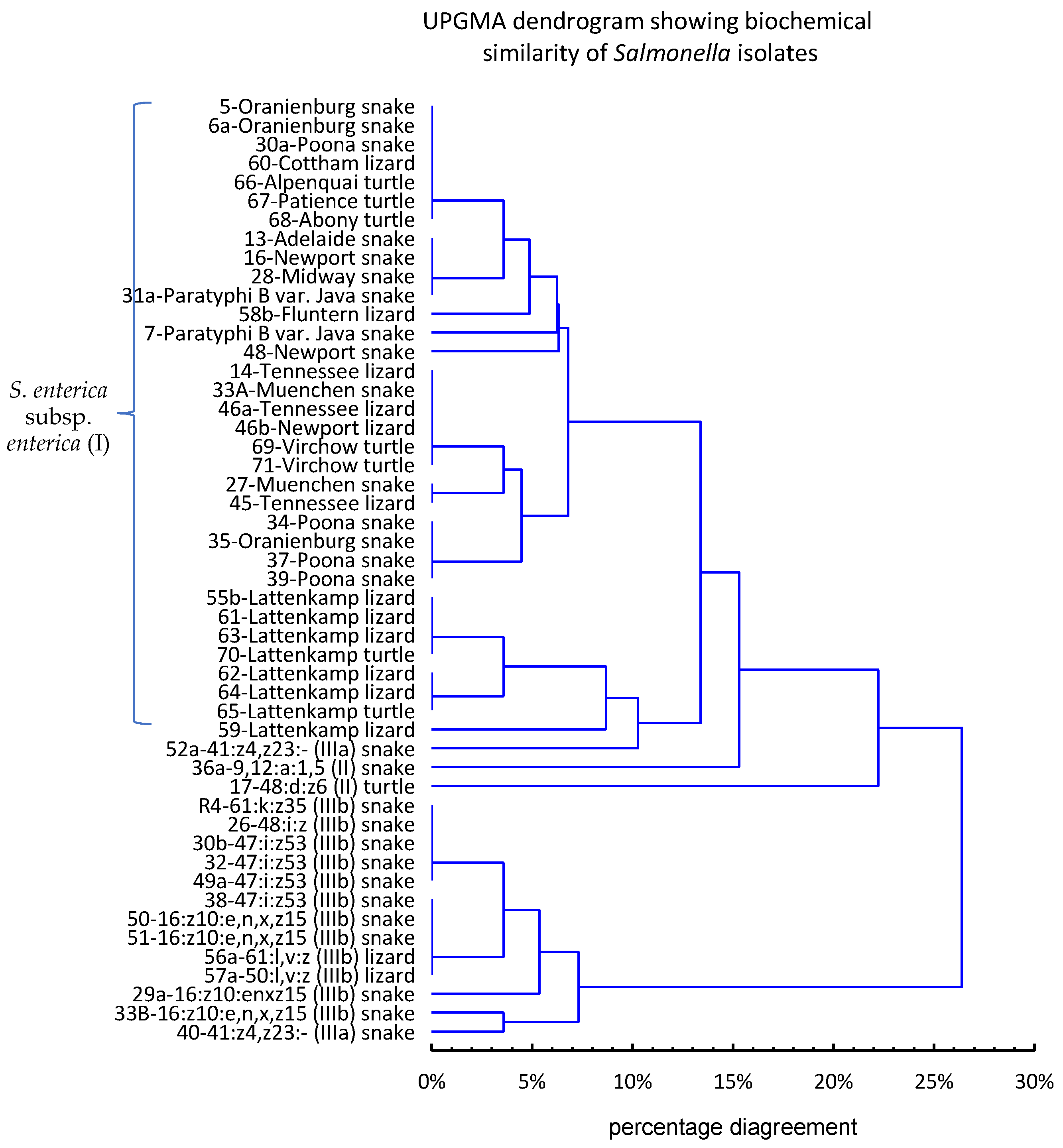
The similarity between Salmonella isolates based on their biochemical profiles, obtained from the results of 28 biochemical assays, is shown in the dendrogram derived from UPGMA cluster analysis (Figure 2).
At the level of 23% dissimilarity, the strains formed two main clusters—one comprised all S. enterica subsp. I and S. enterica subsp. II strains and one strain of S. enterica subsp. IIIa, while the other comprised S. enterica subsp. IIIb strains and one strain of S. enterica subsp. IIIa. This analysis showed that the biochemical tests used in the study are helpful in establishing the taxonomic identity of Salmonella strains, but they do not always allow for the unambiguous assignment of a strain to a subspecies.

3.3. Serotyping

Forty-eight of 51 Salmonella isolates were successfully serotyped by the agglutination test. The serotypic pattern of the three other isolates, designated 33B, 50, and 51, was specified as S. enterica subsp. diarizonae 16:?:? (33B) or S. enterica subsp. diarizonae 16:z10:- (50, 51). Finally, the serotype of these strains was determined to be S. enterica subsp. diarizonae 16:z10:e,n,x,z15 based on whole genome sequence analysis. Ultimately, 25 serotypes were identified among 51 Salmonella isolates, and all of them were non-typhoidal. S. Lattenkamp was the most frequently detected (15.7% of isolates, n = 8), followed by S. Poona (7.8%, n = 4), S. enterica subsp. diarizonae 47:i:z53 (7.8%, n = 4), S. enterica subsp. diarizonae 16:z10:e,n,x,z15 (7.8%, n = 4), S. Newport (5.9%, n = 3), S. Oranienburg (5.9%, n = 3), S. Tennessee (5.9%, n = 3), S. enterica subsp. arizonae 41:z4,z23:- (3.9%, n = 2), S. Paratyphi B var. Java (3.9%, n = 2), and S. Virchow (3.9%, n = 2). The other 14 serovars were found in single isolates (Table 3).
Some serotypes were found only in snakes, e.g., Oranienburg (n = 3), Poona (n = 4), 16:z10:e,n,x,z15 (n = 4), and 47:i:z53 (n = 4); S. Tennessee (n = 3) was characteristic of lizards, and S. Virchow (n = 2) was found only in turtles.

3.4. Antimicrobial Testing

As many as 90.2% of Salmonella isolates (n = 46/51) were sensitive to all antimicrobial substances used in the study (ampicillin, cefotaxime, ceftazidime, meropenem, nalidixic acid, ciprofloxacin, chloramphenicol, gentamicin, trimethoprim, sulfamethoxazole, tetracycline, colistin, nitrofurantoin, streptomycin, amikacin, and nitrofurantoin). Only 5 isolates (9.8%) were resistant to streptomycin, and 52.9% (n = 27/51) showed intermediate susceptibility to this antibiotic. Four of five streptomycin-resistant strains belonged to different serovars of Salmonella enterica subsp. diarizonae; the other was S. Paratyphi B v. Java (Table S3).

3.5. WGS

The total length of the final assemblies of four S. enterica strains ranged from 5,376,408 bp to 5,984,163 bp. The three strains, whose serotype could not be determined by agglutination (nos. 33B, 50, and 51), were identified as S. enterica subsp. diarizonae 16:z10:e,n,x,z15 (Table 4).
The presence/absence of the lacZ gene encoding β-galactosidase was correlated with the results of the ONPG test for the four analysed strains. Interestingly, despite the presence of the gene encoding the MFS oligosaccharide transporter, strain 33B did not ferment lactose within 48 h of incubation. Comparative analysis of the lactose operon sequence of strains 33A, 50 and 51 revealed the presence of a non-synonymous mutation (substitution CCG→CTG at nt position 290) in the gene encoding MFS transporter in strain 33B, leading to a Pro97→Leu change.
The aac(6′)-Iaa gene, coding for chromosomal-encoded aminoglycoside acetyltransferase, and the kdeA, mdtM, emrD and acrf genes, encoding multidrug efflux transporters, were detected in all strains (Table 4). However, the presence of these genes did not correlate with the phenotypic antimicrobial susceptibility profiles.

3.6. Pathogenic and Zoonotic Potential of Salmonella Serovars Identified in Reptiles

Reports of human Salmonella infections in the USA [25], the EU [1] and Queensland, Australia [26], confirm the prevalence of salmonellosis caused by 19 (76%) of the 25 serotypes identified in this work in reptiles (Table 5). The most accurate data come from the USA, where in 2006–2016 salmonellosis was most often caused by S. Newport (~1.4/100,000), S. Oranienburg (~0.23/100,000), S. Paratyphi B var. Java (~0.13/100,000) and S. Poona (~0.11/100,000). S. Tennessee (~0.04/100,000) and S. Muenchen (~0.03/100,000) were recorded slightly less frequently. Infections caused by serotypes of the subspecies arizonae, diarizonae and salamae identified in this study were reported sporadically (<14 cases per year, <0.004/100,000). In the EU (2018–2020), only 2 of the 25 serotypes, i.e., S. Newport (~0.17/100,000) and Muenchen (~0.05/100,000), were confirmed to have caused infections in humans. In Queensland, over 10 years (2007–2016), infections caused by S. Virchow were recorded with high frequency (~5.6/100,000), and significantly fewer cases were caused by S. Paratyphi B var. Java, S. Newport and S. Oranienburg (>0.17/100,000) (Table 5). In summary, on the basis of available reports and the adopted criteria, the serotypes with high pathogenic potential (>0.05 cases/100,000 annually) include Newport, Oranienburg, Paratyphi B var. Java, Poona, Virchow, and Muenchen.
Ten (40%) of the 25 serotypes under consideration have documented zoonotic potential. These are the eight serotypes representing subspecies I, i.e., Abony, Cotham, Muenchen, Newport, Oranienburg, Paratyphi B var. Java and Poona, and Tennessee, as well as one serotype of S. enterica subsp. arizonae, i.e., 41:z4,z23:-, and one serotype of S. enterica subsp. diarizonae, i.e., 47:i:z53. The pathogen was most often transmitted as a result of contact with turtles, less often with snakes or lizards. The age of patients with RAS ranged from one month to 87 years, but the median estimated in many reports indicates that children aged 3–10 years were most often affected (Table 5).

4. Discussion

4.1. Prevalence of Salmonella spp. in Captive Reptiles in Poland

Over the past 20 years, there have been a number of reports showing that reptiles, both in the wild and in captivity, are carriers of Salmonella [6]. However, the frequency of occurrence of these zoonotic bacteria is highly varied, ranging from 2.1% [38] to 87.5% [39], depending on the geographic region and type of reptiles analysed [6]. The results of the present study showed that pet reptiles bred in Poland are asymptomatic carriers of Salmonella enterica strains (71.64% positive samples), with a prevalence of 87%, 78% and 39% in snakes, lizards and turtles, respectively. These findings are largely consistent with the observations of Zając et al. [9], who showed a very high frequency of Salmonella in captive reptiles bred in Poland; it was highest in snakes (92.2%), followed by lizards (83.7%) and turtles (60.0%). Our results are also partially in line with the findings of Geue and Löschner [40] and Nakadai et al. [41], who showed the presence of Salmonella in 54.1% and 74.1% of faecal samples, respectively, from caged reptiles reared in Germany and Austria and in Japan. The trend of snakes as the most common carrier of Salmonella spp. (69.7–76.0% ± 9.3%), lizards as slightly less common (61–69.0% ± 6.7%) and turtles as the least common carriers (2.6–24.3%) has also been confirmed by several other teams of researchers [40,42,43]. A significantly lower frequency of S. enterica than that recorded in this study was reported in captive reptiles in Italy (13.61%) [44], Taiwan (30.9%) [42], Indonesia (10%) [45] and Croatia (13%) [46]. Moreover, the Croatian study showed a much higher Salmonella prevalence in lizards (48.4%) than in snakes (8.9%) and chelonians (3.8%). The large variation in the frequency of Salmonella prevalence in reptiles in different countries may depend on the type of reptile included in the study and the type of diet associated with it. In carnivorous reptiles, Salmonella is more likely to occur, as these bacteria may be found in the meat (mice, rats) that these animals are fed. In addition, antibiotics are commonly used on reptilian farms in some countries; antimicrobial substances can destroy the gut microflora of reptiles, including Salmonella.

4.2. Subspecies and Serovars of Reptile Salmonella Strains

Salmonella strains derived from reptiles are very diverse and represent different subspecies and serotypes [9,40,42]. Our findings are consistent with the findings of some other authors [9,40,43], who in captive reptiles in Poland, Germany, Austria and Spain showed predominance of strains of the enterica (I) subspecies (53–66.4%) and a lower prevalence of Salmonella strains representing the IIIb (11.2–30.3%), IIIa (2–6%), II (3–14.6%) and IV (2–19.6%) subspecies. A lower frequency (34.6%) of S. enterica subsp. enterica strains and more frequent identification of arizonae (IIIa) strains (23.1%) were recorded in captive reptiles (snakes, lizards, and chelonians) in Croatia [46]. A higher prevalence of subspecies IIIb isolates in snakes compared to lizards and turtles was previously also demonstrated by Zając et al. [9]. On the other hand, Geue and Löschner [40] showed that Salmonella enterica subsp. diarizonae strains occur with a similar frequency in snakes (27%) and lizards (29%).
The lactose fermentation capacity of Lattenkamp strains noted in the present study is a feature rarely found in S. enterica subsp. I, in contrast to strains of subspecies IIIa and IIIb [47]. The lack of correlation in several strains between the presence of β-galactosidase and the capacity to utilize lactose may be the result of mutations in the genes encoding lactose transporter or in the regulatory genes of the lactose operon [48]. More detailed studies are needed to confirm the effect of the Pro97→Leu mutation in the MFS transporter gene on the protein’s ability to transport lactose into Salmonella cells.
Five of the 25 serotypes described in this study, i.e., Oranienburg, Tennessee, Fluntern, Muenchen and Newport, belong to the 10 most frequently identified serotypes in Salmonella strains derived from reptiles in Poland, while the Abony, Adelaide, Cotham, Lattenkamp, Fluntern, Paratyphi B v. Java, Patience, Poona, 47:z10:z35 (IIIb), 48:i:z (IIIb), and 41:z4,z23:- (IIIa) serotypes have been detected less frequently [9]. The relatively high frequency of Lattenkamp strains noted in our research is probably explained by the fact that several samples were collected from the same pet shop (six of eight Lattenkamp strains were obtained from lizards from the same shop). S. Midway and S. Virchow, S. enterica subsp. arizonae 61:k:z35, and S. enterica subsp. diarizonae 48:d:z6, 16: z10:e,n,x,z15, and 47:i:z53 have been reported in reptiles in other countries [41,43,49,50,51,52,53]. The remaining four serotypes recorded in this study, i.e., S. Alpenquai, S. II 9,12:a:1,5, S. IIIb 50:l,v:z, and S. IIIb 61:l,v:z, to the best of our knowledge have not yet been described in Salmonella strains from reptiles.

4.3. Zoonotic Potential of Identified Salmonella Serovars

All serotypes detected in this study were non-typhoid and belonged to either S. enterica subsp. enterica, which is responsible for over 99% of human infections, and to subspecies II, IIIa and IIIb, which are much less likely to cause human salmonellosis [6]. The Virchow and Newport serovars identified in the study are among the seven serotypes most frequently causing infections in Poland (in 2016 and 2017) [3], while S. Newport and S. Muenchen rank among the top 11 serotypes most commonly recorded in humans in the EU [1].
Statistical data on the occurrence of RAS in the EU, including Poland, are not available, as there is no obligation to interview patients regarding possible contact with reptiles. The scale of the problem is therefore unknown, although cases of Salmonella infections attributed to direct or indirect contact with captive reptiles have been reported in many European countries (in 2008–2017) [28,30,33,34,35,36,54,55,56,57]. Meletiadis et al. [58] estimated the frequency of RAS in Italy at 3% to 7%, and in the 0–21 age group even up to 11.7%. In the USA, regular reports of reptile-associated salmonellosis were conducted from 1994 to 2002 [37,59,60]. Based on the available reports and case studies, some serotypes found in reptiles in Poland, i.e., S. Abony, S. Cotham, S. Muenchen, S. Newport, S. Oranienburg, S. Paratyphi B var. Java, S. Poona, S. enterica subsp. arizonae 41:z4,z23:- and S. enterica subsp. diarizone 47:i:z53, can be considered to have high zoonotic potential, causing infections not only in infants and babies (under 2 years old) but also in older children and adults with normal immunity [10,11,27,36,61]. Occasionally, RAS in infants and young children (<1–5 years) caused by some of these serotypes (S. Abony, S. Cotham, S. Muenchen, S. Newport, and S. Oranienburg) has occurred as invasive infections such as septicaemia, meningitis and colitis [28,29,30].
Many RAS reports point to turtles as the most common source of the pathogen [10,11,27,28,34,35]. However, our research shows that only 38.9% of turtles kept in Poland are Salmonella carriers, and in Germany, snakes or lizards are most often identified as transmitters of Salmonella causing RAS [36].
The diversity of Salmonella strains in terms of pathogenic potential is confirmed not only by the analyses of salmonellosis in humans but also by the results of in vitro studies. McWhorter et al. [62] showed that all considered reptile Salmonella strains representing 30 different serotypes (including strains of the subspecies I, II, IIa and IIIb) were invasive into both human intestinal epithelial (Caco2) and mouse macrophage cell lines (J774A.1). One of the most invasive was the Paratyphi B ver. Java serotype, and a strain of another serotype identified in our study, S. Adelaide, was moderately invasive. Similar results were obtained by Pasmans et al. [63], who demonstrated that all tested reptile Salmonella strains representing 44 serotypes belonging to subspecies I, II, IIIb, and IV were able to invade human intestinal epithelial Caco-2 cells, although isolates belonging to subspecies I invaded the Caco-2 cells to a higher extent than those from the other subspecies. What is more, the human isolates invaded the Caco-2 cells to a lesser degree than their saurian counterparts. These in vitro findings correlate with reports of more hospitalizations and cases of invasive infections among RAS patients than in non-RAS ones [30,64].

4.4. Antimicrobial Susceptibility

In the present study, we demonstrated that the majority of Salmonella strains were sensitive to all tested antimicrobials, except for single strains resistant to streptomycin. Our findings are largely in line with the results of Chen et al. [42], who reported that the prevalence of streptomycin-intermediate-resistant and resistant Salmonella strains in captive reptiles in Taiwan was 23.1% and 14%, respectively. The frequency of resistance to other antimicrobial substances was low, ranging from 0% to 9.2%. A relatively low frequency (32.8%) of drug-resistant Salmonella strains in captive reptiles in Poland was also shown by Zając et al. [9]. The prevalence of streptomycin-resistant strains reported by these authors was twice as high (25%) as that recorded in this study, while the percentage of strains showing resistance to other antimicrobial substances ranged from 0.2% to 8.1%. A low frequency (12.8%, n = 34/39) of resistant Salmonella strains was also reported in captive reptiles in the Czech Republic [65]. Completely different results regarding the drug susceptibility of reptilian Salmonella strains were obtained in Italy and Spain, where the frequency of drug-resistant strains (to at least one antimicrobial substance) ranged from 93.1% to 100% [43,44], and the percentage of multi-drug resistant strains was about 70%. Moreover, the Italian study [44] showed a very high percentage of Salmonella isolates with resistance (89.7%) and intermediate susceptibility (79%) to streptomycin; however, in contrast to our results, this was not the only type of resistance in the strains tested. The presence of streptomycin-resistant Salmonella strains in reptiles (75%) was also reported by Arnafia et al. [45].
The resistance of Salmonella strains to streptomycin noted in this study cannot be explained by the use of antibiotic therapy in reptiles, as streptomycin is not used in treatment of these animals in Poland. It also appears that the reduced susceptibility to streptomycin recorded in the study is not a result of acquired resistance, as none of the strains contained the genes that determine streptomycin resistance in Salmonella (strA, strB, aadA, aac(3)-IVa, and aphA1) [66,67]. For all strains, a small zone of inhibition of growth was recorded around the streptomycin discs, i.e., up to 13.5 mm, while a diameter ≤11 mm indicates resistance. Hence, it appears that this reduced susceptibility of reptilian Salmonella strains to streptomycin may have an evolutionary basis. The same trait has previously been observed in reptilian strains of E. coli [24]. The lack of correlation between the presence of the aac(6′)-Iaa gene encoding chromosomal aminoglycoside N(6′)-acetyltransferase (conferring resistance to tobramycin, kanamycin and amikacin) and the phenotypic susceptibility of the Salmonella strains (none of the strains were resistant to amikacin) confirms previous reports about the non-functionality of this gene in Salmonella [68]. Common occurrence of the aac(6′)-Iaa cryptic gene in Salmonella enterica strains, including subsp. diarizonae, has previously been confirmed by several authors [69,70].

5. Conclusions

The research provided valuable data complementing existing knowledge of the prevalence of Salmonella strains in reptiles, as well as knowledge of their serotypes, drug susceptibility, and biochemical characteristics. We showed that the pet reptiles kept in households in Poland are common carriers of Salmonella strains, including serotypes that had previously been confirmed to cause reptile-associated salmonellosis in humans. Identification of the Salmonella reservoir is important in assessing the risk associated with the spread of this zoonotic agent and potential infection.
Reptiles should always be considered a potential source of Salmonella transmission, and special hygiene rules should be followed when handling these animals and cleaning terrariums. It is advisable to wash your hands after each contact with reptiles or objects in their living areas. Reptiles should not move freely around the house, especially in the food preparation area, due to possible environmental contamination. When washing terrariums, use disinfectants and disposable gloves. Due to the increased risk of RAS and hospitalization, children under the age of five, immunocompromised people, and the elderly should not touch reptiles or their environment. There is also a need to educate the public, especially reptile owners, about RAS and related preventive measures. Despite the zoonotic risk, the low level of drug resistance of reptilian Salmonella strains gives hope for effective antibiotic therapy in the event of infection.

Supplementary Materials

The following supporting information can be downloaded at: https://www.mdpi.com/article/10.3390/pathogens11101125/s1, Table S1: Sequences of primers used to detect resistance genes; Table S2: Biochemical profiles of Salmonella strains; Table S3. Salmonella susceptibility test results.

Author Contributions

Conceptualization, M.D. and K.S.; methodology, M.D. and M.Z.; validation, M.D. and M.Z.; formal analysis, M.D. and M.Z.; investigation, M.D., M.Z. and A.P.; resources, M.D. and K.S.; data curation, M.D. and M.Z; writing—original draft preparation, M.D.; writing—review and editing, R.U.-C. and M.Z; visualization, M.D.; funding acquisition, R.U.-C. and M.Z. All authors have read and agreed to the published version of the manuscript.

Funding

This research received no external funding.

Institutional Review Board Statement

Ethical review and approval were waived for this study as no animal procedures were performed. According to Polish law (Act of 15 January 2015 on the protection of animals used for scientific or educational purposes, Journal of Laws 2015 item 266) as well as EU law (2010/63/EU Directive of the European Parliament and of the Council of 22 September 2010 on the protection of animals used for scientific purposes), collecting stool samples is not a procedure and thus does not require the consent of the bioethics committee. Pet owners and pet shop owners agreed to collect reptile faeces while cleaning terrariums without interfering with animal welfare.

Informed Consent Statement

Not applicable.

Data Availability Statement

All the data presented in the study are included in the article. Further enquiries can be directed to the corresponding author.

Conflicts of Interest

The authors declare no conflict of interest.

References

  1. The European Union One Health 2020 Zoonoses Report. EFSA J. 2021, 19, 6971. Available online: https://efsa.onlinelibrary.wiley.com/doi/epdf/10.2903/j.efsa.2021.6971 (accessed on 12 September 2022).
  2. Report of Centers for Disease Control and Prevention, USA. Available online: https://www.cdc.gov/salmonella/ (accessed on 20 August 2022).
  3. Milczarek, M.; Sadkowska-Todys, M.; Czarkowski, M.P.; Kitowska, W. Salmonellosis in Poland in 2017. Prz. Epidemiol. 2019, 73, 463–477. [Google Scholar]
  4. Ruszel, K.; Dubel, R.; Chodun, W.; Nieradko-Iwanicka, B. Salmonella—Still a threat? Epidemiological analysis of infecion. J. Educ. Health Sport 2021, 11, 38–43. [Google Scholar] [CrossRef]
  5. Issenhuth-Jeanjean, S.; Roggentin, P.; Mikoleit, M.; Guibourdenche, M.; de Pinna, E.; Nair, S.; Fields, P.I.; Weill, F.X. Supplement 2008–2010 (no. 48) to the White-Kauffmann-Le Minor scheme. Res. Microbiol. 2014, 165, 526–530. [Google Scholar] [CrossRef] [PubMed]
  6. Dróżdż, M.; Małaszczuk, M.; Paluch, E.; Pawlak, A. Zoonotic potential and prevalence of Salmonella serovars isolated from pets. Infect. Ecol. Epidemiol. 2021, 11, 1975530. [Google Scholar] [CrossRef]
  7. Mermin, J.; Hutwagner, L.; Vugia, D.; Shallow, S.; Daily, P.; Bender, J.; Koehler, J.; Marcus, R.; Angulo, F.J. Emerging Infections Program FoodNet Working Group. Reptiles, amphibians, and human Salmonella infection: A population-based, case-control study. Clin. Infect. Dis. 2004, 38 (Suppl. S3), S253–S261. [Google Scholar] [CrossRef]
  8. FEDIAF (The European Pet Food Industry). FEDIAF Annual Report 2021. p. 44, 50. Available online: https://europeanpetfood.org/about/statistics/ (accessed on 20 January 2022).
  9. Zając, M.; Skarżyńska, M.; Lalak, A.; Kwit, R.; Śmiałowska-Węglińska, A.; Pasim, P.; Szulowski, K.; Wasyl, D. Salmonella in Captive Reptiles and Their Environment-Can We Tame the Dragon? Microorganisms 2021, 9, 1012. [Google Scholar] [CrossRef]
  10. Bosch, S.; Tauxe, R.V.; Behravesh, C.B. Turtle-Associated Salmonellosis, United States, 2006–2014. Emerg. Infect. Dis. 2016, 22, 1149–1155. [Google Scholar] [CrossRef]
  11. Gambino-Shirley, K.; Stevenson, L.; Concepción-Acevedo, J.; Trees, E.; Wagner, D.; Whitlock, L.; Roberts, J.; Garrett, N.; Van Duyne, S.; McAllister, G.; et al. Flea market finds and global exports: Four multistate outbreaks of human Salmonella infections linked to small turtles, United States-2015. Zoonoses Public Health 2018, 65, 560–568. [Google Scholar] [CrossRef]
  12. Dec, M.; Stępień-Pyśniak, D.; Puchalski, A.; Hauschild, T.; Pietras-Ożga, D.; Ignaciuk, S.; Urban-Chmiel, R. Biodiversity of Ligilactobacillus salivarius Strains from Poultry and Domestic Pigeons. Animals 2021, 11, 972. [Google Scholar] [CrossRef]
  13. Grimont, P.A.D.; Weill, F.X. Antigenic Formulae of Salmonella Serovars, 9th ed.; WHO Collaborating Centre for Research on Salmonella, Institute Pasteur: Paris, France, 2017; Available online: https://www.pasteur.fr/sites/default/files/veng_0.pdf (accessed on 12 September 2022).
  14. Ewing, W.H. Edwards and Ewing’s Identification of Enterobacteriaceae, 4th ed.; Elsevier: New York, NY, USA, 1986. [Google Scholar]
  15. Malorny, B.; Bunge, C.; Helmuth, R. Discrimination of d-tartrate-fermenting and -nonfermenting Salmonella enterica subsp. enterica isolates by genotypic and phenotypic methods. J. Clin. Microbiol. 2003, 41, 4292–4297. [Google Scholar] [CrossRef]
  16. Hoszowski, A.; Wasyl, D. Pożywka Agarowa AKG do Ekspresji faz Antygenów Rzęskowych Salmonella, Sposób jej Wytwarzania Oraz Zastosowanie Pożywki do Identyfikacji Antygenów Rzęskowych Salmonella; Państwowy Instytut Weterynaryjny—Państwowy Instytut Badawczy: Puławy, Poland, 2012; p. 401498. [Google Scholar]
  17. Bolger, A.M.; Lohse, M.; Usadel, B. Trimmomatic: A flexible trimmer for Illumina sequence data. Bioinformatics 2014, 30, 2114–2120. [Google Scholar] [CrossRef] [PubMed] [Green Version]
  18. Bankevich, A.; Nurk, S.; Antipov, D.; Gurevich, A.A.; Dvorkin, M.; Kulikov, A.S.; Lesin, V.M.; Nikolenko, S.I.; Pham, S.; Prjibelski, A.D.; et al. SPAdes: A new genome assembly algorithm and its applications to single-cell sequencing. J. Comput. Biol. 2012, 19, 455–477. [Google Scholar] [CrossRef] [PubMed]
  19. Zhang, S.; Yin, Y.; Jones, M.B.; Zhang, Z.; Deatherage Kaiser, B.L.; Dinsmore, B.A.; Fitzgerald, C.; Fields, P.I.; Deng, X. Salmonella serotype determination utilizing high-throughput genome sequencing data. J. Clin. Microbiol. 2015, 53, 1685–1692. [Google Scholar] [CrossRef]
  20. Bortolaia, V.; Kaas, R.F.; Ruppe, E.; Roberts, M.C.; Schwarz, S.; Cattoir, V.; Philippon, A.; Allesoe, R.L.; Rebelo, A.R.; Florensa, A.R.; et al. ResFinder 4.0 for predictions of phenotypes from genotypes. J. Antimicrob. Chemother. 2020, 75, 3491–3500. [Google Scholar] [CrossRef] [PubMed]
  21. EUCAST, Antimicrobial wild type distributions of microorganisms. Available online: https://mic.eucast.org/search/ (accessed on 20 August 2022).
  22. CLSI Supplement M100; Performance Standards for Antimicrobial Susceptibility Testing, 28th ed. Clinical and Laboratory Standards Institute: Wayne, PA, USA, 2018.
  23. EUCAST. The European Committee on Antimicrobial Susceptibility Testing. Breakpoint Tables for Interpretation of MICs and Zone Diameters. Version 11.0. 2021. Available online: http://www.eucast.org (accessed on 20 August 2022).
  24. Dec, M.; Stępień-Pyśniak, D.; Szczepaniak, K.; Turchi, B.; Urban-Chmiel, R. Virulence Profiles and Antibiotic Susceptibility of Escherichia coli Strains from Pet Reptiles. Pathogens 2022, 11, 127. [Google Scholar] [CrossRef] [PubMed]
  25. CDC. National enteric disease surveillance: Salmonella annual report 2016. Available online: https://www.cdc.gov/nationalsurveillance/pdfs/2016-Salmonella-report-508.pdf (accessed on 20 August 2022).
  26. Parisi, A.; Crump, J.A.; Stafford, R.; Glass, K.; Howden, B.P.; Kirk, M.D. Increasing incidence of invasive nontyphoidal Salmonella infections in Queensland, Australia, 2007–2016. PLOS Negl. Trop. Dis. 2019, 13, e0007187. [Google Scholar] [CrossRef]
  27. Kuroki, T.; Ito, K.; Ishihara, T.; Furukawa, I.; Kaneko, A.; Suzuki, Y.; Seto, J.; Kamiyama, T. Turtle-Associated Salmonella Infections in Kanagawa, Japan. Jpn. J. Infect. Dis. 2015, 68, 333–337. [Google Scholar] [CrossRef]
  28. Van Meervenne, E.; Botteldoorn, N.; Lokietek, S.; Vatlet, M.; Cupa, A.; Naranjo, M.; Dierick, K.; Bertrand, S. Turtle-associated Salmonella septicaemia and meningitis in a 2-month-old baby. J. Med. Microbiol. 2009, 58, 1379–1381. [Google Scholar] [CrossRef]
  29. Kiebler, C.A.; Bottichio, L.; Simmons, L.; Basler, C.; Klos, R.; Gurfield, N.; Roberts, E.; Kimura, A.; Lewis, L.S.; Bird, K.; et al. Outbreak of human infections with uncommon Salmonella serotypes linked to pet bearded dragons. Zoonoses Public Health 2020, 67, 425–434. [Google Scholar] [CrossRef]
  30. Murphy, D.; Oshin, F. Reptile-associated salmonellosis in children aged under 5 years in South West England. Arch. Dis. Child. 2015, 100, 364–365. [Google Scholar] [CrossRef] [PubMed]
  31. Braun, S.; Spalloni, W.; Ferreccio, F.; Postigo, J.; Fernández, A.; Porte, L.; Saldivia, A.; Wigant, W.; Triantafilo, V. Salmonella spp. gastroenteritis associated to pet turtles in three infants. Rev. Chilena. Infectol. 2015, 32, 334–338. [Google Scholar] [CrossRef] [PubMed] [Green Version]
  32. CDC’s 2019 Salmonella Outbreak Report. Available online: https://www.cdc.gov/salmonella/oranienburg-10-19/index.html (accessed on 20 August 2022).
  33. Tomaštíková, Z.; Mrázková, M.; Kaňáková, M.; Karpíšková, R. Salmonellosis in an infant as a result of indirect contact with reptiles. Klin. Mikrobiol. Infekc. Lek. 2017, 23, 61–63. [Google Scholar]
  34. Hernandez, E.; Rodriguez, J.L.; Herrera-Leon, S.; Garcia, I.; de Castro, V.; Muniozguren, N. Salmonella Paratyphi B var Java infections associated with exposure to turtles in Bizkaia, Spain, September 2010 to October 2011. Euro Surveill. 2012, 17, 20201. [Google Scholar] [CrossRef] [PubMed]
  35. Lafuente, S.; Bellido, J.B.; Moraga, F.A.; Herrera, S.; Yagüe, A.; Montalvo, T.; de Simó, M.; Simón, P.; Caylà, J.A. Salmonella paratyphi B and Salmonella litchfield outbreaks associated with pet turtle exposure in Spain. Enferm. Infecc. Microbiol. Clin. 2013, 31, 32–35. [Google Scholar] [CrossRef] [PubMed]
  36. Editorial team; Bertrand, S.; Rimhanen-Finne, R.; Weill, F.X.; Rabsch, W.; Thornton, L.; Perevoscikovs, J.; van Pelt, W.; Heck, M. Salmonella infections associated with reptiles: The current situation in Europe. Euro. Surveill. 2008, 13, 18902. [Google Scholar]
  37. CDC. Reptile-associated salmonellosis—Selected states, 1998–2002. MMWR 2003, 52, 1206–1209. [Google Scholar]
  38. Sumiyama, D.; Shimizu, A.; Kanazawa, T.; Anzai, H.; Murata, K. Prevalence of Salmonella in green anoles (Anolis Carolinensis), an invasive alien species in Naha and Tomigusuku Cities, Okinawa Main Island, Japan. J. Vet. Med. Sci. 2020, 82, 678–680. [Google Scholar] [CrossRef]
  39. Zając, M.; Wasyl, D.; Różycki, M.; Ewa Bilska-Zając, E.; Fafiński, Z.; Iwaniak, W.; Krajewska, M.; Hoszowski, A.; Konieczna, O.; Fafińska, P.; et al. Free-living snakes as a source and possible vector of Salmonella spp. and parasites. Eur. J. Wildl. Res. 2016, 62, 161–166. [Google Scholar] [CrossRef]
  40. Geue, L.; Löschner, U. Salmonella enterica in reptiles of German and Austrian origin. Vet. Microbiol. 2002, 84, 79–91. [Google Scholar] [CrossRef]
  41. Nakadai, A.; Kuroki, T.; Kato, Y.; Suzuki, R.; Yamai, S.; Yaginuma, C.; Shiotani, R.; Yamanouchi, A.; Hayashidani, H. Prevalence of Salmonella spp. in pet reptiles in Japan. J. Vet. Med. Sci. 2005, 67, 97–101. [Google Scholar] [CrossRef] [PubMed]
  42. Chen, C.Y.; Chen, W.C.; Chin, S.C.; Lai, Y.H.; Tung, K.C.; Chiou, C.S.; Hsu, Y.M.; Chang, C.C. Prevalence and antimicrobial susceptibility of salmonellae isolates from reptiles in Taiwan. J. Vet. Diagn. Investig. 2010, 22, 44–50. [Google Scholar] [CrossRef] [PubMed]
  43. Marin, C.; Lorenzo-Rebenaque, L.; Laso, O.; Villora-Gonzalez, J.; Vega, S. Pet Reptiles: A Potential Source of Transmission of Multidrug-Resistant Salmonella. Front. Vet. Sci. 2021, 7, 613718. [Google Scholar] [CrossRef] [PubMed]
  44. Bertelloni, F.; Chemaly, M.; Cerri, D.; Gall, F.L.; Ebani, V.V. Salmonella infection in healthy pet reptiles: Bacteriological isolation and study of some pathogenic characters. Acta Microbiol. Immunol. Hung. 2016, 63, 203–216. [Google Scholar] [CrossRef]
  45. Arnafia, W.; Ningrum, S.G.; Adji, R.S.; Lukman, D.W.; Pasaribu, F.H.; Wibawan, I.T. Isolation of Salmonella from reptiles in pet shop and its susceptibility to antibiotics in Indonesia. Inter. J. Bioflux. Soc. 2016, 8, 177–181. [Google Scholar]
  46. Lukac, M.; Pedersen, K.; Prukner-Radovcic, E. Prevalence of salmonella in captive reptiles from Croatia. J. Zoo Wildl. Med. 2015, 46, 234–240. [Google Scholar] [CrossRef]
  47. Leonard, S.R.; Lacher, D.W.; Lampel, K.A. Acquisition of the lac operon by Salmonella enterica. BMC Microbiol. 2015, 15, 173. [Google Scholar] [CrossRef]
  48. Smirnova, I.N.; Kaback, H.R. A mutation in the lactose permease of Escherichia coli that decreases conformational flexibility and increases protein stability. Biochemistry 2003, 42, 3025–3031. [Google Scholar] [CrossRef]
  49. Callaway, Z.; Thomas, A.; Melrose, W.; Buttner, P.; Speare, R. Salmonella Virchow and Salmonella Weltevreden in a random survey of the Asian house gecko, Hemidactylus frenatus, in houses in northern Australia. Vector Borne Zoonotic Dis. 2011, 11, 621–625. [Google Scholar] [CrossRef]
  50. Smith, K.F.; Yabsley, M.J.; Sanchez, S.; Casey, C.L.; Behrens, M.D.; Hernandez, S.M. Salmonella isolates from wild-caught Tokay geckos (Gekko gecko) imported to the U.S. from Indonesia. Vector Borne Zoonotic Dis. 2012, 12, 575–582. [Google Scholar] [CrossRef]
  51. Ebani, V.V.; Cerri, D.; Fratini, F.; Meille, N.; Valentini, P.; Andreani, E. Salmonella enterica isolates from faeces of domestic reptiles and a study of their antimicrobial in vitro sensitivity. Res. Vet. Sci. 2005, 78, 117–121. [Google Scholar] [CrossRef] [PubMed]
  52. Gorski, L.; Jay-Russell, M.T.; Liang, A.S.; Walker, S.; Bengson, Y.; Govoni, J.; Mandrell, R.E. Diversity of pulsed-field gel electrophoresis pulsotypes, serovars, and antibiotic resistance among Salmonella isolates from wild amphibians and reptiles in the California Central Coast. Foodborne Pathog. Dis. 2013, 10, 540–548. [Google Scholar] [CrossRef]
  53. Report of the Sweedesh National Veterinary Institute to the Commission 2001. Available online: https://www.sva.se/media/da2px1k2/zoonos-2000-eng.pdf (accessed on 12 September 2022).
  54. Horvath, L.; Kraft, M.; Fostiropoulos, K.; Falkowski, A.; Tarr, P.E. Salmonella enterica Subspecies diarizonae Maxillary Sinusitis in a Snake Handler: First Report. Open Forum Infect. Dis. 2016, 3, ofw066. [Google Scholar] [CrossRef] [PubMed]
  55. Gavrilovici, C.; Pânzaru, C.V.; Cozma, S.; Mârţu, C.; Lupu, V.V.; Ignat, A.; Miron, I.; Stârcea, M. Message from a turtle: Otitis with salmonella arizonae in children: Case report. Medicine 2017, 96, e8455. [Google Scholar] [CrossRef] [PubMed]
  56. Baranzelli, A.; Loïez, C.; Bervar, J.F.; Scherpereel, A.; Wallet, F. The snake raiser lung: An unusual cause of Salmonella enterica subspecies arizonae pneumonia. Med. Mal. Infect. 2017, 47, 424–425. [Google Scholar] [CrossRef]
  57. Hervás, J.A.; Rosell, A.; Hervás, D.; Rubio, R.; Dueñas, J.; Mena, A. Reptile pets-associated Salmonella enterica subspecies diarizonae gastroenteritis in a neonate. Pediatr. Infect. Dis. J. 2012, 31, 1102–1103. [Google Scholar] [CrossRef]
  58. Meletiadis, A.; Biolatti, C.; Mugetti, D.; Zaccaria, T.; Cipriani, R.; Pitti, M.; Decastelli, L.; Cimino, F.; Dondo, A.; Maurella, C.; et al. Surveys on Exposure to Reptile-Associated Salmonellosis (RAS) in the Piedmont Region-Italy. Animals 2022, 12, 906. [Google Scholar] [CrossRef]
  59. CDC. Reptile-associated salmonellosis—Selected states, 1994–1995. MMWR 1995, 44, 347–350. [Google Scholar]
  60. CDC. Reptile-associated salmonellosis—Selected states, 1996–1998. MMWR 1999, 48, 1009–1013. [Google Scholar]
  61. Bjelland, A.M.; Sandvik, L.M.; Skarstein, M.M.; Svendal, L.; Debenham, J.J. Prevalence of Salmonella serovars isolated from reptiles in Norwegian zoos. Acta Vet. Scand. 2020, 62, 3. [Google Scholar] [CrossRef]
  62. McWhorter, A.; Owens, J.; Valcanis, M.; Olds, L.; Myers, C.; Smith, I.; Trott, D.; McLelland, D. In vitro invasiveness and antimicrobial resistance of Salmonella enterica subspecies isolated from wild and captive reptiles. Zoonoses Public Health 2021, 68, 402–412. [Google Scholar] [CrossRef] [PubMed]
  63. Pasmans, F.; Martel, A.; Boyen, F.; Vandekerchove, D.; Wybo, I.; Immerseel, F.V.; Heyndrickx, M.; Collard, J.M.; Ducatelle, R.; Haesebrouck, F. Characterization of Salmonella isolates from captive lizards. Vet. Microbiol. 2005, 110, 285–291. [Google Scholar] [CrossRef]
  64. Guyomard-Rabenirina, S.; Weill, F.X.; Le Hello, S.; Bastian, S.; Berger, F.; Ferdinand, S.; Legreneur, P.; Loraux, C.; Malpote, E.; Muanza, B.; et al. Reptiles in Guadeloupe (French West Indies) are a reservoir of major human Salmonella enterica serovars. PLoS ONE 2019, 14, e0220145. [Google Scholar] [CrossRef]
  65. Tomastikova, Z.; Barazorda Romero, S.; Knotek, Z.; Karpiskova, R. Prevalence and characteristics of Salmonella species isolated from captive reptiles in the Czech Republic. Vet. Med. 2017, 62, 456–469. [Google Scholar] [CrossRef]
  66. Pławińska-Czarnak, J.; Wódz, K.; Kizerwetter-Swida, M.; Bogdan, J.; Kwieciński, P.; Nowak, T.; Strzałkowska, Z.; Anusz, K. Multi-Drug Resistance to Salmonella spp. When Isolated from Raw Meat Products. Antibiotics 2022, 11, 876. [Google Scholar] [CrossRef] [PubMed]
  67. Qin, X.; Yang, M.; Cai, H.; Liu, Y.; Gorris, L.; Aslam, M.Z.; Jia, K.; Sun, T.; Wang, X.; Dong, Q. Antibiotic Resistance of Salmonella Typhimurium Monophasic Variant 1,4,[5],12:i:-in China: A Systematic Review and Meta-Analysis. Antibiotics 2022, 11, 532. [Google Scholar] [CrossRef]
  68. Magnet, S.; Courvalin, P.; Lambert, T. Activation of the Cryptic aac(6′)-Iy Aminoglycoside Resistance Gene of Salmonella by a Chromosomal Deletion Generating a Transcriptional Fusion. J. Bacteriol. 1999, 181, 6650–6655. [Google Scholar] [CrossRef]
  69. Zhou, M.; Shi, Q.; Zhang, X.; Mei, L.; Ye, Y.; Fang, C.; Shang, S. Salmonella enterica subsp. diarizonae Harboring ST233, ST1263, and ST1845 in Children. Front. Cell Infect. Microbiol. 2021, 11, 727811. [Google Scholar] [CrossRef]
  70. García-Soto, S.; Abdel-Glil, M.Y.; Tomaso, H.; Linde, J.; Methner, U. Emergence of Multidrug-Resistant Salmonella enterica Subspecies enterica Serovar Infantis of Multilocus Sequence Type 2283 in German Broiler Farms. Front. Microbiol. 2020, 11, 1741. [Google Scholar] [CrossRef]
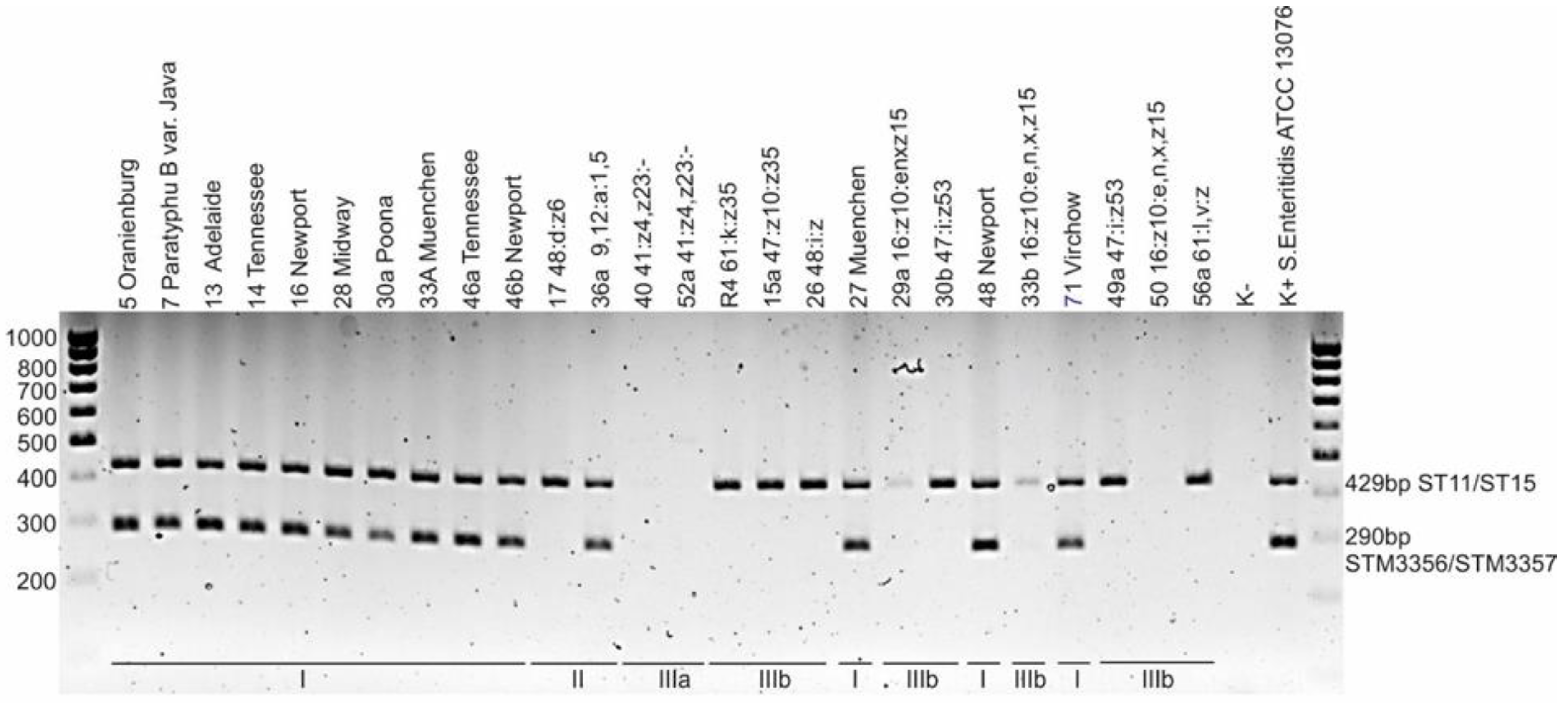
Figure 1. Agarose gel electrophoresis of multiplex PCR employed for discrimination of dT+ and dT- Salmonella strains. The ST11/ST15 amplicon (429 bp) is characteristic of the genus Salmonella, and the STM3356/STM3357 amplicon (290 bp) indicates the ability of the strain to utilize sodium potassium tartrate; M—DNA marker, K+—Salmonella Enteritidis ATCC 13076. The photo shows profiles of selected Salmonella strains.
Pathogens 11 01125 g001
Figure 2. UPGMA dendrogram showing the biochemical similarity of Salmonella isolates based on the results of 28 assays (detailed results of individual biochemical tests are presented in Table S1).
Pathogens 11 01125 g002
Table 1. Frequency of isolation of Salmonella from captive reptiles in Poland.
Animals Number of SamplesSpecies (Number of Samples) Diet GroupNumber of Salmonella Positive Samples Number of Salmonella Isolates
Snakes31Pantheropsis guttatus (14)
Python regius (3)
Boa constrictor (4)
Lampropeltis triangulum (4)
Morelia pilota (1)
Orthrophis teaniurus (5)
carnivore
carnivore
carnivore
carnivore
carnivore
carnivore
12 (86%)
3 (100%)
4 (100%)
3 (75%)
1 (100%)
4 (80%)
13
3
4
4
1
4
Total: 27/31 (87.1%)Total: 29/51 (56.9%)
Lizards18Pogona vitticeps (6)
Iguana iguana (4)
Eublepharis macularius (3)
Furcifer pardali (5)
omnivore
herbivore
carnivore
omnivore
6 (100%)
3 (75%)
3 (100%)
2 (40%)
7
3
3
2
Total: 14/18 (77.8%)Total: 15/51 (29.4%)
Turtles 18Testudo horsfieldii (8)
Testudo hermanni (9)
Chelonoidis carbonaria (1)
herbivore
herbivore
herbivore
4 (50%)
3 (33%)
0
4
3
0
Total: 7/18 (38.9%)Total: 7/51 (13.7%)
Total:67 48/67 (71.6%)51 (100%)
Table 2. Summarized data on the biochemical analysis of Salmonella isolates.
SpeciesSalmonella enterica
Subspecies
Number of Strains
enterica
n = 34
salamae
n = 2
arizonae
n = 2
diarizonae
n = 13
ONPG (24 h)10 [29%]1 [50%]2 [100%]13 [100%]
Lactose8 [23.5%] *01 [50%]10 [77%]
Salicin0000
Dulcitol34 [100%]2 [100%]00
Sorbitol34 [100%]1 [50%]2 [100%]11 [85%]
L(+)-tartrate **34 [100%]1 [50%]00
Mucate34 [100%]2 [100%]1 [50%]0
Malonate2 [6%]2 [100%]2 [100%]13 [100%]
Galacturonate02 [100%]1 [50%]13 [100%]
β-glucuronidase11 [32%]1 [50%]013 [100%]
Gelatinase0000
* Eight strains of S. enterica subsp. enterica that were able to utilize lactose represented the Lattenkamp serotype. ** The ability of Salmonella strains to utilize L(+)-tartrate was determined indirectly by detecting the sequence located in the region between the STM 3357 and STM 3356 genes.
Table 3. Salmonella serovars in captive snakes, lizards, and turtles in Poland.
No.SerovarSubsp.Number of Strains (%)Snakes
n = 31
Lizards
n = 18
Turtles
n = 18
HostStrain ID
19,12:a:1,5II1 (2.0)1 boa constrictor36a
248:d:z6II1 (2.0) 1steppe tortoise17
341:z4,z23:-IIIa2 (3.9)2 milk snake
beauty rat snake
40
52a
416:z10:e,n,x,z15IIIb4 (7.8)4 corn snake
milk snake
beauty rat snake
beauty rat snake
29a
33B
50
51
547:i:z53IIIb4 (7.8)4 corn snake
milk snake
corn snake
beauty rat snake
30b
32
38
49a
647:z10:z35IIIb1 (2.0)1 ball python15a
748:i:zIIIb1 (2.0)1 boa constrictor26
850:l,v:zIIIb1 (2.0) 1 panther chameleon57a
961:k:z35IIIb1 (2.0)1 corn snakeR4
1061:l,v:zIIIb1 (2.0) 1 panther chameleon56a
11AbonyI1 (2.0) 1Greek tortoise68
12AdelaideI1 (2.0)1 ball python13
13AlpenquaiI1 (2.0) 1Greek tortoise66
14CothamI1 (2.0) 1 leopard gecko60
15FlunternI1 (2.0) 1 leopard gecko58b
16LattenkampI8 (15.7) 71bearded dragon
leopard gecko
bearded dragon
bearded dragon
bearded dragon
green iguana
green iguana
steppe tortoise
55b
59
61
62
63
64
65
70
17MidwayI1 (2.0)1 corn snake28
18MuenchenI2 (3.9)2 boa constrictor
milk snake
27
33A
19NewportI3 (5.9)21 corn snake
bearded dragon
carpet python
16
46b
48
20OranienburgI3 (5.9)3 corn snake
corn snake
boa constrictor
5
6a
35
21Paratyphi B var. JavaI2 (3.9)2 corn snake
corn snake
7
31a
22PatienceI1 (2.0) 1Greek tortoise67
23PoonaI4 (7.8)4 corn snake
ball python
corn snake
corn snake
30a
34
37
39
24TennesseeI3 (5.9) 3 bearded dragon
green iguana
bearded dragon
14
45
46a
25VirchowI2 (3.9) 2steppe tortoise
Greek tortoise
69
71
Total number (%) 51 (100) 29 (57) 15 (29)7 (14)
Table 4. Results of genomic analysis of selected Salmonella isolates.
Isolate ID50
(S21_0833)
51
(S21_0834)
33B
(S21_1654)
33A
(S21_0821)
SubspeciesIIIbIIIbIIIbI
Serotype 16:z10:e,n,x,z1516:z10:e,n,x,z1516:z10:e,n,x,z15Muenchen (6,8:d:1,2)
Sourcebeauty rat snake
(Orthriophis taeniurus)
beauty rat snake
(Orthriophis taeniurus)
milk snake
(Lampropeltis triangulum)
milk snake
(Lampropeltis triangulum)
Acc. No. SAMN25211032SAMN25211031SAMN25211030SAMN25046248
Genome size5921031 bp5922870 bp5984163 bp5376408 bp
Contigs 339293245115
Resistance
genes
aac(6′)-Iaa *, kdeA, mdtM, acrF, emrDaac(6′)-Iaa, kdeA, mdtM, acrF, emrDaac(6′)-Iaa, kdeA, mdtM, acrF, emrDaac(6′)-Iaa, kdeA, mdtM, acrF, emrD mdsA, mdsB
Lactose operon genes lacZ **, locus_tag L4A35_20235 (oligosaccharide MFS transporter), lacA ***, lacI **** (Acc. No. JAKJQB010000022.1)lacZ, locus_tag L4B25_00380 (oligosaccharide MFS transporter), lacA, lacI (Acc. No. JAKJQC010000001.1)lacZ, locus_tag L4A43_06620 (oligosaccharide MFS transporter), lacA, lacI (Acc. No. JAKJQD010000004.1)Not found
* cryptic gene; ** lacZ coding for β-galactosidase; *** lacA coding for galactoside O-acetyltransferase; **** lacI coding for transcriptional regulator.
Table 5. Pathogenic and zoonotic potential of Salmonella serovars identified in the study.
Pathogenic PotentialZoonotic Potential
SerovarSubsp.Number of Infections in US in 2006–2016 [25]Number of Infections in EU in 2018–2020 [1]Number of Infections in Queensland in 2007–2016 [26]HostAge of Patients with RAS/Median [years]Number of InfectionsCountryYearReference
AbonyI62aturtle
turtle
4
<1
1
1
Japan
Belgium
2007–8
2008
[27]
[28]
AdelaideI1001
AlpenquaiI
CothamI429lizards<1–79/3160USA2012–14[29]
FlunternI77
LattenkampI17
MidwayI
MuenchenI1216703reptiles b
turtle
<5
<1–60/10
48
132
UK
USA
2015
2009
[30]
[10]
NewportI47,4812233160reptiles b
turtle
turtles
<5
1.25
<1–85/6
48
1
124
UK
Chile
USA
2015
?
2012
[30]
[31]
[10]
OranienburgI801288reptiles b
turtle
bearded dragon
<5
<5
?
48
26
1
UK
USA
Czech Republic
2015
2019
2016
[30]
[32]
[33]
Paratyphi B var. JavaI4486355turtle
turtle
turtle
turtle
snake b
<1–10
<1–4
1–87/7
1–75/6
50
8
4
107
132
1
Spain
Spain
USA
USA
Finland
2010–11
2009
2007
2011
2005–8
[34]
[35]
[10]
[10]
[36]
PatienceI
PoonaI3844turtle
turtle
turtle
turtle
turtle
snake
iguana
<1 to 82/6
5
<1–84/3.5
<1–83/3
<1–75/5
<1
2
61
1
58
78
40
1
1
USA
Japan
USA
USA
USA
Germany
USA
2015
2007–8
2012
2012
2014
2008
2002
[11]
[27]
[10]
[10]
[10]
[36]
[37]
TennesseeI1326bearded dragon<11USA2000[37]
VirchowI9802804
9,12:a:1,5II2
48:d:z6II10
41:z4,z23:-IIIa146snake or lizard
snake
<1
<1–57
2
3
Germany
Belgium
2007–8
2008
[36]
[36]
16:z10:e,n,x,z15IIIb21
47:i:z53IIIbsnake251Germany2006[36]
47:z10:z35IIIb3
48:i:zIIIb69
50:l,v:zIIIb3
61:k:z35IIIb
61:l,v:zIIIb
a—the serotype was not identified or listed in the report; cases of infections caused by rarely identified or unknown Salmonella serotypes were included in the report as one group. b—probable source of infection, not scientifically proven
Publisher’s Note: MDPI stays neutral with regard to jurisdictional claims in published maps and institutional affiliations.