
A Review of Zoonotic Babesiosis as an Emerging Public Health Threat in Asia

 ,
,  , , and
Abstract
1. Introduction
2. Materials and Methods
3. Results and Discussion
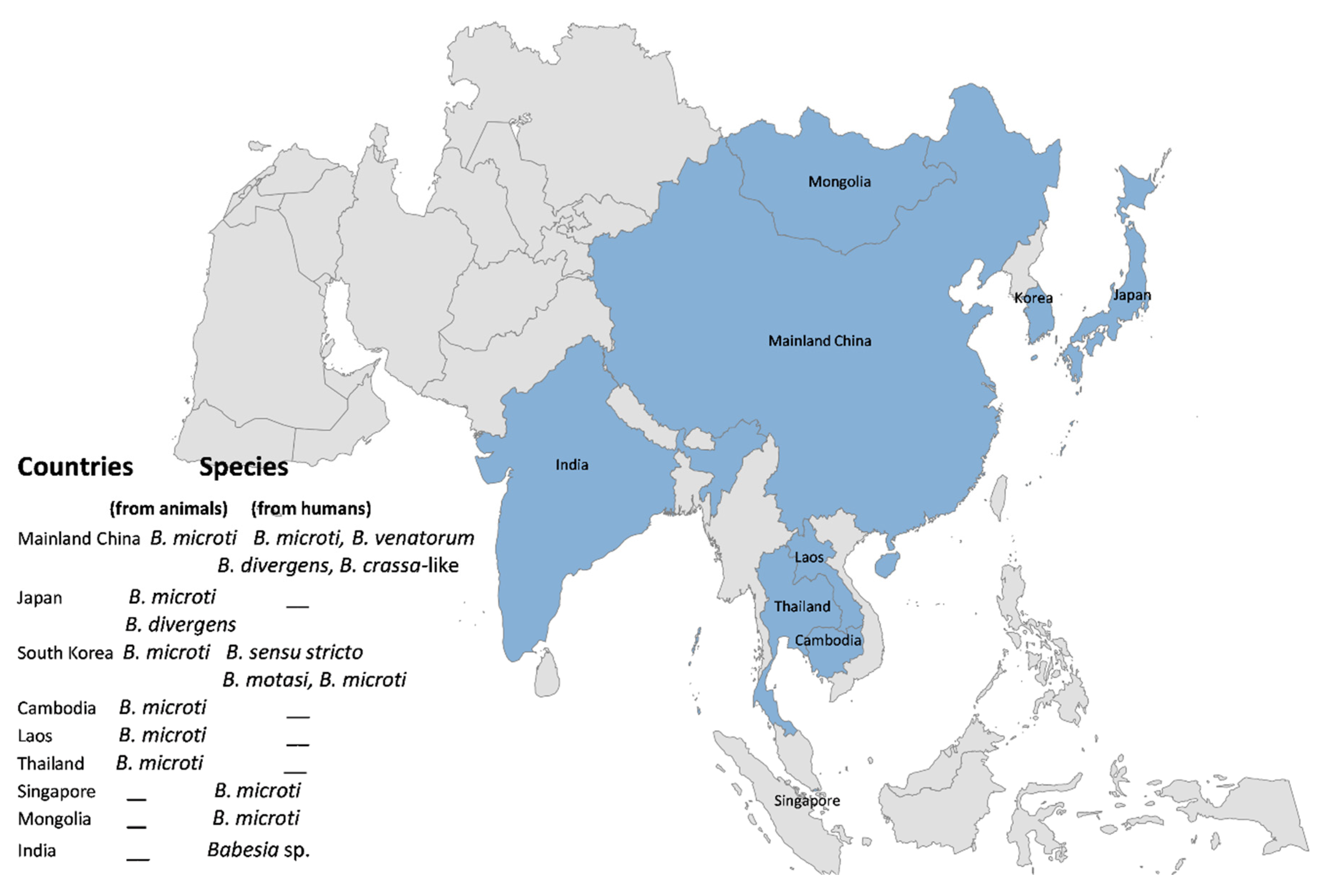
3.1. China
3.2. Korea
3.3. Japan
3.4. Singapore
3.5. Other Countries
4. Conclusions
Author Contributions
Funding
Institutional Review Board Statement
Informed Consent Statement
Data Availability Statement
Acknowledgments
Conflicts of Interest
References
- Homer, M.J.; Aguilar-Delfin, I.; Telford, S.R., III; Krause, P.J.; Persing, D.H. Babesiosis. Clin. Microbiol. Rev. 2000, 13, 451–469. [Google Scholar] [CrossRef] [PubMed]
- Gray, J.; Zintl, A.; Hildebrandt, A.; Hunfeld, K.-P.; Weiss, L. Zoonotic babesiosis: Overview of the disease and novel aspects of pathogen identity. Ticks Tick-Borne Dis. 2010, 1, 3–10. [Google Scholar] [CrossRef]
- Centers for Disease Control and Prevention. Babesiosis surveillance-18 states, 2011. MMWR Morb. Mortal. Wkly. Rep. 2012, 61, 505–509. [Google Scholar]
- Young, K.M.; Corrin, T.; Wilhelm, B.; Uhland, C.; Greig, J.; Mascarenhas, M.; Waddell, L.A. Zoonotic Babesia: A scoping review of the global evidence. PLoS ONE 2019, 14, e0226781. [Google Scholar] [CrossRef] [PubMed]
- Dumic, I.; Severnini, E. “Ticking bomb”: The impact of climate change on the incidence of Lyme disease. Can. J. Infect. Dis. Med. Microbiol. 2018, 2018, 5719081. [Google Scholar] [CrossRef]
- Dumic, I.; Madrid, C.; Rueda Prada, L.; Nordstrom, C.W.; Taweesedt, P.T.; Ramanan, P. Splenic Complications of Babesia microti Infection in Humans: A Systematic Review. Can. J. Infect. Dis. Med. Microbiol. 2020, 2020, 6934149. [Google Scholar] [CrossRef]
- Piesman, J.; Spielman, A. Human Babesiosis on Nantucket Island: Prevalence of Babesia microti in Ticks. Am. J. Trop. Med. Hyg. 1980, 29, 742–746. [Google Scholar] [CrossRef]
- Asante, E.A.; Linehan, J.M.; Smidak, M.; Tomlinson, A.; Grimshaw, A.; Jeelani, A.; Jakubcova, T.; Hamdan, S.; Powell, C.; Brandner, S.; et al. Inherited Prion Disease A117V Is Not Simply a Proteinopathy but Produces Prions Transmissible to Transgenic Mice Expressing Homologous Prion Protein. PLoS Pathog. 2013, 9, e1003643. [Google Scholar] [CrossRef]
- Nzenze, S.A.; Shiri, T.; Nunes, M.C.; Klugman, K.P.; Kahn, K.; Twine, R.; de Gouveia, L.; von Gottberg, A.; Madhi, S.A. Temporal Changes in Pneumococcal Colonization in a Rural African Community with High HIV Prevalence Following Routine Infant Pneumococcal Immunization. Pediatr. Infect. Dis. J. 2013, 32, 1270–1278. [Google Scholar] [CrossRef]
- Sanchez, E.; Vannier, E.; Wormser, G.P.; Hu, L.T. Diagnosis, treatment, and prevention of Lyme disease, human granulocytic anaplasmosis, and babesiosis: A review. JAMA 2016, 315, 1767–1777. [Google Scholar] [CrossRef]
- Krause, P.; McKay, K.; Gadbaw, J.; Christianson, D.; Closter, L.; Lepore, T.; Telford, S.R., 3rd; Sikand, V.; Ryan, R.; Persing, D.; et al. Increasing health burden of human babesiosis in endemic sites. Am. J. Trop. Med. Hyg. 2003, 68, 431–436. [Google Scholar] [CrossRef]
- Purnell, R.; Brocklesby, D.; Kitchenham, B.; Young, E. A statistical comparison of the behaviour of five British isolates of Babesia divergens in splenectomized calves. J. Comp. Pathol. 1976, 86, 609–614. [Google Scholar] [CrossRef]
- Silaghi, C.; Woll, D.; Hamel, D.; Pfister, K.; Mahling, M.; Pfeffer, M. Babesia spp. and Anaplasma phagocytophilum in questing ticks, ticks parasitizing rodents and the parasitized rodents—Analyzing the host-pathogen-vector interface in a metropolitan area. Parasites Vectors 2012, 5, 191. [Google Scholar] [CrossRef] [PubMed]
- Wilcove, D.S.; Giam, X.; Edwards, D.P.; Fisher, B.; Koh, L.P. Navjot’s nightmare revisited: Logging, agriculture, and biodiversity in Southeast Asia. Trends Ecol. Evol. 2013, 28, 531–540. [Google Scholar] [CrossRef] [PubMed]
- Morand, S. (macro-) Evolutionary ecology of parasite diversity: From determinants of parasite species richness to host diversification. Int. J. Parasitol. Parasites Wildl. 2015, 4, 80–87. [Google Scholar] [CrossRef]
- Zhou, X.; Xia, S.; Huang, J.-L.; Tambo, E.; Zhuge, H.-X.; Zhou, X.-N. Human babesiosis, an emerging tick-borne disease in the People’s Republic of China. Parasites Vectors 2014, 7, 509. [Google Scholar] [CrossRef]
- Tsuji, M.; Wei, Q.; Zamoto, A.; Morita, C.; Arai, S.; Shiota, T.; Fujimagari, M.; Itagaki, A.; Fujita, H.; Ishihara, C. Human Babesiosis in Japan: Epizootiologic Survey of Rodent Reservoir and Isolation of New Type of Babesia microti -Like Parasite. J. Clin. Microbiol. 2001, 39, 4316–4322. [Google Scholar] [CrossRef] [PubMed]
- Kim, J.-Y.; Cho, S.-H.; Joo, H.-N.; Tsuji, M.; Cho, S.-R.; Park, I.-J.; Chung, G.-T.; Ju, J.-W.; Cheun, H.-I.; Lee, H.-W. First case of human babesiosis in Korea: Detection and characterization of a novel type of Babesia sp. (KO1) similar to ovine babesia. J. Clin. Microbiol. 2007, 45, 2084–2087. [Google Scholar] [CrossRef]
- Shih, C.-M.; Liu, L.-P.; Chung, W.-C.; Ong, S.; Wang, C.-C. Human babesiosis in Taiwan: Asymptomatic infection with a Babesia microti-like organism in a Taiwanese woman. J. Clin. Microbiol. 1997, 35, 450–454. [Google Scholar] [CrossRef] [PubMed]
- Zhou, X.; Li, S.-G.; Chen, S.-B.; Wang, J.-Z.; Xu, B.; Zhou, H.-J.; Ge, H.-X.Z.; Chen, J.-H.; Hu, W. Co-infections with Babesia microti and Plasmodium parasites along the China-Myanmar border. Infect. Dis. Poverty 2013, 2, 24. [Google Scholar] [CrossRef] [PubMed]
- Rar, V.; Yakimenko, V.; Makenov, M.; Tikunov, A.; Epikhina, T.; Tancev, A.; Bobrova, O.; Tikunova, N. High prevalence of Babesia microti ‘Munich’ type in small mammals from an Ixodes persulcatus/Ixodes trianguliceps sympatric area in the Omsk region, Russia. Parasitol. Res. 2016, 115, 3619–3629. [Google Scholar] [CrossRef]
- Chen, X.-R.; Ye, L.; Fan, J.-W.; Li, C.; Tang, F.; Liu, W.; Ren, L.-Z.; Bai, J.-Y. Detection of Kobe-type and Otsu-type Babesia microti in wild rodents in China’s Yunnan province. Epidemiol. Infect. 2017, 145, 2704–2710. [Google Scholar] [CrossRef]
- Gao, Z.-H.; Huang, T.-H.; Jiang, B.-G.; Jia, N.; Liu, Z.-X.; Shao, Z.-T.; Jiang, R.-R.; Liu, H.-B.; Wei, R.; Li, Y.-Q. Wide distribution and genetic diversity of Babesia microti in small mammals from Yunnan province, Southwestern China. PLoS Negl. Trop. Dis. 2017, 11, e0005898. [Google Scholar] [CrossRef] [PubMed]
- Wei, C.-Y.; Wang, X.-M.; Wang, Z.-S.; Wang, Z.-H.; Guan, Z.-Z.; Zhang, L.-H.; Dou, X.-F.; Wang, H. High prevalence of Babesia microti in small mammals in Beijing. Infect. Dis. Poverty 2020, 9, 155. [Google Scholar] [CrossRef]
- Saito-Ito, A.; Takada, N.; Ishiguro, F.; Fujita, H.; Yano, Y.; Ma, X.-H.; Chen, E.-R. Detection of Kobe-type Babesia microti associated with Japanese human babesiosis in field rodents in central Taiwan and southeastern mainland China. Parasitology 2008, 135, 691–699. [Google Scholar] [CrossRef] [PubMed]
- Hong, S.-H.; Lee, S.-E.; Jeong, Y.-I.; Kim, H.-C.; Chong, S.-T.; Klein, T.A.; Song, J.-W.; Gu, S.H.; Cho, S.-H.; Lee, W.-J. Prevalence and molecular characterizations of Toxoplasma gondii and Babesia microti from small mammals captured in Gyeonggi and Gangwon Provinces, Republic of Korea. Vet. Parasitol. 2014, 205, 512–517. [Google Scholar] [CrossRef] [PubMed]
- Hong, S.-H.; Kim, H.-J.; Jeong, Y.-I.; Cho, S.-H.; Lee, W.-J.; Kim, J.-T.; Lee, S.-E. Serological and molecular detection of Toxoplasma gondii and Babesia microti in the blood of rescued wild animals in Gangwon-do (Province), Korea. Korean J. Parasitol. 2017, 55, 207–212. [Google Scholar] [CrossRef]
- Karnchanabanthoeng, A.; Morand, S.; Jittapalapong, S.; Carcy, B. Babesia Occurrence in Rodents in Relation to Landscapes of Mainland Southeast Asia. Vector-Borne Zoonotic Dis. 2018, 18, 121–130. [Google Scholar] [CrossRef]
- Okabayashi, T.; Hagiya, J.; Tsuji, M.; Ishihara, C.; Satoh, H.; Morita, C. Detection of Babesia microti-like Parasite in Filter Paper-Absorbed Blood of Wild Rodents. J. Vet. Med. Sci. 2002, 64, 145–147. [Google Scholar] [CrossRef][Green Version]
- Saito-Ito, A.; Kasahara, M.; Kasai, M.; Dantrakool, A.; Kawai, A.; Fujita, H.; Yano, Y.; Kawabata, H.; Takada, N. Survey of Babesia microti Infection in Field Rodents in Japan: Records of the Kobe-Type in New Foci and Findings of a New Type Related to the Otsu-Type. Microbiol. Immunol. 2007, 51, 15–24. [Google Scholar] [CrossRef]
- Tabara, K.; Arai, S.; Kawabuchi, T.; Itagaki, A.; Ishihara, C.; Satoh, H.; Okabe, N.; Tsuji, M. Molecular survey of Babesia microti, Ehrlichia species and Candidatus Neoehrlichia mikurensis in wild rodents from Shimane Prefecture, Japan. Microbiol. Immunol. 2007, 51, 359–367. [Google Scholar] [CrossRef]
- Zamoto-Niikura, A.; Hagiwara, K.; Imaoka, K.; Morikawa, S.; Ishihara, C.; Hanaki, K.-I. Epidemiological Survey of Babesia divergens Asia Lineage in Wild Sika Deer (Cervus nippon) by Using Direct PCR in Japan. Jpn. J. Infect. Dis. 2020, 73, 68–71. [Google Scholar] [CrossRef] [PubMed]
- Hong, S.-H.; Kim, S.-Y.; Song, B.G.; Roh, J.Y.; Cho, C.R.; Kim, C.-N.; Um, T.-H.; Kwak, Y.G.; Cho, S.-H.; Lee, S.-E. Detection and characterization of an emerging type of Babesia sp. similar to Babesia motasi for the first case of human babesiosis and ticks in Korea. Emerg. Microbes Infect. 2019, 8, 869–878. [Google Scholar] [CrossRef]
- Kwon, H.Y.; Im, J.H.; Park, Y.-K.; Durey, A.; Lee, J.-S.; Baek, J.H. Two imported cases of babesiosis with complication or co-infection with lyme disease in republic of Korea. Korean J. Parasitol. 2018, 56, 609. [Google Scholar] [CrossRef] [PubMed]
- Marathe, A.; Tripathi, J.; Handa, V.; Date, V. Human babesiosis—A case report. Indian J. Med. Microbiol. 2005, 23, 267. [Google Scholar] [CrossRef]
- Hong, S.-H.; Anu, D.; Jeong, Y.-I.; Abmed, D.; Cho, S.-H.; Lee, W.-J.; Lee, S.-E. Molecular detection and seroprevalence of Babesia microti among stock farmers in Khutul City, Selenge Province, Mongolia. Korean J. Parasitol. 2014, 52, 443. [Google Scholar] [CrossRef]
- Saito-Ito, A.; Tsuji, M.; Wei, Q.; He, S.; Matsui, T.; Kohsaki, M.; Arai, S.; Kamiyama, T.; Hioki, K.; Ishihara, C. Transfusion-Acquired, Autochthonous Human Babesiosis in Japan: Isolation of Babesia microti -Like Parasites with hu-RBC-SCID Mice. J. Clin. Microbiol. 2000, 38, 4511–4516. [Google Scholar] [CrossRef] [PubMed]
- Su, G. A case report of babesiosis. Chin. J. Zoonoses 2002, 18, 112. [Google Scholar]
- Qi, C.; Zhou, D.; Liu, J.; Cheng, Z.; Zhang, L.; Wang, L.; Wang, Z.; Yang, D.; Wang, S.; Chai, T. Detection of Babesia divergens using molecular methods in anemic patients in Shandong Province, China. Parasitol. Res. 2011, 109, 241–245. [Google Scholar] [CrossRef]
- Sun, Y.; Li, S.-G.; Jiang, J.-F.; Wang, X.; Zhang, Y.; Wang, H.; Cao, W.-C. Babesia venatorum infection in child, China. Emerg. Infect. Dis. 2014, 20, 896. [Google Scholar] [CrossRef]
- Jiang, J.-F.; Zheng, Y.-C.; Jiang, R.-R.; Li, H.; Huo, Q.-B.; Jiang, B.-G.; Sun, Y.; Jia, N.; Wang, Y.-W.; Ma, L. Epidemiological, clinical, and laboratory characteristics of 48 cases of “Babesia venatorum” infection in China: A descriptive study. Lancet Infect. Dis. 2015, 15, 196–203. [Google Scholar] [CrossRef]
- Man, S.-Q.; Qiao, K.; Cui, J.; Feng, M.; Fu, Y.-F.; Cheng, X.-J. A case of human infection with a novel Babesia species in China. Infect. Dis. Poverty 2016, 5, 28. [Google Scholar] [CrossRef] [PubMed]
- Wang, H. Investigation of Babesia spp. infections in blood donors in Guangxi, China. Acad. J. Second Mil. Med. Univ. 2016, 37, 283–287. [Google Scholar]
- Jia, N.; Zheng, Y.-C.; Jiang, J.-F.; Jiang, R.-R.; Jiang, B.-G.; Wei, R.; Liu, H.-B.; Huo, Q.-B.; Sun, Y.; Chu, Y.-L. Human Babesiosis Caused by a Babesia crassa–Like Pathogen: A Case Series. Clin. Infect. Dis. 2018, 67, 1110–1119. [Google Scholar] [CrossRef]
- Yao, L.-N.; Ruan, W.; Zeng, C.-Y.; Li, Z.-H.; Zhang, X.; Lei, Y.-L.; Lu, Q.-Y.; Che, H.-L. Pathogen identification and clinical diagnosis for one case infected with Babesia. Zhongguo Ji Sheng Chong Xue Yu Ji Sheng Chong Bing Za Zhi Chin. J. Parasitol. Parasit. Dis. 2012, 30, 118–121. [Google Scholar]
- Birkenheuer, A.J.; Horney, B.; Bailey, M.; Scott, M.; Sherbert, B.; Catto, V.; Marr, H.S.; Camacho, A.-T.; Ballman, A.E. Babesia microti-like infections are prevalent in North American foxes. Vet. Parasitol. 2010, 172, 179–182. [Google Scholar] [CrossRef]
- Telford, S.R., III; Mather, T.N.; Adler, G.H.; Spielman, A. Short-Tailed Shrews as Reservoirs of the Agents of Lyme Disease and Human Babesiosis. J. Parasitol. 1990, 76, 681–683. [Google Scholar]
- Birkenheuer, A.J.; Marr, H.S.; Hladio, N.; Acton, A.E. Molecular evidence of prevalent dual piroplasma infections in North American raccoons (Procyon lotor). Parasitology 2008, 135, 33–37. [Google Scholar] [CrossRef]
- Spielman, A.; Etkind, P.; Piesman, J.; Ruebush, T., 2nd; Juranek, D.D.; Jacobs, M.S. Reservoir hosts of human babesiosis on Nantucket Island. Am. J. Trop. Med. Hyg. 1981, 30, 560–565. [Google Scholar] [CrossRef]
- Siński, E. Enzootic reservoir for new Ixodes ricinus-transmitted infections. Wiad. Parazytol. 1999, 45, 135–142. [Google Scholar]
- Wei, Q.; Tsuji, M.; Zamoto, A.; Kohsaki, M.; Matsui, T.; Shiota, T.; Telford, S.R., III; Ishihara, C. Human Babesiosis in Japan: Isolation of Babesia microti-Like Parasites from an Asymptomatic Transfusion Donor and from a Rodent from an Area Where Babesiosis Is Endemic. J. Clin. Microbiol. 2001, 39, 2178–2183. [Google Scholar] [CrossRef] [PubMed]
- Saito-Ito, A.; Yano, Y.; Dantrakool, A.; Hashimoto, T.; Takada, N. Survey of Rodents and Ticks in Human Babesiosis Emergence Area in Japan: First Detection of Babesia microti-Like Parasites in Ixodes ovatus. J. Clin. Microbiol. 2004, 42, 2268–2270. [Google Scholar] [CrossRef]
- Beck, R.; Vojta, L.; Ćurković, S.; Mrljak, V.; Margaletić, J.; Habrun, B. Molecular Survey of Babesia microti in Wild Rodents in Central Croatia. Vector-Borne Zoonotic Dis. 2011, 11, 81–83. [Google Scholar] [CrossRef] [PubMed]
- Bajer, A.; Bednarska, M.; Pawelczyk, A.; Konopka, E.; Karbowiak, G.; Siñski, E. Blood parasites in a wild rodent community of Mazury Lakes District, Poland. Wiadomości Parazytol. 1998, 44, 426. [Google Scholar]
- Lim, P.-L.; Chavatte, J.-M.; Vasoo, S.; Yang, J. Imported human babesiosis, Singapore, 2018. Emerg. Infect. Dis. 2020, 26, 826. [Google Scholar] [CrossRef] [PubMed]
- Jabłońska, J.; Żarnowska-Prymek, H.; Stańczak, J.; Kozłowska, J.; Wiercińska-Drapało, A. Symptomatic co-infection with Babesia microti and Borrelia burgdorferi in patient after international exposure; a challenging case in Poland. Ann. Agric. Environ. Med. 2016, 23, 387–389. [Google Scholar] [CrossRef]
- Schleinitz, N.; Harlé, J.-R.; Piarroux, R.; Carcy, B.; Bernit, E.; Faucher, B.; Berda-Haddad, Y.; Ebbo, M.; Poisnel, E. Babesia microti: An unusual travel-related disease. BMC Infect. Dis. 2013, 13, 99. [Google Scholar]
- Kwak, M.L. Ticks in the Lion City: A preliminary review of the tick fauna of Singapore. Exp. Appl. Acarol. 2018, 76, 263–267. [Google Scholar] [CrossRef]
- Negi, T.; Kandari, L.S.; Arunachalam, K. Update on prevalence and distribution pattern of tick-borne diseases among humans in India: A review. Parasitol. Res. 2021, 120, 1523–1539. [Google Scholar] [CrossRef]


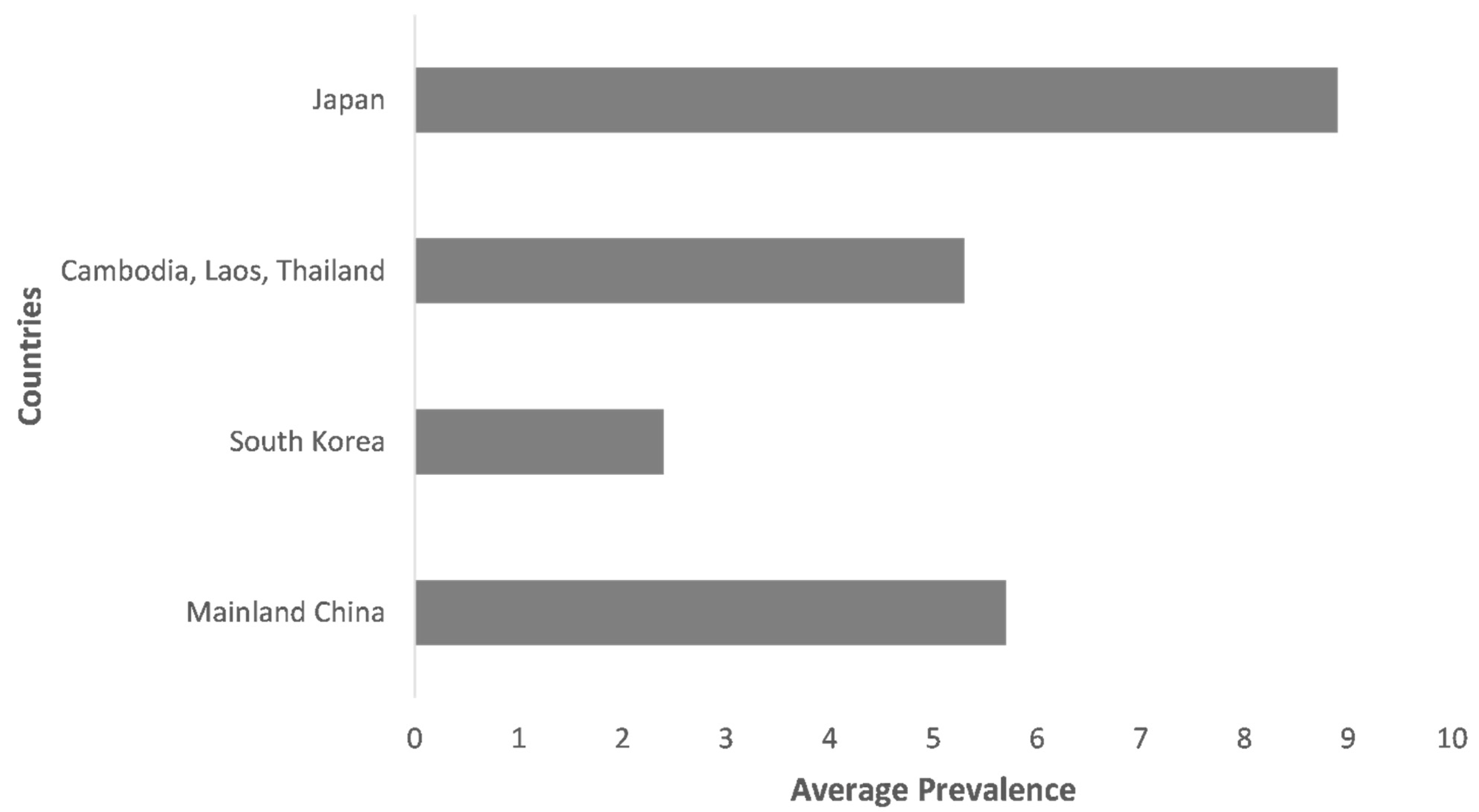
| Year of Study | Country and Region | Continent | Method of Identification | Sample Site/Host | Species | Sample Size | Positive | Prevalence % | CI 95% | Reference |
|---|---|---|---|---|---|---|---|---|---|---|
| 2009–2011 | China | Asia | PCR/Seq | Wild rodents | B. microti | 1672 | 72 | 4.3 | 3.4–5.4 | [22] |
| 2009–2011 | China | Asia | PCR/Seq | Small mammals | B. microti | 2204 | 53 | 2.4 | 1.8–3.1 | [23] |
| 2018 | China | Asia | PCR/Seq | Small mammals | B. microti | 1391 | 168 | 12.1 | 10.4–13.9 | [24] |
| 2002–2005 | China and Taiwan | Asia | PCR/Seq | Field rodents | B. microti | 68 | 15 | 22.1 | 12.9–33.8 | [25] |
| 2008 | Korea | Asia | PCR/Seq | Small mammals | B. microti | 667 | 14 | 2.1 | 1.2–3.5 | [26] |
| 2008–2009 | Korea | Asia | PCR/Seq | Rescued wild animals | B. microti | 70 | 4 | 5.7 | 1.6–14.0 | [27] |
| 2008–2009 | Cambodia, Laos, Thailand | Asia | Nested PCR | Wild rodents | B. microti | 1439 | 76 | 5.3 | 4.2–6.6 | [28] |
| 1998 | Japan | Asia | PCR/Seq | Wild rodents | B. microti | 97 | 13 | 13.4 | 7.3–21.8 | [29] |
| 2003–2005 | Japan | Asia | PCR/Seq | Field rodents | B. microti | 247 | 36 | 14.6 | 10.4–19.6 | [30] |
| 2000–2004 | Japan | Asia | PCR/Seq | Wild rodents | B. microti | 62 | 28 | 45.2 | 32.5–58.3 | [31] |
| 2012–2018 | Japan | Asia | PCR | Wild Sika deer | B. divergens | 1747 | 116 | 6.6 | 5.5–7.9 | [32] |
| Year | Species | Geographical Location | Country | Number of Cases | Diagnostic Technique | Potential Transmission Route | Gender | Age Range (Years) | Reference |
|---|---|---|---|---|---|---|---|---|---|
| 2006 | Babesia sensu stricto | Gurae, Jeon-nam | Korea | 1 | Microscopy, PCR | Blood transfusion | Female | 75 | [18,33] |
| 2005 | B. motasi | Hoengseong-gun, Gangwon-do | Korea | 2 | Microscopy, PCR | tick bite | Male | 70 | [32] |
| 2018 | B. microti | Incheon | Korea | 2 | PCR | – | Female | 50–72 | [34] |
| 2004 | Babesia sp. | Baroda (Gujarat) | India | 1 | Microscopy | – | Male | 51 | [35] |
| 2011 | B. microti | Selenge | Mongolia | 3 | IFA, PCR | – | Male | – | [36] |
| 1999 | B. microti | Kobe, Hyogo Prefecture | Japan | 1 | IFA, PCR | Blood transfusion | Male | 40 | [37] |
| 2018 | B. microti | Tan Tock Seng Hospital | Singapore | 1 | Microscopy, PCR | Tick bites | Male | 37 | [16] |
| 1994 | B. microti-like | Southern Taiwan | China | 2 | Microscopy, IFA, inoculation | – | Female | 51, − | [19] |
| 2000 | Babesia sp. | Hangzhou, Zhejiang | China | 1 | Microscopy | Blood transfusion | Male | 36 | [38] |
| 2009 | B. divergens | Tai’an, Shandong | China | 2 | PCR | – | Male | – | [39] |
| 2010 | B. microti-like | Yunnan | China | 1 | Microscopy, IFA | Tick bites | Female | 46 | [40] |
| 2012–2013 | B. microti | Tengchong, Yunnan | China | 10 | Microscopy, PCR | Blood transfusion, tick bites | 6 males | 22–45 | [20] |
| 2012 | B. venatorum | Pishan, Xinjiang | China | 1 | Microscopy, PCR, inoculation | Tick bites | Male | 8 | [40] |
| 2011–2014 | B. venatorum | Heilongjiang | China | 48 a | PCR, microscopy, FISH, inoculation | Tick bites | – | 0.6–75 | [41] |
| 2015 | B. sp. XXB/Hangzhou | Hangzhou, Zhejiang | China | 1 | Microscopy, PCR | – | Male | 42 | [42] |
| 2013–2015 | B. microti | Guangxi Zhuang | China | 48 | Microscopy, PCR, IFA | – | – | – | [43] |
| 2015–2016 | B. crassa-like | Heilongjiang | China | 58 b | Microscopy, PCR | Tick bites | 19 males | 4–72 | [44] |
Publisher’s Note: MDPI stays neutral with regard to jurisdictional claims in published maps and institutional affiliations. |
© 2021 by the authors. Licensee MDPI, Basel, Switzerland. This article is an open access article distributed under the terms and conditions of the Creative Commons Attribution (CC BY) license (https://creativecommons.org/licenses/by/4.0/).
Share and Cite
Hussain, S.; Hussain, A.; Aziz, M.U.; Song, B.; Zeb, J.; George, D.; Li, J.; Sparagano, O. A Review of Zoonotic Babesiosis as an Emerging Public Health Threat in Asia. Pathogens 2022, 11, 23. https://doi.org/10.3390/pathogens11010023
Hussain S, Hussain A, Aziz MU, Song B, Zeb J, George D, Li J, Sparagano O. A Review of Zoonotic Babesiosis as an Emerging Public Health Threat in Asia. Pathogens. 2022; 11(1):23. https://doi.org/10.3390/pathogens11010023
Chicago/Turabian StyleHussain, Sabir, Abrar Hussain, Muhammad Umair Aziz, Baolin Song, Jehan Zeb, David George, Jun Li, and Olivier Sparagano. 2022. "A Review of Zoonotic Babesiosis as an Emerging Public Health Threat in Asia" Pathogens 11, no. 1: 23. https://doi.org/10.3390/pathogens11010023
APA StyleHussain, S., Hussain, A., Aziz, M. U., Song, B., Zeb, J., George, D., Li, J., & Sparagano, O. (2022). A Review of Zoonotic Babesiosis as an Emerging Public Health Threat in Asia. Pathogens, 11(1), 23. https://doi.org/10.3390/pathogens11010023