Article

Amplicon-Based Next Generation Sequencing for Rapid Identification of Rickettsia and Ectoparasite Species from Entomological Surveillance in Thailand

by
Suwanna Chaorattanakawee
1,2,
Achareeya Korkusol
1,
Bousaraporn Tippayachai
1,
Sommai Promsathaporn
1,
Betty K. Poole-Smith
1 and
Ratree Takhampunya
1,*
1
Department of Entomology, US Army Medical Directorate of the Armed Forces Research Institute of Medical Sciences (USAMD-AFRIMS), Bangkok 10400, Thailand
2
Department of Parasitology and Entomology, Faculty of Public Health, Mahidol University, Ratchawithi Road, Bangkok 10400, Thailand
*
Author to whom correspondence should be addressed.
Pathogens 2021, 10(2), 215; https://doi.org/10.3390/pathogens10020215
Submission received: 31 December 2020 / Revised: 28 January 2021 / Accepted: 2 February 2021 / Published: 16 February 2021
(This article belongs to the Collection Updates on Rickettsia and Coxiella)

Abstract

:
Background: Next generation sequencing (NGS) technology has been used for a wide range of epidemiological and surveillance studies. Here, we used amplicon-based NGS to species identify Rickettsia and their arthropod hosts from entomological surveillance. Methods: During 2015–2016, we screened 1825 samples of rodents and ectoparasites collected from rodents and domestic mammals (dog, cat, and cattle) across Thailand for Rickettsia. The citrate synthase gene was amplified to identify Rickettsia to species, while the Cytochrome Oxidase subunit I (COI) and subunit II (COII) genes were used as target genes for ectoparasite identification. All target gene amplicons were pooled for library preparation and sequenced with Illumina MiSeq platform. Result: The highest percentage of Rickettsia DNA was observed in fleas collected from domestic animals (56%) predominantly dogs. Only a few samples of ticks from domestic animals, rodent fleas, and rodent tissue were positive for Rickettisia DNA. NGS based characterization of Rickettsia by host identified Rickettsia asembonensis as the most common bacteria in positive fleas collected from dogs (83.2%) while “Candidatus Rickettsia senegalensis” was detected in only 16.8% of Rickettsia positive dog fleas. Sequence analysis of COI and COII revealed that almost all fleas collected from dogs were Ctenocephalides felis orientis. Other Rickettsia species were detected by NGS including Rickettsia heilongjiangensis from two Haemaphysalis hystricis ticks, and Rickettsia typhi in two rodent tissue samples. Conclusion: This study demonstrates the utility of NGS for high-throughput sequencing in the species characterization/identification of bacteria and ectoparasite for entomological surveillance of rickettsiae. A high percentage of C. f. orientis are positive for R. asembonensis. In addition, our findings indicate there is a risk of tick-borne Spotted Fever Group rickettsiosis, and flea-borne murine typhus transmission in Tak and Phangnga provinces of Thailand.

1. Introduction

The genus Rickettsia is comprised of obligate intracellular Gram-negative organisms that cause diseases in people throughout the world. Rickettsioses are zoonotic diseases that can be transmitted to human by ectoparasites including fleas, ticks, mites, and lice. Mammals including rodents, domestic animals, and wildlife species have been recognized as reservoirs of rickettsioses. Currently there are 30 identified Rickettsia species [1], and some of these species have been recognized as human pathogens [2]. Rickettsia are classically characterized into two groups, the spotted fever group (i.e., Rickettsia rickettsii, Rickettsia africae) which cause the tick-borne spotted fever group (SFG) rickettsioses, and typhus group (Rickettsia prowazekii, and Rickettsia typhi) which cause louse-borne epidemic typhus, and flea-borne murine or endemic typhus, respectively. There are also some pathogenic rickettsial species classified into the transitional group, as they share features between SFG and typhus group. The transitional rickettsiae includes Rickettsia akari, Rickettsia australis, Rickettsia felis which are causative agents of mite-borne rickettsialpox, Queensland tick typhus, and flea-borne spotted fever, respectively [2].
The epidemiology of Rickettsia spp. worldwide is defined by a combination of climate, the geographical distribution of the primary vector and distribution of the natural animal hosts required to amplify and maintain its presence in the environment. Most SFG Rickettsia are distributed in the limited geographic regions where their specific tick host species is found [2,3,4]. For example, Rocky Mountain spotted fever (RMSF) is caused by R. rickettsii, is distributed throughout the Americas, and Dermacentor ticks serve as the primary vector for RMSF in southeast and south central United States [5], while Amblyomma cajennense sensu lato species complex transmit RMSF in central and South America [6]. However, Rhipicephalus sanguineus sensu lato, which commonly infests dogs, has been implicated in an RMSF outbreak in the tribal lands of Arizona and in Sonora, Mexico [7,8]. Mediterranean spotted fever, caused by Rickettsia conorii, is distributed across southern Europe, northern Africa, with occasional cases reported in Asia, with the potential vector R. sanguineus s.l. (brown dog tick) [5,9]. Rickettsia africae causes African tick bite fever in sub-Saharan Africa which correlates with the distribution of its known vectors Amblyomma hebraeum and Amblyomma variegatum [10].
Although, there are a number of published reports of Rickettsia in Thailand, SFG Rickettsia are considered to be an under recognized cause of febrile illness in Thailand [11] and broader multi-province surveillance for Rickettsia vectors and animal reservoirs to better understand transmission and epidemiology is lacking. Briefly, there is serologic and molecular evidence that Rickettsia honei, and Rickettsia japonica cause humans infections in Thailand, [12,13,14]. In addition R. honei has been identified from Ixodes spp. and Rhipicephalus spp. larval ticks, and detection of R. japonica from Haemaphysalis hystricis suggesting these tick species as potential vectors [15,16]. Flea-borne murine typhus caused by R. typhi, and flea-borne SFG disease caused by R. felis are distributed worldwide. Murine typhus is endemic in Thailand, as shown to be the second leading cause of acute undifferentiated febrile illness in Bangkok [17]. Rickettsia typhi is transmitted by rat flea (Xenopsylla cheopis), and Rattus spp. serve as the primary reservoir [18]. In some regions, R. typhi can also be transmitted by cat flea (Ctenocephalides felis) [19,20]. The detection of R. felis and R. felis-like Rickettsia spp. DNA in dog, flea, and febrile patients suggests these bacteria can infect in human and domestic animals in Thailand [21,22,23].
To better understand the epidemiology of rickettsiosis and assess the risk for emerging rickettsial zoonoses in human, we performed surveillance for Rickettsia in wildlife and domestic animals, as well as their ectoparasites. Most studies perform PCR and sequence analysis of target genes to screen and characterize Rickettsia species and other interested pathogens [21,22,23,24,25]. While other studies applied a metagenomic approach using universal 16S rRNA sequencing to characterize a large spectrum of bacterial pathogens and bacterial communities in vectors and wildlife [26,27,28,29]. Due to high number of samples collected from surveys, high-throughput sequencing would have an advantage over the conventional Sanger sequencing for bacteria species characterization in large number of positive samples. Moreover, it would allow for molecular identification of ectoparasites and host species in parallel. In our previous surveillance during 2015–2016 in Thailand, a high percentage of flea samples were positive for Rickettsia DNA by real-time PCR assay. Here, we develop the amplicon-based next generation sequencing (NGS) to characterize Rickettsia and their arthropod hosts into species. Amplicons of the target genes for Rickettsia and ectoparasites species identification were sequenced simultaneously using Miseq platform. This alternative approach facilitates identification of Rickettsia spp. from a large number of positive samples acquired from our multi-province surveillance. The identification of bacteria, vector, and animal reservoirs would aid our understanding of the risk of rickettsial disease transmission and epidemiology.

2. Results

2.1. A High Percentage of Rickettsia DNA in Fleas Collected from Domestic Dogs

One hundred and three samples were PCR-positive for Rickettsia DNA from a total of 1825 samples including rodents, and ectoparasites collected from rodents and domestic animals, dog, cat, and cattle, (2015–2016). Table 1 shows the number of samples and the percentage of Rickettsia DNA by sample type and province where samples were collected. There was a high percentage of Rickettsia DNA in fleas were collected from domestic animals (97/173 = 56%), all positive fleas were collected from dogs. Of other sample types, only a few tick pools from domestic animals (3/157 = 1.9%), rodent flea pools (1/52 = 1.9%), and rodent tissue samples (2/1384 = 0.1%) were Rickettsia positive. No Rickettsia were found in lice collected from domestic animals or ticks collected from rodents. The data of domestic animals infested with ectoparasites were provided as Supplementary Materials (Tables S1 and S2).

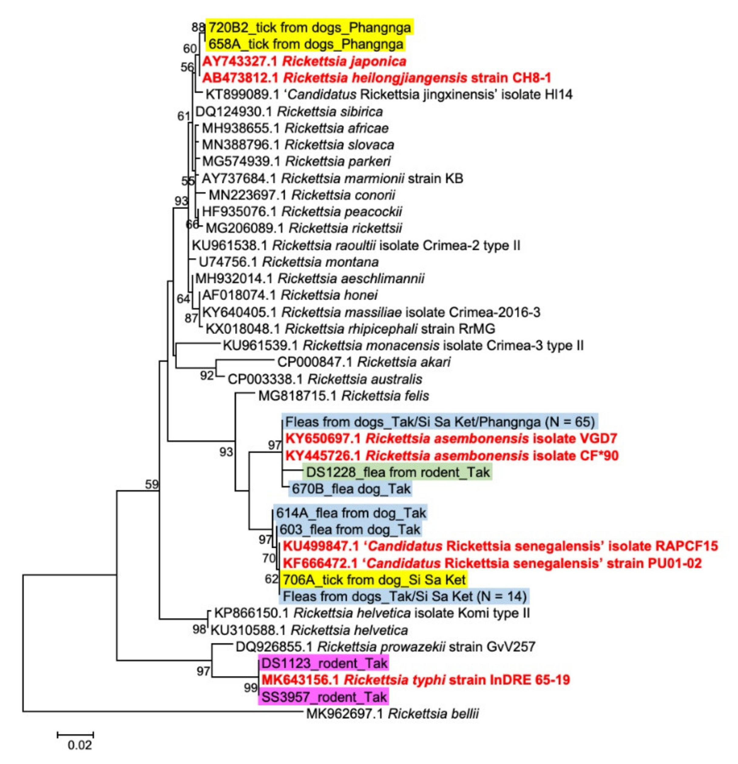
2.2. Rickettsia Species in Rodent and Ectoparasite Pools

From a total of 103 Rickettsia-positive samples, 82 samples were selected for amplicon-based NGS species identification for Rickettsia species and Sanger sequencing was used for the remaining pools. Rickettsia gltA sequences were obtained from 101 samples. Phylogenetic analysis of gltA sequences for Rickettsia species characterization is shown in Figure 1. The Rickettsia species were identified based on their highest DNA sequence identity to the reference sequences retrieved from Genbank database. There were four Rickettsia species identified from our samples, R. asembonensis, “Candidatus Rickettsia senegalensis”, R. helongjiangensis and R. typhi with 99.2–100% identity to references sequences. The result of Rickettsia species characterization by gltA gene is shown in Table 2. The majority of Rickettsia species found were identified as R. asembonensis (80/101, 79.2%) followed by “Ca. R. senegalensis” (16.8%), R. helongjiangensis (2%) and R. typhi (2%). All Rickettsia from rodents (n = 2) and rodent fleas (n = 1) were R. typhi and R. asembonensis, respectively. The majority of Rickettsia identified from fleas collected from dogs (n = 95) were R. asembonensis (79/95, 83.2%), while “Ca. R. senegalensis” was detected in 16.8% (16/95) of fleas from dogs. Of three Rickettsia-positive ticks collected from dogs, two from Phangnga province were R. helongjiangensis and one from Si Sa Ket province was “Ca. R. senegalensis”.

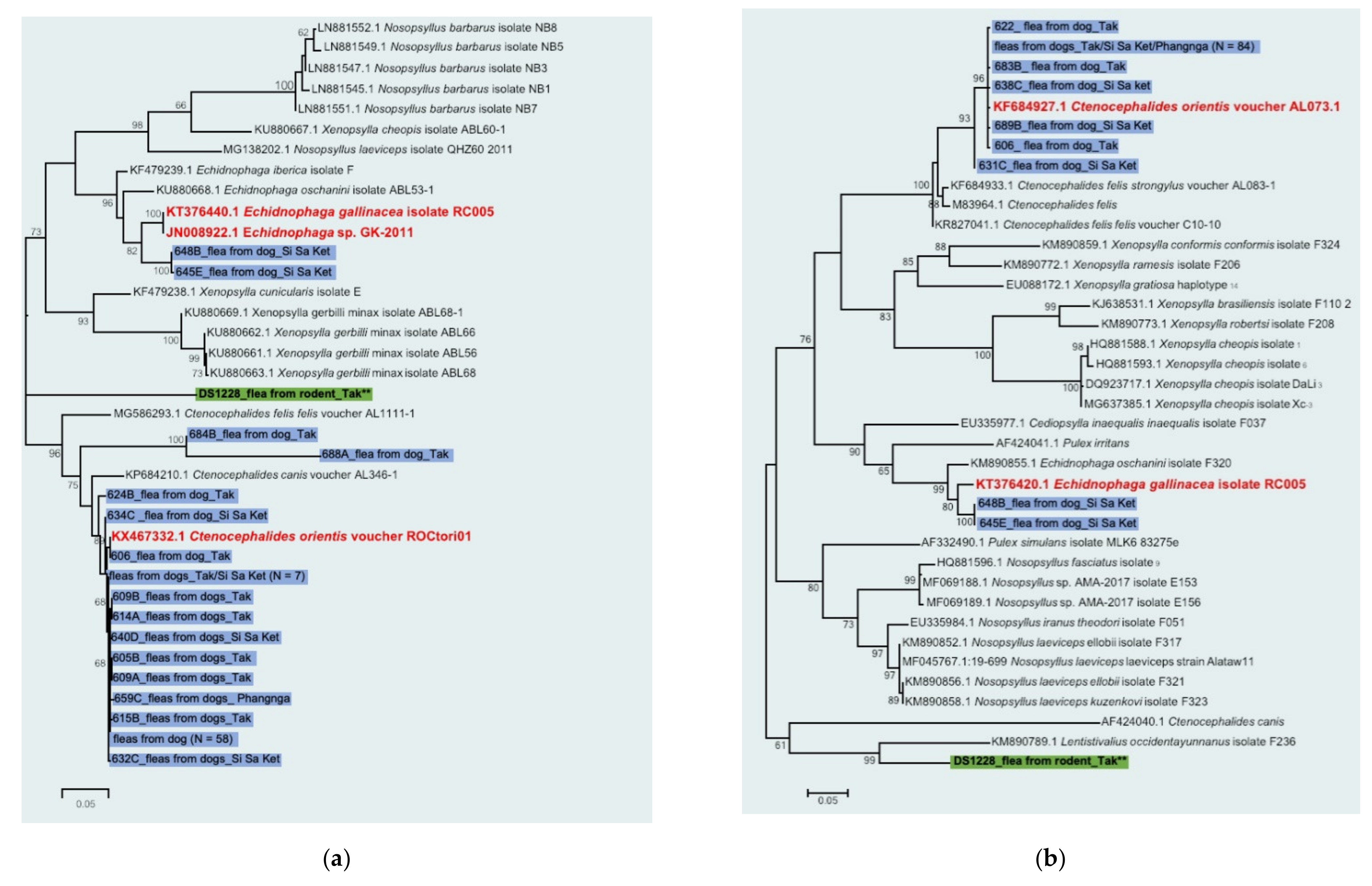
2.3. Ectoparasite Species Identification in Rickettsia-Positive Pools

Results of flea species identification using COI and COII sequences by NGS and Sanger sequencing were available for 92 Rickettsia-positive pools (91 flea pools from dogs and 1 flea pool from rodent), and phylogenetic analysis was shown in Figure 2a,b respectively. The following results were obtained from COI and COII characterization for almost all flea samples (Table 3). The majority of Rickettsia-positive fleas from dogs (89/91 = 97.8%) were identified as Ctenocephalides felis orientis (C. f. orientis) with 98–100% identity to the reference sequence, only 2 of them (2.2%) were Echidnophaga gallinacea. (95–97% identity). Interestingly, two flea pools from dogs were unidentified in which their COI sequences have only 91 and 82% identity to C. f. orientis sequence reference, but their COII sequences are 100% identical to C. f. orientis. Thus, they are classified as C. f. orientis. One positive rat flea was classified as an unidentified group by both COI and COII gene sequences.
For tick species identification, only COI sequence was used to characterize three Rickettsia-positive tick pools by Sanger sequencing. Two tick pools were characterized as H. hystricis with 99% identity to the reference sequence (data not shown), while one tick pool that had been previously identified as R. sanguineus s.l. by morphology failed in sequencing. When the distribution of Rickettsia species among ectoparasite species was examined, R. asembonensis and “Ca. R. senegalensis” were found in various species of fleas and Rhipicephalus ticks from dogs. Among C. f. orientis population (n = 89), 83% (74/89) carried R. asembonensis and 17% (15/89) had “Ca. R. senegalensis”. However, it was noted that R. heilongjiangensis was found in only H. hystricis ticks.

3. Discussion

Here, we demonstrated the utility of next generation sequencing (NGS) for high-throughput sequencing to identify Rickettsia and ectoparasite species in parallel. In our previous surveillance during 2015–2016 in Thailand, high percentage of flea samples were PCR-positive for Rickettsia DNA. Instead of using Sanger sequencing to characterize Rickettsia and flea species using their specific target genes which would require large amounts of time to assemble and analyze each individual sequence, we applied the amplicon-based NGS technology. The Citrate synthase (gltA) gene, Cytochrome Oxidase subunit I (COI) and subunit II (COII) amplicons were sequenced simultaneously using Miseq platform. The advantage of NGS over traditional Sanger sequencing is that amplicons of several target genes could be sequenced simultaneously and up to 384 samples could be analyzed in one experimental run, if dual indexes were used in library preparation (Illumina). NGS sequencing strategies are used widely and provide a range of applications for epidemiological and surveillance analysis. For example, whole genome sequencing and genome-wide screening for single nucleotide polymorphisms have been used for microbial typing for outbreak analysis [30]. Other researchers have used a metagenomic approach based on universal 16S rRNA sequencing to survey a large spectrum of bacterial pathogens and analyze bacterial communities in human, wildlife, and vectors [26,27,28,29]. The results of metagenomic approaches help elucidate the complexities of transmission and circulation of bacteria in the environment. Our study illustrates another application of NGS for simultaneous characterization of Rickettsia and flea species, to enable rapid analysis of a large number of samples collected from a surveillance study.
More than a half of all fleas from dogs during our 2015–2016 surveillance were PCR-positive for Rickettsia species and most were identified later as R. asembonensis. Molecular identification of flea species indicates all flea from dogs are Ctenocephalides felis orientis, except for two Echidnophaga gallinacea (sticktight flea) that may occasionally be found in dogs [31]. The evidence of high percentage of C. f. orientis positive for R. asembonensis correspond to the study in India, suggesting a specific host-bacteria relationship [32,33]. Similarly, the high percentage of R. asembonensis DNA were also detected among fleas from cats in Khon Kaen province, northeastern Thailand [23], although all fleas were identified as C. felis by morphology rather than molecular identification. Rickettsia asembonensis belongs to a group of R. felis-like organisms which are ubiquitously distributes. It was firstly detected as Rickettsia sp. RF2125 in Ctenocephalides canis in western Thailand near the Myanmar border [34]. Its host includes various arthropods but mostly in fleas of three families: Pulicidae, Ceratophyllidae, and Coptopsyllidae [33]. Rickettsia asembonensis could be a potential human pathogen as it has been reported from patients with fever of unknown origin in Thailand, Malaysia, and Peru [22,35,36,37]. Rickettsia asembonensis was also detected in healthy dogs in South Africa [38], sick dog in Thailand [22], and health monkey in Malaysia [39]. Nonetheless, its pathogenicity to humans and animals remains unknown.
A low percentage of Rickettsia DNA (<2%) was observed in dog tick, rodent flea, and rodent samples. Interestingly, R. heilongjiangensis was found in only H. hystricis ticks from dog in Phangnga province, while R. typhi was found in rodent samples from Tak province. R. heilongjiangensis belonging to Spotted Fever group (SFG), cause Far-Eastern spotted fever which is endemic in northeastern China [40], Siberia, and far-eastern Russia [41]. In Thailand, there has been one case report of SFG rickettsiosis in human related to R. japonica, later confirmed to be R. heilongjiangensis [14,16]. However, the identification of this agent in tick vector has not been documented. To our knowledge, this is the first report identifying R. heilongjiangensis, causative agent of SFG rickettsiosesis, in H. hystricis ticks in Thailand. Spotted Fever Group Rickettsia apparently adapts itself to a specific tick species, and the relationship between SFG Rickettsia, tick, and vertebrate host have been documented [5].
This study had several limitations: no access to blood from domestic animals and phylogenetic analysis was limited. The conclusions that can be drawn from the animal infections was limited as we were unable to collect blood samples from domestic cats and dogs. The phylogenetic analysis was limited to one or two genes with analyses of Rickettsia based on the Citrate synthase (gltA) gene (Table 1) and fleas on the Cytochrome Oxidase subunit I and II genes (Table 2). The gltA gene is best for distinguishing between Rickettsia species while the COI gene is the gold standard for barcoding or distinguishing between species. This limits the breadth of the conclusions that can be drawn from these data to species identification. Therefore, we recommend that selection of genes be tailored to the study goal.
In conclusion, a high percentage of R. asembonensis DNA (>50%) was observed in fleas from dogs (C. f. orientis). Rickettsia asembonensis belongs to a group of R. felis-like organisms which distribute ubiquitously, but its definite pathogenicity to human remains unknown. We also identified R. heilongjiangensis, causative agent of SFG rickettsioses, in H. hystricis ticks from dog in Phangnga province and R. typhi in rodents from Tak province. Our findings demonstrate the circulation of tick- and flea-borne SFG rickettsioses, and flea-borne murine typhus, and thus risk of transmission from potential vectors and animal reservoir in Tak and Phangnga provinces of Thailand. Moreover, our study illustrates another application of NGS for simultaneous characterization of Rickettsia and flea species, to enable rapid analysis of a large number of samples collected from a surveillance study.

4. Materials and Methods

4.1. The Field Surveillance for Rickettsia in Rodents and Ectoparasites

During field surveillance for Rickettsia species (2015–2016), rodents and ectoparasites collected from rodents and domestic mammals (dog, cat, and cattle) in Phangnga, Ranong, Si Sa Ket, Tak, and Udon Thani provinces of Thailand were screened for Rickettsia spp. Rodents were captured from plantations, cultivated rice-fields, and grassland areas using live traps, as previously described [28]. Captured rodents were euthanized using carbon dioxide and processed immediately at the site of collection. Liver, spleen, kidney, and lung samples were collected and stored on dry ice. Ticks, fleas, and lice were collected and kept in absolute ethanol. Ectoparasites were also collected from domestic animals (dog, cat, and cattle). There were pools of ectoparasites (ticks, fleas, and lice) collected from individual rodent or domestic animal. All rodent and ectoparasite samples were transported to the AFRIMS laboratory, Bangkok, Thailand, for further processing. Rodent species were identified to species level as described previously [42]. Ectoparasites (ticks, fleas, and lice) were morphologically identified [43,44,45] and pooled by genus, stage, gender, and their animal host origin. Up to five ticks were included in a pool, while there were as many as 29 fleas and 11 lice from a domestic mammal included in a pool.

4.2. Genomic DNA Extraction from Rodents and Ectoparasite Samples

Genomic DNA was extracted from rodent tissue, and ectoparasite pools, as previously described [28]. Briefly, kidney and spleen tissue from each rodent were cut into small pieces, homogenized, and extracted for genomic DNA using a QIAsymphony® SP instrument (Qiagen, Hombrechtikon, Switzerland) with QIAsymphony® DNA Mini Kit (Qiagen, Germany). For ectoparasites, a modified protocol of QIAamp DNA Mini Kit (Qiagen) was used to extract genomic DNA from each pool as previously described [28]. DNA was kept in −20 °C until use.

4.3. Real-Time PCR Detection of Rickettsia

Rodent samples and ectoparasite pools were screened for Rickettsia using TaqMan real-time PCR (qPCR) targeting 17 kDa protein gene and some of the positive signals from qPCR were confirmed by conventional PCR of Rickettsia citrate synthase (gltA) gene. Details of primers, probes and PCR conditions were previously published by Takhampunya et al. 2019 [28].

4.4. Amplicon-Based NGS Characterization of Rickettsia and Ectoparasite Species

Most Rickettsia-positive ectoparasite pools were selected for NGS characterization of Rickettsia (82/103) and flea species (92/103), and the remaining pools was characterized by Sanger sequencing, while there was not enough DNA to do sequencing for two samples. Citrate synthase (gltA) gene was amplified for Rickettsia species characterization, while Cytochrome Oxidase subunit I (COI) and subunit II (COII) genes were used as target genes for flea species identification. The gltA amplification was done following the protocol for the Rickettsia confirmation assay as mention earlier [28]. Primer sequences for flea COI and COII and PCR conditions were described in the previous study [46,47]. The gltA, COI, and COII amplicons of each sample were pooled and subjected to NGS. Since the length of these amplicons is more than 600 bp, direct amplicon sequencing on Illumina system could not provide sequence information across the entire length. So, the Nextera XT DNA library prep kit was used to prepare amplicon libraries, according to the manufacturer’s protocol (Illumina). Briefly, amplicons were fragmented and tagged with adapter sequences by Nextera transposome. Then, library was amplified using a limit-cycle PCR to tag DNA fragments with dual indices and Illumina sequencing adapters. The final products were purified using Agencourt AMPure XP beads to remove excess primers, nucleotides, salts, and enzymes. The purity of the libraries was verified using the QIAxcel Advanced System (Qiagen) with a QIAxcel DNA High Resolution Cartridge. The purified Index library concentration was quantified using the Qubit dsDNA HS Assay Kit (Invitrogen) and DNA concentration was normalized to 4.0 nM for each library. Five microliters of DNA from each library at 4.0 nM were pooled for one NGS run. Pooled libraries were denatured with NaOH, according to the manufacturer’s protocol (Illumina). Denatured libraries were diluted to a final concentration of 4 pM. A 10% Phix control adapter-ligated library (Illumina) was included to serve as internal control of each run for low-diversity library. Sequencing was performed on the Illumina MiSeq System using the MiSeq Reagent Kit v2 (300-cycles), following manufacturer’s instructions. High throughput sequencing (HTS) datasets was deposited in the NCBI’s Sequence Read Archive under BioProject # PRJNA691717, accession number SRR13425361-SRR13425442.
For Sanger sequencing, the PCR product of each gene was sequenced. Excess primers and nucleotides were removed from PCR amplicons using ExoSAP-IT™ (Applied Biosystem) according to the manufacturer’s instructions. The purified amplicons were sequenced using an ABI BigDye™ Terminator v3.1 Cycle Sequencing Kit, ethanol precipitated, and run on a SeqStudio Genetic Analyzer (Applied Biosystems Thermo Fisher, Thailand). Sequences were assembled using Sequencher™ ver. 5.1 (Gene Codes Corp., Ann Arbor, MI, USA). Nucleic acid sequences generated during this study (Sanger sequences and consensus sequences from NGS) were deposited in the GenBank database (http://www.ncbi.nlm.nih.gov/Genbank/) under accession number MW492078-MW492178, MW492179-MW492261, and MW492262-MW492354. The full list of sequences and their database identifiers are given as Supplementary Materials (Table S3).

4.5. Sequence Analysis

The sequence reads generated on MiSeq sequencers were processed on the CLC Genomics workbench software v 20.0.3 (Qiagen, Aarhus A/S1). High-throughput sequences were imported into CLC Genomics Workbench according to quality scores of Illumina pipeline 1.8 (using a Phred scale encoded using ASCII 33 to 93). The sequences were trimmed to remove adaptor sequences, low quality sequence (limit = 0.05), ambiguous nucleotides (maximal 2 nucleotides allowed), and read lengths shorter than 100 bp. Trimmed reads were mapped to reference sequences of Rickettsia gltA, flea COI, and COII retrieved from Genbank (KY650697.1, KY865419.1, and M83964.1, respectively). Consensus sequences were extracted from read mapping, according to default settings, and used for phylogenetic tree analysis. Sequences from NGS and Sanger sequencing were aligned with reference sequences retrieved from the GenBank database using the MUSCLE codon alignment algorithm [48]. Maximum likelihood phylogenetic tree was then constructed using the best fit model of nucleotide substitution with bootstrapping (1000 replicates) in MEGA 6 [49].

Supplementary Materials

The following are available online at https://www.mdpi.com/2076-0817/10/2/215/s1, Table S1: The number of animals infested with ectoparasites as well as the number (percentage) of animals infested with Rickettsia-PCR positive fleas or ticks, Table S2: The number of dogs infested with ectoparasites and screened for Rickettsia DNA as well as Rickettsia species detected from ectoparasites collected from them, Table S3: The GenBank (http://www.ncbi.nlm.nih.gov/Genbank/) accession numbers for the Rickettsia gltA gene, Cytochrome Oxidase subunit I and II genes of Rickettsia-positive fleas described in this paper.

Author Contributions

Conceptualization, R.T. and A.K.; Methodology, R.T. and A.K.; Formal Analysis, S.C. and A.K.; Investigation, A.K., B.T., and S.P.; Data Curation, S.C. and R.T.; Writing—Original Draft Preparation, S.C.; Writing—Review and Editing, R.T. and B.K.P.-S.; Visualization, S.C. and A.K.; Supervision, R.T. All authors have read and agreed to the published version of the manuscript.

Funding

This work was funded by the Armed Forces Health Surveillance Division (AFHSD) Global Emerging Infections Surveillance Branch (GEIS), ProMIS ID # P0108_19_AF_03.02.

Institutional Review Board Statement

Rodents were trapped according to the institutional animal collection protocol entitled “Field Sampling of Small Mammal (orders: Erinaceomorpha, Soricomorpha, Scandentia, Macroscelidea, and Rodentia) Populations to Support Zoonotic Diseases Surveillance and Ectoparasite Collection” (PN no. 18-04), which had been reviewed and approved by the USAMC-AFRIMS Institutional Animal Care and Use Committee. All sampling procedures and experimental manipulations were reviewed and approved as part of the animal collection protocol (PN no. 18-04). Research was conducted in compliance with the Animal Welfare Act and other federal statutes and regulations relating to animals and experiments involving animals, and adhered to principles outlined in the Guide for the Care and Use of Laboratory Animals, NRC Publication, 2011 edition.

Informed Consent Statement

Not applicable.

Data Availability Statement

The data presented in this study are available in the materials and methods section as well as in the Supplementary Materials (Table S3). High throughput sequencing (HTS) datasets was deposited in the NCBI’s Sequence Read Archive under BioProject # PRJNA691717, accession number SRR13425361-SRR13425442.

Acknowledgments

We are grateful for all staffs in Diagnostic and Reemerging Disease section and Mite and Rodent Support section, Entomology Department, AFRIMS for their continuing support throughout the study.

Conflicts of Interest

The authors declare no conflict of interest. The funders had no role in the design of the study; in the collection, analyses, or interpretation of data; in the writing of the manuscript, or in the decision to publish the results.

Disclaimer

Material has been reviewed by the Walter Reed Army Institute of Research. There is no objection to its presentation and/or publication. The opinions or assertions contained herein are the private views of the author, and are not to be construed as official, or as reflecting true views of the Department of the Army or the Department of Defense. Research was conducted under an approved animal use protocol in an AAALACi accredited facility in compliance with the Animal Welfare Act and other federal statutes and regulations relating to animals and experiments involving animals and adheres to principles stated in the Guide for the Care and Use of Laboratory Animals, NRC.

References

  1. Parte, A.C.; Sarda Carbasse, J.; Meier-Kolthoff, J.P.; Reimer, L.C.; Goker, M. List of Prokaryotic names with Standing in Nomenclature (LPSN) moves to the DSMZ. Int. J. Syst. Evol. Microbiol. 2020, 70, 5607–5612. [Google Scholar] [CrossRef]
  2. Blanton, L.S. The Rickettsioses: A Practical Update. Infect. Dis. Clin. N. Am. 2019, 33, 213–229. [Google Scholar] [CrossRef] [PubMed]
  3. Graves, S.R.; Stenos, J. Tick-borne infectious diseases in Australia. Med. J. Aust. 2017, 206, 320–324. [Google Scholar] [CrossRef] [PubMed]
  4. Zaharia, M.; Popescu, C.P.; Florescu, S.A.; Ceausu, E.; Raoult, D.; Parola, P.; Socolovschi, C. Rickettsia massiliae infection and SENLAT syndrome in Romania. Ticks Tick Borne Dis. 2016, 7, 759–762. [Google Scholar] [CrossRef]
  5. Parola, P.; Paddock, C.D.; Socolovschi, C.; Labruna, M.B.; Mediannikov, O.; Kernif, T.; Abdad, M.Y.; Stenos, J.; Bitam, I.; Fournier, P.E.; et al. Update on tick-borne rickettsioses around the world: A geographic approach. Clin. Microbiol. Rev. 2013, 26, 657–702. [Google Scholar] [CrossRef] [Green Version]
  6. Tarragona, E.L.; Soares, J.F.; Costa, F.B.; Labruna, M.B.; Nava, S. Vectorial competence of Amblyomma tonelliae to transmit Rickettsia rickettsii. Med. Vet. Entomol. 2016, 30, 410–415. [Google Scholar] [CrossRef]
  7. Drexler, N.A.; Yaglom, H.; Casal, M.; Fierro, M.; Kriner, P.; Murphy, B.; Kjemtrup, A.; Paddock, C.D. Fatal Rocky Mountain Spotted Fever along the United States-Mexico Border, 2013–2016. Emerg. Infect. Dis. 2017, 23, 1621–1626. [Google Scholar] [CrossRef] [Green Version]
  8. Straily, A.; Drexler, N.; Cruz-Loustaunau, D.; Paddock, C.D.; Alvarez-Hernandez, G. Notes from the Field: Community-Based Prevention of Rocky Mountain Spotted Fever—Sonora, Mexico, 2016. MMWR Morb. Mortal. Wkly. Rep. 2016, 65, 1302–1303. [Google Scholar] [CrossRef] [Green Version]
  9. Levin, M.L.; Killmaster, L.F.; Zemtsova, G.E. Domestic dogs (Canis familiaris) as reservoir hosts for Rickettsia conorii. Vector Borne Zoonotic Dis. 2012, 12, 28–33. [Google Scholar] [CrossRef]
  10. Jensenius, M.; Fournier, P.E.; Kelly, P.; Myrvang, B.; Raoult, D. African tick bite fever. Lancet Infect. Dis. 2003, 3, 557–564. [Google Scholar] [CrossRef]
  11. Robinson, M.T.; Satjanadumrong, J.; Hughes, T.; Stenos, J.; Blacksell, S.D. Diagnosis of spotted fever group Rickettsia infections: The Asian perspective. Epidemiol. Infect. 2019, 147, e286. [Google Scholar] [CrossRef] [Green Version]
  12. Sirisanthana, T.; Pinyopornpanit, V.; Sirisanthana, V.; Strickman, D.; Kelly, D.J.; Dasch, G.A. First cases of spotted fever group rickettsiosis in Thailand. Am. J. Trop. Med. Hyg. 1994, 50, 682–686. [Google Scholar] [CrossRef] [Green Version]
  13. Jiang, J.; Sangkasuwan, V.; Lerdthusnee, K.; Sukwit, S.; Chuenchitra, T.; Rozmajzl, P.J.; Eamsila, C.; Jones, J.W.; Richards, A.L. Human Infection with Rickettsia honei, Thailand. Emerg. Infect. Dis. 2005, 11, 1473–1475. [Google Scholar] [CrossRef] [PubMed]
  14. Gaywee, J.; Sunyakumthorn, P.; Rodkvamtook, W.; Ruang-areerate, T.; Mason, C.J.; Sirisopana, N. Human infection with Rickettsia sp. related to R. japonica, Thailand. Emerg. Infect. Dis. 2007, 13, 657–659. [Google Scholar] [CrossRef] [Green Version]
  15. Robertson, R.G.; Wisseman, C.L., Jr. Tick-borne rickettsiae of the spotted fever group in West Pakistan. II. Serological classification of isolates from West Pakistan and Thailand: Evidence for two new species. Am. J. Epidemiol. 1973, 97, 55–64. [Google Scholar] [CrossRef] [PubMed]
  16. Takada, N.; Fujita, H.; Kawabata, H.; Ando, S.; Sakata, A.; Takano, A.; Chaithong, U. Spotted fever group Rickettsia sp. closely related to Rickettsia japonica, Thailand. Emerg. Infect. Dis. 2009, 15, 610–611. [Google Scholar] [CrossRef] [PubMed]
  17. Luvira, V.; Silachamroon, U.; Piyaphanee, W.; Lawpoolsri, S.; Chierakul, W.; Leaungwutiwong, P.; Thawornkuno, C.; Wattanagoon, Y. Etiologies of Acute Undifferentiated Febrile Illness in Bangkok, Thailand. Am. J. Trop. Med. Hyg. 2019, 100, 622–629. [Google Scholar] [CrossRef] [Green Version]
  18. Kuo, C.C.; Wardrop, N.; Chang, C.T.; Wang, H.C.; Atkinson, P.M. Significance of major international seaports in the distribution of murine typhus in Taiwan. PLoS Negl. Trop. Dis. 2017, 11, e0005430. [Google Scholar] [CrossRef] [Green Version]
  19. Blanton, L.S.; Idowu, B.M.; Tatsch, T.N.; Henderson, J.M.; Bouyer, D.H.; Walker, D.H. Opossums and Cat Fleas: New Insights in the Ecology of Murine Typhus in Galveston, Texas. Am. J. Trop. Med. Hyg. 2016, 95, 457–461. [Google Scholar] [CrossRef]
  20. Maina, A.N.; Fogarty, C.; Krueger, L.; Macaluso, K.R.; Odhiambo, A.; Nguyen, K.; Farris, C.M.; Luce-Fedrow, A.; Bennett, S.; Jiang, J.; et al. Rickettsial Infections among Ctenocephalides felis and Host Animals during a Flea-Borne Rickettsioses Outbreak in Orange County, California. PLoS ONE 2016, 11, e0160604. [Google Scholar] [CrossRef] [PubMed] [Green Version]
  21. Foongladda, S.; Inthawong, D.; Kositanont, U.; Gaywee, J. Rickettsia, Ehrlichia, Anaplasma, and Bartonella in ticks and fleas from dogs and cats in Bangkok. Vector Borne Zoonotic Dis. 2011, 11, 1335–1341. [Google Scholar] [CrossRef] [PubMed]
  22. Mongkol, N.; Suputtamongkol, Y.; Taweethavonsawat, P.; Foongladda, S. Molecular Evidence of Rickettsia in Human and Dog Blood in Bangkok. Vector Borne Zoonotic Dis. 2018, 18, 297–302. [Google Scholar] [CrossRef] [PubMed]
  23. Phomjareet, S.; Chaveerach, P.; Suksawat, F.; Jiang, J.; Richards, A.L. Spotted Fever Group Rickettsia Infection of Cats and Cat Fleas in Northeast Thailand. Vector Borne Zoonotic Dis. 2020, 20, 566–571. [Google Scholar] [CrossRef]
  24. Papa, A.; Tsioka, K.; Kontana, A.; Papadopoulos, C.; Giadinis, N. Bacterial pathogens and endosymbionts in ticks. Ticks Tick Borne Dis. 2017, 8, 31–35. [Google Scholar] [CrossRef]
  25. Pesquera, C.; Portillo, A.; Palomar, A.M.; Oteo, J.A. Investigation of tick-borne bacteria (Rickettsia spp., Anaplasma spp., Ehrlichia spp. and Borrelia spp.) in ticks collected from Andean tapirs, cattle and vegetation from a protected area in Ecuador. Parasit. Vectors 2015, 8, 46. [Google Scholar] [CrossRef] [Green Version]
  26. Kurilshikov, A.; Livanova, N.N.; Fomenko, N.V.; Tupikin, A.E.; Rar, V.A.; Kabilov, M.R.; Livanov, S.G.; Tikunova, N.V. Comparative Metagenomic Profiling of Symbiotic Bacterial Communities Associated with Ixodes persulcatus, Ixodes pavlovskyi and Dermacentor reticulatus Ticks. PLoS ONE 2015, 10, e0131413. [Google Scholar] [CrossRef] [Green Version]
  27. Portillo, A.; Palomar, A.M.; de Toro, M.; Santibanez, S.; Santibanez, P.; Oteo, J.A. Exploring the bacteriome in anthropophilic ticks: To investigate the vectors for diagnosis. PLoS ONE 2019, 14, e0213384. [Google Scholar] [CrossRef] [Green Version]
  28. Takhampunya, R.; Korkusol, A.; Pongpichit, C.; Yodin, K.; Rungrojn, A.; Chanarat, N.; Promsathaporn, S.; Monkanna, T.; Thaloengsok, S.; Tippayachai, B.; et al. Metagenomic Approach to Characterizing Disease Epidemiology in a Disease-Endemic Environment in Northern Thailand. Front. Microbiol. 2019, 10, 319. [Google Scholar] [CrossRef] [Green Version]
  29. Galan, M.; Razzauti, M.; Bard, E.; Bernard, M.; Brouat, C.; Charbonnel, N.; Dehne-Garcia, A.; Loiseau, A.; Tatard, C.; Tamisier, L.; et al. 16S rRNA Amplicon Sequencing for Epidemiological Surveys of Bacteria in Wildlife. mSystems 2016, 1. [Google Scholar] [CrossRef] [Green Version]
  30. Perez-Losada, M.; Arenas, M.; Castro-Nallar, E. Microbial sequence typing in the genomic era. Infect. Genet. Evol. 2018, 63, 346–359. [Google Scholar] [CrossRef]
  31. Kalkofen, U.P.; Greenberg, J. Echidnophaga gallinacea infestation in dogs. J. Am. Vet. Med. Assoc. 1974, 165, 447–448. [Google Scholar]
  32. Hii, S.F.; Lawrence, A.L.; Cuttell, L.; Tynas, R.; Abd Rani, P.A.; Slapeta, J.; Traub, R.J. Evidence for a specific host-endosymbiont relationship between ‘Rickettsia sp. genotype RF2125’ and Ctenocephalides felis orientis infesting dogs in India. Parasit. Vectors 2015, 8, 169. [Google Scholar] [CrossRef] [Green Version]
  33. Maina, A.N.; Jiang, J.; Luce-Fedrow, A.; St John, H.K.; Farris, C.M.; Richards, A.L. Worldwide Presence and Features of Flea-Borne Rickettsia asembonensis. Front. Vet. Sci. 2018, 5, 334. [Google Scholar] [CrossRef] [Green Version]
  34. Parola, P.; Cornet, J.P.; Sanogo, Y.O.; Miller, R.S.; Thien, H.V.; Gonzalez, J.P.; Raoult, D.; Telford, I.S.; Wongsrichanalai, C. Detection of Ehrlichia spp., Anaplasma spp., Rickettsia spp., and other eubacteria in ticks from the Thai-Myanmar border and Vietnam. J. Clin. Microbiol. 2003, 41, 1600–1608. [Google Scholar] [CrossRef] [Green Version]
  35. Rodkvamtook, W.; Pholpetch, S.; Yokanit, W.; Thongphun, A.; Somsri, M.; Somsri, K.; Boonsom, V.; Thimhaipol, C.; Siriwatthanakul, K.; Amradee, B.; et al. Rickettsia asembonensis causing febrile disease in southern Thailand, 2016–2018. In Proceedings of the 29th Meeting of the American Society for Rickettsiology, Milwaukee, WI, USA, 16–19 June 2018. [Google Scholar]
  36. Palacios-Salvatierra, R.; Caceres-Rey, O.; Vasquez-Dominguez, A.; Mosquera-Visaloth, P.; Anaya-Ramirez, E. Rickettsial species in human cases with non-specific acute febrile syndrome in Peru. Rev. Peru Med. Exp. Salud. Publica 2018, 35, 630–635. [Google Scholar] [CrossRef] [Green Version]
  37. Kho, K.L.; Koh, F.X.; Singh, H.K.; Zan, H.A.; Kukreja, A.; Ponnampalavanar, S.; Tay, S.T. Spotted Fever Group Rickettsioses and Murine Typhus in a Malaysian Teaching Hospital. Am. J. Trop. Med. Hyg. 2016, 95, 765–768. [Google Scholar] [CrossRef] [Green Version]
  38. Kolo, A.O.; Sibeko-Matjila, K.P.; Maina, A.N.; Richards, A.L.; Knobel, D.L.; Matjila, P.T. Molecular Detection of Zoonotic Rickettsiae and Anaplasma spp. in Domestic Dogs and Their Ectoparasites in Bushbuckridge, South Africa. Vector Borne Zoonotic Dis. 2016, 16, 245–252. [Google Scholar] [CrossRef] [Green Version]
  39. Tay, S.T.; Koh, F.X.; Kho, K.L.; Sitam, F.T. Rickettsial infections in monkeys, Malaysia. Emerg. Infect. Dis. 2015, 21, 545–547. [Google Scholar] [CrossRef]
  40. Liu, H.; Li, Q.; Zhang, X.; Li, Z.; Wang, Z.; Song, M.; Wei, F.; Wang, S.; Liu, Q. Characterization of rickettsiae in ticks in northeastern China. Parasit. Vectors 2016, 9, 498. [Google Scholar] [CrossRef] [Green Version]
  41. Igolkina, Y.; Rar, V.; Vysochina, N.; Ivanov, L.; Tikunov, A.; Pukhovskaya, N.; Epikhina, T.; Golovljova, I.; Tikunova, N. Genetic variability of Rickettsia spp. in Dermacentor and Haemaphysalis ticks from the Russian Far East. Ticks Tick Borne Dis. 2018, 9, 1594–1603. [Google Scholar] [CrossRef]
  42. Muul, I. Lekagul, Boonsong, Jeffrey, A. McNeely. Mammals of thailand. Association for the Conservation of Wildlife*, Bangkok, Thailand. J. Mammal. 1979, 60, 241–242. [Google Scholar] [CrossRef]
  43. Fleas, B.M.R.C.O.; Hopkins, G.H.E.; Siphonaptera, B.M.D.O.E.; Collection, B.M.R.; Rothschild, M.; Mardon, D.K.; Smit, F.G.A.M. An Illustrated Catalogue of the Rothschild Collection of Fleas (Siphonaptera) in the British Museum (Natural History): With Keys and Short Descriptions for the Identification of Families, Genera, Species and Subspecies of the Order. Hystrichopsyllidae (Ctenophthalminae, Dinopsyllinae, Doratopsyllinae and Listropsyllinae); Trustees of the British Museum (Natural History): London, UK, 1966. [Google Scholar]
  44. Tanskull, P.; Inlao, I. Keys to the adult ticks of Haemaphysalis Koch, 1844, in Thailand with notes on changes in taxonomy (Acari: Ixodoidea: Ixodidae). J. Med. Entomol. 1989, 26, 573–601. [Google Scholar] [CrossRef] [Green Version]
  45. Durden, L.A.M.; Guy, G. The sucking lice (Insecta, Anoplura) of the world: A taxonomic checklist with records of mammalian hosts and geographical distributions. Bull. AMNH 1994, 218, 1–90. [Google Scholar]
  46. Whiting, M.F. Mecoptera is paraphyletic: Multiple genes and phylogeny of Mecoptera and Siphonaptera. Zool. Scr. 2002, 31, 93–104. [Google Scholar] [CrossRef] [Green Version]
  47. Folmer, O.; Black, M.; Hoeh, W.; Lutz, R.; Vrijenhoek, R. DNA primers for amplification of mitochondrial cytochrome c oxidase subunit I from diverse metazoan invertebrates. Mol. Mar. Biol. Biotechnol. 1994, 3, 294–299. [Google Scholar]
  48. Edgar, R.C. MUSCLE: Multiple sequence alignment with high accuracy and high throughput. Nucleic Acids Res. 2004, 32, 1792–1797. [Google Scholar] [CrossRef] [Green Version]
  49. Tamura, K.; Stecher, G.; Peterson, D.; Filipski, A.; Kumar, S. MEGA6: Molecular Evolutionary Genetics Analysis version 6.0. Mol. Biol. Evol. 2013, 30, 2725–2729. [Google Scholar] [CrossRef] [Green Version]
Figure 1. Phylogenetic tree of Rickettsia gltA gene detected in rodents and ectoparasites were analyzed with reference sequences retrieved from GenBank database. A maximum likelihood (ML) tree was constructed using the T92 + G models of nucleotide substitution in the MEGA 6 program with bootstrapping (1000 replicates). Rickettsia sequences from rodent, rodent fleas, flea, and ticks from dogs are highlighted in pink, green, blue, and yellow, respectively, and the provinces samples collected are indicated. The Rickettsia reference sequences of species identified in the present study are marked in red.
Pathogens 10 00215 g001
Figure 2. Phylogenetic trees of Cytochrome Oxidase subunit I (a) and II (b) sequences of Rickettsia positive fleas and reference sequences retrieved from GenBank database. A ML tree was constructed using the GTR + G models of nucleotide substitution in the MEGA 6 program with bootstrapping (1000 replicates). Sequences from fleas collected from rodent and dogs are highlighted in green and blue, respectively, and the provinces samples collected are indicated. The reference sequences of flea species identified in the present study are marked in red. Sequence classified as unknown species is noted**.
Pathogens 10 00215 g002
Table 1. A high percentage of Rickettsia DNA in rodent and ectoparasite pools collected in field surveillance during 2015–2016.
Sample Type/ProvincesRickettsia-PCR Positive Pools (% Positive)
Phangnga
8°41′08.5″ N 98°21′07.8″ E
Ranong
10°21′54.7″ N 98°49′01.8″ E
Si Sa Ket
14°34′13.9″ N 104°20′25.9″ E
Tak
16°23′41.4″ N 98°44′03.7″ E
Udon Thani
17°36′44.3″ N 102°20′38.3″ E
Total
Rodent0/516 (0%)0/137 (0%)0/220 (0%)1/452* (0.2%)1/59* (1.7%)2/1384 (0.1%)
Rodent Fleas0/3 (0%)NA.0/15 (0%)1/22 (4.5%)0/12 (0%)1/52 (1.9%)
Rodent Tick0/8 (0%)NA.0/12 (0%)0/17 (0%)NA.0/37 (0%)
Rodent Lice0/2 (0%)NA.0/1 (0%)0/3 (0%)NA.0/6 (0%)
DA Flea116/21 (76.2%)NA.31/57 (54.4%)50/95 (52.6%)NA.97/173 (56.1)
DA Tick22/88 (2%)NA.1/69 (1.4%)NA.NA.3/157 (1.9%)
DA Lice30/5 (0%)NA.0/6 (0%)0/5 (0%)NA.0/16 (0%)
Total18/643 (2.8%)0/137 (0%)32/380 (8.4%)52/594 (8.8%)1/71 (1.4%)103/1825 (5.6%)
DA = domestic animals (dog, cat, and cattle), NA. = no samples collected; 1 Of a total 173 DA Flea pools, 20 and 153 pools were from cats, and dogs, respectively; 2 Of a total 157 DA Tick pools, 3, and 154 pools were from cattle and dogs, respectively; 3 All 16 DA Lice pools were from dogs; *No ectoparasites were collected from two Rickettsia-PCR positive rodents.
Table 2. Rickettsia species identification by Rickettsia citrate synthase (gltA) gene in rodent samples and ectoparasite pools.
Rickettsia Species
Identified
Sample TypeTotal Positive Samples (% of Overall Positive Samples)
RodentRodent FleaDA FleaDA Tick
Ca. R. senegalensis”0016117 (16.8%)
R. asembonensis0179080 (79.2%)
R. helongjiangensis00022 (2.0%)
R. typhi20002 (2.0%)
Total21953101 (100%)
DA = domestic animals (dog).
Table 3. Ectoparasite species identification by Cytochrome Oxidase subunit I (COI) and subunit II (COII) genes in Rickettsia-positive pools.
HostEctoparasite SpeciesRickettsia Species Total
Ca.
R. senegalensis”
R. asembonensisR. heilongjiangensis
RodentFlea- Unknown species0101
DogFlea- Ctenocephalides f. orientis1574089
Flea- Echidnophaga gallinacea1102
Tick- Unknown *1001
Tick- Haemaphysalis hystricis0022
TotalAll species1776295
Note, (*) tick was identified as Rhipicephalus sanguineus sensu lato by morphology, but sequencing failed.
Publisher’s Note: MDPI stays neutral with regard to jurisdictional claims in published maps and institutional affiliations.

