
Seroprevalence of Toxoplasma gondii in Pinnipeds under Human Care and in Wild Pinnipeds


 ,
,  ,
,  and
and
Abstract
:1. Introduction
2. Results
3. Discussion
4. Materials and Methods
4.1. Animals and Samples
4.2. Serological Examination
4.3. Statistical Analysis
5. Conclusions
Author Contributions
Funding
Institutional Review Board Statement
Informed Consent Statement
Data Availability Statement
Acknowledgments
Conflicts of Interest
References
- Dubey, J.P. Toxoplasmosis of Animals and Humans, 2nd ed.; CRC Press: Boca Raton, FL, USA, 2010; pp. 52–216. [Google Scholar]
- Miller, M.A.; Gardner, I.A.; Kreuder, C.; Paradies, D.M.; Worcester, K.R.; Jessup, D.A.; Dodd, E.; Harris, M.D.; Ames, J.A.; Packham, A.E.; et al. Coastal freshwater runoff is a risk factor for Toxoplasma gondii infection of southern sea otters (Enhydra lutris nereis). Int. J. Parasitol. 2002, 32, 997–1006. [Google Scholar] [CrossRef]
- Miller, M.A. Tissue Cyst-Forming Coccidia of Marine Mammals. In Zoo and Wild Animal Medicine Current Therapy, 6th ed.; Fowler, M.E., Miller, R.E., Eds.; Saunders: St. Louis, MI, USA, 2008; Volume 6, pp. 319–340. [Google Scholar] [CrossRef]
- Lindsay, D.S.; Phelps, K.K.; Smith, S.A.; Flick, G.; Sumner, S.S.; Dubey, J.P. Removal of Toxoplasma gondii oocysts from sea water by eastern oysters (Crassostrea virginica). J. Eukaryot. Microbiol. 2001, 48, 197S–198S. [Google Scholar] [CrossRef]
- Arkush, K.D.; Miller, M.A.; Leutenegger, C.M.; Gardner, I.A.; Packham, A.E.; Heckeroth, A.R.; Tenter, A.M.; Barr, B.C.; Conrad, P.A. Molecular and bioassay-based detection of Toxoplasma gondii oocyst uptake by mussels (Mytilus galloprovincialis). Int. J. Parasitol. 2003, 33, 1087–1097. [Google Scholar] [CrossRef]
- Lindsay, D.S.; Collins, M.V.; Mitchell, S.M.; Wetch, C.N.; Rosypal, A.C.; Flick, G.J.; Zajac, A.M.; Lindquist, A.; Dubey, J.P. Survival of Toxoplasma gondii oocysts in eastern oysters (Crassostrea virginica). J. Parasitol. 2004, 90, 1054–1057. [Google Scholar] [CrossRef]
- Massie, G.N.; Ware, M.W.; Villegas, E.N.; Black, M.W. Uptake and transmission of Toxoplasma gondii oocysts by migratory, filter-feeding fish. Vet. Parasitol. 2010, 169, 296–303. [Google Scholar] [CrossRef] [PubMed]
- Donahoe, S.L.; Rose, K.; Šlapeta, J. Multisystemic toxoplasmosis associated with a type II-like Toxoplasma gondii strain in a New Zealand fur seal (Arctocephalus forsteri) from New South Wales, Australia. Vet. Parasitol. 2014, 205, 347–353. [Google Scholar] [CrossRef] [PubMed]
- Carlson-Bremer, D.; Colegrove, K.M.; Gulland, F.M.D.; Conrad, P.A.; Mazet, J.A.K.; Johnson, C.K. Epidemiology and pathology of Toxoplasma gondii in free-ranging California sea lions (Zalophus californianus). J. Wildl. Dis. 2015, 51, 362–373. [Google Scholar] [CrossRef] [Green Version]
- Barbieri, M.M.; Kashinsky, L.; Rotstein, D.S.; Colegrove, K.M.; Haman, K.H.; Magargal, S.L.; Sweeny, A.R.; Kaufman, A.C.; Grigg, M.E.; Littnan, C.L. Protozoal-related mortalities in endangered Hawaiian monk seals Neomonachus schauinslandi. Dis. Aquat. Org. 2016, 121, 85–95. [Google Scholar] [CrossRef]
- Dubey, J.P.; Murata, F.H.A.; Cerqueira-Cézar, C.K.; Kwok, O.C.H.; Grigg, M.E. Recent epidemiologic and clinical importance of Toxoplasma gondii infections in marine mammals: 2009–2020. Vet. Parasitol. 2020, 288, 109296. [Google Scholar] [CrossRef] [PubMed]
- Roe, W.D.; Michael, S.; Fyfe, J.; Burrows, E.; Hunter, S.A.; Howe, L. First report of systemic toxoplasmosis in a New Zealand sea lion (Phocarctos hookeri). NZ Vet. J. 2016, 46–50. [Google Scholar] [CrossRef]
- Miller, M.A.; Shapiro, K.; Murray, M.J.; Haulena, M.; Raverty, S. Protozoan parasites of marine mammals. In CRC Handbook of Marine Mammal Medicine, 3rd ed.; Gulland, F.M.D., Dierauf, L.A., Whitman, K.L., Eds.; CRC Press Publisher: Boca Raton, FL, USA, 2018; Volume 3, pp. 425–469. [Google Scholar] [CrossRef]
- Dubey, J.P.; Zarnke, R.; Thomas, N.J.; Wong, S.K.; Van Bonn, W.; Briggs, M.; Davis, J.W.; Ewing, R.; Mense, M.; Kwok, O.C.H.; et al. Toxoplasma gondii, Neospora caninum, Sarcocystis neurona, and Sarcocystis canis-like infections in marine mammals. Vet. Parasitol. 2003, 116, 275–296. [Google Scholar] [CrossRef]
- Measures, L.N.; Dubey, J.P.; Labelle, P.; Martineau, D. Seroprevalence of Toxoplasma gondii in Canadian pinnipeds. J. Wildl. Dis. 2004, 40, 294–300. [Google Scholar] [CrossRef] [PubMed] [Green Version]
- Cabezón, O.; Hall, A.J.; Vincent, C.; Pabón, M.; García-Bocanegra, I.; Dubey, J.P.; Almería, S. Seroprevalence of Toxoplasma gondii in North-eastern Atlantic harbor seal (Phoca vitulina vitulina) and grey seal (Halichoerus grypus). Vet. Parasitol. 2011, 179, 253–256. [Google Scholar] [CrossRef]
- Simon, A.; Chambellant, M.; Ward, B.J.; Simard, M.; Proulx, J.F.; Levesque, B.; Bigras-Poulin, M.; Rousseau, A.N.; Ogden, N.H. Spatio-temporal variations and age effect on Toxoplasma gondii seroprevalence in seals from the Canadian Arctic. Parasitology 2011, 138, 1362–1368. [Google Scholar] [CrossRef] [PubMed] [Green Version]
- Alvarado-Esquivel, C.; Sánchez-Okrucky, R.; Dubey, J.P. Serological evidence of Toxoplasma gondii infection in captive marine mammals in Mexico. Vet. Parasitol. 2012, 184, 321–324. [Google Scholar] [CrossRef]
- Rengifo-Herrera, C.; Ortega-Mora, L.M.; Álvarez-García, G.; Gómez-Bautista, M.; García-Párraga, D.; García-Peña, F.J.; Pedraza-Díaz, S. Detection of Toxoplasma gondii antibodies in Antarctic pinnipeds. Vet. Parasitol. 2012, 190, 259–262. [Google Scholar] [CrossRef] [PubMed]
- Namroodi, S.; Shirazi, A.S.; Khaleghi, S.R.; Mills, J.N.; Kheirabady, V. Frequency of exposure of endangered Caspian seals to Canine distemper virus, Leptospira interrogans, and Toxoplasma gondii. PLoS ONE 2018, 13, 1–10. [Google Scholar] [CrossRef] [Green Version]
- Cano-Terriza, D.; Almería, S.; Caballero-Gómez, J.; Jiménez-Martín, D.; Castro-Scholten, S.; Dubey, J.P.; García-Bocanegra, I. Exposure to Toxoplasma gondii in zoo animals in Spain. Prev. Vet. Med. 2020, 176. [Google Scholar] [CrossRef]
- Dumètre, A.; Le Bras, C.; Baffet, M.; Meneceur, P.; Dubey, J.P.; Derouin, F.; Duguet, J.; Joyeux, M.; Moulin, L. Effects of ozone and ultraviolet radiation treatments on the infectivity of Toxoplasma gondii oocysts. Vet. Parasitol. 2008, 153, 209–213. [Google Scholar] [CrossRef]
- Wainwright, K.E.; Miller, M.A.; Barr, B.C.; Gardner, I.A.; Melli, A.C.; Essert, T.; Packham, A.E.; Truong, T.; Lagunas-Solar, M.; Conrad, P.A. Chemical inactivation of Toxoplasma gondii oocysts in water. J. Parasitol. 2007, 93, 925–931. [Google Scholar] [CrossRef]
- Jensen, S.K.; Aars, J.; Lydersen, C.; Kovacs, K.M.; Åsbakk, K. The prevalence of Toxoplasma gondii in polar bears and their marine mammal prey: Evidence for a marine transmission pathway? Polar Biol. 2010, 33, 599–606. [Google Scholar] [CrossRef]
- Hueffer, K.; Holcomb, D.; Ballweber, L.R.; Gende, S.M.; Blundell, G.; O’Hara, T.M. Serologic surveillance of pathogens in a declining harbor seal (Phoca vitulina) population in Glacier Bay National Park, Alaska, USA and a Reference Site. J. Wildl. Dis. 2011, 47, 984–988. [Google Scholar] [CrossRef] [PubMed]
- Van de Velde, N.; Devleesschauwer, B.; Leopold, M.; Begeman, L.; IJsseldijk, L.; Hiemstra, S.; IJzer, J.; Brownlow, A.; Davison, N.; Haelters, J.; et al. Toxoplasma gondii in stranded marine mammals from the North Sea and Eastern Atlantic Ocean: Findings and diagnostic difficulties. Vet. Parasitol. 2016, 230, 25–32. [Google Scholar] [CrossRef] [Green Version]
- Mazzariol, S.; Centelleghe, C.; Petrella, A.; Marcer, F.; Beverelli, M.; Di Francesco, C.E.; Di Francesco, G.; Di Renzo, L.; Di Guardo, G.; Audino, T.; et al. Atypical Toxoplasmosis in a Mediterranean Monk Seal (Monachus monachus) Pup. J. Comp. Pathol. 2021, 184, 65–71. [Google Scholar] [CrossRef]
- Reiling, S.J.; Measures, L.; Feng, S.; Boone, R.; Merks, H.; Dixon, B.R. Toxoplasma gondii, Sarcocystis sp. and Neospora caninum-like parasites in seals from northern and eastern Canada: Potential risk to consumers. Food Waterborne Parasitol. 2019, 17, 1–11. [Google Scholar] [CrossRef]
- Berta, A. Pinnipedia, Overview. In Encyclcopedia of Marine Mammals, 2nd ed.; Perrin, W., Würsig, B., Thewissen, J.G.M., Eds.; Academic Press: Cambridge, CA, USA, 2009; pp. 878–890. [Google Scholar]
- Michael, S.A.; Howe, L.; Chilvers, B.L.; Morel, P.C.H.; Roe, W.D. Seroprevalence of Toxoplasma gondii in mainland and sub-Antarctic New Zealand sea lion (Phocarctos hookeri) populations. N. Z. Vet. J. 2016, 64, 293–297. [Google Scholar] [CrossRef] [PubMed]
- Lagrée, A. Les Infections Par Toxoplasma Gondii Chez Les Mammifères. Bull. De L’académie Vétérinaire De Fr. 2015, 168, 148–155. [Google Scholar]
- Aguirre, A.A.; Keefe, T.J.; Reif, J.S.; Kashinsky, L.; Yochem, P.K.; Saliki, J.T.; Stott, J.L.; Goldstein, T.; Dubey, J.P.; Braun, R.; et al. Infectious disease monitoring of the endangered Hawaiian monk seal. J. Wildl. Dis. 2007, 43, 229–241. [Google Scholar] [CrossRef] [Green Version]
- Fujii, K.; Kakumoto, C.; Kobayashi, M.; Saito, S.; Kariya, T.; Watanabe, Y.; Xuan, X.; Igarashi, I.; Suzuki, M. Seroepidemiology of Toxoplasma gondii and Neospora caninum in Seals around Hokkaido, Japan. J. Vet. Med Sci. 2007, 69, 393–398. [Google Scholar] [CrossRef] [Green Version]
- Jensen, S.K.; Nymo, I.H.; Forcada, J.; Godfroid, J.; Hall, A. Prevalence of Toxoplasma gondii antibodies in pinnipeds from Antarctica. Vet. Rec. 2012, 171, 249. [Google Scholar] [CrossRef]
- Lambourn, D.M.; Jeffries, S.J.; Dubey, J.P. Seroprevalence of Toxoplasma gondii in Harbor Seals (Phoca vitulina) in Southern Puget Sound, Washington. J. Parasitol. 2001, 87, 1196–1197. [Google Scholar] [CrossRef]
- Greig, D.J.; Gulland, F.M.D.; Smith, W.A.; Conrad, P.A.; Field, C.L.; Fleetwood, M.; Harvey, J.T.; Ip, H.S.; Jang, S.; Packham, A.; et al. Surveillance for zoonotic and selected pathogens in harbor seals Phoca vitulina from central California. Dis. Aquat. Org. 2014, 111, 93–106. [Google Scholar] [CrossRef] [Green Version]
- Mohamed, Z.; Hajissa, K. Effective Diagnostic Marker for Serodiagnosis of Toxoplasma gondii Infection: New Developments and Perspectives. In Toxoplasmosis, 1st ed.; Akyar, I., Ed.; IntechOpen: London, UK, 2017; pp. 105–117. [Google Scholar] [CrossRef] [Green Version]
- Villard, O.; Cimon, B.; L’Ollivier, C.; Fricker-Hidalgo, H.; Godineau, N.; Houze, S.; Paris, L.; Pelloux, H.; Villena, I.; Candolfi, E. Serological diagnosis of Toxoplasma gondii infection. Recommendations from the French National Reference Center for Toxoplasmosis. Diagn. Microbiol. Infect. Dis. 2016, 84, 22–33. [Google Scholar] [CrossRef] [Green Version]
- Scribble Maps. Available online: https://www.scribblemaps.com/ (accessed on 17 October 2021).
- Hasselmeier, I.; Fonfara, S.; Driver, J.; Siebert, U. Differential Hematology Profiles of Free-Ranging, Rehabilitated, and Captive Harbor Seals (Phoca vitulina) of the German North Sea. Aquat. Mamm. 2008, 34, 149–156. [Google Scholar] [CrossRef]
- Gajadhar, A.A.; Measures, L.; Forbes, L.B.; Kapel, C.; Dubey, J.P. Experimental Toxoplasma gondii infection in grey seals (Halichoerus grypus). J. Parasitol. 2004, 90, 255–259. [Google Scholar] [CrossRef]
- Liu, Q.; Wang, Z.D.; Huang, S.Y.; Zhu, X.Q. Diagnosis of toxoplasmosis and typing of Toxoplasma gondii. Parasites Vectors 2015, 8, 1–14. [Google Scholar] [CrossRef] [PubMed] [Green Version]
- Wyrosdick, H.M.; Schaefer, J.J. Toxoplasma gondii: History and diagnostic test development. Anim. Health Res. Rev. 2015, 16, 150–162. [Google Scholar] [CrossRef] [PubMed]

| Common Name (Scientific name) | No. MAT Positive/No. Tested (n) | Antibody Titres | ||||||||
|---|---|---|---|---|---|---|---|---|---|---|
| <25 | 25 | 50 | 100 | 200 | 400 | 800 | 1600 | ≥3200 | ||
| Pinnipeds under human care | 12/60 | 48 | 4 | 1 | 5 | 0 | 0 | 0 | 1 | 1 |
| California sea lion (Zalophus californianus) | 2/12 | 10 | 2 | 0 | 0 | 0 | 0 | 0 | 0 | 0 |
| Grey seal (Halichoerus grypus) | 0/7 | 7 | 0 | 0 | 0 | 0 | 0 | 0 | 0 | 0 |
| Harp seal (Pagophilus groenlandicus) | 0/2 | 2 | 0 | 0 | 0 | 0 | 0 | 0 | 0 | 0 |
| Harbor seal (Phoca vitulina) | 2/13 | 11 | 2 | 0 | 0 | 0 | 0 | 0 | 0 | 0 |
| Ringed seal (Pusa hispida) | 0/1 | 1 | 0 | 0 | 0 | 0 | 0 | 0 | 0 | 0 |
| South African fur seal (Arctocephalus pusillus pusillus) | 4/20 | 16 | 0 | 1 | 1 | 0 | 0 | 0 | 1 | 1 |
| South American sea lion (Otaria flavescens) | 4/5 | 1 | 0 | 0 | 4 | 0 | 0 | 0 | 0 | 0 |
| Wild pinnipeds | 3/109 | 106 | 1 | 1 | 0 | 0 | 0 | 0 | 0 | 1 |
| Grey seal (Halichoerus grypus) | 0/4 | 4 | 0 | 0 | 0 | 0 | 0 | 0 | 0 | 0 |
| Harbor seal (Phoca vitulina) | 2/99 | 97 | 0 | 1 | 0 | 0 | 0 | 0 | 0 | 1 |
| Hooded seal (Cystophora cristata) | 1/6 | 5 | 1 | 0 | 0 | 0 | 0 | 0 | 0 | 0 |
| TOTAL | 15/169 | 154 | 5 | 2 | 5 | 0 | 0 | 0 | 1 | 2 |
| Independent Variable | Animals Tested (n) | Relative Distribution (%) | MAT-Positive (n) | Prevalence (%) | 95% CI |
|---|---|---|---|---|---|
| Provenance | |||||
| Under human care | 60 | 35.5 | 12 | 20.0 | 10.8–32.3 |
| Wild | 109 | 64.5 | 3 | 2.8 | 0.6–7.8 |
| Family | |||||
| Otariidae | 37 | 21.9 | 10 | 27.0 | 13.8–44.1 |
| Phocidae | 132 | 78.1 | 5 | 3.8 | 1.2–8.6 |
| Diet | |||||
| Fresh and/or frozen fish | 109 | 64.4 | 3 | 2.8 | 0.6–7.8 |
| Frozen fish | 60 | 35.6 | 12 | 20.0 | 10.8–32.3 |
| Sex | |||||
| Female | 70 | 41.4 | 7 | 10.0 | 4.1–19.5 |
| Male | 99 | 58.6 | 8 | 8.1 | 3.6–15.3 |
| Age | |||||
| Subadult | 21 | 12.4 | 1 | 4.8 | 0.1–23.8 |
| Adult | 148 | 87.6 | 14 | 9.5 | 5.3–15.4 |
| Clinical status | |||||
| Apparently healthy | 57 | 81.4 | 8 | 14.0 | 6.3–25.8 |
| Clinically sick | 13 | 18.6 | 5 | 38.5 | 13.9–68.4 |
| Birthplace * | |||||
| Zoomarine | 10 | 16.7 | 1 | 10.0 | 0.3–44.5 |
| Other location | 50 | 83.3 | 11 | 22.0 | 11.5–36.0 |
| Presence of felids around habitats * | |||||
| Absent | 0 | 0 | 0 | 0 | ND |
| Present | 60 | 100 | 12 | 20.0 | 10.8–32.3 |
| Pregnancy * | |||||
| Non-pregnant | 14 | 60.9 | 5 | 35.7 | 12.8–64.9 |
| Pregnant | 9 | 39.1 | 1 | 11.1 | 0.3–48.3 |
| Abortion * | |||||
| No miscarriage | 21 | 91.3 | 6 | 28.6 | 11.3–52.2 |
| ≥1 miscarriage | 2 | 8.7 | 0 | 0 | 0.0–84.2 |
| TOTAL | 169 | 100 | 15 | 8.9 | 5.1–14.2 |
| Common Name (Scientific name) | 2001 | 2002 | 2003 | 2004 | 2005 | 2007 | 2013 | 2017 | 2019 |
|---|---|---|---|---|---|---|---|---|---|
| California sea lion (Zalophus californianus) | <25 | - | - | - | 25 | - | - | - | - |
| California sea lion | - | - | - | 50 | 25 | - | - | - | - |
| Harbor seal (Phoca vitulina) | - | <25 | - | - | 25 | - | - | - | - |
| Harbor seal | - | <25 | - | 25 | - | - | - | - | - |
| South African fur seal (Arctocephalus pusillus pusillus) | - | - | 800 | - | 50 | - | - | - | - |
| South African fur seal | - | - | - | - | ≥3200 | 100 | - | - | - |
| South African fur seal | - | - | - | - | - | - | - | ≥3200 | ≥3200 |
| South American sea lion (Otaria flavescens) | - | - | - | 100 | 100 | - | - | - | - |
| South American sea lion | - | - | <25 | - | 100 | - | - | - | - |
| South American sea lion | <25 | - | - | - | 100 | - | - | - | - |
| South American sea lion | - | 50 | - | - | 100 | - | - | - | - |
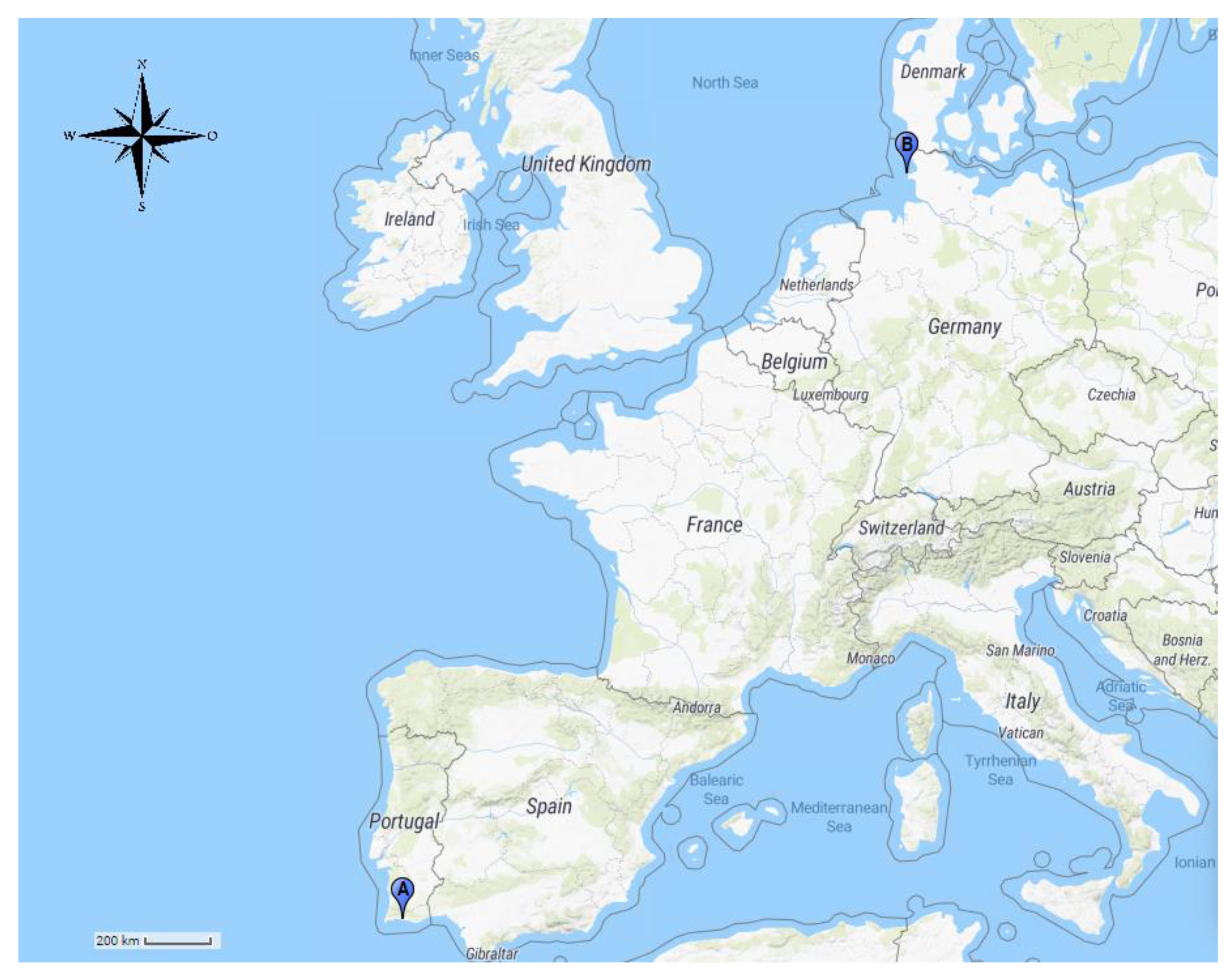
| Common Name (Scientific name) | Provenance | Location | No. Animals Tested |
|---|---|---|---|
| Family Otariidae | |||
| California sea lion (Zalophus californianus) | Human care | Zoomarine Portugal | 12 |
| South African fur seal (Arctocephalus pusillus pusillus) | Human care | Zoomarine Portugal | 20 |
| South American sea lion (Otaria flavescens) | Human care | Zoomarine Portugal | 5 |
| Family Phocidae | |||
| Grey seal (Halichoerus grypus) | Human care | Zoomarine Portugal | 7 |
| Wild | South Portuguese coast | 4 | |
| Harbor seal (Phoca vitulina) | Human care | Zoomarine Portugal | 13 |
| Wild | Lorenzensplate, Wadden Sea | 99 | |
| Harp seal (Pagophilus groenlandicus) | Human care | Zoomarine Portugal | 2 |
| Hooded seal (Cystophora cristata) | Wild | South Portuguese coast | 6 |
| Ringed seal (Pusa hispida) | Human care | Zoomarine Portugal | 1 |
Publisher’s Note: MDPI stays neutral with regard to jurisdictional claims in published maps and institutional affiliations. |
© 2021 by the authors. Licensee MDPI, Basel, Switzerland. This article is an open access article distributed under the terms and conditions of the Creative Commons Attribution (CC BY) license (https://creativecommons.org/licenses/by/4.0/).
Share and Cite
Martins, M.; Urbani, N.; Flanagan, C.; Siebert, U.; Gross, S.; Dubey, J.P.; Cardoso, L.; Lopes, A.P. Seroprevalence of Toxoplasma gondii in Pinnipeds under Human Care and in Wild Pinnipeds. Pathogens 2021, 10, 1415. https://doi.org/10.3390/pathogens10111415
Martins M, Urbani N, Flanagan C, Siebert U, Gross S, Dubey JP, Cardoso L, Lopes AP. Seroprevalence of Toxoplasma gondii in Pinnipeds under Human Care and in Wild Pinnipeds. Pathogens. 2021; 10(11):1415. https://doi.org/10.3390/pathogens10111415
Chicago/Turabian StyleMartins, Micaela, Nuno Urbani, Carla Flanagan, Ursula Siebert, Stephanie Gross, Jitender P. Dubey, Luís Cardoso, and Ana Patrícia Lopes. 2021. "Seroprevalence of Toxoplasma gondii in Pinnipeds under Human Care and in Wild Pinnipeds" Pathogens 10, no. 11: 1415. https://doi.org/10.3390/pathogens10111415
APA StyleMartins, M., Urbani, N., Flanagan, C., Siebert, U., Gross, S., Dubey, J. P., Cardoso, L., & Lopes, A. P. (2021). Seroprevalence of Toxoplasma gondii in Pinnipeds under Human Care and in Wild Pinnipeds. Pathogens, 10(11), 1415. https://doi.org/10.3390/pathogens10111415