Article

Sea Turtles in the Cancer Risk Landscape: A Global Meta-Analysis of Fibropapillomatosis Prevalence and Associated Risk Factors

1
Centre for Integrative Ecology, School of Life and Environmental Sciences, Deakin University, Geelong Waurn Ponds, Vic 3216, Australia
2
CANECEV-Centre de Recherches Ecologiques et Evolutives sur le Cancer (CREEC), 34090 Montpellier, France
3
MIVEGEC, CREEC, UMR IRD 224-CNRS 5290-Université de Montpellier, 34295 Montpellier, France
4
School of Biological and Chemical Sciences, Queen Mary University of London, London E1 4NS, UK
*
Author to whom correspondence should be addressed.
Pathogens 2021, 10(10), 1295; https://doi.org/10.3390/pathogens10101295
Submission received: 11 August 2021 / Revised: 29 September 2021 / Accepted: 4 October 2021 / Published: 8 October 2021

Abstract

:
Several cancer risk factors (exposure to ultraviolet-B, pollution, toxins and pathogens) have been identified for wildlife, to form a “cancer risk landscape.” However, information remains limited on how the spatiotemporal variability of these factors impacts the prevalence of cancer in wildlife. Here, we evaluated the cancer risk landscape at 49 foraging sites of the globally distributed green turtle (Chelonia mydas), a species affected by fibropapillomatosis, by integrating data from a global meta-analysis of 31 publications (1994–2019). Evaluated risk factors included ultraviolet light exposure, eutrophication, toxic phytoplanktonic blooms, sea surface temperature, and the presence of mechanical vectors (parasites and symbiotic species). Prevalence was highest in areas where nutrient concentrations facilitated the emergence of toxic phytoplankton blooms. In contrast, ultraviolet light exposure and the presence of parasitic and/or symbiotic species did not appear to impact disease prevalence. Our results indicate that, to counter outbreaks of fibropapillomatosis, management actions that reduce eutrophication in foraging areas should be implemented.

1. Introduction

Cancers are a group of diseases that emerge in multicellular organism hosts when cells stop collaborating with surrounding cells, and obtain the ability to proliferate in an uncontrolled manner [1,2]. The emergence of cancers mirrors speciation events, in which new parasitic species emerge and consume the resources of hosts, reducing overall fitness [3,4,5]. Cancers have the potential to drive already threatened wildlife towards extinction (i.e., the Tasmanian devil, Sarcophilus harrisii, [6]; California sea lion, Zalophus californianus, [7]; Santa Catalina fox, Urocyon littoralis catalinae, [8]; Cape mountain Zebra, Equus zebra zebra, [9]). Cancers can also cause significant changes to the ecosystems of impacted wildlife through trophic cascades and reshaping of biotic interactions, leading to mass mortalities [10,11,12,13,14,15]. For example, one transmissible cancer depleted > 80% of Tasmanian devils, a keystone predator, from the ecosystem of the island of Tasmania (Australia) [16]. This catastrophic event caused the complete reorganization of the trophic chain, releasing meso-predators from competition with Tasmanian devils, and allowing invasive species, such as feral cats (Felis catus), to proliferate [11,12].
Thus, it is important to quantify the impact of cancers on wildlife populations and associated ecosystems [17,18,19,20]. For instance, several studies have delineated cancer-causing risk factors for wildlife populations [19,20,21], which are directly associated with increasing anthropogenic pressures in both terrestrial and aquatic ecosystems [22]. These risk factors, which include exposure to chemicals, ultraviolet light, ionizing particles, and habitat fragmentation, form a landscape in which species are exposed to cumulative impacts of varying spatiotemporal variability [23]. Thus, evaluating historical data within this framework provides an opportunity to examine the drivers of cancer in wildlife.
Sea turtles are a group of seven threatened species at risk of various diseases, including a type of cancer called fibropapillomatosis [24,25,26]. This disease was first documented in Key West, Florida, USA in 1938 in green turtles (Chelonia mydas, [27]), but has now been detected in all seven sea turtle species and across all ocean basins in which the species are present, reaching panzoonotic status [25,26]. Fibropapillomatosis manifests as cutaneous tumors on the body surface and internal organs [24,28]. Fibropapillomatosis is thought to be initiated by the Chelonid Alphaherpesvirus 5 (ChHV5, also known as Chelonid herpesvirus 5 in the literature) [29,30,31], and is likely horizontally transmitted via shedding of virus particles contained in epithelial cells, and possibly in urine and/or bodily secretions [29,32].
Fibropapillomatosis tumors range in size from 0.1 cm to greater than 30 cm in diameter [24]. These tumors debilitate the host by impeding movement and foraging ability, and reducing host immune function, while consuming energetic resources [33,34]. These tumors can be concomitant to other parasitic infections (i.e., trematodes [28]), but also become secondary infection sites for other parasitic species (i.e., bacterial infections, [35]). All of these factors increase the risk of death to individuals with this cancer [24,26,36]. However, not all sea turtles infected with the virus develop tumors, with regression and recovery also being possible [25,37,38]. Therefore, this disease is likely multifactorial [39], with sea turtles possibly being more likely to develop the disease when occupying stressful environments that exacerbate immunosuppression and inflammation [40], which are both tumor promotors. Because sea turtles contribute to ecosystem functioning, in addition to being of economic (e.g., wildlife watching) and cultural importance, it is important to quantify the risk of this disease to population resilience. Such knowledge could then be integrated in risk assessments to implement appropriate protection actions [41,42].
Here, we conducted a comprehensive meta-analysis on the prevalence of fibropapillomatosis in green turtles on a global scale. Out of the seven sea turtle species, green turtles have the overall highest prevalence of fibropapillomatosis, the largest number of publications on this subject, and some epidemiological studies spanning decades [28,43,44,45], making them the most suitable for meta-analysis. We used mixed-effect models to identify the risk factors most closely associated with the prevalence of fibropapillomatosis across populations. Our results are expected to demonstrate the importance of delineating the cancer risk landscape for integration in the conservation management of threatened species.

2. Material and Methods

2.1. Literature Review and Data Consolidation

Using a previously established protocol (see [46,47]), we performed a comprehensive literature review. We searched the Thomson Reuters ISI Web of ScienceTM database, the Scopus database, and Google Scholar for relevant publications spanning 1994–2019. Specific terms were used in the topic field, which included the title, abstract, keywords and keywords plus (i.e., words that frequently appear in the titles of articles cited within a publication). The terms included: “sea turtle”, “green turtle”, “Chelonia mydas”, “cancer”, “fibropapilloma”, “fibropapillomatosis”, and “neoplasia”. To locate potential articles missed by the initial search, we checked the reference and citation lists of relevant papers based on the predefined keywords. We also screened and included conference proceedings and reports in French, English, and Spanish [48]. See references [49,50,51,52,53,54,55,56,57,58,59,60,61,62,63,64,65,66,67,68,69,70,71,72,73,74,75,76] and Supplementary Table S1 for the full list of publications included in the meta-analysis.
For each document, which contained at least one year of data, we extracted the sample size (expressed as number of green turtles surveyed), the number of individuals infected with fibropapillomatosis, coordinates of the study sites (approximated using Google Earth if exact coordinates were not available in the publication), and the year(s) of sampling. We focused only on foraging sites, where sea turtles spend the majority of their immature and adult life-stages; thus, representing where they are most likely to be infected by the virus (see [77]). We also classified each study depending on the condition of the surveyed sea turtles, i.e., “stranded” (alive or dead on a beach), “live” (free in the water), or “mix” (if the survey contained a mixture of live, free, and dead or stranded turtles in the water or on the beach at the study sites). Measurements of sea turtle size (i.e., carapace length) were not consistently reported across studies (i.e., curved, straight, or estimated lengths), and a breakdown per year was rarely available; thus, we classified individuals into three size groups: “immature”, “adult”, or “mix” (if both groups were present) based on the information provided in each publication. Since size at maturity varies across sites and ocean basins, we relied on the classification provided by the authors and experts of each publication included in this meta-analysis. For many studies, it was not possible to distinguish between male and female turtles; therefore, they were grouped as adults only. There is currently no evidence in the literature that males and females are affected differently by ChHV5 [26]. A publication was excluded from the meta-analysis if it lacked any of the specified key information, and if the reported prevalence was computed by aggregating individuals sampled over a period of time of more than two adjacent years.

2.2. Identification of Potential Risk Factors

To define the cancer risk landscape of green turtles, we identified potential risk factors associated with fibropapillomatosis from the published literature [23]. We then obtained data for those risk factors from various online databases (where available) to investigate their effect on the prevalence of fibropapillomatosis in sea turtle foraging areas.

2.2.1. Exposure to the Chelonid Herpesvirus 5 Virus

ChHV5 is strongly suspected to cause fibropapillomatosis in sea turtles, although Koch’s postulates have yet to be definitively confirmed because of difficulty in culturing the virus in a laboratory setting [78] (but see recent progress [79]). Laboratory experiments showed that green turtles develop tumors between 15 and 43 weeks after being exposed to cell-free tumor extracts [29]. Thus, sea turtles infected by ChHV5 can be observed in the wild that do not yet present tumors [25,38]. Since ChHV5 is globally distributed, we assumed that all populations included in this study had been exposed [25,30].

2.2.2. Ultraviolet Light

Ultraviolet light (UV), especially UVB, is a well-documented cancer risk factor causing damage to DNA and increasing its mutation rate, including in aquatic ecosystems [80,81]. In at least one publication, UV was suspected to contribute to the emergence of tumors in ChHV5-infected sea turtles [82]; however, this hypothesis was not tested at a global scale. To quantify exposure to UV, annual minimum, maximum, and mean exposure data, along with seasonal data (all expressed in J·m−2·day−1), were obtained from the global UV-B radiation dataset for macroecological studies (glUV). This dataset included a series of climate surfaces containing information on various aspects of the global distribution of ultraviolet-B radiation [80].

2.2.3. Seabed Depth

Green turtles are primarily benthic foragers feeding on algae and seagrass beds to depths typically up to 40 m [83,84]. Using the ETOPO1 1 Arc-Minute Global Relief Model (https://www.ngdc.noaa.gov/mgg/global accessed on 25 July 2021), the mean and median seabed depth of each study area was calculated, restricting the analysis to depths < 40 m (the area most likely frequented by turtles). Seabed depth is an interesting variable for inclusion here, because UV light is rapidly absorbed as sea water depth increases. Ninety-percent of the irradiance of UV is absorbed in the first 0.2 to 15 m of the water column, depending on the type of marine ecosystem [85].

2.2.4. Eutrophication

Eutrophication, the excessive richness of nutrients [86,87] presents a potential risk factor for fibropapillomatosis because it promotes the growth of algae and phytoplankton that produce tumor-promoting components, such as biotoxins [88,89,90,91]. For example, exposure to biotoxins produced by benthic filamentous cyanobacterium (Lyngbya majuscule) and dinoflagellates (Prorocentrum spp.), as well as brevetoxins produced by red tides, represent potential risk factors for fibropapillomatosis [88,89,90,91].
To quantify eutrophication in the marine ecosystem, monthly surface concentrations of nitrate (NO3−), phosphorus (PO4−3), silicate (Si), and phytoplankton (in µmol·L−1) were obtained at a resolution of ¼° from the global ocean biogeochemistry hindcast (GLOBAL_REANALYSIS_BIO_001_029, https://resources.marine.copernicus.eu accessed on 25 July 2021) for the period between 1993 to 2019. Datasets on nitrate, phosphate, silicate, and phytoplankton concentrations were assimilated to compute metrics on an annual time scale, including minimum, maximum, mean, median, and standard deviations.

2.2.5. Sea Surface Temperature

Transfection experiments in green turtles suggest that temperature influences the speed at which tumors appear after infection with ChHV5 [29]. Page-Kajan et al. [36] found that green turtles at rehabilitation centers developed more tumors during warmer months. In addition, a rise in temperature often increases the virulence of herpesviruses in other aquatic species (e.g., [92,93]). Temperature also impacts tumor growth in other poikilothermic species. For example, relatively cold temperatures (< 10 °C) promote the growth of papilloma tumors in the Japanese fire belly newt (Cynops pyrrhogaster, [94]), while relatively warm temperatures (> 10 °C) promote the growth of dermal sarcoma tumors in walleye (Stizostedion vitreum, [95]). Thus, temperature likely impacts fibropapillomatosis occurrence in sea turtles.
We obtained global daily sea surface temperature data (SST, expressed as degrees Celsius) from the Optimum Interpolation Sea Surface Temperature (OISST) v2.1 website (https://www.ncdc.noaa.gov/oisst/data-access accessed on 25 July 2021). The OISST dataset extends from September 1981 to the present at a resolution of ¼° [96]. The daily SST dataset was assimilated to compute the following metrics on an annual time scale: minimum, maximum, mean, median, and standard deviation. To determine a threshold for a potential effect of temperature, we also computed the number of days in each year with an SST greater than a given threshold, from 20 °C to 30 °C (using 1 °C steps).

2.2.6. Potential ChHV5 Vectors

While horizontal transmission most likely occurs directly between individual green turtles through the shedding of viral particles [77], several species have been identified as potential ChHV5 mechanical vectors. At least two taxa represent potential mechanical vectors of ChHV5 that are able to infect sea turtles. Specifically, leeches of the genus Ozobranchus that attach to sea turtles are present in the Caribbean Sea and the Gulf of Mexico, while saddleback wrasse (Thalassoma duperrey) are present in the Hawaiian archipelago. Polymerase chain reaction (PCR) diagnostics confirmed that individuals of these two taxa carry high ChHV5 loads [97,98,99,100]. We obtained the global geographical distribution of both taxa from www.marinespecies.org accessed on 25 July 2021 and https://www.fishbase.se accessed on 25 July 2021. We classified the study sites as “present” and “absent” for these two taxa.

2.3. Data Consolidation and Spatial Scale Selection

Most published studies on green turtles were conducted at the spatial scale of a bay (up to ~80 km coastline, e.g., Moreton Bay in Australia [76]), island (up to ~85 km coastline, e.g., in La Martinique, France [66]) or along a relatively long stretch of coastline (up to ~60 km coastline, e.g., in Brazil [70]). Thus, to facilitate comparison, we computed the potential risk factors at a consistent scale, by calculating the metric of interest (min, max, mean, median, and standard deviation, etc.) of the values within two circles of ½° and 1° diameter around the coordinates of each study site, encompassing all study site scales. The environmental values associated to each prevalence measurement were computed for a time period starting one year before the start of a study to its end (over the 1994–2019 period). This approach took into account the fact that 15 to 45 weeks are required for tumors to develop in sea turtles observed with fibropapillomatosis after infection [29]. There was good agreement between the computed variables for the two spatial scales (92% of pairwise comparisons had a Pearson’s R > 0.85). Thus, we conducted all subsequent statistical analyses using the metrics computed at a 1° spatial scale.

2.4. Statistical Analysis

We used a series of logistic mixed effect regression models to quantify the effects of the potential risk factors on the prevalence of fibropapillomatosis [101]. All models used in our study were mixed-effect models. To avoid losing temporal resolution, we only included studies that overlapped a maximum of two calendar years in the model. In all models, we included a random intercept to account for variability between study sites, and a random slope to account for temporal variability within sites (using the temporal mid-point of each study [102]).

2.4.1. Data Exploration

We first investigated collinearity between the environmental variables computed for each risk factor using a Pearson correlation matrix. Almost all variables computed for a potential risk factor were highly collinear (Pearson’s R > 0.80). Therefore, to determine which variables to retain in the subsequent analyses for each risk factor, we fitted separate models and computed the associated odds ratio, Akaike Information Criterion (AIC) and weights (see Supplementary Material 2). Within each group, we identified the variable with the highest predictive potential (lowest AIC, highest AIC weight, and odds ratio confidence intervals excluding one) [103,104,105]. This method eliminates potential risk factors with little explanatory power, with the aim to fit models as parsimoniously as possible. At the end of the procedure, the median nitrate concentration, maximum silicate concentration, and number of days with a temperature above 30 °C were retained. The variables computed for the other risk factors (seabed depth, ultraviolet exposure, phosphate concentration) had non-significant odds ratios, and were excluded from the subsequent analyses (see Supplementary Material 2 for full details).

2.4.2. Quantification of the Effect of Each Risk Factor
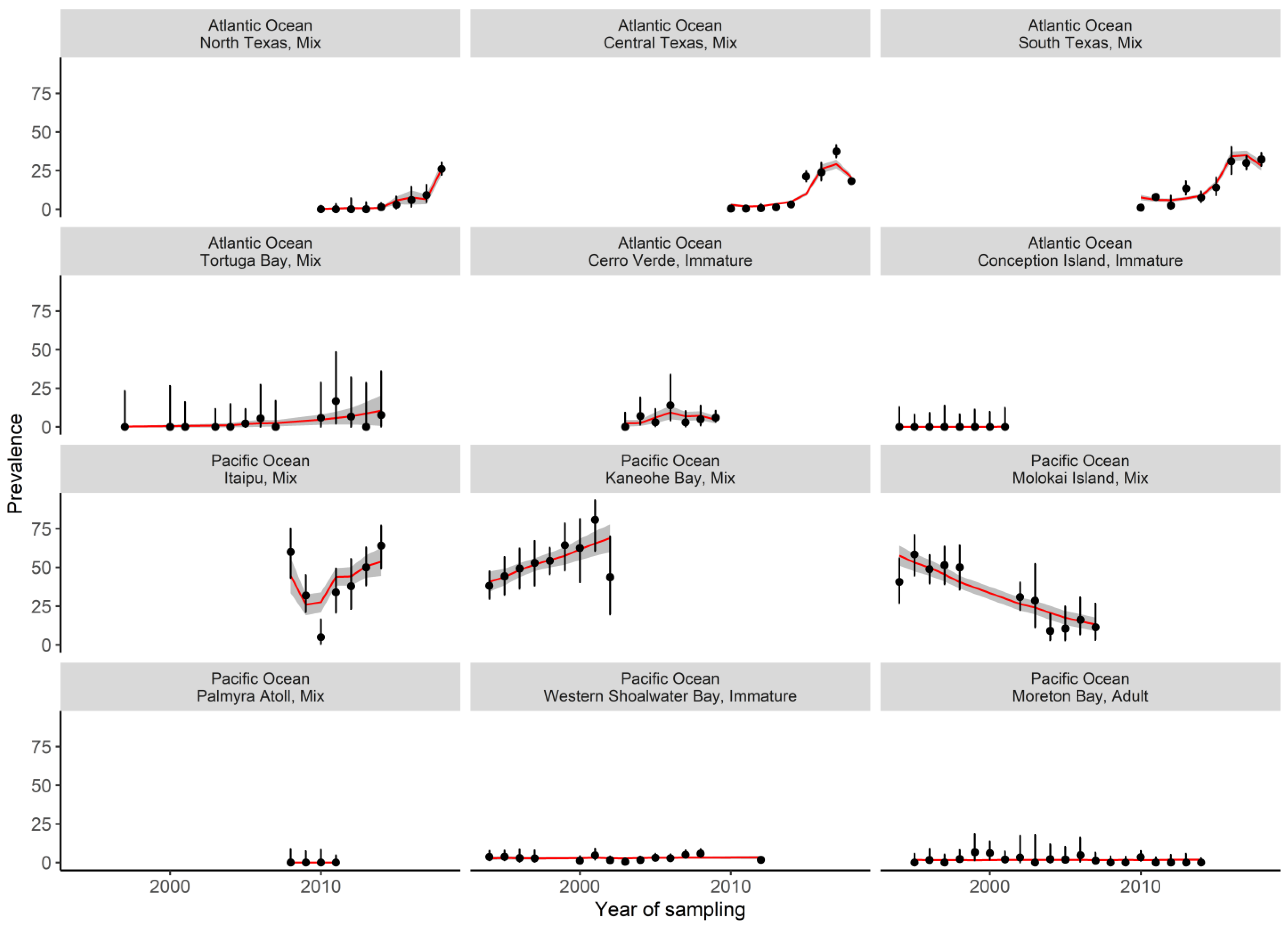
Using the selected environmental variables, we implemented a series of mixed-effect models to quantify the effects of various potential risk factors on the prevalence of fibropapillomatosis. To obtain an optimal model, we tested different combinations of the following risk factors: maturity of turtle (i.e., the study included immature, adult, or a mix of both), sea turtle status (live, stranded, or a mix of both), median nitrate concentration, maximum silicate concentration, number of days SST > 30 °C, presence/absence of wrasse and leeches, and the mid-year point of each study. The body size of sea turtles has a well-documented positive effect on the prevalence of fibropapillomatosis [26,106]; thus, this variable was included in all models (i.e., it was used as a null model to compare the effect of other risk factors). These models were fitted using maximum likelihood (ML). The model with the lowest AIC and highest AIC Weight was retained.
The optimal model was then refitted using the restricted maximum likelihood method (REML) to generate predictions and interpret its coefficients [102]. To visualize the effect of a risk factor on fibropapillomatosis prevalence, we computed marginal effects, which involve varying one risk factor at a time, while keeping all other factors constant (see [107]). The computation of marginal effects allows potential trends in the dataset to be visually represented. Since the computation of p-values is not reliable for mixed-effect models (see [108,109]), we reported the 95% confidence intervals (95% CI) of odds ratios (OR). Odds ratios were considered significant if one 95% CI did not overlap with another [104]. Means were reported with their standard deviation and associated range. All models were fitted in R software V.4.0.2 [110] using the glmmTMB package [111], the marginal effect was fitted using the ggeffects package [107] and usual validation diagnostics for mixed-effect models (see [102]) were performed using the DHARma package [112].

3. Results

We retained 31 published studies in our meta-analysis, identifying 49 study sites (Figure 1) and 265 prevalence measurements (computed from a mean sample size of 187 ± 387 individuals, range:11–4407, for a total of 49,606 surveyed turtles globally) between 1994 and 2019. Fibropapillomatosis prevalence was estimated once at 23 sites, and between 2 and 21 times (average 7.6 ± 4.3) at the other 26 sites (see Supplementary Material 1 for a list of study sites.). Out of the 265 prevalence measurements, 38 (14%) were calculated on adults, 71 (27%) on immature turtles, and 156 (59%) on a mix of adult and immature turtles. Similarly, 225 (85%) prevalence measurements were calculated on live turtles, 13 (5%) on stranded turtles, and 27 (10%) on a mix of live and stranded turtles.
On a global scale, the prevalence of fibropapillomatosis increased in green turtles, on average, between 1994 and 2019 (OR: 1.20, 95% CI: 1.10–1.32, Figure 2a). As expected, there were significant differences in prevalence between the body size groups. Studies including a mix of adult and immature turtles reported the highest prevalence, on average. Studies that only evaluated immature (OR: 0.08, 95% CI: 0.01–0.21) or adult turtles (OR: 0.01, 95% CI: 0.00–0.02) had a lower prevalence compared to studies that included both (Figure 2b). There was a strong interaction between nutrient availability, phytoplankton concentration, and fibropapillomatosis prevalence (OR:0.96, 95% CI: 0.94–0.99 for the interaction between maximum silicates and phytoplankton concentrations; OR:1.24, 95% CI: 1.14–1.34 for the interaction between median nitrates and phytoplankton concentrations. Supplementary Tables S9 and S10). Prevalence was higher in areas with combined low silicate concentrations, high nitrate concentrations, and high phytoplankton concentrations. Regardless of nitrate and silicate concentrations, low fibropapillomatosis prevalence was observed in areas of low phytoplankton concentrations (Figure 2c,d). We also found moderate evidence that prevalence increases when the number of days of SST > 30 °C rises (OR:1.02, 95% CI: 1.01–1.04, Figure 2e, Supplementary Tables S9 and S10). Overall, there was good agreement between observed and modelled prevalence globally (Figure 3).
There was no evidence that fibropapillomatosis prevalence in green turtles at a global scale was impacted by: exposure to ultraviolet radiation (OR: 1.00, 95% CI: 1.00–1.00 for mean annual UV exposure), seabed depth (OR: 0.99, 95% CI: 0.84–1.17 for mean depth), phosphate concentration (OR: 0.44, 95% CI: 0.03–6.75 for mean phosphate concentration), or sea turtle status (see Supplementary Table S9). Similarly, we found no evidence that the presence of saddleback wrasses (OR: 6.22, 95% CI: 0.34–113.6) or leeches (OR: 0.49, 95% CI: 0.10–2.42) increased the prevalence of fibropapillomatosis on a global scale.

4. Discussion

This study delineates the cancer risk landscape of green turtles by combining a global meta-analysis with models of potential risk factors. Our results indicate that fibropapillomatosis is more prevalent in areas exposed to greater eutrophication, with cancer promotors (e.g. biotoxins) appearing to be present in these areas. In contrast, there was no evidence that wrasses and leeches, or UV exposure, contributed to the observed prevalence of fibropapillomatosis. Our findings highlight the risk factors that managers should monitor at all coastal foraging sites of green turtles globally [113] to mitigate fibropapillomatosis, and could potentially be used to forecast the likelihood of incidence.

4.1. Effect of Eutrophication

Our results indicate that high prevalence of fibropapillomatosis is associated with an exposure to toxic phytoplankton blooms. While the abundance of toxic phytoplankton could not be directly measured in our study, nutrient availability is usually a good indicator of the risk of toxic blooms [114]. Indeed, in marine ecosystems, high concentrations of nitrogen and phosphorus promote the development of diatom blooms until silicates are depleted [115,116]. Consequently, nonsiliceous taxa (such as cyanobacteria and dinoflagellates), which are more efficient at growing in nutrient-poor and silica-limited environments, tend to outcompete diatoms to become dominant, and form toxic blooms covering areas up to several hundreds of kilometers [117,118]. We found that the prevalence of fibropapillomatosis was greater in areas with both high phytoplankton concentration and low silicate concentration. In addition, extended periods of high temperatures increase stratification in the water column, potentially favoring toxic phytoplanktonic blooms [116]. Our results suggest that green turtles exposed to high oceanic temperatures (> 30 °C) were associated with a higher prevalence of fibropapillomatosis. This finding supports the observation that rehabilitating turtles (here sea turtles treated at the Georgia Sea Turtle Center, Jekyll Island, USA) tend to develop new tumors faster during warmer months [36].
Thus, fibropapillomatosis is likely associated with exposure to harmful algal blooms that are promoted by higher water temperatures and eutrophication. Indeed, biotoxin-producing algae can generate large quantities of contact irritants causing inflammatory responses (a cancer promoter) or neurotoxins [88,119]. Exposure to these biotoxins likely occurs when green turtles ingest seagrass, during gular pumping, or are possibly inhaled during pre-dive breathing [91,120,121]. Certain molecules, such as brevetoxin (a neurotoxin), accumulate in the plasma of green turtles, and might bind with albumin, causing long-term inflammation and exacerbating oxidative stress responses (another cancer promotor [91,120,122,123])

4.2. Transmission of the Virus

The most likely route of transmission for ChHV5 is through the shedding of viral particles in the water column [100,124]. At a local scale, the dispersal of such particles depends on certain key environmental factors, such as oceanic currents (which physically disperse the virus) and sea water temperatures (which affect infectivity and virulence). For example, lung–eye–trachea disease-associated herpesvirus remains infective in natural sea water for at least 120 h at 23 °C [125]. Similarly, the infectivity of another herpesvirus that causes grey-patch disease in green turtles increases as temperature rises from 25 °C to 30 °C [93], likely because higher temperatures enhance herpesviruses replication. If ChHV5 particles that shed in the water column have similar dispersal traits, infection would primarily occur at a given coastal foraging site, with relatively limited transfer across spatially distinct sites (as suggested by genetic analyses of the virus in infected sea turtles [77,100,126]). Alternatively, transfer across sites might be driven by potential mechanical vectors, like wrasses or leeches. However, we found no evidence, on a global scale, that either of these two vectors are associated with the prevalence of fibropapillomatosis. These potential mechanical vectors may contribute to the transmission of the virus at a local scale, because it has been detected in them [97,98,99]. Thus, continued monitoring of the relationship between temperature and fibropapillomatosis is required, as well as studies on the mechanisms driving the transmission routes and rates of this virus across foraging sites, including data on leech loads on turtles, the frequency of fish cleaning station visits, and the frequency of movement and interactions with turtles from other sites (which can be quantified, see [127,128,129]). Knowledge on the ChHV5 variants documented globally also remains limited, including transmission rates. Different strains could drive different immune responses in turtles across regions, as observed between the Pacific (Hawaii) and Atlantic (Florida) green turtle stocks, potentially driving differences in prevalence [30,130]. Thus, epidemiological studies on the co-evolution of the virus and its hosts are required across regions.

4.3. Implications for the Conservation of Green Turtles

Efficient treatments of fibropapillomatosis in green turtles are lacking [82]. While the regression of tumors has been detected in some individuals in the wild [37], many die, with the surgical removal of tumors having limited effect because they often regrow [131]. In particular, survival is strongly associated with the body parts on which tumors develop [132]. For example, sea turtles with ocular tumors are eight times more likely to die compared to those with tumors on other body parts [36]. Our study also showed that environmental factors exacerbate the progression and prevalence of disease; thus, actions towards restoring ecosystem quality (for example, by reducing eutrophication) could help to limit, or entirely prevent outbreaks of fibropapillomatosis in sea turtles. For instance, actively monitoring the cancer risk landscape in areas where green turtles forage could help detect emerging hotspots. Focus could be placed on mitigating nutrient input (and hence toxic phytoplanktonic blooms) to these areas by using indicators to predict and forestall fibropapillomatosis outbreaks, implementing dynamic ocean management to prevent the overlap of threatened marine wildlife, similar to that already implemented for fisheries and shipping routes [133,134].

4.4. Limitations and Conclusions

The presence of a significant temporal trend in the prevalence of fibropapillomatosis in our best model indicates that some risk factors have yet to be identified for fibropapillomatosis in green turtles. We found that the reported prevalence was higher in studies with a mix of immature and adult turtles, as previously documented [26,106]. However, data on separate immature and adult size classes were limited, with previous comparisons between studies using size as a continuous variable, rather than a categorical classification as done in this study, being difficult to conduct. The low prevalence of fibropapillomatosis in studies of immature turtles might be attributed to relatively smaller (i.e., younger) individuals being encountered, which are less likely to harbor tumors. In contrast, studies that evaluated both immature and mature sea turtles likely screened for a larger proportion of the population (and age classes), leading to more individuals with fibropapillomatosis being detected. Cross-discipline studies integrating information on epidemiology, environment, and turtle characteristics (e.g. demography, genetic diversity, behavior, movement and habitat use) at large scales are required to establish causality links [46,135,136,137]. There is currently no evidence that organic chemical contamination increases the prevalence of fibropapillomatosis in sea turtles [40,138]. However, data are lacking on chemical contamination on a global scale [139], particularly for emerging contaminants.
Our study focused on establishing trends in the prevalence of fibropapillomatosis for green turtles on a global scale, and could be replicated at different scales or on a different species to facilitate management actions (i.e., from site scale to regional management unit scale [RMU] [140]). Because fibropapillomatosis appears to be associated with multifactorial risks [39], with sea turtles inhabiting a complex and dynamic cancer risk landscape [23], it is important to assimilate longitudinal datasets investigating multiple risk factors as a whole (e.g., phytoplankton bloom occurrence, composition, and associated biotoxins), along with any other environmental variables of relevance at a given site (e.g., other cancer promotors in the environment, ChHV5 shedding rates from environmental DNA [100], genetics or immune responses of sea turtles, emerging contaminants). The sharing of holistic datasets collected from multiple study sites within a strong collaborative network could facilitate rapid advances in our understanding of the disease, similar to those developed for cancer in humans [141] and transmissible cancers in wildlife [46]. In conclusion, through identifying key environmental parameters associated with the prevalence of fibropapillomatosis, this study provides a basis for developing direct monitoring and management strategies to mitigate this cancer.

Supplementary Materials

The following are available online at https://www.mdpi.com/article/10.3390/pathogens10101295/s1, Supplementary Materials 1 “List of publication included in the meta-analysis”, Supplementary Materials 2 “Selection of variables for inclusion in the mixed effect models”, Supplementary Result 1 “Summary the logistic mixed effect models”. Supplementary Table S1: Summary of the published literature (1994–2019) on fibropapillomatosis in green sea turtles at foraging areas globally and included in the meta-analysis; Supplementary Table S2: Summary results of the models used to explore the variables derived from nitrate concentration data; Supplementary Table S3: Summary results of the models used to explore the variables derived from phosphate concentration data; Supplementary Table S4: Summary results of the models used to explore the variables derived from silicate concentration data; Supplementary Table S5: Summary results of the models used to explore the variables derived from phytoplanktonic concentration data; Supplementary Table S6: Summary results of the models used to explore the variables derived from seabed depth data; Supplementary Table S7: Summary results of the models used to explore the variables derived from ultraviolet exposure data; Supplementary Table S8: Summary results of the models used to explore the variables derived from SST data; Supplementary Table S9: Summary table of the logistic mixed effect models used to investigate the effect of risk factors on fibropapillomatosis in green sea turtles to determine an optimal model; Supplementary Table S10: Odd ratios computed from the optimal model linking fibropapillomatosis prevalence in green sea turtles to a range of risk factors.

Author Contributions

A.M.D. designed the study, conducted the literature review, and performed statistical analyses. R.M.V. provided expertise in environmental data collection. G.S. provided expertise in sea turtles biology. A.M.D., F.T. and B.U. provided expertise in the ecology of cancer. A.M.D. wrote the first draft of the manuscript. All authors provided input during manuscript editing and gave final approval for publication. All authors have read and agreed to the published version of the manuscript.

Funding

This study was supported by the MAVA foundation and the ANR TRANSCAN and Deakin University PRESS2021 Funding scheme.

Institutional Review Board Statement

Not applicable.

Informed Consent Statement

Not applicable.

Data Availability Statement

All data sources used in this publication are cited in the manuscript or in the Supplementary Materials.

Conflicts of Interest

The authors declare no conflict of interest.

References

  1. Aktipis, C.A.; Boddy, A.M.; Jansen, G.; Hibner, U.; Hochberg, M.E.; Maley, C.C.; Wilkinson, G. Cancer across the tree of life: Cooperation and cheating in multicellularity. Philos. Trans. R. Soc. B Biol. Sci. 2015, 370, 20140219. [Google Scholar] [CrossRef] [Green Version]
  2. Albuquerque, T.A.F.; Val, L.D.D.; Doherty, A.; De Magalhães, J.P. From humans to hydra: Patterns of cancer across the tree of life. Biol. Rev. 2018, 93, 1715–1734. [Google Scholar] [CrossRef]
  3. Duesberg, P.; Mandrioli, D.; McCormack, A.; Nicholson, J.M. Is carcinogenesis a form of speciation? Cell Cycle 2011, 10, 2100–2114. [Google Scholar] [CrossRef] [Green Version]
  4. Capp, J.-P.; Thomas, F. A Similar Speciation process relying on cellular stochasticity in microbial and cancer cell populations. iScience 2020, 23, 101531. [Google Scholar] [CrossRef]
  5. Pienta, K.J.; Hammarlund, E.U.; Axelrod, R.; Amend, S.R.; Brown, J.S. Convergent evolution, evolving evolvability, and the origins of lethal cancer. Mol. Cancer Res. 2020, 18, 801–810. [Google Scholar] [CrossRef] [PubMed] [Green Version]
  6. McCallum, H.; Jones, M.; Hawkins, C.; Hamede, R.; Lachish, S.; Sinn, D.L.; Beeton, N.; Lazenby, B. Transmission dynamics of Tasmanian devil facial tumor disease may lead to disease-induced extinction. Ecology 2009, 90, 3379–3392. [Google Scholar] [CrossRef] [PubMed] [Green Version]
  7. Browning, H.M.; Gulland, F.M.D.; Hammond, J.A.; Colegrove, K.M.; Hall, A.J. Common cancer in a wild animal: The California sea lion (Zalophus californianus) as an emerging model for carcinogenesis. Philos. Trans. R. Soc. B Biol. Sci. 2015, 370, 20140228. [Google Scholar] [CrossRef] [PubMed] [Green Version]
  8. Vickers, T.W.; Clifford, D.L.; Garcelon, D.K.; King, J.L.; Duncan, C.L.; Gaffney, P.M.; Boyce, W.M. Pathology and epidemiology of ceruminous gland tumors among endangered Santa Catalina Island foxes (Urocyon littoralis catalinae) in the Channel Islands, USA. PLoS ONE 2015, 10, e0143211. [Google Scholar] [CrossRef] [PubMed]
  9. Sasidharan, S.P.; Ludwig, A.; Harper, C.; Moodley, Y.; Bertschinger, H.J.; Guthrie, A.J. Comparative genetics of sarcoid tumour-affected and non-affected mountain zebra (Equus zebra) populations. South. Afr. J. Wildl. Res. 2011, 41, 36–49. [Google Scholar] [CrossRef]
  10. Mateo, D.R.; Maccallum, G.S.; Davidson, J. Field and laboratory transmission studies of haemic neoplasia in the soft-shell clam, Mya arenaria, from Atlantic Canada. J. Fish. Dis. 2016, 39, 913–927. [Google Scholar] [CrossRef]
  11. Hollings, T.; Jones, M.; Mooney, N.; Mccallum, H. Trophic cascades following the disease-induced fecline of an apex predator, the Tasmanian devil. Conserv. Biol. 2014, 28, 63–75. [Google Scholar] [CrossRef]
  12. Hollings, T.; Jones, M.; Mooney, N.; McCallum, H. Disease-induced decline of an apex predator drives invasive dominated states and threatens biodiversity. Ecology 2016, 97, 394–405. [Google Scholar] [CrossRef] [Green Version]
  13. Cunningham, C.X.; Johnson, C.N.; Barmuta, L.; Hollings, T.; Woehler, E.; Jones, M. Top carnivore decline has cascading effects on scavengers and carrion persistence. Proc. R. Soc. B Biol. Sci. 2018, 285, 20181582. [Google Scholar] [CrossRef] [Green Version]
  14. Perret, C.; Gidoin, C.; Ujvari, B.; Thomas, F.; Roche, B. Predation shapes the impact of cancer on population dynamics and the evolution of cancer resistance. Evol. Appl. 2020, 13, 1–12. [Google Scholar] [CrossRef] [PubMed]
  15. Boutry, J.; Mistral, J.; Berlioz, L.; Klimovich, A.; Tökölyi, J.; Fontenille, L.; Ujvari, B.; Dujon, A.M.; Giraudeau, M.; Thomas, F. Tumors (re)shape biotic interactions within ecosystems: Experimental evidence from the freshwater cnidarian Hydra. Sci. Total Environ. 2021, 803, 149923. [Google Scholar] [CrossRef] [PubMed]
  16. Hawkins, C.; Baars, C.; Hesterman, H.; Hocking, G.; Jones, M.; Lazenby, B.; Mann, D.; Mooney, N.; Pemberton, D.; Pyecroft, S.; et al. Emerging disease and population decline of an island endemic, the Tasmanian devil Sarcophilus harrisii. Biol. Conserv. 2006, 131, 307–324. [Google Scholar] [CrossRef]
  17. Dujon, A.M.; Aktipis, A.; Alix-Panabières, C.; Amend, S.R.; Boddy, A.M.; Brown, J.S.; Capp, J.; DeGregori, J.; Ewald, P.; Gatenby, R.; et al. Identifying key questions in the ecology and evolution of cancer. Evol. Appl. 2021, 14, 877–892. [Google Scholar] [CrossRef]
  18. Vittecoq, M.; Roche, B.; Daoust, S.P.; Ducasse, H.; Missé, D.; Abadie, J.; Labrut, S.; Renaud, F.; Gauthier-Clerc, M.; Thomas, F. Cancer: A missing link in ecosystem functioning? Trends Ecol. Evol. 2013, 28, 628–635. [Google Scholar] [CrossRef]
  19. Giraudeau, M.; Sepp, T.; Ujvari, B.; Ewald, P.W.; Thomas, F. Human activities might influence oncogenic processes in wild animal populations. Nat. Ecol. Evol. 2018, 2, 1065–1070. [Google Scholar] [CrossRef]
  20. Pesavento, P.A.; Agnew, D.; Keel, M.K.; Woolard, K.D. Cancer in wildlife: Patterns of emergence. Nat. Rev. Cancer 2018, 18, 646–661. [Google Scholar] [CrossRef] [PubMed]
  21. Hochberg, M.E.; Noble, R. A framework for how environment contributes to cancer risk. Ecol. Lett. 2017, 20, 117–134. [Google Scholar] [CrossRef]
  22. Halpern, B.S.; Walbridge, S.; Selkoe, K.A.; Kappel, C.V.; Micheli, F.; D’Agrosa, C.; Bruno, J.F.; Casey, K.S.; Ebert, C.; Fox, H.E.; et al. A global map of human impact on marine ecosystems. Science 2008, 319, 948–953. [Google Scholar] [CrossRef] [Green Version]
  23. Dujon, A.M.; Ujvari, B.; Thomas, F. Cancer risk landscapes: A framework to study cancer in ecosystems. Sci. Total Environ. 2021, 763, 142955. [Google Scholar] [CrossRef]
  24. Herbst, L.H. Fibropapillomatosis of marine turtles. Annu. Rev. Fish. Dis. 1994, 4, 389–425. [Google Scholar] [CrossRef]
  25. Alfaro-Núñez, A.; Bertelsen, M.F.; Bojesen, A.M.; Rasmussen, I.; Zepeda-Mendoza, L.; Olsen, M.T.; Gilbert, M.T.P. Global distribution of Chelonid fibropapilloma-associated herpesvirus among clinically healthy sea turtles. BMC Evol. Biol. 2014, 14, 206. [Google Scholar] [CrossRef] [PubMed]
  26. Jones, K.; Ariel, E.; Burgess, G.; Read, M. A review of fibropapillomatosis in Green turtles (Chelonia mydas). Vet. J. 2016, 212, 48–57. [Google Scholar] [CrossRef] [PubMed]
  27. Smith, G.M.; Coates, C.W. Fibro-epithelial growths of the skin in large marine turtles, Chelonia mydas (Linnaeus). Zoologica 1938, 23, 93–98. [Google Scholar] [CrossRef]
  28. Work, T.M.; Work, T.M.; Balazs, G.H.; Rameyer, R.A.; Morris, R.A. Retrospective pathology survey of green turtles Chelonia mydas with fibropapillomatosis in the Hawaiian Islands, 1993–2003. Dis. Aquat. Organ. 2004, 62, 163–176. [Google Scholar] [CrossRef] [Green Version]
  29. Herbst, L.H.; Jacobson, E.R.; Moretti, R.; Brown, T.; Sundberg, J.P.; Klein, P.A. Experimental transmission of green turtle fibropapillomatosis using cell-free tumor extracts. Dis. Aquat. Org. 1995, 22, 1–12. [Google Scholar] [CrossRef] [Green Version]
  30. Patrício, A.R.; Herbst, L.H.; Duarte, A.; Vélez-Zuazo, X.; Loureiro, N.; Pereira, N.; Tavares, L.; Toranzos, G.A. Global phylogeography and evolution of chelonid fibropapilloma-associated herpesvirus. J. Gen. Virol. 2012, 93, 1035–1045. [Google Scholar] [CrossRef]
  31. Ackermann, M.; Koriabine, M.; Hartmann-Fritsch, F.; De Jong, P.J.; Lewis, T.D.; Schetle, N.; Work, T.M.; Dagenais, J.; Balazs, G.H.; Leong, J.-A.C. The genome of chelonid herpesvirus 5 harbors atypical genes. PLoS ONE 2012, 7, e46623. [Google Scholar] [CrossRef] [Green Version]
  32. Page-Karjian, A.; Norton, T.M.; Ritchie, B.; Brown, C.; Mancia, C.; Jackwood, M.; Gottdenker, N.L. Quantifying chelonid herpesvirus 5 in symptomatic and asymptomatic rehabilitating green sea turtles. Endanger. Species Res. 2015, 28, 135–146. [Google Scholar] [CrossRef] [Green Version]
  33. Brill, R.W.; Balazs, G.H.; Holland, K.N.; Chang, R.K.; Sullivan, S.; George, J.C. Daily movements, habitat use, and submergence intervals of normal and tumor-bearing juvenile green turtles (Chelonia mydas L.) within a foraging area in the Hawaiian Islands. J. Exp. Mar. Biol. Ecol. 1995, 185, 203–218. [Google Scholar] [CrossRef]
  34. Perrault, J.; Levin, M.; Mott, C.; Bovery, C.; Bresette, M.; Chabot, R.; Gregory, C.; Guertin, J.; Hirsch, S.; Ritchie, B.; et al. Insights on immune function in free-ranging green sea turtles (Chelonia mydas) with and without Fibropapillomatosis. Animals 2021, 11, 861. [Google Scholar] [CrossRef] [PubMed]
  35. Work, T.M.; Balazs, G.H.; Wolcott, M.; Morris, R. Bacteraemia in free-ranging Hawaiian green turtles Chelonia mydas with fibropapillomatosis. Dis. Aquat. Org. 2003, 53, 41–46. [Google Scholar] [CrossRef] [PubMed] [Green Version]
  36. Page-Karjian, A.; Norton, T.M.; Krimer, P.; Groner, M.; Nelson, S.E.; Gottdenker, N. Factors influencing survivorship of rehabilitating green sea turtles (Chelonia mydas) with fibropapillomatosis. J. Zoo Wildl. Med. 2014, 45, 507–519. [Google Scholar] [CrossRef] [PubMed]
  37. Guimarães, S.M.; Gitirana, H.M.; Wanderley, A.V.; Monteiro-Neto, C.; Lobo-Hajdu, G. Evidence of regression of fibropapillomas in juvenile green turtles Chelonia mydas caught in Niterói, southeast Brazil. Dis. Aquat. Org. 2013, 102, 243–247. [Google Scholar] [CrossRef] [Green Version]
  38. James, A.; Page-Karjian, A.; Charles, K.; Edwards, J.; Gregory, C.; Cheetham, S.; Buter, B.; Marancik, D. Chelonid alphaherpesvirus 5 prevalence and first confirmed case of sea turtle fibropapillomatosis in Grenada, West Indies. Animals 2021, 11, 1490. [Google Scholar] [CrossRef]
  39. Herbst, L.H.; Klein, A.P. Green turtle fibropapillomatosis: Challenges to assessing the role of environmental cofactors. Environ. Health Perspect. 1995, 103, 27–30. [Google Scholar] [CrossRef] [Green Version]
  40. Keller, J.M.; Balazs, G.H.; Nilsen, F.; Rice, M.; Work, T.; Jensen, B.A. Investigating the potential role of persistent organic pollutants in Hawaiian green sea turtle fibropapillomatosis. Environ. Sci. Technol. 2014, 48, 7807–7816. [Google Scholar] [CrossRef]
  41. Gore, M.L.; Wilson, R.; Siemer, W.F.; Hudenko, H.W.; Clarke, C.E.; Hart, P.S.; Maguire, L.A.; Muter, B.A. Application of Risk Concepts to wildlife management: Special issue introduction. Hum. Dimens. Wildl. 2009, 14, 301–313. [Google Scholar] [CrossRef]
  42. Perez, E.A.; Marco, A.; Martins, S.; Hawkes, L. Is this what a climate change-resilient population of marine turtles looks like? Biol. Conserv. 2016, 193, 124–132. [Google Scholar] [CrossRef]
  43. Aguirre, A.A.; Lutz, P.L. Marine Turtles as Sentinels of Ecosystem Health: Is Fibropapillomatosis an indicator? EcoHealth 2004, 1, 275–283. [Google Scholar] [CrossRef]
  44. Chaloupka, M.; Balazs, G.H.; Work, T.M. Rise and fall over 26 years of a marine epizootic in Hawaiian green sea turtles. J. Wildl. Dis. 2009, 45, 1138–1142. [Google Scholar] [CrossRef] [PubMed] [Green Version]
  45. Foley, A.M.; Schroeder, B.A.; Redlow, A.E.; Fick-Child, K.J.; Teas, W.G. Fibropapillomatosis in stranded green turtles (Chelonia mydas) from the eastern united states (1980–1998): Trends and associations with environmental factors. J. Wildl. Dis. 2005, 41, 29–41. [Google Scholar] [CrossRef] [PubMed] [Green Version]
  46. Dujon, A.M.; Schofield, G.; Bramwell, G.; Raven, N.; Hamede, R.; Thomas, F.; Ujvari, B. Global meta-analysis of over 50 years of multidisciplinary and international collaborations on transmissible cancers. Evol. Appl. 2020, 13, 1745–1755. [Google Scholar] [CrossRef]
  47. Dujon, A.; Schofield, G. Importance of machine learning for enhancing ecological studies using information-rich imagery. Endanger. Species Res. 2019, 39, 91–104. [Google Scholar] [CrossRef] [Green Version]
  48. Nuñez, M.A.; Amano, T. Monolingual searches can limit and bias results in global literature reviews. Nat. Ecol. Evol. 2021, 5, 2021. [Google Scholar] [CrossRef] [PubMed]
  49. Rodenbusch, C.; Baptistotte, C.; Werneck, M.R.; Pires, T.T.; Melo, M.T.D.; De Ataíde, M.W.; Dos Reis, K.; Testa, P.; Alieve, M.M.; Canal, C. Fibropapillomatosis in green turtles Chelonia mydas in Brazil: Characteristics of tumors and virus. Dis. Aquat. Org. 2014, 111, 207–217. [Google Scholar] [CrossRef] [Green Version]
  50. Adnyana, W.; Ladds, P.W.; Blair, D. Observations of fibropapillomatosis in green turtles (Chelonia mydas) in Indonesia. Aust. Vet. J. 1997, 75, 737–742. [Google Scholar] [CrossRef]
  51. Tagliolatto, A.; Guimarães, S.; Lobo-Hajdu, G.; Monteiro-Neto, C. Characterization of fibropapillomatosis in green turtles Chelonia mydas (Cheloniidae) captured in a foraging area in Southeastern Brazil. Dis. Aquat. Org. 2016, 121, 233–240. [Google Scholar] [CrossRef] [PubMed]
  52. Murakawa, S.K.K. Hawaiian archipelago fibropapillomatosis data. In Proceedings of the 2015 International Summit on Fibropapillomatosis: Global Status, Trends, and Population Impacts, Honolulu, HI, USA, 11–14 June 2015; pp. 36–56. [Google Scholar]
  53. Mejía-Radillo, R.; Zavala-Norzagaray, A.; Chávez-Medina, J.; Aguirre, A.; Escobedo-Bonilla, C. Presence of chelonid herpesvirus 5 (ChHV5) in sea turtles in northern Sinaloa, Mexico. Dis. Aquat. Org. 2019, 132, 99–108. [Google Scholar] [CrossRef]
  54. Hirama, S.; Ehrhart, L.M. Description, prevalence and severity of green turtle fibropapillomatosis in three developmental habitats on the East Coast of Florida. Fla. Sci. 2007, 70, 435–448. [Google Scholar]
  55. Avens, L.; Goshe, L.; Harms, C.; Anderson, E.; Hall, A.G.; Cluse, W.; Godfrey, M.; Braun-McNeill, J.; Stacy, B.; Bailey, R.; et al. Population characteristics, age structure, and growth dynamics of neritic juvenile green turtles in the northeastern Gulf of Mexico. Mar. Ecol. Prog. Ser. 2012, 458, 213–229. [Google Scholar] [CrossRef] [Green Version]
  56. Campillo, A. Projet Origine, Répartition et Evolution du Fibropapillomas aux îles Barren; Association Caouanne, 2012. [Google Scholar]
  57. Work, T.M.; Balazs, G.H. Relating Tumor score to hematology in green turtles with fibropapillomatosis in Hawaii. J. Wildl. Dis. 1999, 35, 804–807. [Google Scholar] [CrossRef] [PubMed] [Green Version]
  58. Sterling, E.J.; McFadden, K.W.; Holmes, K.E.; Vintinner, E.C.; Arengo, F.; Naro-Maciel, E. Ecology and conservation of marine turtles in a central pacific foraging ground. Chelonian Conserv. Biol. 2013, 12, 2–16. [Google Scholar] [CrossRef]
  59. Loureiro, N.D.S.; Damião, M. Presence of fibropapillomatosis in green turtles Chelonia mydas at Príncipe Island in the Gulf of Guinea. Arquipélago. Life Mar. Sci. 2009, 26, 79–83. [Google Scholar]
  60. Patrício, A.; Diez, C.; Van Dam, R.; Godley, B. Novel insights into the dynamics of green turtle fibropapillomatosis. Mar. Ecol. Prog. Ser. 2016, 547, 247–255. [Google Scholar] [CrossRef]
  61. Gillis, J.A. Foraging ecology and diet selection of juvenile green turtles (Chelonia mydas) in the Western Bahamas: Insights from stable isotope analysis and prey mapping. Master’s Thesis, Florida State University, Tallahassee, FL, USA, 2018. [Google Scholar]
  62. Hamann, M.; Schäuble, C.S.; Simon, T.; Evans, S. Demographic and health parameters of green sea turtles Chelonia mydas foraging in the Gulf of Carpentaria, Australia. Endanger. Species Res. 2006, 2, 81–88. [Google Scholar] [CrossRef]
  63. Foley, A.M.; Singel, K.E.; Dutton, P.H.; Summers, T.M.; Redlow, A.E.; Lessman, J. Characteristics of a green turtle (Chelonia mydas) assemblage in Northwestern Florida determined during a hypothermic stunning event. Gulf Mex. Sci. 2007, 25, 131–143. [Google Scholar] [CrossRef]
  64. Stringell, T.B.; Clerveaux, W.V.; Godley, B.J.; Phillips, Q.; Ranger, S.; Richardson, P.B.; Sanghera, A.; Broderick, A.C. Fisher choice may increase prevalence of green turtle fibropapillomatosis disease. Front. Mar. Sci. 2015, 2, 1–8. [Google Scholar] [CrossRef] [Green Version]
  65. Piovano, S.; Lemons, G.; Ciriyawa, A.; Batibasaga, A.; Seminoff, J. Diet and recruitment of green turtles in Fiji, South Pacific, inferred from in-water capture and stable isotope analysis. Mar. Ecol. Prog. Ser. 2020, 640, 201–213. [Google Scholar] [CrossRef]
  66. Liebart, M. Photo-Identification des Tortues Vertes (Chelonia mydas) et Son Application Dans L’indice D’abondance ou de Fidélité aux Sites D’alimentation en Martinique; Ecole Nationale Vétérinaire de Toulouse: Toulouse, France, 2019. [Google Scholar]
  67. Patricio, A.R.; Centre, E.S. Fibropapillomatosis in marine turtles of the caribbean region: The case study of Puerto Rico. In Proceedings of the International Summit on Fibropapillomatosis: Global Status, Trends, and Population Impacts, Honolulu, HI, USA, 11–14 June 2016. [Google Scholar]
  68. Van Mil, C. Fibropapillomatosis Affecting Green Turtles (Chelonia mydas); Sea Turtle Conservation Bonaire: Gainesville, FL, USA, 2014. [Google Scholar]
  69. Albareda, D.A.; Garne, M.; Prosdocimi, L.; Rodriguez, H.S.; Di, P.J.L.; Loureiro, J. Pathological studies in green sea turtles (Chelonia mydas) and loggerhead sea turtles (Caretta caretta) from the northern coastal area of Buenos Aires, Argentina. In Proceedings of the Twenty-Seventh Annual Symposium on Sea Turtle Biology and Conservation, Myrtle Beach, SC, USA, 22–28 February 2008. [Google Scholar]
  70. Da Silva-Júnior, E.S.; De Farias, D.S.D.; Bomfim, A.D.C.; Freire, A.C.D.B.; Revorêdo, R.Â.; Rossi, S.; Matushima, E.R.; Grisi-Filho, J.H.H.; Silva, F.J.D.L.; Gavilan, S.A. Stranded marine turtles in Northeastern Brazil: Incidence and spatial–temporal distribution of fibropapillomatosis. Chelonian Conserv. Biol. 2019, 18, 249. [Google Scholar] [CrossRef]
  71. Shaver, D.J.; Walker, J.S.; Backof, T.F. Fibropapillomatosis prevalence and distribution in green turtles Chelonia mydas in Texas (USA). Dis. Aquat. Org. 2019, 136, 175–182. [Google Scholar] [CrossRef] [Green Version]
  72. López-Mendilaharsu, M.; Vélez-Rubio, G.M.; Lezama, C.; Aisenberg, A.; Bauzá, A.; Berrondo, L.; Calvo, V.; Caraccio, N.; Estrades, A.; Hernández, M.; et al. Demographic and tumour prevalence data for juvenile green turtles at the Coastal-Marine Protected Area of Cerro Verde, Uruguay. Mar. Biol. Res. 2016, 12, 541–550. [Google Scholar] [CrossRef]
  73. Bjorndal, K.; Bolten, A.; Chaloupka, M.Y. Evaluating trends in abundance of immature green turtles, Chelonia mydas, in the greater caribbean. Ecol. Appl. 2005, 15, 304–314. [Google Scholar] [CrossRef]
  74. Formia, A.; Deem, S.; Billes, A.; Ngouessono, S.; Parnell, R.; Collinis, T.; Sounguet, G.P.; Gibudi, A.; Villarubia, A.; Balazs, G.H.; et al. Fibropapillomatosis confirmed in Chelonia mydas in the Gulf of Guinea, West Africa. Mar. Turt. Newsl. 2007, 116, 20–22. [Google Scholar]
  75. Baptistotte, C.; Sclfoni, J.T.; Gallo, B.M.G.; dos Santos, A.S.; de Castilhos, J.C.; Lima, E.H.S.M.; Bellini, C.; Barata, P.C.R. Prevalence of sea turtle fribropapillomatosis in Brazil. In Proceedings of the 21st Annual Symposium on Sea Turtle Biology and Conservation, Philadelphia, PA, USA, 24–28 February 2001. [Google Scholar]
  76. Jones, K. Environmental Influences on the Epidemiology of Fibropapillomatosis in Green Turtles (Chelonia mydas) and Consequences for Management of Inshore Areas of the Great Barrier Reef; James Cook University: Townsville, Australia, 2019. [Google Scholar]
  77. Jones, K.; Burgess, G.; Budd, A.M.; Huerlimann, R.; Mashkour, N.; Ariel, E. Molecular evidence for horizontal transmission of chelonid alphaherpesvirus 5 at green turtle (Chelonia mydas) foraging grounds in Queensland, Australia. PLoS ONE 2020, 15, 1–22. [Google Scholar] [CrossRef] [PubMed]
  78. Work, T.; Dagenais, J.; Balazs, G.H.; Schumacher, J.; Lewis, T.D.; Leong, J.-A.C.; Casey, R.N.; Casey, J.W. In vitro biology of fibropapilloma-associated turtle herpesvirus and host cells in Hawaiian green turtles (Chelonia mydas). J. Gen. Virol. 2009, 90, 1943–1950. [Google Scholar] [CrossRef] [PubMed]
  79. Work, T.M.; Dagenais, J.; Weatherby, T.M.; Balazs, G.H.; Ackermann, M. In Vitro Replication of Chelonid Herpesvirus 5 in organotypic skin cultures from hawaiian green turtles (Chelonia mydas). J. Virol. 2017, 91, e00404-17. [Google Scholar] [CrossRef] [PubMed] [Green Version]
  80. Beckmann, M.; Vaclavik, T.; Manceur, A.M.; Šprtová, L.; Von Wehrden, H.; Welk, E.; Cord, A. glUV: A global UV-B radiation data set for macroecological studies. Methods Ecol. Evol. 2014, 5, 372–383. [Google Scholar] [CrossRef]
  81. Häder, D.-P.; Williamson, C.E.; Wängberg, S.-Å.; Rautio, M.; Rose, K.C.; Gao, K.; Helbling, E.W.; Sinha, R.P.; Worrest, R. Effects of UV radiation on aquatic ecosystems and interactions with other environmental factors. Photochem. Photobiol. Sci. 2015, 14, 108–126. [Google Scholar] [CrossRef] [Green Version]
  82. Duffy, D.J.; Schnitzler, C.; Karpinski, L.; Thomas, R.; Whilde, J.; Eastman, C.; Yang, C.; Krstic, A.; Rollinson, D.; Zirkelbach, B.; et al. Sea turtle fibropapilloma tumors share genomic drivers and therapeutic vulnerabilities with human cancers. Commun. Biol. 2018, 1, 1–13. [Google Scholar] [CrossRef]
  83. Seminoff, J.; Resendiz, A.; Nichols, W. Home range of green turtles Chelonia mydas at a coastal foraging area in the Gulf of California, Mexico. Mar. Ecol. Prog. Ser. 2002, 242, 253–265. [Google Scholar] [CrossRef] [Green Version]
  84. Tucker, A.D.; Lutz, P.L.; Musick, J.A. The Biology of Sea Turtles. Copeia 1998, 1998, 803. [Google Scholar] [CrossRef]
  85. Tedetti, M.; Sempéré, R. Penetration of ultraviolet radiation in the marine environment: A review. Photochem. Photobiol. 2006, 82, 389. [Google Scholar] [CrossRef] [PubMed]
  86. Smith, V.H. Eutrophication of freshwater and coastal marine ecosystems a global problem. Environ. Sci. Pollut. Res. 2003, 10, 126–139. [Google Scholar] [CrossRef] [PubMed]
  87. Grall, J.; Chauvaud, L. Marine eutrophication and benthos: The need for new approaches and concepts. Glob. Chang. Biol. 2002, 8, 813–830. [Google Scholar] [CrossRef]
  88. Arthur, K.; Limpus, C.; Balazs, G.; Capper, A.; Udy, J.; Shaw, G.; Keuper-Bennett, U.; Bennett, P. The exposure of green turtles (Chelonia mydas) to tumour promoting compounds produced by the cyanobacterium Lyngbya majuscula and their potential role in the aetiology of fibropapillomatosis. Harmful Algae 2008, 7, 114–125. [Google Scholar] [CrossRef]
  89. Arthur, K.; Shaw, G.; Limpus, C.; Udy, J. A review of the potential role of tumour-promoting compounds produced by Lyngbya majusculain in marine turtle fibropapillomatosis. Afr. J. Mar. Sci. 2006, 28, 441–446. [Google Scholar] [CrossRef]
  90. Landsberg, J.H.; Balazs, G.H.; Steidinger, K.A.; Baden, D.G.; Work, T.M.; Russell, D.J. The Potential role of natural tumor promoters in marine turtle fibropapillomatosis. J. Aquat. Anim. Health 1999, 11, 199–210. [Google Scholar] [CrossRef]
  91. Perrault, J.R.; Stacy, N.; Lehner, A.F.; Mott, C.R.; Hirsch, S.; Gorham, J.C.; Buchweitz, J.P.; Bresette, M.J.; Walsh, C.J. Potential effects of brevetoxins and toxic elements on various health variables in Kemp’s ridley (Lepidochelys kempii) and green (Chelonia mydas) sea turtles after a red tide bloom event. Sci. Total. Environ. 2017, 605, 967–979. [Google Scholar] [CrossRef] [PubMed]
  92. Delisle, L.; Petton, B.; Burguin, J.F.; Morga, B.; Corporeau, C.; Pernet, F. Temperature modulate disease susceptibility of the Pacific oyster Crassostrea gigas and virulence of the Ostreid herpesvirus type Fish. Shellfish Immunol. 2018, 80, 71–79. [Google Scholar] [CrossRef] [Green Version]
  93. Haines, H.; Kleese, W.C. Effect of water temperature on a herpesvirus infection of sea turtles. Infect. Immun. 1977, 15, 756–759. [Google Scholar] [CrossRef] [PubMed] [Green Version]
  94. Asashima, M.; Oinuma, T.; Matsuyama, H.; Nagano, M. Effects of temperature on papilloma growth in the Newt, Cynops pyrrhogaster. Cancer Res. 1985, 45, 1198–1205. [Google Scholar] [PubMed]
  95. Bowser, P.R.; Martineau, D.; Wooster, G.A. Effects of water temperature on experimental transmission of dermal sarcoma in fingerling walleyes. J. Aquat. Anim. Health 1990, 2, 157–161. [Google Scholar] [CrossRef] [Green Version]
  96. Huang, B.; Liu, C.; Banzon, V.; Freeman, E.; Graham, G.; Hankins, B.; Smith, T.; Zhang, H.-M. Improvements of the daily optimum interpolation sea surface temperature (DOISST) version 2. J. Clim. 2021, 34, 1–47. [Google Scholar] [CrossRef]
  97. Greenblatt, R.J.; Work, T.; Balazs, G.H.; Sutton, C.A.; Casey, R.N.; Casey, J.W. The Ozobranchus leech is a candidate mechanical vector for the fibropapilloma-associated turtle herpesvirus found latently infecting skin tumors on Hawaiian green turtles (Chelonia mydas). Virology 2004, 321, 101–110. [Google Scholar] [CrossRef] [Green Version]
  98. Lu, Y.; Yu, Q.; Zamzow, J.P.; Wang, Y.; Losey, G.S.; Balazs, G.H.; Nerurkar, V.R.; Yanagihara, R. Detection of Green Turtle Herpesviral Sequence in Saddleback Wrasse Thalassoma duperrey: A Possible Mode of Transmission of Green Turtle Fibropapilloma. J. Aquat. Anim. Health 2000, 12, 58–63. [Google Scholar] [CrossRef]
  99. Rittenburg, L.T.; Kelley, J.R.; Mansfield, K.L.; Savage, E.A. Marine leech parasitism of sea turtles varies across host species, seasons, and the tumor disease fibropapillomatosis. Dis. Aquat. Org. 2021, 143, 1–12. [Google Scholar] [CrossRef]
  100. Farrell, J.A.; Yetsko, K.; Whitmore, L.; Whilde, J.; Eastman, C.B.; Ramia, D.R.; Thomas, R.; Linser, P.; Creer, S.; Burkhalter, B.; et al. Environmental DNA monitoring of oncogenic viral shedding and genomic profiling of sea turtle fibropapillomatosis reveals unusual viral dynamics. Commun. Biol. 2021, 4, 1–17. [Google Scholar] [CrossRef] [PubMed]
  101. Lin, L.; Chu, H. Meta-analysis of proportions using generalized linear mixed models. Epidemiology 2020, 31, 713–717. [Google Scholar] [CrossRef]
  102. Zuur, A.F.; Ieno, E.N.; Walker, N.J.; Saveliev, A.A.; Smith, G.M. Statistics for biology and health. In Mixed Effects Models and Extensions in Ecology with R.; Springer: Berlin, Germany, 2009. [Google Scholar] [CrossRef] [Green Version]
  103. Symonds, M.R.E.; Moussalli, A. A brief guide to model selection, multimodel inference and model averaging in behavioural ecology using Akaike’s information criterion. Behav. Ecol. Sociobiol. 2011, 65, 13–21. [Google Scholar] [CrossRef]
  104. Szumilas, M. Explaining odds ratios. J. Can. Acad. Child. Adolesc. Psychiatry 2010, 19, 227–229. [Google Scholar]
  105. Tredennick, A.T.; Hooker, G.; Ellner, S.P.; Adler, P.B. A practical guide to selecting models for exploration, inference, and prediction in ecology. Ecology 2021, 102, e03336. [Google Scholar] [CrossRef]
  106. Van Houtan, K.S.; Hargrove, S.K.; Balazs, G.H. Land use, macroalgae, and a tumor-forming disease in marine turtles. PLoS ONE 2010, 5, e12900. [Google Scholar] [CrossRef]
  107. Lüdecke, D. Ggeffects: Tidy data frames of marginal effects from regression models. J. Open Source Softw. 2018, 3, 772. [Google Scholar] [CrossRef] [Green Version]
  108. Zuur, A.F.; Ieno, E.N. A protocol for conducting and presenting results of regression-type analyses. Methods Ecol. Evol. 2016, 7, 636–645. [Google Scholar] [CrossRef]
  109. Halsey, L.G.; Curran-Everett, D.; Vowler, S.L.; Drummond, G.B. The fickle p-value generates irreproducible results. Nat. Methods 2015, 12, 179–185. [Google Scholar] [CrossRef]
  110. R Development Core Team. R: A Language and Environment for Statistical Computing; R Development Core Team: Vienna, Austria, 2020. [Google Scholar]
  111. Mollie, E.B.; Kristensen, K.; Koen, J.; Magnusson, A.; Casper, W.B.; Nielsen, A.; Hans, J.S.; Mächler, M.; Benjamin, M.B. Glmmtmb balances speed and flexibility among packages for zero-inflated generalized linear mixed modeling. R J. 2017, 9, 378–400. [Google Scholar] [CrossRef] [Green Version]
  112. Hartig, F. DHARMa: Residual Diagnostics for Hierarchical (Multi-Level/Mixed) Regression Models; University of Regensburg: Regensburg, Germany, 2020. [Google Scholar]
  113. Esteban, N.; Mortimer, J.A.; Stokes, H.J.; Laloë, J.-O.; Unsworth, R.K.F.; Hays, G.C. A global review of green turtle diet: Sea surface temperature as a potential driver of omnivory levels. Mar. Biol. 2020, 167, 1–17. [Google Scholar] [CrossRef]
  114. Heisler, J.; Glibert, P.; Burkholder, J.; Anderson, D.; Cochlan, W.; Dennison, W.; Dortch, Q.; Gobler, C.; Heil, C.; Humphries, E.; et al. Eutrophication and harmful algal blooms: A scientific consensus. Harmful Algae 2008, 8, 3–13. [Google Scholar] [CrossRef] [Green Version]
  115. Hecky, R.E.; Kilham, P. Nutrient limitation of phytoplankton in freshwater and marine environments: A review of recent evidence on the effects of enrichment. Limnol. Oceanogr. 1988, 33, 796–822. [Google Scholar] [CrossRef] [Green Version]
  116. O’Neil, J.; Davis, T.; Burford, M.; Gobler, C. The rise of harmful cyanobacteria blooms: The potential roles of eutrophication and climate change. Harmful Algae 2012, 14, 313–334. [Google Scholar] [CrossRef]
  117. Egge, J.; Aksnes, D. Silicate as regulating nutrient in phytoplankton competition. Mar. Ecol. Prog. Ser. 1992, 83, 281–289. [Google Scholar] [CrossRef]
  118. Anderson, C.R.; Berdalet, E.; Kudela, R.M.; Cusack, C.K.; Silke, J.; O’Rourke, E.; Dugan, D.; McCammon, M.; Newton, J.A.; Moore, S.K.; et al. Scaling up from regional case studies to a global harmful algal bloom observing system. Front. Mar. Sci. 2019, 6, 250. [Google Scholar] [CrossRef]
  119. Bláha, L.; Babica, P.; Maršálek, B. Toxins produced in cyanobacterial water blooms-toxicity and risks. Interdiscip. Toxicol. 2009, 2, 36–41. [Google Scholar] [CrossRef] [Green Version]
  120. Walsh, C.J.; Leggett, S.R.; Carter, B.J.; Colle, C. Effects of brevetoxin exposure on the immune system of loggerhead sea turtles. Aquat. Toxicol. 2010, 97, 293–303. [Google Scholar] [CrossRef]
  121. Capper, A.; Flewelling, L.J.; Arthur, K. Dietary exposure to harmful algal bloom (HAB) toxins in the endangered manatee (Trichechus manatus latirostris) and green sea turtle (Chelonia mydas) in Florida, USA. Harmful Algae 2013, 28, 1–9. [Google Scholar] [CrossRef]
  122. Nechifor, M.; Neagu, T.-M.; Manda, G. Reactive oxygen species, cancer and anti-cancer therapies. Curr. Chem. Biol. 2012, 3, 22–46. [Google Scholar] [CrossRef]
  123. Perrault, J.R.; Perkins, C.R.; Ajemian, M.J.; Bresette, M.J.; Mott, C.R.; Page-Karjian, A. Harmful algal and cyanobacterial toxins in foraging green turtles (Chelonia mydas) in Florida’s Big Bend. Toxicon X 2020, 5, 100020. [Google Scholar] [CrossRef] [PubMed]
  124. Work, T.M.; Dagenais, J.; Balazs, G.H.; Schettle, N.; Ackermann, M. Dynamics of Virus shedding and in situ confirmation of chelonid herpesvirus 5 in Hawaiian green turtles with fibropapillomatosis. Vet. Pathol. 2015, 52, 1195–1201. [Google Scholar] [CrossRef] [PubMed]
  125. Curry, S.S.; Brown, D.R.; Gaskin, J.M.; Jacobson, E.R.; Ehrhart, L.M.; Blahak, S.; Herbst, L.H.; Klein, P.A. Persistent infectivity of a disease-associated herpesvirus in green turtles after exposure to seawater. J. Wildl. Dis. 2000, 36, 792–797. [Google Scholar] [CrossRef]
  126. Ene, A.; Su, M.; Lemaire, S.; Rose, C.; Schaff, S.; Moretti, R.; Lenz, J.; Herbst, L.H. Distribution of chelonid fibro papillomatosis-associated herpesvirus variants in Florida: Molecular genetic evidence for infection of turtles following recruitment to neritic developmental habitats. J. Wildl. Dis. 2005, 41, 489–497. [Google Scholar] [CrossRef] [PubMed] [Green Version]
  127. Schofield, G.; Papafitsoros, K.; Haughey, R.; Katselidis, K. Aerial and underwater surveys reveal temporal variation in cleaning-station use by sea turtles at a temperate breeding area. Mar. Ecol. Prog. Ser. 2017, 575, 153–164. [Google Scholar] [CrossRef]
  128. Losey, G.S.; Balazs, G.H.; Privitera, L.A. Cleaning symbiosis between the wrasse, Thalassoma duperry, and the green turtle, Chelonia mydas. Copeia 1994, 1994, 684–690. [Google Scholar] [CrossRef]
  129. Dujon, A.M.; Ierodiaconou, D.; Geeson, J.J.; Arnould, J.P.Y.; Allan, B.M.; Katselidis, K.A.; Schofield, G. Machine learning to detect marine animals in UAV imagery: Effect of morphology, spacing, behaviour and habitat. Remote. Sens. Ecol. Conserv. 2021, 7, 341–354. [Google Scholar] [CrossRef]
  130. Work, T.M.; Dagenais, J.; Willimann, A.; Balazs, G.; Mansfield, K.; Ackermann, M. Differences in antibody responses against Chelonid Alpha herpesvirus 5 (ChHV5) suggest differences in virus biology in ChHV5-seropositive green turtles from Hawaii and ChHV5-seropositive green turtles from Florida. J. Virol. 2019, 94, 1–15. [Google Scholar] [CrossRef]
  131. Page-Karjian, A.; Perrault, J.; Zirkelbach, B.; Pescatore, J.; Riley, R.; Stadler, M.; Zachariah, T.T.; Marks, W.; Norton, T.M. Tumor re-growth, case outcome, and tumor scoring systems in rehabilitated green turtles with fibropapillomatosis. Dis. Aquat. Org. 2019, 137, 101–108. [Google Scholar] [CrossRef]
  132. Yetsko, K.; Farrell, J.A.; Blackburn, N.B.; Whitmore, L.; Stammnitz, M.R.; Whilde, J.; Eastman, C.B.; Ramia, D.R.; Thomas, R.; Krstic, A.; et al. Molecular characterization of a marine turtle tumor epizootic, profiling external, internal and postsurgical regrowth tumors. Commun. Biol. 2021, 4, 1–16. [Google Scholar] [CrossRef]
  133. Hazen, E.L.; Scales, K.L.; Maxwell, S.M.; Briscoe, D.K.; Welch, H.; Bograd, S.J.; Bailey, H.; Benson, S.R.; Eguchi, T.; Dewar, H.; et al. A dynamic ocean management tool to reduce bycatch and support sustainable fisheries. Sci. Adv. 2018, 4, eaar3001. [Google Scholar] [CrossRef] [Green Version]
  134. Howell, E.A.; Hoover, A.; Benson, S.R.; Bailey, H.; Polovina, J.J.; Seminoff, J.A.; Dutton, P.H. Enhancing the TurtleWatch product for leatherback sea turtles, a dynamic habitat model for ecosystem-based management. Fish. Oceanogr. 2015, 24, 57–68. [Google Scholar] [CrossRef]
  135. Hamede, R.; Owen, R.; Siddle, H.; Peck, S.; Jones, M.; Dujon, A.; Giraudeau, M.; Roche, B.; Ujvari, B.; Thomas, F. The ecology and evolution of wildlife cancers: Applications for management and conservation. Evol. Appl. 2020, 13, 1719–1732. [Google Scholar] [CrossRef] [Green Version]
  136. Dheilly, N.M.; Ewald, P.W.; Brindley, P.J.; Fichorova, R.N.; Thomas, F. Parasite-microbe-host interactions and cancer risk. PLoS Pathog. 2019, 15, 1–10. [Google Scholar] [CrossRef] [PubMed] [Green Version]
  137. Mazaris, A.D.; Gkazinou, C.; Almpanidou, V.; Balazs, G. The sociology of sea turtle research: Evidence on a global expansion of co-authorship networks. Biodivers. Conserv. 2018, 27, 1503–1516. [Google Scholar] [CrossRef]
  138. Vilca, F.Z.; Rossi, S.; Olinda, R.; Sánchez-Sarmiento, A.M.; Prioste, F.E.S.; Matushima, E.R.; Tornisielo, V.L. Concentrations of polycyclic aromatic hydrocarbons in liver samples of juvenile green sea turtles from Brazil: Can these compounds play a role in the development of fibropapillomatosis? Mar. Pollut. Bull. 2018, 130, 215–222. [Google Scholar] [CrossRef] [PubMed]
  139. Boehm, A.B.; Ismail, N.; Sassoubre, L.M.; Andruszkiewicz, E.A. Oceans in peril: Grand Challenges in applied water quality research for the 21st century. Environ. Eng. Sci. 2017, 34, 3–15. [Google Scholar] [CrossRef]
  140. Wallace, B.P.; DiMatteo, A.D.; Hurley, B.J.; Finkbeiner, E.M.; Bolten, A.; Chaloupka, M.Y.; Hutchinson, B.J.; Abreu-Grobois, F.A.; Amorocho, D.; Bjorndal, K.; et al. Regional management units for marine turtles: A Novel framework for prioritizing conservation and research across multiple scales. PLoS ONE 2010, 5, 1–11. [Google Scholar] [CrossRef] [Green Version]
  141. The International Cancer Genome Consortium. International network of cancer genome projects. Nature 2010, 464, 993–998. [Google Scholar] [CrossRef] [Green Version]
Figure 1. Location of the study sites (black circles) included in our meta-analysis of green turtles (Chelonia mydas) frequenting foraging areas for which the prevalence of fibropapillomatosis was reported. Red circles represent green turtle foraging sites adapted from the most recent comprehensive global meta-analysis of diet for this species [113], showing the spread of our sampling effort.
Pathogens 10 01295 g001
Figure 2. Marginal effect of fibropapillomatosis risk factors on green turtles (Chelonia mydas) computed on a global scale from the optimal logistic regression mixed-effect model for: (a) mid-year of each survey; (b) turtle size group; (c) Median nitrate concentration in seawater; (d) Maximum silicate concentration in sea water; and (e) number of days with a sea surface temperature >30 °C. Marginal effects indicate by how much, on average, the prevalence of fibropapillomatosis is expected to increase when exposure to a given risk factor increases (while keeping all other risk factors constant). In (c,d), a high concentration of nitrate and low concentration of silicate favour the proliferation of non-siliceous phytoplanktonic blooms (i.e., toxic dinoflagellates).
Pathogens 10 01295 g002
Figure 3. Measured fibropapillomatosis prevalence (black dots) and associated 95% CI intervals (black error bars) for 12 foraging sites of green turtles (Chelonia mydas) globally. The prevalence estimated by the optimal mixed-effect model is represented by a red line, and the associated 95% CI as grey shading. The size group (immature, adult, mix) of surveyed green turtles is stated with the site name. Sources for prevalence data: north, south and central Texas, USA [71]; Palmyra Atoll, USA [58]; Cerro Verde, Uruguay [72]; Conception Island, Bahamas [73]; Moreton Bay and Western Shoalwater Bay, Australia [76], Kaneohe Bay and Molokai Island, USA [52]; Tortuga Bay, Puerto Rico [60]; and Itapu, Brazil [51].
Pathogens 10 01295 g003
Publisher’s Note: MDPI stays neutral with regard to jurisdictional claims in published maps and institutional affiliations.

