Abstract
This paper summarizes the main results of the study, diagnostics, and restorations conducted and applied to a 16th century wall painting (a portion of the frieze and the Riario coat of arm) in Palazzo Gallo (Bagnaia, Viterbo district—central Italy) recently concluded, which was also the subject of a master’s degree thesis in Conservation and Restoration of Cultural Heritage at the University of Tuscia, Viterbo. Innovative imaging techniques were used for the first time on a wall painting on-site: hypercolorimetric multispectral imaging (HMI) and pulse-compression thermography (PuCT), combined with more traditional analysis such as X-ray fluorescence spectroscopy, Fourier transform infrared spectroscopy, and cross-section investigation. HMI allowed for mapping the conservation status before and after the removal of the scialbo layer that covered the original paintings. It also allowed different areas of the painting to be compared and for verifying the effectiveness of the cleaning. PuCT enabled the detection of cracks and discontinuities in the ground layers and to evaluate the depth of such anomalies, giving valuable support in the consolidation step. Moreover, passive thermography was used to monitor the penetration level of a hydraulic mortar in real time, a technique that was greatly helpful for verifying the successful fill and consolidation of voids beneath the pictorial layer. Overall, the multi-technique approach reported here was of considerable assistance for restoration of the mentioned artwork, the result of which has also been documented.
1. Introduction
In this paper, the main phases of the restoration process of a valuable 16th century wall painting are presented and discussed, which are highly interconnected and essential in any intervention on artwork: (i) the study of the historical and artistical context of the painting; (ii) the analysis of the materials and techniques of the artwork through direct observation, comparison with similar and coeval cases, diagnostics, and laboratory analysis; and (iii) interventions on the painting (consolidation, cleaning, final aesthetical restitution).
Following the outline described above, the artistical context of the wall paintings are provided. These are in Palazzo Gallo, the Riario room, in Bagnaia—a small town in the Viterbo district (central Italy)—see Figure 1 [1]. Palazzo Gallo was built within 1500–1520 AC, probably commissioned by Giuliano Gallo and financed by Cardinal Raffaele Riario, lord of Bagnaia. The presence of the Cardinal Riario was here fundamental for a renewal to take place, in line with the most refined instances of the Renaissance. In fact, together with interventions on the urban plan, which revolve around the idea of noble residence, Raffaele Riario also imprinted a hunting boat in what would become Villa Lante, a famous attraction that still makes Bagnaia a well-known tourist destination and a worldwide reference in art history. Many popes, including Leo X, engaged in hunting trips in the boat inaugurated by Cardinal Riario, and have visited the park and the bishop’s palace, which is also richly decorated by the powerful bishops to whom the prestigious Bagnaia seat was assigned. In Palazzo Gallo, there are paintings embodying a style favored by high-caliber clients, where the interpenetration between architecture and painting merges into the façade and wherein the internal rooms are enriched by friezes and precious furnishings. The artwork is historically attributed to the same workshop which painted similar classical motifs combined with the representative coats of arms of various families linked to Cardinal Riario. These themes also decorate the ceilings (Figure 2), with elements of great impact and taste, created together with the paintings on the walls.
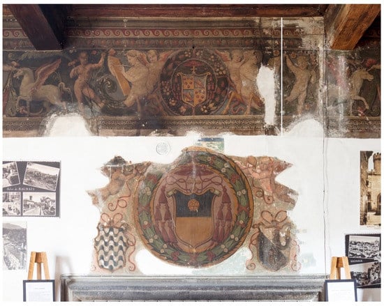
Figure 1.
The wall paintings before the restoration (photograph by Gaetano Alfano).
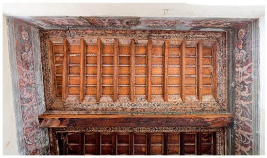
Figure 2.
Ceiling of the Sala de Rossi, adjacent to that of Sala Riario.
The paintings in the Sala Riario, the second most important in Palazzo Gallo and the most richly decorated, consist of an antique-style frieze and the great coat of arms of Raffaele Riario, flanked by that of the Gallo’s family and the civic coat of arms of Bagnaia, just above the fireplace.
The coats of arms are carried by putti through ribbons, and the decoration, in its simplicity, is rich and varied. In 1576, Sulpizio Gallo, Giuliano’s heir, sold the building to the Municipality—the building was described as “a complex of over twenty rooms (…) with beautiful paintings in the reception rooms, a huge fireplace in the living room and a delightful porch on the square” ([2], p. 61). The importance of Bagnaia is given by the abundance and refinement of the artwork created there. The wall paintings were the focus of ancient restorations that could be considered renovations, identified in several points of the Sala Riario. On the portion being restored here, these ancient interventions (presumably carried out between the end of the 16th century and the beginning of the 17th century) run through the areas of the painting between the flue of the fireplace and the surrounding solid masonry. The remaking is carried out by an expert hand who engages in a mural painting with the necessary iconographic analogies but with different executive procedures. The style inevitably deviates from the original painting, showing characters closer to a more mature mannerism (Figure 3 and Figure 4).
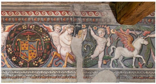
Figure 3.
Detail of the frieze decoration on the south wall after restoration, original painting.
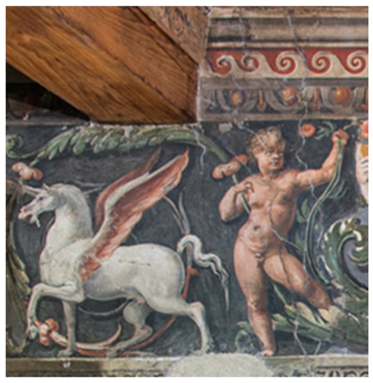
Figure 4.
Detail of the ancient restoration after the present intervention.
Around the 1620s, the Sala Riario was probably intended and used as a religious environment. This hypothesis is based on the presence of two mural paintings on the short walls of the room of a sacred nature: the Crucifixion, and the Virgin between Saint Sebastiano and Saint Millerio. After that date, there is not much information on the history of the Palace, but it is certain that the rooms were used as private residences after 1943 for those displaced by the war, and consequently adapted by adding balconies, partitions, and installing a stove. Furthermore, during this period, many superimposed layers were added to cover the original paintings in the area above the fireplace, increasingly heavy so as to hide them largely. Concerning the preparatory layers, the antiquity of the Palace and its conservation events led to a massive presence of adhesion defects and deep cracks, provoking a deformation of the plaster in the upper right area mainly due to the presence of the fireplace and the flue. Finally, the use of the fireplace and the kitchen produced carbon black deposits on the painting surface which, together with an altered protective applied in the past, made it difficult to read the artwork, as visible in Figure 1. Civil use has led to a significant deterioration of the conservation conditions. The tampering of the room, in fact, has been added to operations such as the posting of electric wires with nails. In addition to having caused or aggravated local detachments of the preparatory layers, these also resulted in minor cracks. The recently ended restoration of the paintings was a very important chance to investigate the materials and the employed realization techniques, and to closely observe all the details, giving new light on these not-still-attributed wall paintings. The restoration allowed for making a comparison with other paintings, to suggest possible dating (within 1530), and possible artists’ workshop for the realization of the artworks ([1], pp. 71–79). The greatest similarity was found between the decorations on the architectural partitions in the church of Santa Maria delle Fortezze (Viterbo, Italy), a place also linked in the foundation (1514) to Ottaviano Riario, Raffaele’s favorite nephew ([3], pp. 9–12) (see Figure 5).
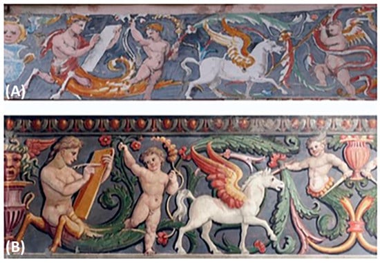
Figure 5.
Frieze in the church of S. Maria della Fortezze (Viterbo) (A) compared with that of the Sala Riario of Palazzo Gallo (B).
The frieze of S. Maria delle Fortezze shows an iconographic composition very close to that of Bagnaia room. Except for a few details, the figure of the putto is central and can also be found slavishly in another panel of the church’s paintings. Some differences can be noticed in both the sizes and orientation of the figures. Consequently, a very similar executive technique is deduced, in particular concerning the use of cardboard and on the way the pictorial layers are drafted, using very similar colors.
Furthermore, owing to the restoration, it was possible to compare faithfully the paintings above the fireplace with those of the frieze, allowing for a clearer juxtaposition of common iconographic features between those in the church of S. Maria delle Fortezze and those found in other contemporary paintings in the area of Bagnaia.
The study of the materials of the paintings and of their conservation status was supported by the diagnostics that combined innovative imaging techniques, i.e., hypercolorimetric multispectral imaging (HMI) and pulse-compression thermography (PuCT), used for the first time together to investigate a wall painting on-site, with traditional analysis such as passive thermography, X-ray fluorescence spectroscopy, microstratigraphic analysis, and Fourier transform infrared (FTIR) spectroscopy. Clearly, a careful observation of the paintings during the restoration was fundamental to study the execution technique and to address the diagnostics and the laboratory analysis [4].
The investigation of wall paintings might be carried out by several techniques on-site and in the laboratory using microsamples. In the most recent approaches applied in this framework during restoration activities, it has become praxis in our research group to start from noninvasive imaging techniques associated with portable spectroscopic methods, so as to gather as much information as possible on the materials and on the conservation status, and to address possible sampling of microfragments or powders from the artwork for laboratory investigation [4,5]. We hope that this approach would become increasingly more consolidated in the field of cultural heritage investigation and pivotal if restoration is foreseen. However, the use of imaging techniques applied to large wall paintings is not as widespread as for easel paintings, probably due to the dimensions and sometimes to the difficulties linked to the necessity of scaffoldings, or other technical solutions, to reach the whole painting. Note that this is demonstrated by the few papers available in the literature related to the use of multispectral and hyperspectral imaging techniques, applied to a limited portion of wall painting only [6,7,8]. Recent review papers showed that the most used techniques for the analysis of wall paintings are those operating at microscale level such as XRF, SEM-EDS, GC-MS, FTIR, micro-Raman, etc. [9,10,11]. In no cases are multispectral imaging and active thermography techniques reported in the reviews, demonstrating the innovative approach used in this work.
The use of innovative imaging techniques yields several advantages on different aspects: (i) in terms of information on the materials and procedures for creating the wall paintings; (ii) in terms of time needed to obtain useful data during the restoration steps; (iii) in terms of costs related to the limited amount of sampling for traditional spectroscopic and chemical analysis.
Concerning point (i), the information gathered from imaging techniques concern the entire surface and interface of the artwork, not single points or areas. In the case of innovative techniques such as HMI and PuCT, the combination of the image acquisition with powerful calibration and processing software supplies a great amount of data and information, which could be used to know the state of conservation, but also to choose and select points where further analyses are deemed necessary [12].
Concerning point (ii), these techniques gives immediate response, which is a highly important aspect to consider in a restoration project.
Concerning (iii), the use of imaging techniques as first step in the diagnostics of a wall painting allows economic resources available for the restoration to be saved. In fact, these techniques can supply several useful data for the intervention to start and can help addressing possible further analysis, thus limiting the extent of further processing. In situ spectroscopic analysis and laboratory methods are expensive and time-consuming, especially in the case of large wall paintings where the surfaces to be examined are relevant and would require several samples to be examined. This can be avoided with a preliminary investigation through innovative multispectral and imaging methods.
2. The Execution Technique of the Wall Paintings
Two different wall supports were identified: a brick one for the flue of the fireplace, and a solid wall based on peperino stones, the typical stones used in Bagnaia. Different preparatory layers were also distinguished. A rough layer was found only on the wall made of peperino blocks. The arriccio of the preparatory layers appears slightly different in the mixture and in the thickness in relation to the area: that of the flue appears as being finer and thinner than that of the solid wall. On this layer, a sinopia was identified, which divides the frieze and the paintings of the fireplace with two parallel lines. In the area subject to restoration, four large plaster areas were identified and carried out at the same time as the organization of the spaces. These were divided using direct incisions, and tracing a beading of thread to obtain the twelve bands of the frieze. By centering the painting through the crossing of cords between the fireplace frame and the internal side of the beams, the median and the encumbrance of the garlands with the heraldic shields were identified. An elaborate system of transferring the drawing was used, using cardboard to draw the dust and the indirect incisions. With the traced lines, the colors were applied to create the paintings. Specifically, the backgrounds were firstly applied: red in the central scene of the frieze and white on the remaining surface. The blue background was applied to the red background so as to complete the flesh tones of the figures, the winged horses, and the spirals.
The paintings were made by a fresco technique with some finishing applied by lime, with the probable addition of an organic binder. Finally, the bands of the frames were probably built by superimposing several stencils to complete the decoration.
3. Materials and Methods
The wall paintings were investigated before and after the various restoration steps. The adopted approach was aimed at obtaining detailed information through noninvasive and nondestructive analysis on the entire surfaces, this approach is fundamental when examining artwork [13,14]. For this reason, multispectral imaging and thermography were selected to investigate both the painting layers and the ground. Multispectral imaging was performed by the HMI technique based on a modified digital camera, a Nikon D800 (full range from 300 to 1000 nm), equipped with a 17–35 zoom lens. The HMI apparatus is composed also of three filters, namely A (UV-Vis), B (Vis-NIR), and UV-IR cut, the latter used in combination with filter A for obtaining the ultraviolet fluorescence (UVF) acquisition [15]. The acquisition set was completed with standard white patches and a color-checker positioned in the camera field of view, and an illuminating system based on modified flashes, as described in [16]. Two CR230B-HP 10W UV LED projectors, peak emission at 365 nm, were used to obtain the UVF images. The acquired images are then calibrated through SpectraPick® software (V. 1.1) that produces seven high resolution spectral images in .tiff format from UV to NIR at 350, 450, 550, 650, 750, 850 and 950 nm, and the RGB image. The entire folder is then loaded into the processing software Pickviewer® (V. 1.1), which provides several diagnostic imaging and statistical tools to produce various outputs that are useful for the restoration.
The PuCT setup is composed of a portable TiePie HS5 Handyscope (TiePie Engineering, Sneek, The Netherlands) for both generating the coded waveform and grabbing the thermograms from an FLIR T660 thermal camera. In particular, a pseudonoise code modulates the ON/OFF state of a TDK Lambda GEN 750 W (TDK, Tokyo, Japan) power supply. In this way, ten halogen lamps have been driven to emit light following the coded sequence for an overall heating stimulus duration of 440 s.
The heating system and the thermal camera were placed on the same side at about 600 mm from the inspected surface, so that the resulting inspection was performed in reflection mode. A laptop was used to store the data using an in-house routine developed in LabVIEW. The results are reported using a gray colormap, where the darker pixels are related to lower temperature/emissivity. Further details of the experimental setup and of the technique’s principles are reported in [17,18,19].
X-ray fluorescence analysis was performed by Dr. Claudio Falcucci of the MIDA society, on twenty-two points with a portable apparatus equipped with an Au anode operating at 40 kV; acquisition time was about 50 s.
FTIR spectroscopy was performed on microsamples with a Nicolet Avatar 360 instrument equipped (GMI Inc., Ramsey, MN, USA) with a diffuse reflectance accessory. The spectrometer operates in the 400–4000 cm−1 region with a resolution of 4 cm−1. Potassium bromide (KBr) was used as background.
Cross-sections were prepared by embedding the microsamples in a polyester resin (X60 NXMET), suitably cut and polished. The prepared cross-section was observed with a polarized-light microscope Zeiss Axioskop equipped (Carl Zeiss, Heidenheim an der Brenz, Germany) with reflected, transmitted, and UV lighting. The images were acquired by using a Zeiss AxioCam NRc camera (Carl Zeiss, Heidenheim an der Brenz, Germany) and processed with AxioVision software (Carl Zeiss, Heidenheim an der Brenz, Germany) to trace the sequence of stratigraphic layers and to recognize possible traces of organic material or characteristic fluorescence of materials by means of observation with ultraviolet radiation.
4. Results and Discussion
The first relevant result was obtained with the UVF acquisition performed before the painting cleaning. In fact, UVF images give fundamental information about the conservation state of the painting surfaces, on the presence of retouched areas and grouting, etc. [13]. By observing the UVF images of the two paintings, the first evidence is related to an intense yellow response in the coat of arms of Bagnaia and Gallo’s family that can be associated with a repainting, see Figure 6. The areas characterized by a blue fluorescence correspond to grouting made to compensate for the lack of painted plaster. UVF also shows a general dark brown response of the surface due to a superimposed layer over the original paintings.
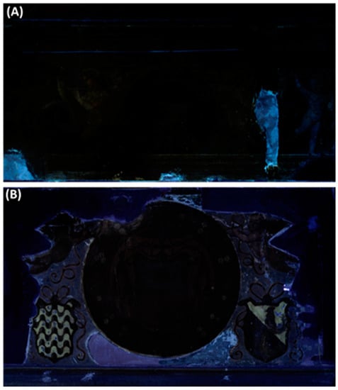
Figure 6.
UVF photomosaic of the wall paintings before the restoration. The upper frieze (A) and the Riario coat of arms (B).
UVF was repeated at the end of the intervention, when the cleaning operation for removing the different scialbo (slaked-lime) layers, applied in previous restoration and remaking on the wall, was almost completed. At this stage, UVF highlighted a general increase in the painting visibility due to the removal of the brown layer, especially in correspondence with the background in the lower painting, see Figure 7.
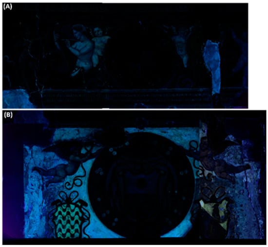
Figure 7.
UVF photomosaic of the wall paintings after the almost complete removal of the scialbo. The upper frieze (A) and the Riario coat of arms (B).
This part, in fact, acquired both a blue and pale-yellow fluorescence probably associated with organic binders. In the last step of the cleaning, UVF was pivotal for addressing the final interventions on the surfaces. In the same stage of the restoration, HMI was also applied to investigate the material distribution, to map the scialbo layer still present on the surface, and to check the effectiveness of the cleaning intervention. Specifically, this last goal was reached by applying the color measurement algorithm in PickViewer® (V. 1.1) software, proprietary software of the HMI equipment used here, to uncleaned and cleaned areas for comparing the chromatic coordinates. First of all, PickViewer® (V. 1.1) was used for obtaining the infrared and ultraviolet false color (IRFC and UVFC) images, see Figure 8 and Figure 9, of the paintings that allowed for hypothesizing the composition of some pigments: blue smalt that appears as vine color in IRFC (Figure 8B), vermilion/cinnabar that becomes yellow, and different greens (earths and emerald green) that exhibited different response in false colors, especially in UVFC (Figure 9C) as better visible in a detail of the frieze, see Figure 10.
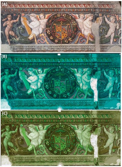
Figure 8.
RGB (A), IRFC (B), and UVFC (C) images of the frieze after the complete removal of the scialbo, obtained with PickViewer®.
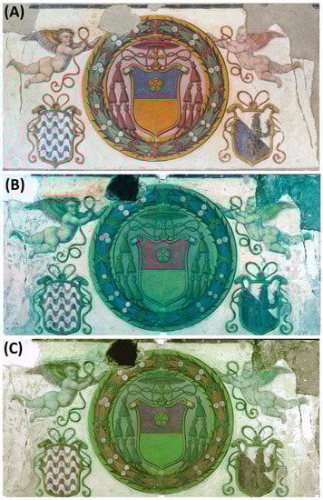
Figure 9.
RGB (A), IRFC (B), and UVFC (C) images of the Riario coat of arms after the complete removal of the scialbo, obtained with PickViewer®.
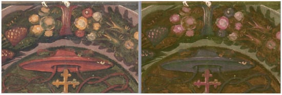
Figure 10.
Detail of the frieze with the Cardinal’s hat. RGB image on the left and UVFC on the right, as obtained by PickViewer®.
Green earth has a violet color in UVFC, whereas the emerald/Scheele’s green appears dark brown (see the background of the coat of arms behind the red plumes). This appearance has been reported by other researchers as well [20].
PickViewer® was used also for mapping different areas on the paintings, such as the blue color (due to smalt) in the Riario coat of arms and the yellow (characterized as ochre) in the frieze, by applying the spectral similarity algorithm (see Figure 11).
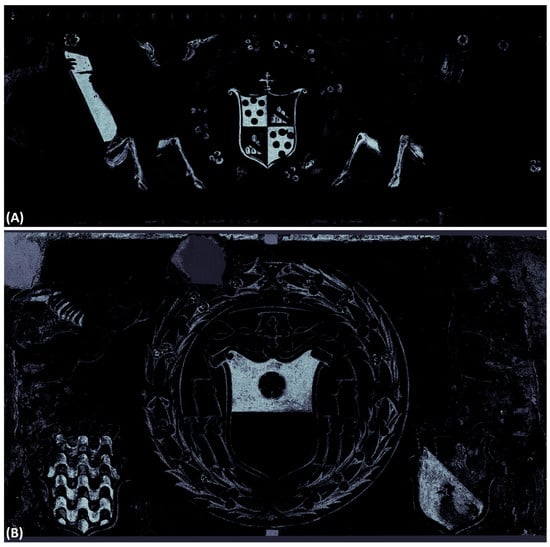
Figure 11.
Result of the spectral similarity algorithm applied to the yellow ochre in the frieze (A) and in the blue smalt in the Riario coat of arms (B).
This algorithm allows for choosing a point in the RGB image and it compares the spectral reflectance at this point with those of the entire image. The result, as shown Figure 11, is a black and white image where the areas having the same spectral and chromatic characteristics of the selected point appear white. In this way, it is possible to map the areas of the painting with the same spectral and chromatic characteristics.
Pointwise XRF analysis was applied to confirm the hypotheses about the possible composition of pigments on the base of the UVF response and for the obtained false colors. In the areas exhibiting a yellow fluorescence under UV, XRF analysis revealed the presence of zinc, which confirms the use of zinc white (ZnO), a nonoriginal pigment ([1], pp. 103–105) (Table 1, Point 5).

Table 1.
Results of the XRF analysis on some of the areas inspected. A small photograph of the area is reported for clarity, together with a pinpoint for easy reading.
In Table 1, five out of twenty-two measurements are reported; the other results are presented and discussed in the text.
In the areas colored in blue, the presence of cobalt associated with arsenic allows blue smalt to be identified, an ancient pigment widely used in wall painting, especially in the Renaissance period [21]. The red colors were obtained both with iron-based pigments and vermilion/cinnabar (mercury sulfide), as shown for Point 2. In the areas painted in green, XRF revealed the presence of iron, copper, and arsenic. The presence of Fe can be associated with green earths, widely used since antiquity as preferred green pigments for wall paintings [22]. In the other case, Cu and As indicate the use of emerald or Scheele green, the first being a copper acetoarsenite, and the other a copper arsenite [23]. Scheele’s green was the first synthetic pigment based on Cu and As, introduced in 1778, but its use as a pigment is little documented [22]. Emerald green was obtained in the laboratory during experiments aimed to improve Scheele’s Green. It was used starting from the beginning of 1800 and was available until recently, when it was removed from the artists’ pigment palette due to its high toxicity [22]. These green pigments are clearly nonoriginal and may be referred to a restoration intervention.
Concerning the other points examined through XRF spectroscopy, in the yellow zone the presence of iron indicates the use of ochre; in the flesh tone, only Ca and Fe were detected (use of lime white mixed with red ochre).
The presence of organic materials has been confirmed by FTIR spectroscopy performed on two microsamples of the paintings. FTIR showed traces of proteins (characteristic bands at 1644, 1563, and 1448 cm−1, partially overlapped with those of gypsum and calcium carbonate), lipidic substances (oils and/or wax with the characteristic strong signatures at 2919 and 2853 cm−1 due to C-H stretching of long-chain molecules) and abundant gypsum (bands at cm−1: 3531, 3400, 2240, 2317, 1623, 1151, 1120, 672, 603, and 467), probably used in the grouting, see Figure 12 [24].
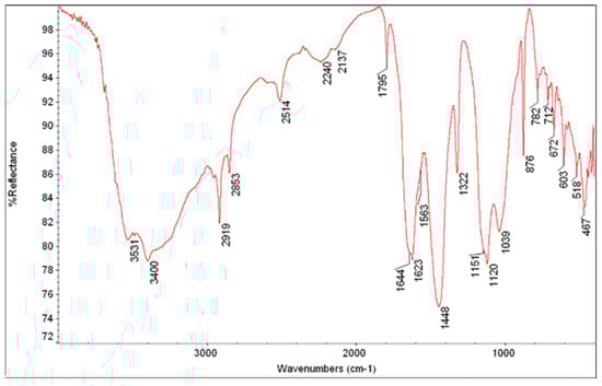
Figure 12.
Spectrum of a sample taken from the upper frieze in correspondence with a brown surface layer.
The presence of oxalates further confirms the organic materials on the surface of the paintings (characteristic bands at cm−1: 1623—in common with gypsum—1322 and 782).
The microsamples were also used for obtaining cross-sections, useful for obtaining information about the stratigraphy of the wall paintings.
In Figure 13, the cross-sections under reflected light and ultraviolet fluorescence of three microsamples are shown. The first sample comes from the light blue background of the frieze: over the dark mortar (made of lime and pozzolanic materials) a red layer was applied as preparatory ground for blue smalt, whose pale blue grains are well visible in the cross-section. Because smalt is a glass, the grains are transparent and characterized by conchoidal fractures with typical thin, sharp edges of glass splinters. The second sample examined as a cross-section is taken from the background of the Riario’s coat of arms that appeared clearly repainted. In the cross section shown in Figure 13B,C, the presence of a white layer is evident, a sort of scialbo, which is in turn covered by thick yellow layers (probably repainting), the surface appearing greenish in color. The third sample comes from the Riario’s coat of arms corresponding to the right foot of the winged putto on the right of the painting area. In this sample, it can also be observed the white scialbo covered by an evidently new layer of repainting.
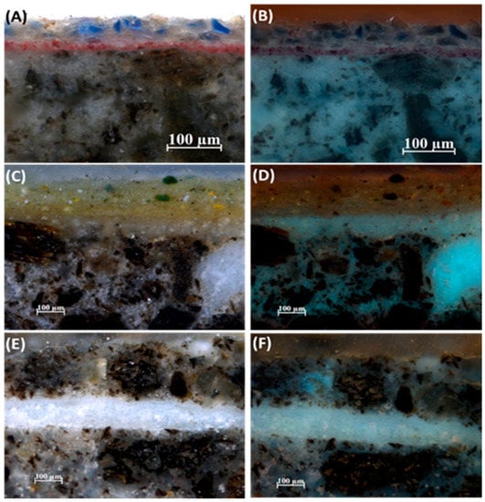
Figure 13.
Microphotographs of cross-sections from the three samples. (A,C,E) reflected light; (B,D,F) ultraviolet fluorescence.
5. The Restoration
The restoration was aimed at first solving the most urgent critical issues of the paintings: the serious adhesion defects in the frieze with the deformation of the plaster, and the removal of the scialbo layer applied around the paintings above the fireplace, visible in the PuCT emissivity images, see Figure 14.
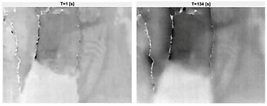
Figure 14.
Emissivity images at 1 s and 134 s of a portion of the frieze obtained using PuCT before consolidation, showing deep cracking, detachment, and deformations.
After a color-fastness test, the cleaning was carried out. The detachments of the pictorial layer were treated with a microacrylic resin (K52) in a solution of water and isopropyl alcohol (1:1) at 20%, when deemed necessary. In this way, the removal of the superimposed layers was performed in safety, fixing the most critical areas between the different layers. The same 10% resin was used to consolidate the vermilion/cinnabar, which had areas of slight surface decohesion. Cleaning was carried out with water and 2% Tween 20 surfactant supported by Japanese paper (17 g). Repeating the operation as necessary, this allowed the nature and quantity of the superimposed layers to be fully examined. A total of 13 recent layers were found, sometimes discontinuous, then mechanically removed with a scalpel. The scialbo removal (descialbo) was performed step-by-step with the use of stratigraphic test areas, small descialbo zones studies on similar decorations for epoch, and context and imaging diagnostics, see Figure 15.
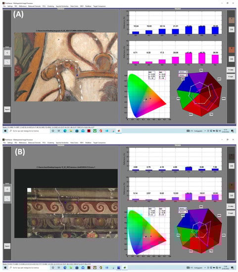
Figure 15.
Chromatic and reflectance comparison between cleaned and uncleaned areas on the yellow color in the Riario coat of arms (A) and on the red color in the frieze (B) showing a clear increase in saturation due to the removal of the surface soot layer.
This operation allows details hidden for a long time to be finally observed, and to fully appreciate the composition. The details that emerged from the descialbo allowed the paintings to be strongly contextualized in the Bagnaia and Viterbo context of the workshop operating in the first decades of the 16th century. Following meticulous stratigraphic analysis, the light background was proposed only after more extensive descialbo test areas. The decision to end at this layer was also undertaken with the support of historical–artistic research, which has identified paintings of this kind above various fireplaces or in salons diffused in various Italian contexts between the end of the 15th and 16th centuries.
In Figure 15, two examples of chromatic variation due to the surface blackish layer are shown: one for the yellow in the Riario coat of arms (Figure 15A) and the other for the red in the frieze decoration (Figure 15B). Yellow underwent a significant increase in saturation with values of a* and b* chromatic coordinates changing from 6.05 and 11.49 to 22.47 and 26.10, respectively; the values were calculated by the specific algorithm in PickViewer® and reported in the bar under the image in the graphical user interface of the software. The values before cleaning are indicated as L1, a1, and b1, and those after cleaning as L2, a2, and b2. The color measurements are possible owing to the procedure used by the HMI system based on calibration of the acquired images with the use of reference white patches and the colorchecker. This procedure allowed for obtaining calibrated images centered at specific wavelengths whose reflectance and color data are characterized by high precision (verified by Profilocolore by using a laboratory spectroradiometer) [25,26].
In Figure 15, the CIE1931 xyY color coordinates are also reported together with the position of the two selected points (the blue is referred to as the uncleaned area and the pink as the cleaned one). The values of reflectance are reported in the upper right side of the graphic user interface of PickViewer® by using blue (uncleaned area) and pink bars (cleaned). The main changes occur at 650 nm (increase in reflectance) and in the IR, further indicating an increase in yellow saturation.
Additionally, in the case of red color in the frieze, the chromatic coordinates underwent a significant increase: L* changed from 33.96 to 39.60, a* from 5.40 to 25.35, and b* from 4.52 to 16.27, indicating a clear increase in saturation.
At the end of the removal intervention, the pictorial layer with the original colors and new details hidden by time had been restored.
The consolidation of the preparatory layers was important to recover the artwork, and it was studied carefully by thermography. The adhesion between the preparatory layers was restored through injections of various hydraulic mortars (from Ledan series), with special attention to the deformed area. The major gaps were fixed here through the creation of pins made of Ledan Adesiva. After setting the product, a filling with a mixture of Ledan Ristat B and Ledan Ristat Extra was applied. This mixture has excellent adhesive, filling, and light characteristics. In the case of well-circumscribed detachments, the Ledan TB1 was used by monitoring the consolidant penetration with the thermal camera to verify the effectiveness of the consolidation, see Figure 16.
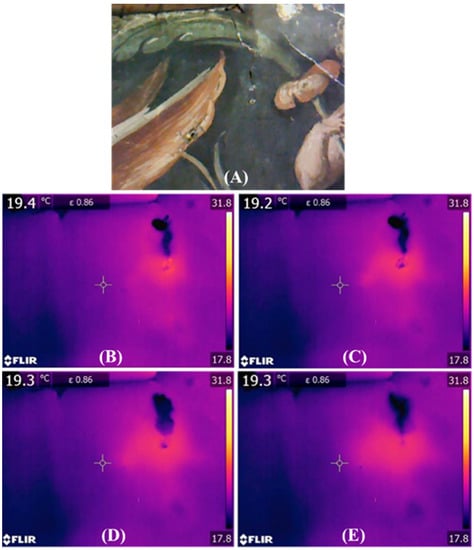
Figure 16.
RGB image (A) and timeline of thermograms (B–E) of the area investigated with passive thermography where the (warmer) Ledan is visible spreading in the preparatory layers after injection by a syringe. Image (E) has been taken after 5 min.
It is worth noting that the quantities of hydraulic mortar used to compensate for the various adhesion defects are 5 kg of Ledan TB1, 1.4 kg of Ledan Ristat B, 1.4 kg of Ledan Ristat Extra, and 100 g of Ledan Adesiva, which suggest the large extent of the detachments and the consolidation work.
The emissivity images over time obtained with PuCT show some anomalies (highlighted by yellow circles): the appearance of the crack after having been infiltrated by water and alcohol, and how the dark areas are less intense after the injection (Figure 17) [27].
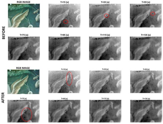
Figure 17.
Sequence of emissivity images at different times obtained through PuCT before and after Ledan injection.
After this consolidation work on the painting ground layers, the previous grouting materials, unsuitable and incompatible with the original materials, were mechanically removed. The brick support was restored in correspondence with the through-hole of the kitchen, and the large grouting was subsequently treated with a sublevel approach. The painting-level grouting was mimetically reintegrated, while all the abrasions, lacunae, whitening, and lack of patina were treated with lowering of tone. With an overall chromatic balance, the painting is once again legible, vivid in the colors, fully showing the compositional rhythm and the refined pictorial taste of the workshop of the artists working in Palazzo Gallo, see Figure 18.
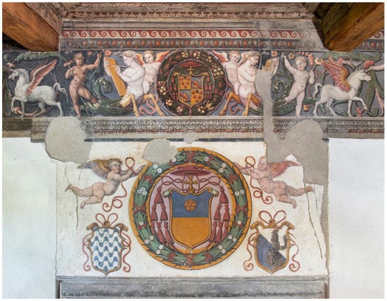
Figure 18.
The wall paintings after the restoration. Photograph by Gaetano Alfano.
6. Conclusions
A series of noninvasive and microinvasive techniques were applied here for aiding restoration of a wall painting at Palazzo Gallo in Bagnaia, Viterbo, Italy. The different information collected by using this multi-technique approach played a key role in aiding the restorers toward the faithful restoration of such an artwork. This work aimed at showing how each technique gave a range of different information to the restorers, and the actions undertaken in light of the acquired knowledge of the artwork. The documentation of the entire diagnostic procedure, i.e., starting from establishing the historical background until the final restoration, passing through the extended range of investigation techniques used, stands out as a rare example in the scientific literature.
Author Contributions
Conceptualization, C.P., R.V., V.V., P.P. and M.R.; methodology, C.P., R.V., V.V., P.P., M.R. and S.L.; software, C.P., L.L., R.Z., M.R. and S.L.; formal analysis, R.V., L.L. and S.C.; investigation, C.P., R.V., V.V., P.P., M.R., S.L.; S.C., R.Z. and L.L., data curation, R.V., R.Z., S.C. and L.L.; writing—original draft preparation, R.V., C.P., S.L. and M.R.; writing—review and editing, all authors. All authors have read and agreed to the published version of the manuscript.
Funding
This research received no external funding.
Data Availability Statement
Data is available upon reasonable request.
Conflicts of Interest
The authors declare no conflict of interest.
References
- Vettraino, R. Il restauro del fregio a grottesca e dei dipinti sulla parete del camino nella Sala Riario di Palazzo Gallo a Bagnaia. Master’s Thesis, University of Tuscia, Viterbo, Italy, 2022. [Google Scholar]
- Frittelli, V. (Ed.) Bagnaia Cronache d’una Terra del Patrimonio; Bagnaia: Viterbo, Italy, 1977. [Google Scholar]
- Padelli, I. La Chiesa ed il Convento di S. Maria delle Fortezze in Viterbo: Documenti e Ipotesi Attributive. Master Thesis, University of Tuscia, Viterbo, Italy, 2003. [Google Scholar]
- Groppi, F.; Vigliotti, D.; Lanteri, L.; Agresti, G.; Casoli, A.; Laureti, S.; Ricci, M.; Pelosi, C. Advanced documentation methodologies combined with multi-analytical approach for the preservation and restoration of 18th century architectural decorative elements at Palazzo Nuzzi in Orte (Central Italy). Int. J. Conserv. Sci. 2021, 12, 921–934. [Google Scholar]
- Lanteri, L.; Agresti, G.; Vaccarella, C.; Lucchetti, L.; Noto, M.; Pelosi, C. The mediaeval crypt of Saint Sepulchre in Acquapendente (Italy). Study and photogrammetric documentation of the painted surfaces. Eur. J. Sci. Theol. 2020, 16, 177–185. [Google Scholar]
- Legnaioli, S.; Lorenzetti, G.; Cavalcanti, G.H.; Grifoni, E.; Marras, L.; Tonazzini, A.; Salerno, E.; Pallecchi, P.; Giachi, G.; Palleschi, V. Recovery of archaeological wall paintings using novel multispectral imaging approaches. Herit. Sci. 2013, 1, 33. [Google Scholar] [CrossRef]
- Cucci, C.; Picollo, M.; Chiarantini, L.; Uda, G.; Fiori, L.; De Nigris, B.; Osanna, M. Remote-sensing hyperspectral imaging for applications in archaeological areas: Non-invasive investigations on wall paintings and on mural inscriptions in the Pompeii site. Microchem. J. 2020, 158, 105082. [Google Scholar] [CrossRef]
- Rahrig, M.; Ángel, M.; Cortell, H.; Lerma, J.L. Multiband Photogrammetry and Hybrid Image Analysis for the Investigation of a Wall Painting by Paolo de San Leocadio and Francesco Pagano in the Cathedral of Valencia. Sensors 2023, 23, 2301. [Google Scholar] [CrossRef] [PubMed]
- Costantini, I.; Castro, K.; Madariaga, J.M. Portable and laboratory analytical instruments for the study of materials, techniques and environmental impacts in mediaeval mural paintings. Anal. Methods 2018, 10, 4854–4870. [Google Scholar] [CrossRef]
- Bracci, S.; Bartolozzi, G. Wall paintings—diagnostic and archaeometric studies. Phys. Sci. Rev. 2019, 4, 20180013. [Google Scholar] [CrossRef]
- Zhu, Z.; Yao, X.; Qin, Y.; Lu, Z.; Ma, Q.; Zhao, X.; Liu, L. Visualization and mapping of literature on the scientific analysis of wall paintings: A bibliometric analysis from 2011 to 2021. Herit. Sci. 2022, 10, 1–13. [Google Scholar] [CrossRef] [PubMed]
- Colantonio, C.; Pelosi, C.; Calabrò, G.; Spizzichino, V.; Partenzi, I.; Lanteri, L. Scientific Investigation of Contemporary Pastel Painting by Roberto Sebastian Matta: Characterization of Original Materials through Multispectral Imaging and Spectroscopic Techniques. Heritage 2023, 6, 2541–2558. [Google Scholar] [CrossRef]
- Miliani, C.; Rosi, F.; Brunetti, B.G.; Sgamellotti, A. In Situ Noninvasive Study of Artworks: The MOLAB Multitechnique Approach. Acc. Chem. Res. 2010, 43, 728–738. [Google Scholar] [CrossRef] [PubMed]
- Thickett, D.; Cheung, C.S.; Liang, H.; Twydle, J.; Maev, R.G.; Gavrilov, D. Using non-invasive non-destructive techniques to monitor cultural heritage objects. Insight Non Destr. Test. Cond. Monit. 2017, 59, 230–234. [Google Scholar] [CrossRef]
- Lanteri, L.; Pelosi, C. 2D and 3D ultraviolet fluorescence applications on cultural heritage paintings and objects through a low-cost approach for diagnostics and documentation. In Proceedings of the SPIE 11784, Optics for Arts, Architecture, and Archaeology VIII, Munich, Germany, 8 July 2021; pp. 1–10. [Google Scholar]
- Laureti, S.; Colantonio, C.; Burrascano, P.; Melis, M.; Calabrò, G.; Malekmohammadi, H.; Sfarra, S.; Ricci, M.; Pelosi, C. Development of integrated innovative techniques for the examination of paintings: The case studies of The Resurrection of Christ attributed to Andrea Mantegna and the Crucifixion of Viterbo attributed to Michelangelo’s workshop. J. Cult. Herit. 2019, 40, 1–16. [Google Scholar] [CrossRef]
- Laureti, S.; Sfarra, S.; Malekmohammadi, H.; Burrascano, P.; Hutchins, D.A.; Senni, L.; Silipigni, G.; Maldague, X.P.V.; Ricci, M. The use of pulse-compression thermography for detecting defects in paintings. NDT E Int. 2018, 98, 147–154. [Google Scholar] [CrossRef]
- Sfarra, S.; Laureti, S.; Gargiulo, G.; Malekmohammadi, H.; Sangiovanni, M.A.; La Russa, M.; Burrascano, P.; Ricci, M. Low Thermal Conductivity Materials and Very Low Heat Power: A Demanding Challenge in the Detection of Flaws in Multi-Layer Wooden Cultural Heritage Objects Solved by Pulse-Compression Thermography Technique. Appl. Sci. 2020, 10, 4233. [Google Scholar] [CrossRef]
- Ricci, M.; Laureti, S.; Malekmohammadi, H.; Sfarra, S.; Lanteri, L.; Colantonio, C.; Calabrò, G.; Pelosi, C. Surface and Interface Investigation of a 15th Century Wall Painting Using Multispectral Imaging and Pulse-Compression Infrared Thermography. Coatings 2021, 11, 546. [Google Scholar] [CrossRef]
- Boust, C.; Wohlgelmuth, A. Database: Pigments under UV and IR Radiations, in Scientific Imaging for Cultural Heritage/Images Scientifiques Pour le Patrimoine. Available online: https://copa.hypotheses.org/552 (accessed on 1 January 2023).
- Mühlethaler, B.; Thissen, J. Smalt. In Artists’ Pigments a Handbook of Their History and Characteristics; Roy, A., Ed.; National Gallery of Art: Washington, WA, USA, 1993; Volume 2, pp. 113–130. [Google Scholar]
- Grissom, C.A. Green Earth. In Artists’ Pigments a Handbook of Their History and Characteristics; Feller, R.L., Ed.; National Gallery of Art: Washington, WA, USA, 1986; Volume 1, pp. 141–167. [Google Scholar]
- Fiedler, I.; Bayard, M. .Emerald Green and Scheele’s Green. In Artists’ Pigments a Handbook of Their History and Characteristics; West Fitzhugh, E., Ed.; National Gallery of Art: Washington, WA, USA, 1997; Volume 3, pp. 219–271. [Google Scholar]
- Derrick, M.R.; Stulik, D.; Landry, J.M. Infrared Spectroscopy in Conservation Science; The Getty Conservation Institute: Los Angeles, CA, USA, 1999; pp. 179–200. [Google Scholar]
- Melis, M.; Miccoli, M.; Quarta, D. Multispectral Hypercolorimetry and automatic guided pigment identification: Some masterpieces case studies. In Proceedings of the SPIE 8790, Optics for Arts, Architecture, and Archaeology IV, Munich, Germany, 13–16 May 2013; pp. 1–14. [Google Scholar]
- Melis, M.; Miccoli, M. Trasformazione Evoluzionistica di una Fotocamera Reflex Digitale in un Sofisticato Strumento per Misure Fotometriche e Colorimetriche. In Colore e Colorimetria Contributi Multidisciplinari; Maggioli Editore: Santarcangelo di Romagna, Italy, 2013; pp. 28–38. [Google Scholar]
- Cadelano, G.; Bison, P.; Bortolin, A.; Ferrarini, G.; Peron, F.; Girotto, M.; Volinia, M. Monitoring of historical frescoes by timed infrared imaging analysis. Opto-Electron. Rev. 2015, 23, 102–108. [Google Scholar] [CrossRef]


Disclaimer/Publisher’s Note: The statements, opinions and data contained in all publications are solely those of the individual author(s) and contributor(s) and not of MDPI and/or the editor(s). MDPI and/or the editor(s) disclaim responsibility for any injury to people or property resulting from any ideas, methods, instructions or products referred to in the content. |
© 2023 by the authors. Licensee MDPI, Basel, Switzerland. This article is an open access article distributed under the terms and conditions of the Creative Commons Attribution (CC BY) license (https://creativecommons.org/licenses/by/4.0/).