Review

Research Progress on Proton Irradiation Damage and Irradiation Resistance of Austenitic Stainless Steel

School of Materials and Chemistry, University of Shanghai for Science and Technology, Shanghai 200093, China
*
Author to whom correspondence should be addressed.
Metals 2026, 16(4), 451; https://doi.org/10.3390/met16040451
Submission received: 2 March 2026 / Revised: 25 March 2026 / Accepted: 30 March 2026 / Published: 21 April 2026

Abstract

Nuclear energy is a clean and efficient energy source crucial for the future energy supply. The harsh conditions in reactors, including high temperature, high pressure, and intense neutron irradiation, cause structural materials to accumulate irradiation damage, leading to performance degradation. Austenitic stainless steel, due to its superior mechanical properties, irradiation resistance, and corrosion resistance, has been extensively utilized as a core structural material in light water reactors and emerged as a candidate material for Generation IV nuclear reactors. Therefore, understanding irradiation damage and macroscopic properties evolution in austenitic stainless steels is critical for enhancing the safety and long-term service life of reactor core materials. This review began by elucidating the application of charged particles in irradiation studies, emphasizing the prevailing substitution of neutron irradiation with proton irradiation experiments in current studies. Subsequently, the work systematically synthesized irradiation damages and their consequential impacts on macroscopic properties. Finally, it consolidated the progress and provided prospects for research on improving the resistance of austenitic stainless steel to irradiation-induced segregation, irradiation hardening, irradiation swelling, and irradiation-corrosion synergies.

1. Introduction

Currently, nuclear power generates approximately 13% of the world’s electricity [1] and has become a vital source of reliable base-load power [2], with applications across a wide array of fields, including industry, agriculture, and medicine [3,4,5,6]. As a new type of energy that is green, clean, and environmentally friendly—and with high energy density—nuclear energy has attracted widespread attention from scholars around the world. However, to achieve the safe and efficient utilization of nuclear energy, materials must meet the extremely demanding requirements imposed by harsh operating conditions, such as those in a pressurized water reactor where the core operating temperature exceeds typical service temperatures, while structural components are simultaneously subjected to long-term high-velocity coolant flow (which is strongly oxidizing and corrosive) and high-dose neutron irradiation [7]. These severe conditions require that candidate materials for nuclear reactors possess excellent resistance to corrosion, high-temperature oxidation, and stable irradiation resistance simultaneously in order to ensure long-term high-velocity coolant flow (which is strongly oxidizing and corrosive), high-dose neutron irradiation [7], and the long-term safe operation of nuclear power facilities. Therefore, one of the primary challenges in the development of nuclear energy is the selection of suitable structural materials [2].
Austenitic stainless steel has been widely used in nuclear reactors due to its excellent mechanical properties at room and high temperatures, superior toughness and ductility, corrosion resistance, and irradiation resistance [8,9], especially in the critical core structure [10]. Austenitic stainless steels such as 304, 304L, 316, 316L, 321, and 347 are employed as core structural materials [11,12,13,14,15,16], while 308 and 309 austenitic stainless steels are used as cladding layers on the inner surfaces of reactor pressure vessels and pressurizers [10]. In recent years, with the rapid development of nuclear power, additive manufacturing (AM) technology has been employed to achieve the integral and rapid fabrication of complex structures [17]. Compared with conventionally manufactured austenitic stainless steels, AM can enhance their performance [18,19], potentially enabling their application in even more severe service environments. Dryepondt et al. [20] investigated the tensile properties of 316L stainless steel fabricated by laser powder bed fusion (LPBF, one of the AM techniques) in the temperature range of 20–700 °C. The results showed that the LPBF 316L stainless steel exhibited superior strength and ductility compared to its conventionally manufactured counterparts at elevated temperatures. Lv et al. [21] compared the corrosion resistance of 316L stainless steel fabricated by selective laser melting (SLM, also known as LPBF) with that of wrought 316L stainless steel. It was found that the refined grain structure of SLM 316L effectively inhibited Cr diffusion and the formation of Cr-depleted regions, thereby significantly enhancing its corrosion resistance. Hou et al. [22] investigated the effects of 0.24 displacements per atom (dpa) helium ion irradiation at 300 °C on SLM 304L and conventionally rolled 304L stainless steel. The results showed that SLM 304L exhibited a narrower helium-bubble damage zone and a lower helium-bubble density, demonstrating superior helium resistance.
For fusion reactors requiring higher irradiation doses, existing structural materials struggle to meet the demands for irradiation resistance. This presents broad opportunities for the design and fabrication of structural materials, warranting further in-depth exploration by researchers. Currently, AM technology, with its high precision, good surface finish, and significant design freedom, has emerged as the most promising novel processing technique for fabricating complex core components from austenitic stainless steel. Studies have shown that the rapid solidification characteristics inherent to the AM process result in the formation of nanoscale cellular substructures and precipitates, which aligns with the mechanism identified in current research of improving the irradiation resistance of stainless steel by introducing interfaces as defect sinks. Therefore, AM technology holds potential to become a novel processing method for enhancing the irradiation resistance of stainless steel.
While austenitic stainless steels exhibit excellent high-temperature mechanical properties and corrosion resistance, their irradiation resistance is a key factor in ensuring the safe operation of core components. Currently, research on improving the irradiation resistance of austenitic stainless steels has attracted attention, yet relevant reports remain relatively limited. The main strategies for enhancing their irradiation resistance include: reducing the formation of dislocation loops [23,24]; utilizing mismatched interfaces to absorb defects, thereby achieving low defect mobility [25,26,27]; and promoting the localized recombination of solutes and defects to continuously annihilate irradiation-induced defects while stabilizing high-density precipitates, thus achieving excellent irradiation resistance and mechanical properties [28]. Based on this, the present study focuses on austenitic stainless steel as the primary research object, investigating the types of proton-irradiation-induced damage defects and the effects of irradiation on macroscopic mechanical properties. Furthermore, future research directions for improving irradiation resistance and the existing challenges are discussed.

2. Types of Irradiation

The choice of irradiation types is crucial in the study of material irradiation properties. Common irradiation methods include neutron irradiation, heavy ion irradiation, and proton irradiation [28,29,30,31]. Table 1 and Table 2 summarize the characteristics of different irradiation types
The reactor core, as the core of a nuclear reactor, is the main site of nuclear reactions and generates a large number of neutrons. Due to its characteristics as a source of high-intensity neutron emission, the study of the damage to the material is of paramount importance. In order to simulate the extreme environments of real reactors, neutron irradiation experiments are performed, which can be divided into two types: Fission-reactor neutron irradiation and scattered-neutron-source irradiation. Scattered neutron source irradiation plays an important role in the field of isotope production and material detection, such as neutron small-angle scattering. Scattered neutron sources in operation and under construction are capable of producing displacement damage higher than 10–20 dpa, which provides a powerful tool for studying irradiation-induced microstructural changes and mechanical property degradation of materials [35,36,37,38,39]. However, neutron irradiation experiments have challenges such as high cost, long cycle time, and high risk, and the irradiated materials are highly radioactive, which is inconvenient for microscopic characterization. Compared with neutron irradiation experiments, ion irradiation gas pedals offer the advantages of a small footprint and a low construction investment. Therefore, particles such as electrons, protons, or heavy ions are often used to simulate neutron irradiation experiments as an effective alternative in current research [32,40].
Ion irradiation is widely used as a substitute for neutron irradiation in laboratory-scale research due to its advantages, such as low cost, operational convenience, and low radioactivity. Ion irradiation allows for the independent investigation of the effects of parameters, including temperature, irradiation spectrum, dose, and dose rate, on materials [41]. For example, Fu et al. [29] investigated the temperature dependence of helium bubbles in SLM 316L stainless steel through helium ion implantation in the range of 350–900 °C and found that as the temperature increased, the density of helium bubbles decreased while their size increased. Diao et al. [42] investigated the microstructural evolution and irradiation hardening of 15Cr-ODS steel following helium ion irradiation. It was found that helium bubbles diffusing along high-angle grain boundaries tended to segregate preferentially around nano-oxides, and that the degree of hardening was exacerbated with increasing irradiation temperature.
Among ion irradiation techniques, proton irradiation is widely employed to simulate neutron irradiation due to its advantages, such as high penetration capability, uniform damage distribution, and relatively minor doping effects [43,44]. It has become one of the most commonly used irradiation methods [33]. Proton irradiation generates a broader damage region compared to heavy ion irradiation, effectively overcoming the limitation of the limited penetration depth associated with the latter [34], as shown in Table 2. The differences between proton irradiation and neutron irradiation are summarized in Table 1. Proton irradiation experiments offer advantages such as low radioactivity, short experimental cycles, centralized irradiation data, and operational convenience, resulting in high comparability across different reactor types. It has been shown that proton irradiation can effectively simulate the material damage effects of neutron irradiation under appropriate incident proton energy, beam density, irradiation dose, and temperature conditions [43], and has become a feasible solution to study the mechanism of irradiation-induced stress corrosion cracking (IASCC) in pressurized water reactor environments [33]. Boisson et al. [43] found that proton irradiation did not affect the biphasic properties of the oxides while enhancing the oxidation kinetics, further verifying its validity as a simulated neutron irradiation. Jin et al. [44] showed that the irradiation-induced segregation phenomenon after injection of 2 MeV protons at 360 °C was in good agreement with the neutron irradiation effect under reactor operating conditions, further supporting the feasibility of using proton irradiation as a surrogate for neutron irradiation.

3. Effects of Proton Irradiation

After proton irradiation, nuclear structural materials undergo significant changes in their internal microstructure and macroscopic properties, including the formation of defects such as point defects, dislocation loops, nanotwins, vacancy clusters, and copper-rich clusters [30,45], along with corresponding alterations in irradiation resistance, corrosion resistance, and mechanical properties. These changes provide an important basis for investigating the mechanisms of irradiation damage mechanisms in materials and for optimizing their irradiation resistance. In recent years, numerous scholars have conducted in-depth studies on the effects of proton irradiation on the microstructure and macroscopic properties of nuclear-grade stainless steels, laying the foundation for the further development of high-performance irradiation-resistant materials [45,46,47,48].

3.1. Effects of Proton Irradiation on the Microstructure of Materials

3.1.1. Point Defect

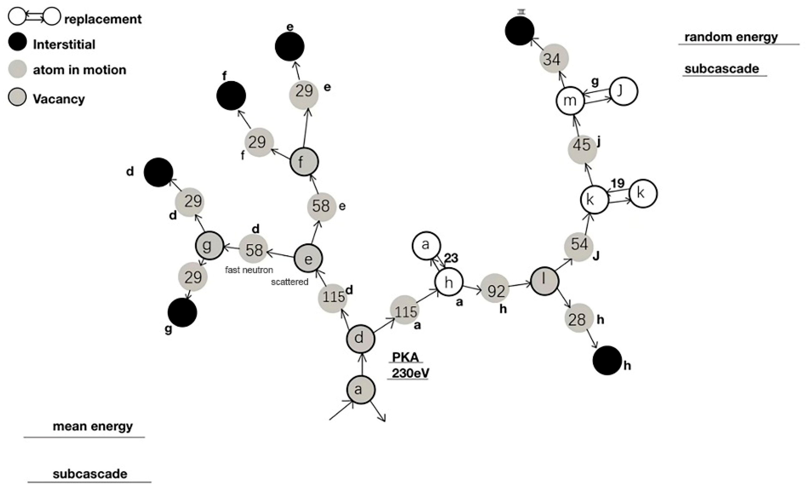
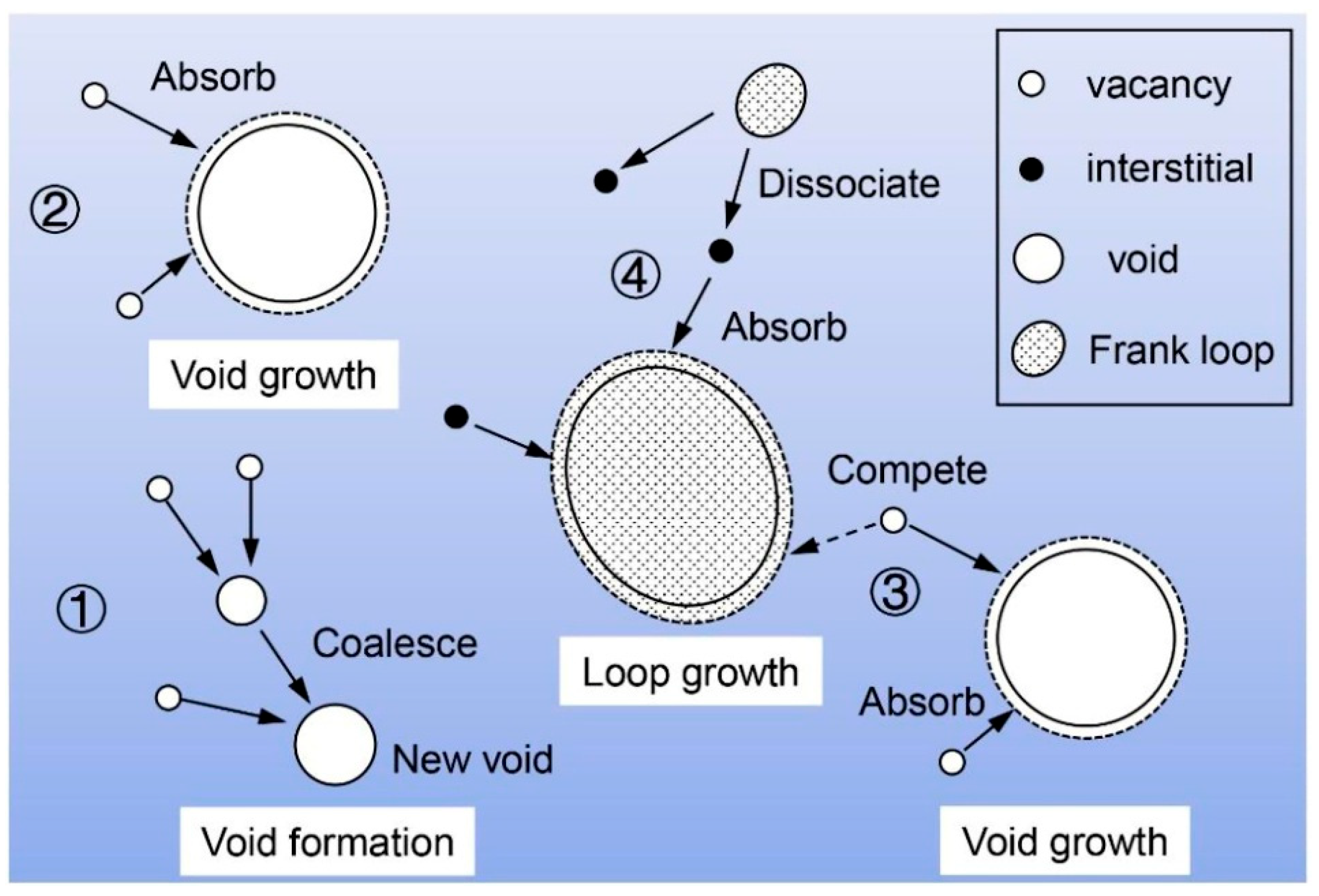
Proton irradiation induces high-density point defects (vacancies and interstitial atoms) in materials through cascade collisions. The diffusion, annihilation, and clustering of these point defects constitute the core mechanisms of irradiation damage and directly drive the subsequent formation of complex defect structures. Figure 1 is a schematic diagram of the primary radiation damage process in materials [49]. When the matrix is subjected to proton bombardment, atoms are displaced from their original lattice sites, creating vacancies. These displaced atoms undergo cascade collisions and eventually come to rest between lattice planes, forming self-interstitial atoms. The resultant vacancy-interstitial pairs, known as Frenkel pairs, lead to hardening and embrittlement of metallic materials. The evolution of small-scale vacancy-type defects can be characterized using positron annihilation spectroscopy (PAS). Liu et al. [47] employed PAS to investigate the vacancy-type point defects generated in domestic A508-3 steel following proton irradiation, further demonstrating the effectiveness of PAS in characterizing vacancy-type defects. Their study also revealed that the size of vacancy-type defects increases with increasing irradiation fluence.

3.1.2. Dislocation Loops

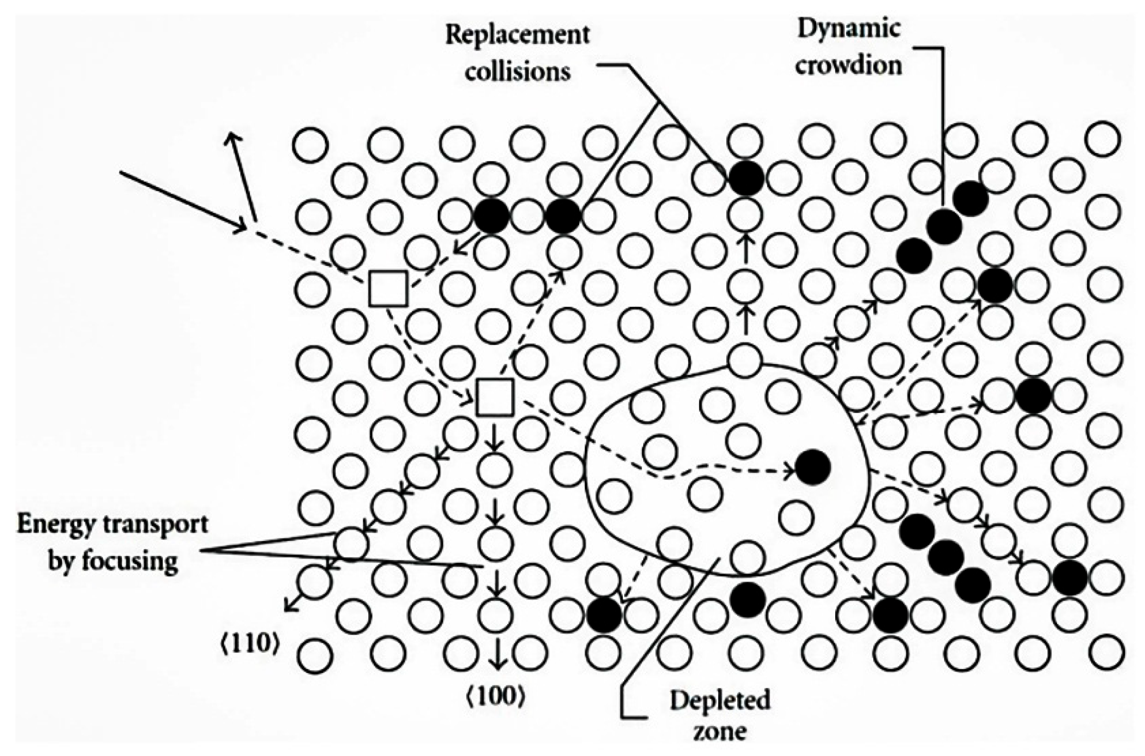
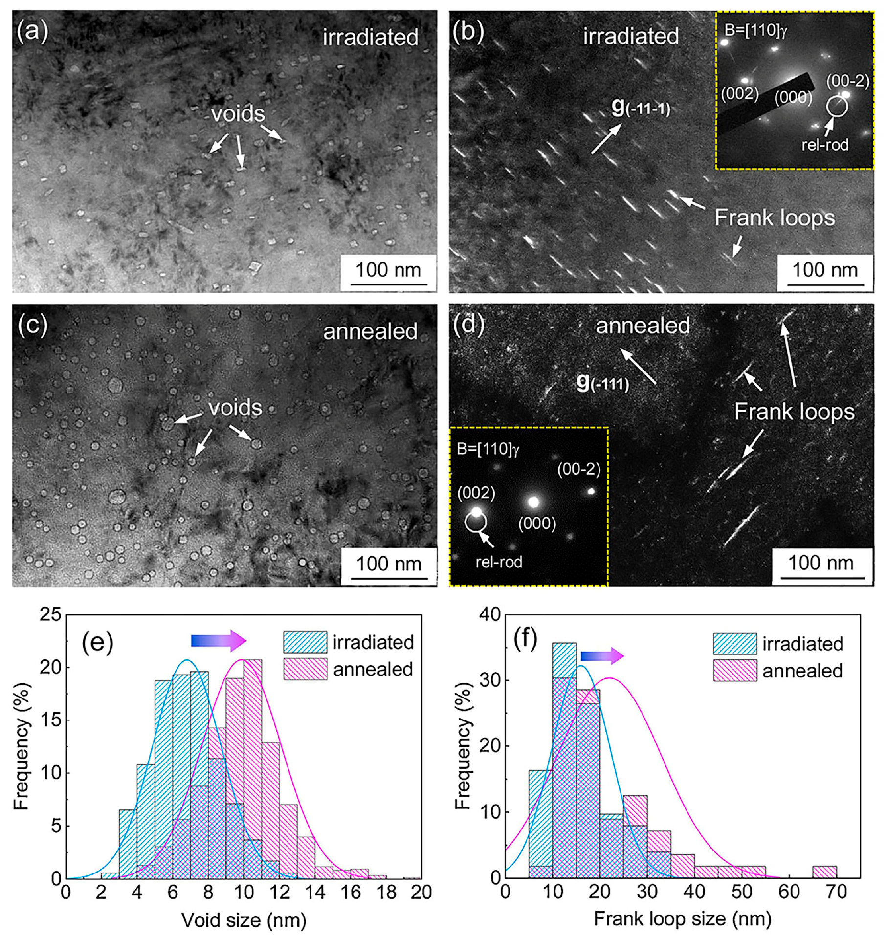
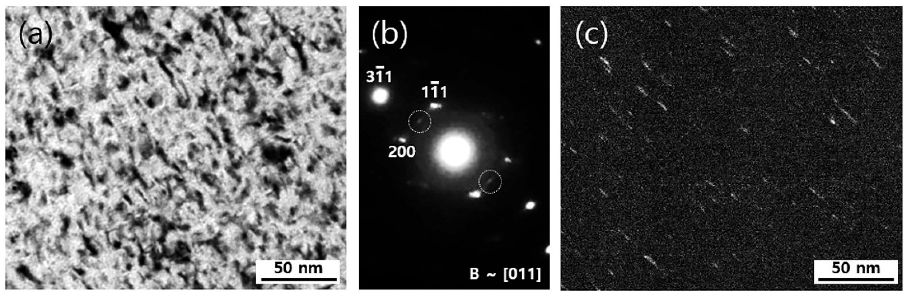
Figure 2 is a schematic diagram of the atomic collision cascade process [50]. Following proton irradiation of structural materials, the generated Frenkel defects undergo cascade collisions during their diffusion and migration, resulting in the annihilation or aggregation of Frenkel defects and the subsequent formation of dislocation loops [51,52]. Specifically, dislocation loops can form either through the aggregation of interstitial atoms or by the collapse of vacancy-rich regions surrounding the cascade zone. These dislocation loops significantly influence the microstructure and mechanical properties of materials. In austenitic stainless steels, proton irradiation-induced structural defects mainly consist of voids and Frank dislocation loops [53]. Lin et al. [46] investigated the effect of annealing on the microstructure of proton-irradiated 308L austenitic stainless steel. During annealing at 550 °C for 1 h, both the size and number density of voids and Frank dislocation loops were observed to continuously increase, as shown in Figure 3. This result indicates that annealing treatment can significantly influence the evolution and distribution of dislocation loops. Shiau et al. [54] investigated the effect of proton irradiation on the microstructure of additively manufactured AM 316L stainless steel. Using bright-field imaging in transmission electron microscopy (TEM), Frank dislocation loops appeared dark, while voids appeared bright, with void diameters ranging from 5 to 10 nm. This study further confirms the presence and morphological characteristics of dislocation loops in irradiation-induced damage of materials. The influence of proton irradiation on 316 stainless steel was systematically examined by Lim et al. [45] as a function of irradiation dose and depth. At a relatively low dose of 4.8 dpa, the irradiated microstructure was found to be dominated by a high density of fine Frank dislocation loops. The morphological features of these loops were further elucidated using dark-field TEM (Figure 4c), which showed that their appearance—ranging from linear to elliptical—depended on their crystallographic orientation within the thin foil.

3.1.3. Vacancy Clusters

Vacancy clusters, as the fundamental carriers of defect evolution at the atomic scale during irradiation damage, directly determine the microstructural degradation and property deterioration of materials through their formation, migration, and aggregation behavior. PAS is characterized by its sensitivity to sub-nanometer-scale defects, enabling the detection of minute defects at extremely low concentrations. TEM is capable of high-resolution imaging but is less sensitive to small-scale defects such as single vacancies, typically requiring defects of larger size or higher concentration for effective observation. Three-dimensional atom probe tomography (3D-APT) offers atomic-scale three-dimensional compositional analysis but requires demanding sample preparation and has a limited analysis volume. Consequently, PAS is particularly suitable for detecting matrix defects such as vacancies and vacancy-solute atom pairs that are difficult to capture using techniques like TEM and 3D-APT. Furthermore, by integrating PAS with complementary methods such as TEM or 3D-APT, researchers can obtain a more comprehensive and accurate characterization of material defects, thereby gaining deeper insights into the microstructure of materials and their influence on properties. Zhang et al. [55] performed proton irradiation at 563 keV on modified 310S stainless steels with additions of Zr and Nb–Ta–W, respectively, and analyzed the vacancy clusters in the irradiated samples using PAS and TEM. The PAS results showed that vacancy clusters were formed in both types of samples after irradiation, with the Zr-added sample exhibiting a lower vacancy cluster density (Zr-added sample: 5.37 × 10²⁰ m−3; Nb–Ta–W-added sample: 1.34 × 10²¹ m−3). Concurrently, TEM observations further revealed the morphology, number density, and size distribution of the vacancy clusters, corroborating the vacancy cluster concentration determined by PAS.

3.1.4. Solute Clusters

Irradiation-induced clusters represent an important manifestation of solute redistribution in austenitic stainless steels under irradiation, primarily including Ni-Si-rich clusters such as the γ’ and G-phase [56]. Conventional wisdom holds that Ni–Si precipitates initially nucleate at interstitial sinks, driven by interstitial diffusion; whereas the radiation-induced segregation of Ni in austenitic stainless steels is attributed to vacancy flux diffusion [57]. Jiao et al. [58] characterized and analyzed the chemical composition both within the grains and at the grain boundaries of neutron-irradiated 304L stainless steel using TEM and atom probe tomography. They observed the formation of Ni-Si-rich atomic clusters within the austenite grains after irradiation. The formation of these two Ni-Si-rich precipitate phases typically exhibits a competitive relationship [59], and which phase dominates depends on a combination of factors such as material composition and irradiation conditions [60,61]. Shim et al. [60] demonstrated through simulation that high dislocation density can provide fast diffusion paths for solute atoms in Ti-stabilized 316 stainless steel, thereby promoting the formation of the γ’. Furthermore, if the sink density is too high, the silicon content near the sinks may fall below the critical value required for γ’ precipitation, in which case the G-phase may preferentially form [61].

3.1.5. Nanotwins

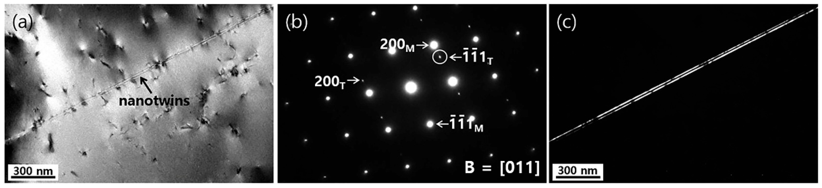
Irradiation-induced formation of nanotwins is one of the important mechanisms for the microstructural evolution of austenitic stainless steel for nuclear applications, and their cross-scale evolutionary behavior has a significant impact on the mechanical properties of the materials [45]. Lim et al. [45] irradiated 316 stainless steel with 2 MeV protons at 360 °C (doses up to 6 dpa) and found that proton irradiation led to the generation of nanotwin crystals, as shown in Figure 5. The generation of nanotwins may be due to the aggregation of dislocation loops, promoting the growth of stacking faults and the formation of twin nuclei at the stacking faults. The introduction of nanotwins has been shown to significantly enhance the mechanical properties of austenitic stainless steel, including ultra-high strength, good plastic elongation, excellent fatigue strength, and resistance to crack extension [62]. The study by Cui et al. [62] focused on the high-cycle fatigue performance of 304 stainless steel with nanotwins. After tensile-compressive fatigue testing, the yield and tensile strengths were found to be as high as 928 MPa and 1312 MPa, respectively, which far exceeded the performance of conventional coarse-grained materials. This showed that irradiation-induced nanotwins may contribute to the improvement of the mechanical properties of the material as well.

3.2. Effect of Proton Irradiation on Material Properties

3.2.1. Forms of Proton Irradiation Damage

Irradiation damage refers to point defects, defect clusters, and their evolved laminar faults, dislocation loops, compositional segregation, and micro-voids that occur when materials are bombarded with energy-carrying particles [2,63,64,65]. These damages can lead to degradation of the performance of in-reactor components, threatening the stable operation of the reactor and increasing economic risks.
Irradiation swelling means that when the concentration of irradiation-generated vacancies reaches saturation, the vacancies aggregate to form three-dimensional defective voids, resulting in material volume expansion and density reduction. This swelling may not only lead to component failure, such as fastener breakage and sleeve bending in the pile, but also significantly reduce the toughness of the material. In addition, irradiation swelling can exacerbate other irradiation damage phenomena, such as irradiation hardening, creep, and stress corrosion cracking (SCC), which ultimately affect the service life and safe operation of the reactor.
Irradiation-induced segregation is a phenomenon that results in an inhomogeneous distribution of elements in a material when the exchange rates of solute atoms with interstitial atoms and vacancies are not equal [66]. In austenitic stainless steel, since the Cr atom size is larger than Fe and Ni, its diffusion rate at grain boundaries is slower, resulting in irradiation-induced segregation at grain boundaries, which is rich in Ni and poor in Cr. The segregation of Fe at grain boundaries depends on the relative diffusion coefficients of Fe and other solute atoms, and the relative contents of Cr and Ni. Within a certain range, the degree of irradiation-induced segregation is positively correlated with the irradiation dose [67]. Irradiation-induced segregation induces precipitation of second phases in austenitic stainless steel, which are usually generated above 350 °C and are mainly chromium-rich carbides. Above 400 °C, γ’-Ni3Si and G phases appear [68]. Irradiation-induced segregation and irradiation-induced precipitation of the second phase can adversely affect the material properties. Deng et al. [69] demonstrated that 304L stainless steel exhibited no grain boundary segregation before proton irradiation. However, after proton irradiation, significant Ni and Si enrichment accompanied by Cr depletion was observed at grain boundaries, with the degree of segregation increasing in proximity to the boundaries. This irradiation-induced Cr depletion compromises corrosion resistance, whereas Si segregation weakens grain boundary strength, thereby facilitating intergranular stress corrosion cracking (IGSCC).

3.2.2. Effect of Proton Irradiation on Mechanical Properties

The mechanical properties of nuclear austenitic stainless steel will change significantly after proton irradiation, which is mainly manifested as irradiation hardening and an irradiation embrittlement phenomenon. Among them, irradiation hardening is the most important phenomenon, which is manifested as an increase in the hardness and yield strength of the material, which is caused by high-density defects, such as dislocation rings hindering the dislocation line slip, produced by proton irradiation of the material [70]. Jin et al. [35] investigated the effect of proton irradiation on austenitic stainless steel at a proton energy of 2 MeV and an irradiation temperature of 360 °C. The formation of Frank loops was observed by TEM, and the average diameter and number density of Frank loops were found to be 7.3 nm and 2.2 × 10²³ m−3, respectively, at 7.6 dpa. Nanoindentation tests showed that the nano-hardness of the material increased gradually with the depth of irradiation at an indentation load of 5 mN and reached a maximum of about 6.5 GPa at a depth of about 20 μm. Furthermore, in pressurized water reactors, the combined effect of irradiation hardening and Cr depletion at grain boundaries may promote the occurrence of IASCC [71].
Regarding the mechanism of irradiation hardening, models such as diffuse barrier hardening and source hardening have been proposed by [49,68,72], thus clarifying the link between irradiation hardening and changes in the microstructure of materials. The source hardening model suggests that changes in the yield strength of a material can be characterized by parameters such as the number density and size of voids and dislocation loops together. The theory of diffuse barrier hardening proposed by Seeger [49] suggests that the amount of change in the hardness of a material due to irradiation is proportional to the arithmetic square root of the product of the dislocation loop number density and size. According to the diffuse barrier hardening model, the change in yield strength caused by dislocation loops or voids can be calculated using Equation (1).
σ y = M α μ b N d 0.5
where ∆σy is the increment in yield strength of the material after irradiation; M is the Taylor factor (i.e., the ratio of the yield strength to the shear strength of the polycrystalline material); α is the strengthening factor of dislocation loops or voids, which depends on the type of obstacles; μ is the shear modulus of the material; b is the Burgers vector; and N and d are the number density and average size of dislocation loops or voids, respectively.
The relationship between ∆σy and the increment in hardness of the material after irradiation (∆H) is given by Equation (2) [64]. Therefore, through Equations (1) and (2), a quantitative relationship can be established between the irradiation-induced hardness change and the number density and size of irradiation-induced defects.
σ y = 3.03 H
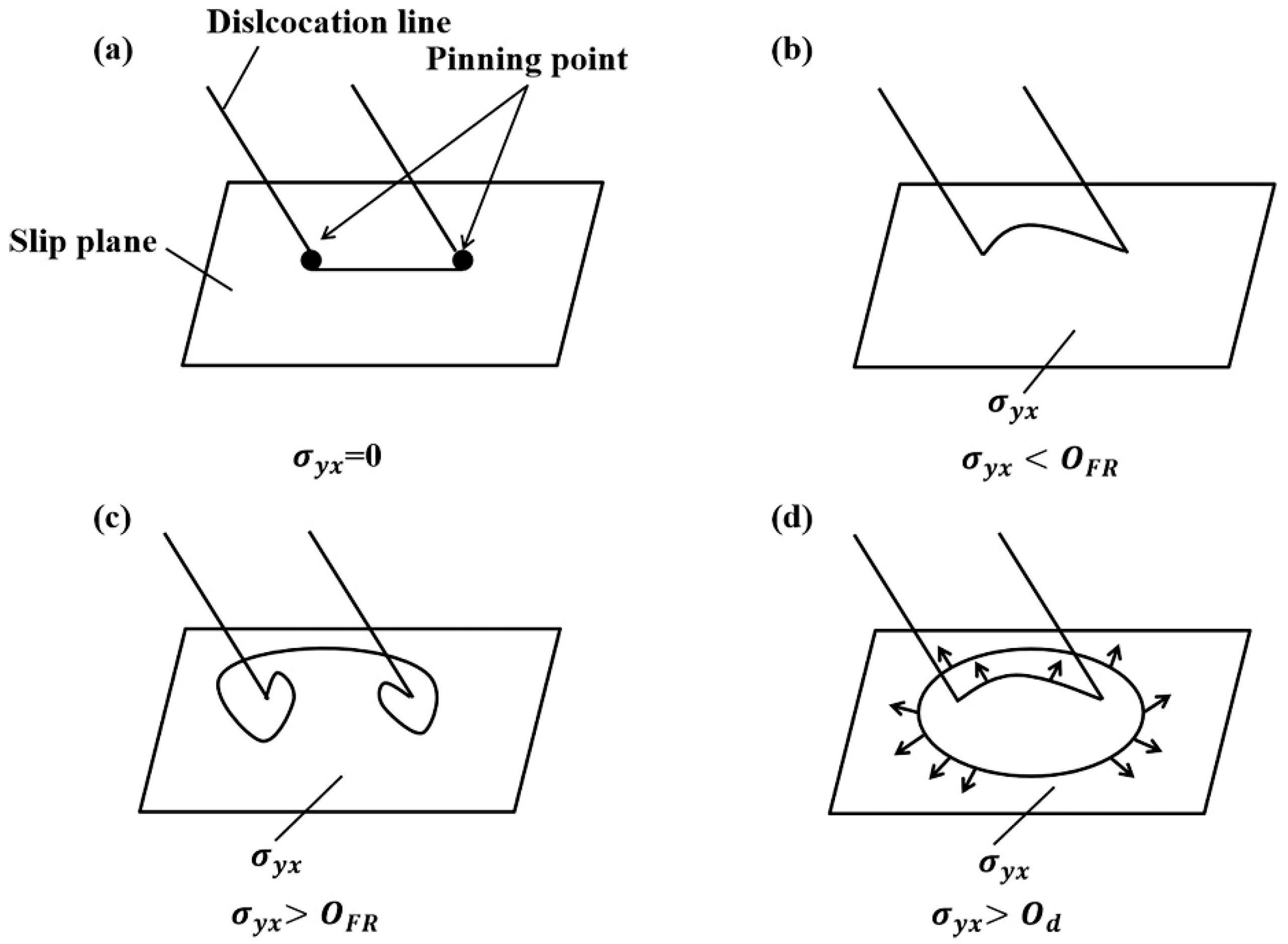
Movement of dislocation in face-centered cubic-structured metals requires unpinning of the Frank–Read (F–R source) dislocation source. As shown in Figure 6 [73], the process of removing the pinning action of the F–R source in a face-centered cubic-structured metal is as follows: the F–R dislocation source starts to bend when the applied stress gradually increases from 0 (Figure 6a). As the applied stress increases, the degree of bending of the F–R dislocation source increases until its radius of curvature decreases to a minimum value of R = 1/2 (Figure 6b). If the applied shear stress continues to increase, the F–R dislocation source will bend further to the state shown in Figure 6c. When P and P’ meet and annihilate each other, a new dislocation loop will be released, while the original F–R dislocation source will continue to proliferate. Under irradiation conditions, a high density of defect clusters is generated near the F–R dislocation source, which acts as an obstacle to increase the applied stress required for dislocation expansion and dislocation source proliferation, leading to the phenomenon of irradiation hardening. If the applied stress increases enough to overcome the pinning effect, the dislocations will proliferate and destroy the small defect clusters, thus reducing the resistance to dislocation proliferation and slip. This process is also the main reason why austenitic stainless steels show a significant yield point after irradiation.

3.2.3. Effect of Irradiation on Corrosion Properties

When austenitic stainless steels are subjected to long-term service under combined harsh conditions of high-temperature pressurized water and intense irradiation, a corrosion product film with a duplex-layer structure forms on the surface. The inner layer is a continuous, protective film, typically formed via solid-state reactions between metallic elements and oxygen at the interface. The outer layer consists of loosely distributed oxide particles, primarily generated through the dissolution-precipitation mechanism of metal ions [69].
Proton irradiation significantly affects austenitic stainless steels, inducing damage such as structural defects, irradiation-induced segregation, and irradiation hardening, which lead to changes in the internal microstructure of the material. These alterations result in modifications to the chemical composition of the inner oxide film and grain boundaries after irradiation [74], thereby causing differences in localized corrosion behavior at grain boundaries. Ultimately, this affects the corrosion performance of the material and may contribute to IASCC. Kuang et al. [75] analyzed the IASCC behavior of 316L stainless steel after proton irradiation to a dose of 2.5 dpa, followed by constant load testing in simulated pressurized water reactor primary water. The results showed that the irradiation damage was consistent with that induced by neutron irradiation at a similar dose, and the microstructural and microchemical characteristics of the IASCC cracks were similar to those observed in neutron-irradiated specimens.
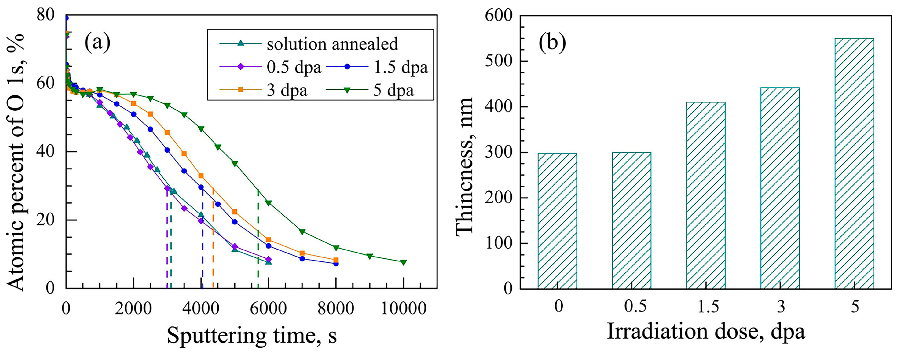
Deng et al. [69] investigated the corrosion mechanisms of nuclear-grade 304 stainless steel under proton irradiation conditions by simulating a pressurized water reactor environment. The findings revealed that (1) the irradiation dose was positively correlated with both the size and number density of dislocation loops; (2) the defects generated by proton irradiation promoted the corrosion process; (3) increasing irradiation dose led to thickening of the oxide layer, as shown in Figure 7; and (4) radiation-induced segregation, including microstructural defects and Cr depletion at grain boundaries, was the primary factor promoting intergranular corrosion. Moreover, the irradiation-induced segregation effect intensified localized corrosion at grain boundaries, thereby facilitating the initiation of SCC sources.

4. Study on the Improvement of Irradiation Properties of Nuclear Stainless Steel

4.1. Improved Corrosion Resistance to Irradiation

The irradiation-corrosion resistance of materials can be significantly improved through approaches such as annealing and AM, as evidenced in relevant research.
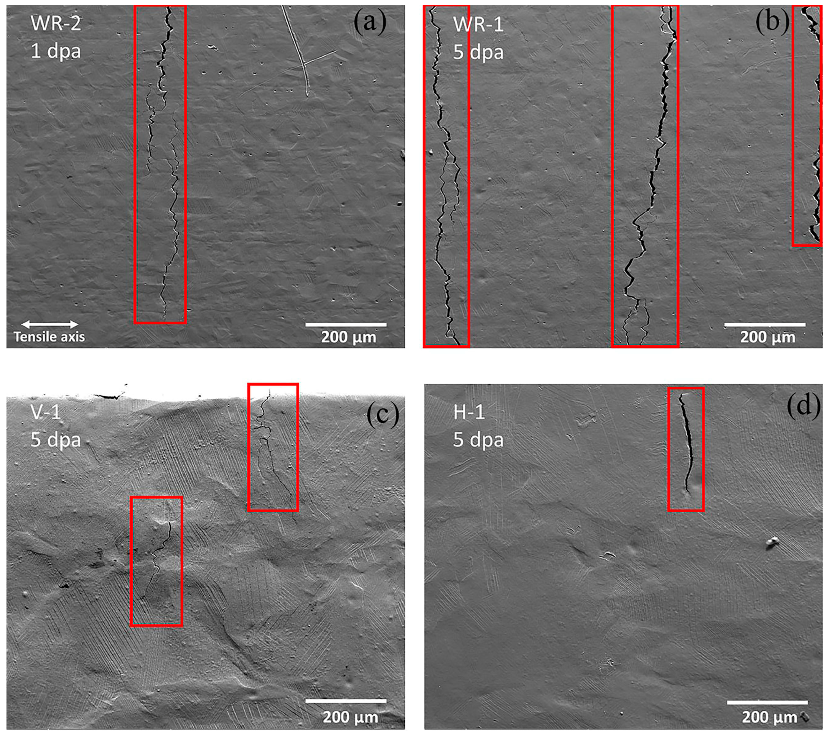
The corrosion behavior of austenite is significantly affected by irradiation-induced structural defects [76,77]. Lin et al. [46] investigated annealing at 550 °C for 1 h on 308L weld metal after proton irradiation to a dose of 3 dpa. The results showed that annealing reduced the number density of irradiation-induced Frank loops (from 6.24 × 10²¹ m−3 to 3.09 × 10²¹ m−3), eliminated other dislocation loops, slowed the corrosion rate, and eliminated localized corrosion at the austenite grain boundaries. The study indicated that voids present in the inner oxide layer play a key role in accelerating corrosion in austenitic materials, suggesting that mitigating void formation represents an effective strategy for improving irradiation corrosion resistance. Recent advances in AM have enabled the production of austenitic stainless steels possessing superior irradiation resistance, with the objective of mitigating their susceptibility to IASCC in nuclear reactor environments. Mcmurtrey et al. [78] investigated the irradiation damage and IASCC behavior of 316L austenitic stainless steel fabricated by two different manufacturing processes (AM and wrought technology) following proton irradiation (2 MeV, 1 dpa and 5 dpa). The results showed that at both 1 dpa and 5 dpa dose levels, the wrought samples (Figure 8a,b) exhibited higher crack number density and length density than the AM samples (Figure 8c,d), indicating that the wrought material is more susceptible to IASCC than the AM material. Zhang et al. [79] further investigated the SCC behavior of SLM 304L stainless steel in high-temperature hydrogenated water. The results demonstrated that, due to its lower silicon and manganese content, SLM 304L stainless steel exhibited significantly higher intergranular oxidation resistance compared to conventionally manufactured 304L stainless steel. The higher dislocation density within the SLM material accelerated the diffusion of solute atoms, further enhancing its resistance to intergranular oxidation. This promoted the precipitation of oxides within cracks, thereby hindering the ingress of corrosive media. Moreover, the combined effect of dislocation cells and nano-oxide inclusions in SLM 304L suppressed dislocation motion and planar slip in the matrix, effectively impeding the propagation of SCC cracks. These findings indicate that the use of AM technology for fabricating austenitic stainless steels can, to a certain extent, improve their irradiation resistance and irradiation-assisted corrosion resistance.
Currently, extensive research has been reported on the irradiation-corrosion resistance of austenitic stainless steels with a single-phase structure. However, relatively few studies have focused on austenitic stainless steel weld metals possessing an austenitic-ferritic duplex structure. Future investigations on such weld materials may lead to breakthroughs in improving irradiation resistance.

4.2. Resistance to Irradiation-Induced Segregation

Approaches to improving irradiation-induced segregation resistance include reducing point-defect concentrations and applying prior unidirectional cold-rolling, both of which have been shown to be effective.
Long et al. [80] conducted experiments on AL-6XN austenitic stainless steel irradiated by 100 keV protons at two temperatures (380 °C and 290 °C) and different doses (1 dpa, 3 dpa, 5 dpa) to investigate the dependence of irradiation-induced segregation at grain boundaries on the irradiation dose and irradiation temperature. The results showed that the irradiation-induced segregation, which was characterized by Cr-depleted and Ni-enriched regions at grain boundaries, became more and more significant with the increase in irradiation dose at the same temperature. The main reason for this phenomenon was the enhancement of irradiation-induced segregation by a high concentration of residual hydrogen: under the condition of high hydrogen content, a large number of hydrogen atoms were captured by vacancies to form H-vacancy complexes, which significantly inhibited the annihilation of irradiated point defects and led to an increase in the concentration of surviving point defects in the steel. And the increase in the concentration of point defects directly promoted the occurrence of segregation. Therefore, controlling the injection amount of H ions became an effective way to reduce the concentration of point defects and thus inhibit the irradiation-induced segregation. On the other hand, it was also found that the degree of irradiation-induced segregation increased with the decrease in irradiation temperature. This may be due to the fact that at lower temperatures, it was more difficult for hydrogen atoms to escape from vacancies, resulting in more hydrogen residue in the material, which enhanced the degree of irradiation-induced segregation. On the other hand, Ahmedabadi et al. [81]. investigated the effect of unidirectional cold rolling process on irradiation-induced segregation of proton irradiated (300 °C, 4.8 MeV, 1 dpa) 304 stainless steels. They found that unidirectional cold rolling introduced a large number of dislocations near grain boundaries, and these high-density dislocations acted as potential wells for point defects, which had a significant effect on the irradiation-induced segregation behavior. Specifically, during plastic deformation of polycrystalline materials, dislocations accumulated near grain boundaries, forming a strain zone immediately adjacent to grain boundaries. This strain zone effectively reduced the point-defect flux toward grain boundaries, and thus the unidirectional cold-rolling process effectively reduced the occurrence of irradiation-induced segregation under low-dose irradiation conditions (<1.8 dpa).
Although some progress has been made in understanding irradiation-induced segregation, more systematic and in-depth studies are still needed. In the future, more optimized kinetic models can be developed to fully elucidate the irradiation-induced segregation mechanism in austenitic stainless steels, which can provide theoretical guidance for improving the irradiation resistance of materials.

4.3. Irradiation-Hardening Resistance

Irradiation hardening is one of the common forms of irradiation damage in nuclear reactor structural materials and directly affects the safe and stable operation of core components [82]. This hardening phenomenon is primarily induced by irradiation-generated defects, including defect clusters, dislocation loops, dislocation lines, voids, and precipitates. Among these, dislocation loops and dislocation lines induced by proton irradiation impede the motion of slip planes within the crystal lattice, serving as the main contributors to material hardening [83]. Two primary strategies have been developed to improve the irradiation hardening resistance of stainless steels for nuclear applications: (i) applying suitable heat treatments to reduce or remove irradiation-induced hardening [84,85]; and (ii) utilizing AM technology to produce austenitic stainless steels with distinctive microstructures—such as cellular sub-grains—that serve as defect sinks, consequently enhancing resistance to irradiation and irradiation-induced hardening.
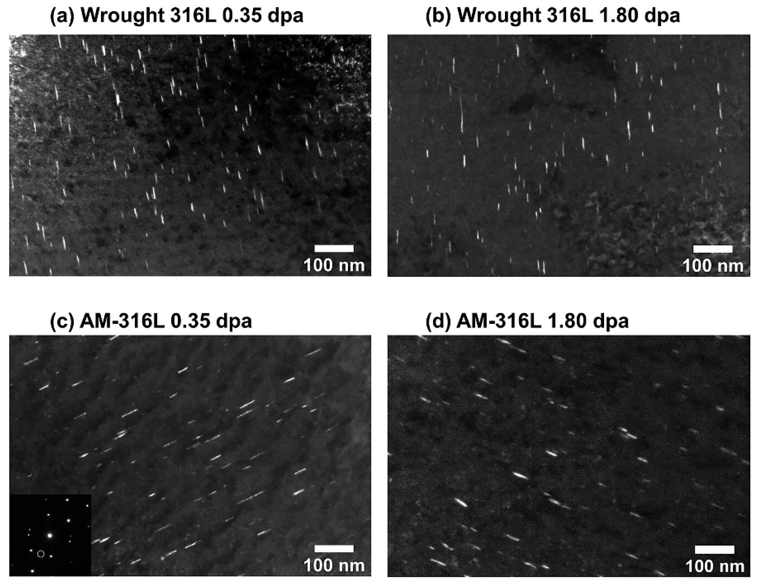
Post-irradiation annealing can partially or fully recover irradiation damage, thereby extending the service life of structural components. Figure 9 is a schematic diagram of the evolution of voids and dislocation loops in austenitic stainless steel during post-irradiation annealing after proton irradiation [46]. Fukumoto et al. [83] performed cumulative isochronous annealing of stainless steel irradiated with ferrous ions at 200 °C, and found that about half of the hardness had been recovered after 30 min of annealing at 400 °C, and that irradiation hardening was almost completely recovered after 30 min of annealing at 500 °C, and decomposition of the Frank loops occurred. Similarly, studies by Van Renterghem et al. [86] and Jiao et al. [87] also observed recovery of neutron-irradiation-induced hardening after annealing at 500 °C. However, it should be noted that current research on post-irradiation annealing is primarily focused on mechanistic exploration at the laboratory scale, aiming to elucidate the evolution of irradiation defects and the laws governing property recovery through controlled annealing. From an engineering application perspective, the implementation of post-irradiation annealing can be categorized into two scenarios: in situ in-core annealing and ex situ annealing after removal. In situ annealing has been applied in engineering practice for non-removable core components such as reactor pressure vessels, but its process requirements are extremely stringent due to the complex in-core environment. Ex situ annealing after removal is technically feasible; however, it necessitates equipment adjustments and disassembly, and its economic cost and maintenance burden limit its routine application. Therefore, the conventional engineering application of post-irradiation annealing in commercial reactors still requires further process optimization and safety validation. Shiau et al. [88] investigated the irradiation resistance of 316L stainless steel fabricated by directed energy deposition (one of the AM techniques). Both wrought and AM 316L stainless steel were subjected to proton irradiation at 360 °C to doses of 0.35 dpa and 1.80 dpa. The experimental results demonstrated that AM 316L stainless steel exhibited a lower number density of dislocation loops, as shown in Figure 10, indicating superior resistance to dislocation loop formation and consequently enhanced irradiation hardening resistance. This improvement is attributed to the cellular sub-grain boundaries generated during the AM process, which act as defect sinks to absorb irradiation-induced defects. Therefore, the adoption of AM technology may offer a promising novel manufacturing approach for improving the irradiation-hardening resistance of austenitic stainless steels.

4.4. Resistance to Irradiation Swelling

Irradiation swelling is closely associated with irradiation-induced void formation. The nucleation and growth of voids lead to volume expansion of the material, thereby causing irradiation swelling. Voids primarily include two types: cavities and gas bubbles. Cavities are three-dimensional volume defects formed by the aggregation, nucleation, and growth of vacancies, while gas bubbles mainly refer to helium bubbles [89]. The irradiation swelling behavior of materials is influenced by multiple factors, including the crystal structure of the material, the type and density of internal defects, the composition of alloying elements, and the irradiation environment (such as irradiation temperature and dose).
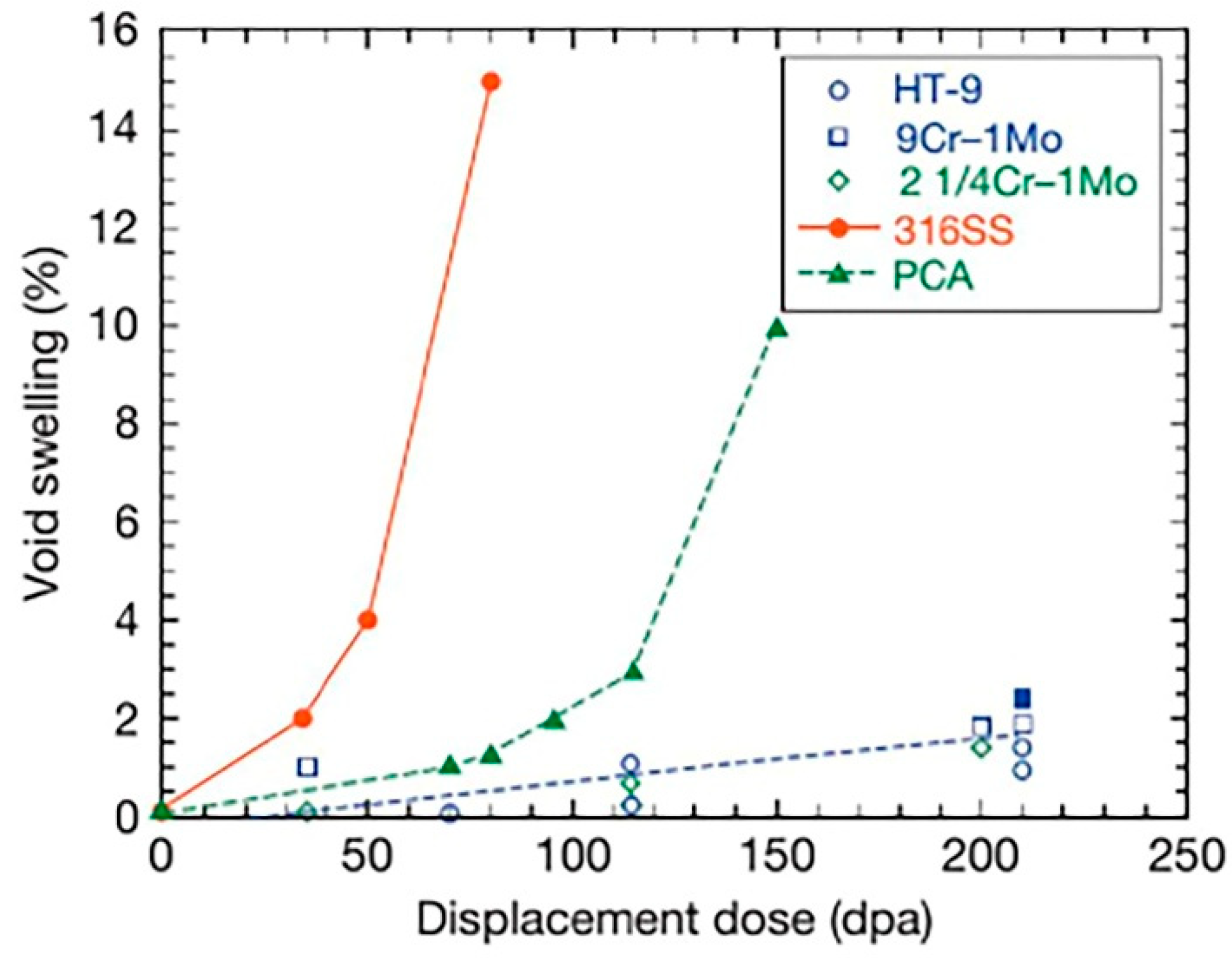
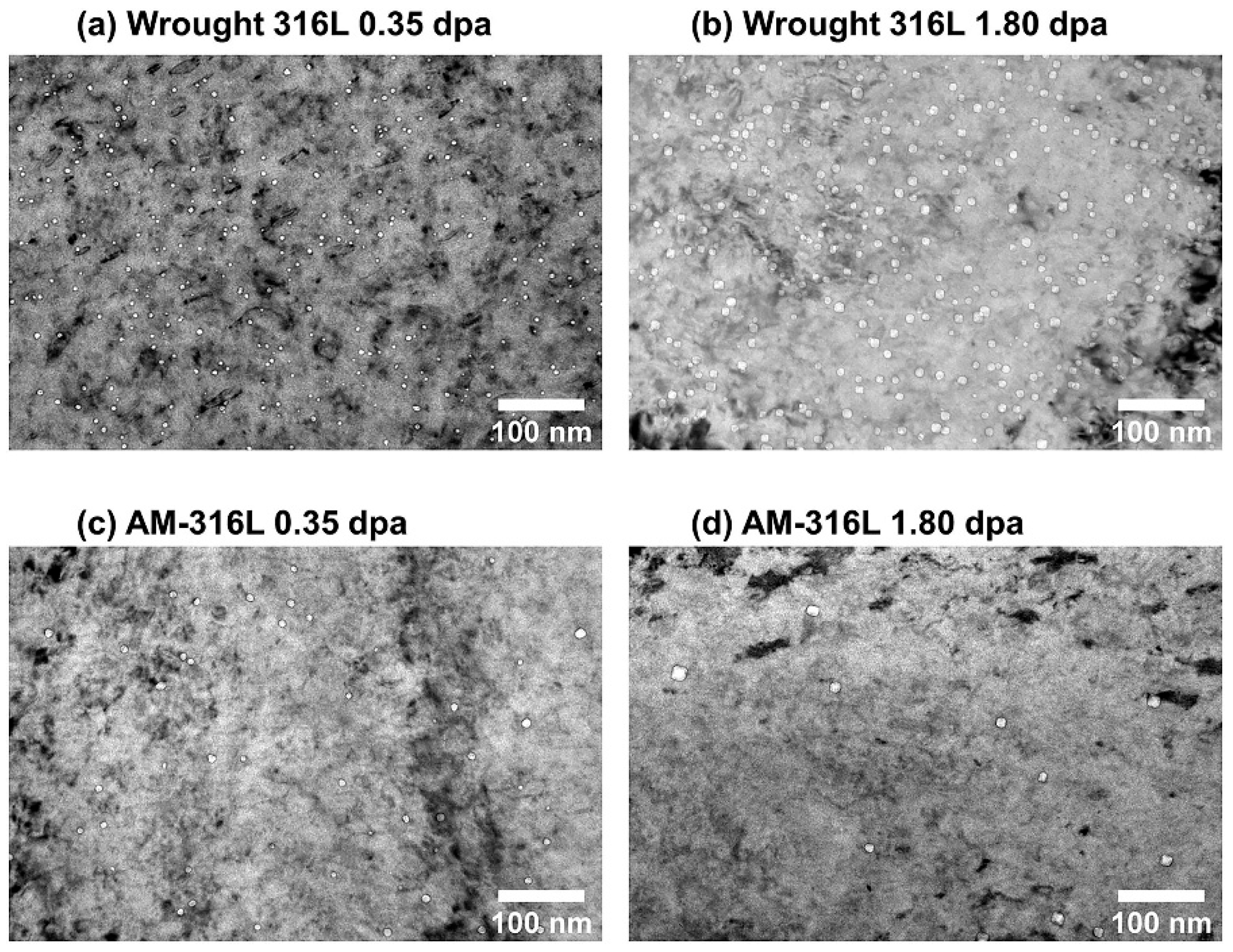
There are significant differences in the irradiation swelling resistance of different materials, as shown in Figure 11 [90]. In general, metals with a body-centered cubic structure, such as vanadium-based alloys and ferritic/martensitic steels, exhibit higher resistance to irradiation swelling compared to materials with a face-centered cubic structure, such as austenitic stainless steels [91,92]. Recent studies have shown that AM can introduce high-density dislocations and interfaces in austenitic stainless steels, which act as defect sinks to promote the recombination and annihilation of irradiation-induced point defects (interstitial atoms and vacancies), thereby significantly reducing the void nucleation rate. Bellefon et al. [93] employed the LPBF method to fabricate 316 austenitic stainless steel and subjected it to 3.5 MeV Fe2+ heavy-ion irradiation tests. The results showed that after post-treatments such as solution annealing and full recrystallization, the material exhibited better swelling resistance. Shiau et al. [88] investigated the irradiation swelling behavior of AM 316L stainless steel subjected to proton irradiation at 360 °C to doses of 0.35 dpa and 1.80 dpa, and compared it with conventionally wrought counterparts. The distribution of voids after irradiation is shown in Figure 12, and the results indicate that the void density in AM 316L stainless steel was significantly lower than that in conventionally wrought specimens. Furthermore, Table 3 summarizes the specific swelling rates of conventionally wrought and AM 316L stainless steel under different irradiation doses, further confirming the significant role of AM technology in improving irradiation swelling resistance. Song et al. [94] investigated the microstructural evolution and IASCC behavior of LPBF 316L stainless steel under irradiation conditions. Proton irradiation experiments were conducted at 360 °C to a dose of 2.5 dpa on LPBF 316L stainless steel in three different conditions: as-built, stress-relief annealed, and hot isostatic pressing (HIP) treated. The experimental results showed that the LPBF 316L stainless steel subjected to HIP treatment exhibited an irradiation swelling rate of 0.0025 ± 0.001%, which was significantly lower than that of conventionally wrought 316L stainless steel (0.156 ± 0.025%). This demonstrates that the combination of LPBF and HIP treatment has a beneficial effect on improving the irradiation swelling resistance of nuclear-grade 316L stainless steel.
Despite the progress made in recent years in research on resistance to irradiated swelling, many challenges remain. Future research will mainly focus on the following aspects: (1) The effect of irradiation conditions: irradiation dose, temperature, and their coupling effect on irradiation swelling still need to be further studied systematically. (2) Alloy design: through the optimization of the composition and proportion of alloying elements, to explore new alloy designs to improve the performance of resistance to irradiation swelling. (3) Molding process and heat treatment: to further study the influence of AM and the heat treatment process on the resistance of materials to irradiation swelling. In addition, the current control technology for irradiation swelling is still insufficient, especially in observing defects during the incubation period of irradiation swelling, which is largely limited by TEM resolution and observation area.

5. Conclusions

In the context of the growing importance of nuclear power technology, austenitic stainless steel, as a commonly used material for core components, must demonstrate performance stability in the complex and harsh core service environment. Given the high difficulty, cost, and analytical challenges of neutron irradiation experiments, proton irradiation has emerged as an effective alternative method for studying irradiation damage and performance improvement in the laboratory. This review combines the current research status of proton irradiation of austenitic stainless steel with a detailed discussion of the microstructural damages induced by proton irradiation, such as point defects, dislocation loops, vacancy clusters, solute clusters, and nano-twins, etc., and explores the impacts of these damages on the mechanical properties, irradiation corrosion, irradiation-induced segregation, irradiation hardening, and irradiation swelling properties of the materials, as well as the methods for improving the relevant irradiation resistance of the materials. Currently, annealing heat treatment and reduction in point defect generation are the main strategies for improving irradiation properties. In addition, AM technology has become a promising method to improve the irradiation resistance of austenitic stainless steel due to its ability to introduce high-density dislocations and interfaces as defect potential sinks, thereby promoting the composite annihilation of irradiated point defects and significantly reducing the formation of voids and dislocation loops.
Looking ahead, with the development of advanced Generation IV nuclear reactors, materials will face more severe service conditions, so the development of new materials or the improvement of existing materials to withstand higher irradiation damage is a key task in the field of nuclear materials. With its advantages of design freedom, high precision, and a short production cycle, AM technology not only meets the demand for one-piece molding of complex components for nuclear reactors but also enhances and improves material properties by controlling the microstructure. In particular, the in situ generation of oxide diffusion strengthened austenitic stainless steel by AM technology allows the introduction of a high density of nano-sized oxide particles, and the interface between these particles and the matrix can not only act as a complex center for irradiation-induced point defects, but also impede the growth of grains and the movement of dislocation lines, which improves the high-temperature mechanical properties of austenitic stainless steel while increasing the resistance to irradiation. Compared to traditional methods such as powder metallurgy production, AM technology is able to avoid complex mechanical alloying steps and achieve a uniform distribution of oxide particles in the matrix. Based on this, combined with current findings on irradiation resistance, AM technology is expected to become a novel processing method that significantly improves the irradiation resistance of core structural materials. However, it should be noted that AM technology still faces challenges in nuclear engineering applications, such as residual stress, microstructural anisotropy, susceptibility to process-induced defects, and manufacturing costs. Its long-term irradiation reliability and performance consistency remain to be further systematically validated. Therefore, to realize the engineering application of AM technology in the nuclear field, systematic research efforts are required to optimize process parameters, gain an in-depth understanding of the relationship between microstructure and properties, and conduct long-term irradiation experiments for validation. In addition, the development of efficient computational models to predict and design irradiation-resistant materials is also an important direction for future research.

Author Contributions

Conceptualization, Y.G. (Yuyu Guo) and J.H.; formal analysis, Y.G. (Yanlin Gu) and Z.Y.; investigation, Y.G. (Yanlin Gu) and Y.G. (Yuyu Guo); data curation, Y.G. (Yuyu Guo) and Z.Y.; writing—original draft, Y.G. (Yuyu Guo); writing—review and editing, J.H. and Y.G. (Yanlin Gu); supervision, J.H. and Z.Y.; resources, J.H.; project administration, J.H.; funding acquisition, J.H. All authors have read and agreed to the published version of the manuscript.

Funding

This work was supported by the National Natural Science Foundation of China (Grant Nos. U22B2067 and 52073176).

Data Availability Statement

No new data were created or analyzed in this study.

Conflicts of Interest

The authors declare that they have no known competing financial interests or personal relationships that could have appeared to influence the work reported in this paper.

References

  1. Chu, S.; Majumdar, A. Opportunities and Challenges for a Sustainable Energy Future. Nature 2012, 488, 294–303. [Google Scholar] [CrossRef]
  2. Zinkle, S.J.; Was, G.S. Materials Challenges in Nuclear Energy. Acta Mater. 2013, 61, 735–758. [Google Scholar] [CrossRef]
  3. Brook, B.W.; Bradshaw, C.J.A. Key Role for Nuclear Energy in Global Biodiversity Conservation: Biodiversity and Sustainable Energy. Conserv. Biol. 2015, 29, 702–712. [Google Scholar] [CrossRef] [PubMed]
  4. Oshima, A.; Shiraki, F.; Fujita, H.; Washio, M. Surface Modification of Polymeric Materials Using Ultra Low Energy Electron Beam Irradiation. Radiat. Phys. Chem. 2011, 80, 196–200. [Google Scholar] [CrossRef]
  5. Ebrahimi Sarcheshmeh, E.; Bijani, M.; Sadighi, H. Adoption Behavior towards the Use of Nuclear Technology in Agriculture: A Causal Analysis. Technol. Soc. 2018, 55, 175–182. [Google Scholar] [CrossRef]
  6. Venugopal, V. Societal Applications of Nuclear Technology in Health Care, Industry and Water Resource Management in India. Energy Procedia 2011, 7, 553–559. [Google Scholar] [CrossRef]
  7. Abram, T.; Ion, S. Generation-IV Nuclear Power: A Review of the State of the Science. Energy Policy 2008, 36, 4323–4330. [Google Scholar] [CrossRef]
  8. Tavassoli, A.A. Assessment of Austenitic Stainless Steels. Fusion Eng. Des. 1995, 29, 371–390. [Google Scholar] [CrossRef]
  9. Loyer-Prost, M.; Puichaud, A.H.; Flament, C.; Rouesne, E.; Béchade, J.L. Impact of Intragranular Misorientation on Void Swelling and Inter-Granular Cavities after Ion Irradiation in Standard and Additive Manufacturing 316L Austenitic Steels. J. Nucl. Mater. 2023, 573, 154102. [Google Scholar] [CrossRef]
  10. Cattivelli, A.; Roy, M.J.; Burke, M.G.; Dhers, J.; Lee, T.L.; Francis, J.A. Internal Stresses in a Clad Pressure Vessel Steel during Post Weld Heat Treatment and Their Relevance to Underclad Cracking. Int. J. Press. Vessel. Pip. 2021, 193, 104448. [Google Scholar] [CrossRef]
  11. Liu, H.; Min, S.; Jiang, M.; Chu, F.; Li, Y.; Chen, Z.; Zhang, K.; Hou, J.; Huang, A. Helium Bubble Growth in He+ Ions Implanted 304L Stainless Steel Processed by Laser Powder Bed Fusion During Post-Irradiation Annealing at 600 °C. Acta Metall. Sin. (Engl. Lett.) 2022, 35, 1509–1518. [Google Scholar] [CrossRef]
  12. Weaver, J.S.; Pathak, S.; Reichardt, A.; Vo, H.T.; Maloy, S.A.; Hosemann, P.; Mara, N.A. Spherical Nanoindentation of Proton Irradiated 304 Stainless Steel: A Comparison of Small Scale Mechanical Test Techniques for Measuring Irradiation Hardening. J. Nucl. Mater. 2017, 493, 368–379. [Google Scholar] [CrossRef]
  13. Tan, L.; Stoller, R.E.; Field, K.G.; Yang, Y.; Nam, H.; Morgan, D.; Wirth, B.D.; Gussev, M.N.; Busby, J.T. Microstructural Evolution of Type 304 and 316 Stainless Steels Under Neutron Irradiation at LWR Relevant Conditions. JOM 2016, 68, 517–529. [Google Scholar] [CrossRef]
  14. Byun, T.S.; Garrison, B.E.; McAlister, M.R.; Chen, X.; Gussev, M.N.; Lach, T.G.; Coq, A.L.; Linton, K.; Joslin, C.B.; Carver, J.K.; et al. Mechanical Behavior of Additively Manufactured and Wrought 316L Stainless Steels before and after Neutron Irradiation. J. Nucl. Mater. 2021, 548, 152849. [Google Scholar] [CrossRef]
  15. Bykov, P.V.; Bayankin, V.Y.; Tcherdyntsev, V.V.; Vorob’ev, V.L.; Pechina, E.A.; Sviridova, T.A.; Shushkov, A.A.; Chukavin, A.I.; Alexandrova, S.S. Effect of Aluminum Ion Irradiation on Chemical and Phase Composition of Surface Layers of Rolled AISI 321 Stainless Steel. Metals 2021, 11, 1706. [Google Scholar] [CrossRef]
  16. Ahmedabadi, P.; Kain, V.; Gupta, M.; Samajdar, I.; Sharma, S.C.; Bhagwat, P.; Chowdhury, R. The Role of Niobium Carbide in Radiation Induced Segregation Behaviour of Type 347 Austenitic Stainless Steel. J. Nucl. Mater. 2011, 415, 123–131. [Google Scholar] [CrossRef]
  17. Liu, J.; Song, Y.; Chen, C.; Wang, X.; Li, H.; Zhou, C.; Wang, J.; Guo, K.; Sun, J. Effect of Scanning Speed on the Microstructure and Mechanical Behavior of 316L Stainless Steel Fabricated by Selective Laser Melting. Mater. Des. 2020, 186, 108355. [Google Scholar] [CrossRef]
  18. Liverani, E.; Toschi, S.; Ceschini, L.; Fortunato, A. Effect of Selective Laser Melting (SLM) Process Parameters on Microstructure and Mechanical Properties of 316L Austenitic Stainless Steel. J. Mater. Process. Technol. 2017, 249, 255–263. [Google Scholar] [CrossRef]
  19. Saeidi, K.; Gao, X.; Zhong, Y.; Shen, Z.J. Hardened Austenite Steel with Columnar Sub-Grain Structure Formed by Laser Melting. Mater. Sci. Eng. A 2015, 625, 221–229. [Google Scholar] [CrossRef]
  20. Dryepondt, S.; Nandwana, P.; Fernandez-Zelaia, P.; List, F. Microstructure and High Temperature Tensile Properties of 316L Fabricated by Laser Powder-Bed Fusion. Addit. Manuf. 2021, 37, 101723. [Google Scholar] [CrossRef]
  21. Lv, J.; Zhou, Z.; Wang, Z. Comparing the Sensitization Behavior and the Corrosion Resistance of the Wrought and Selective Laser Melted 316L Stainless Steels. Mater. Lett. 2022, 328, 133143. [Google Scholar] [CrossRef]
  22. Hou, J.; Dai, B.; Li, Y.; Zhao, J.; Chen, Z.; Pan, D.; Zhu, Y.; Zhang, K.; Huang, A. Helium Bubble Nucleation in Laser Powder Bed Fusion Processed 304L Stainless Steel. J. Nucl. Mater. 2020, 542, 152443. [Google Scholar] [CrossRef]
  23. Cole, J.I.; Allen, T.R. Microstructural Changes Induced by Post-Irradiation Annealing of Neutron-Irradiated Austenitic Stainless Steels. J. Nucl. Mater. 2000, 283–287, 329–333. [Google Scholar] [CrossRef]
  24. Fukuya, K.; Nakano, M.; Fujii, K.; Torimaru, T.; Kitsunai, Y. Separation of Microstructural and Microchemical Effects in Irradiation Assisted Stress Corrosion Cracking Using Post-Irradiation Annealing. J. Nucl. Sci. Technol. 2004, 41, 1218–1227. [Google Scholar] [CrossRef]
  25. Bai, X.-M.; Voter, A.F.; Hoagland, R.G.; Nastasi, M.; Uberuaga, B.P. Efficient Annealing of Radiation Damage Near Grain Boundaries via Interstitial Emission. Science 2010, 327, 1631–1634. [Google Scholar] [CrossRef]
  26. Bai, X.-M.; Uberuaga, B.P. The Influence of Grain Boundaries on Radiation-Induced Point Defect Production in Materials: A Review of Atomistic Studies. JOM 2013, 65, 360–373. [Google Scholar] [CrossRef]
  27. Bai, X.-M.; Vernon, L.J.; Hoagland, R.G.; Voter, A.F.; Nastasi, M.; Uberuaga, B.P. Role of Atomic Structure on Grain Boundary-Defect Interactions in Cu. Phys. Rev. B 2012, 85, 214103. [Google Scholar] [CrossRef]
  28. Du, J.; Jiang, S.; Cao, P.; Xu, C.; Wu, Y.; Chen, H.; Fu, E.; Lu, Z. Superior Radiation Tolerance via Reversible Disordering–Ordering Transition of Coherent Superlattices. Nat. Mater. 2022, 22, 442–449. [Google Scholar] [CrossRef]
  29. Fu, C.; Li, J.; Bai, J.; Lei, Q.; Liu, R.; Lin, J. Evolution of Helium Bubbles in SLM 316L Stainless Steel Irradiated with Helium Ions at Different Temperatures. J. Nucl. Mater. 2022, 562, 153609. [Google Scholar] [CrossRef]
  30. Deng, P.; Peng, Q.; Han, E.-H.; Ke, W.; Sun, C. Proton Irradiation Assisted Localized Corrosion and Stress Corrosion Cracking in 304 Nuclear Grade Stainless Steel in Simulated Primary PWR Water. J. Mater. Sci. Technol. 2021, 65, 61–71. [Google Scholar] [CrossRef]
  31. Was, G.S.; Jiao, Z.; Getto, E.; Sun, K.; Monterrosa, A.M.; Maloy, S.A.; Anderoglu, O.; Sencer, B.H.; Hackett, M. Emulation of Reactor Irradiation Damage Using Ion Beams. Scr. Mater. 2014, 88, 33–36. [Google Scholar] [CrossRef]
  32. Yi, X.; Du, Y.; Geng, D.; Li, Z.; Han, W.; Liu, P.; Chen, J.; Yabuuchi, K.; Yoshida, K.; Ohnuki, S.; et al. Heavy-Ion Irradiation and Post-Irradiation Annealing Effects in Explosion-Welded CuCrZr/316LN Joints for ITER Application. Mater. Charact. 2021, 178, 111252. [Google Scholar] [CrossRef]
  33. Gupta, J.; Hure, J.; Tanguy, B.; Laffont, L.; Lafont, M.-C.; Andrieu, E. Characterization of Ion Irradiation Effects on the Microstructure, Hardness, Deformation and Crack Initiation Behavior of Austenitic Stainless Steel: Heavy Ions vs. Protons. J. Nucl. Mater. 2018, 501, 45–58. [Google Scholar] [CrossRef]
  34. Shiau, C.-H.; Pena, M.; Li, Y.; Xiang, S.; Sun, C.; McMurtrey, M.D.; Shao, L. Micropillar Compression of Additively Manufactured 316L Stainless Steels after 2 MeV Proton Irradiation: A Comparison Study between Planar and Cross-Sectional Micropillars. Metals 2022, 12, 1843. [Google Scholar] [CrossRef]
  35. Lakkam, K.; M.Kerur, S.; Shirahatti, A. Effect of Pitting Corrosion on the Mechanical Properties of 316 Grade Stainless Steel. Mater. Today Proc. 2020, 27, 497–502. [Google Scholar] [CrossRef]
  36. Gussev, M.N.; McClintock, D.A.; Garner, F.A. Analysis of Structure and Deformation Behavior of AISI 316L Tensile Specimens from the Second Operational Target Module at the Spallation Neutron Source. J. Nucl. Mater. 2016, 468, 210–220. [Google Scholar] [CrossRef]
  37. Dai, Y.; Foucher, Y.; James, M.R.; Oliver, B.M. Neutronics Calculation, Dosimetry Analysis and Gas Measurements of the First SINQ Target Irradiation Experiment, STIP-I. J. Nucl. Mater. 2003, 318, 167–175. [Google Scholar] [CrossRef]
  38. Zanini, L.; Dementjev, S.; Gröschel, F.; Leung, W.; Milenkovic, R.; Thomsen, K.; Wagner, W.; Wohlmuther, M.; Cheng, X.; Class, A.; et al. Experience from the Post-Test Analysis of MEGAPIE. J. Nucl. Mater. 2011, 415, 367–377. [Google Scholar] [CrossRef]
  39. Sencer, B.H.; Maloy, S.A.; Hamilton, M.L.; Garner, F.A. Microstructural Evolution of Both As-Irradiated and Subsequently Deformed Microstructures of 316L Stainless Steel Irradiated at 30–160 °C at LANSCE. J. Nucl. Mater. 2005, 345, 136–145. [Google Scholar] [CrossRef]
  40. Zhou, W.; Yang, Y.; Zheng, G.; Woller, K.B.; Stahle, P.W.; Minor, A.M.; Short, M.P. Proton Irradiation-Decelerated Intergranular Corrosion of Ni-Cr Alloys in Molten Salt. Nat. Commun. 2020, 11, 3430. [Google Scholar] [CrossRef]
  41. Mazey, D.J. Fundamental Aspects of High-Energy Ion-Beam Simulation Techniques and Their Relevance to Fusion Materials Studies. J. Nucl. Mater. 1990, 174, 196–209. [Google Scholar] [CrossRef]
  42. Jiang, L.; Song, M.; Yang, L.; Yang, J.; Du, D.; Lou, X.; Chen, Y. A Comparison Study of Void Swelling in Additively Manufactured and Cold-Worked 316L Stainless Steels under Ion Irradiation. J. Nucl. Mater. 2021, 551, 152946. [Google Scholar] [CrossRef]
  43. Boisson, M.; Legras, L.; Andrieu, E.; Laffont, L. Role of Irradiation and Irradiation Defects on the Oxidation First Stages of a 316L Austenitic Stainless Steel. Corros. Sci. 2019, 161, 108194. [Google Scholar] [CrossRef]
  44. Jin, H.-H.; Hwang, S.S.; Choi, M.J.; Lee, G.-G.; Kwon, J. Proton Irradiation for Radiation-Induced Changes in Microstructures and Mechanical Properties of Austenitic Stainless Steel. J. Nucl. Mater. 2019, 513, 271–281. [Google Scholar] [CrossRef]
  45. Lim, Y.S.; Kim, D.J.; Choi, M.J.; Hwang, S.S.; Kim, S.W. Effects of the Dose and Depth on the Microstructural Characteristics of Proton-Irradiated Austenitic 316 Stainless Steel. Mater. Charact. 2022, 194, 112445. [Google Scholar] [CrossRef]
  46. Lin, X.; Han, E.-H.; Peng, Q.; Ke, W.; Jiao, Z. Effect of Post-Irradiation Annealing on Microstructure and Corrosion of Proton-Irradiated 308L Stainless Steel Weld Metal. Corros. Sci. 2020, 175, 108887. [Google Scholar] [CrossRef]
  47. Liu, X.; Wang, R.; Jiang, J.; Wu, Y.; Zhang, C.; Ren, A.; Xu, C.; Qian, W. Slow Positron Beam and Nanoindentation Study of Irradiation-Related Defects in Reactor Vessel Steels. J. Nucl. Mater. 2014, 451, 249–254. [Google Scholar] [CrossRef]
  48. Kočík, J.; Keilová, E.; Čížek, J.; Procházka, I. TEM and PAS Study of Neutron Irradiated VVER-Type RPV Steels. J. Nucl. Mater. 2002, 303, 52–64. [Google Scholar] [CrossRef]
  49. Seeger, A.K. On the theory of radiation damage and radiation hardening. In Proceedings of the Second United Nations International Conference on the Peaceful Uses of Atomic Energy; United Nations Publication: Geneva, Switzerland, 1958. [Google Scholar]
  50. Hyde, J.M.; Nglish, C.A. Microstructural characterisation techniques for the study of reactor pressure vessel (RPV) embrittlement. In Irradiation Embrittlement of Reactor Pressure Vessels (RPVs) in Nuclear Power Plants; Soneda, N., Ed.; Woodhead Publishing: Sawston, UK, 2015; pp. 194–211. [Google Scholar]
  51. Kadoyoshi, T.; Kaburaki, H.; Shimizu, F.; Kimizuka, H.; Jitsukawa, S.; Li, J. Molecular Dynamics Study on the Formation of Stacking Fault Tetrahedra and Unfaulting of Frank Loops in Fcc Metals. Acta Mater. 2007, 55, 3073–3080. [Google Scholar] [CrossRef]
  52. Little, E.A. Development of Radiation Resistant Materials for Advanced Nuclear Power Plant. Mater. Sci. Technol. 2006, 22, 491–518. [Google Scholar] [CrossRef]
  53. Pokor, C.; Brechet, Y.; Dubuisson, P.; Massoud, J.-P.; Barbu, A. Irradiation Damage in 304 and 316 Stainless Steels: Experimental Investigation and Modeling. Part I: Evolution of the Microstructure. J. Nucl. Mater. 2004, 326, 19–29. [Google Scholar] [CrossRef]
  54. Shiau, C.-H.; Sun, C.; McMurtrey, M.; O’Brien, R.; Garner, F.A.; Shao, L. Orientation-Selected Micro-Pillar Compression of Additively Manufactured 316L Stainless Steels: Comparison of as-Manufactured, Annealed, and Proton-Irradiated Variants. J. Nucl. Mater. 2022, 566, 153739. [Google Scholar] [CrossRef]
  55. Zhang, W.; Shen, Z.; Tang, R.; Jin, S.; Song, Y.; Long, Y.; Wei, Y.; Zhou, X.; Chen, C.; Guo, L. Proton-Irradiation Induced Defects in Modified 310S Steels Characterized with Positron Annihilation Spectroscopy and Transmission Electron Microscopy. Methods Phys. Res. B 2018, 427, 1–8. [Google Scholar] [CrossRef]
  56. Maziasz, P.J. Formation and Stability of Radiation-Induced Phases in Neutron-Irradiated Austenitic and Ferritic Steels. J. Nucl. Mater. 1989, 169, 95–115. [Google Scholar] [CrossRef]
  57. Mullurkara, S.V.; Bejawada, A.; Sen, A.; Sun, C.; Bachhav, M.; Wharry, J.P. Nanocluster Evolution in D9 Austenitic Steel under Neutron and Proton Irradiation. Materials 2023, 16, 4852. [Google Scholar] [CrossRef]
  58. Jiao, Z.; Hesterberg, J.; Was, G.S. Effect of Post-Irradiation Annealing on the Irradiated Microstructure of Neutron-Irradiated 304L Stainless Steel. J. Nucl. Mater. 2018, 500, 220–234. [Google Scholar] [CrossRef]
  59. Levine, S.M.; Pareige, C.; Jiao, Z.; Edmondson, P.D.; Was, G.S.; Zinkle, S.J.; Bhattacharya, A. Phase Instabilities in Austenitic Steels during Particle Bombardment at High and Low Dose Rates. Mater. Des. 2022, 217, 110588. [Google Scholar] [CrossRef]
  60. Shim, J.-H.; Povoden-Karadeniz, E.; Kozeschnik, E.; Wirth, B.D. Modeling Precipitation Thermodynamics and Kinetics in Type 316 Austenitic Stainless Steels with Varying Composition as an Initial Step toward Predicting Phase Stability during Irradiation. J. Nucl. Mater. 2015, 462, 250–257. [Google Scholar] [CrossRef]
  61. Pechenkin, V.A.; Epov, G.A. The Influence of Radiation-Induced Segregation on Precipitate Stability in Austenitic Steels. J. Nucl. Mater. 1993, 207, 303–312. [Google Scholar] [CrossRef]
  62. Cui, F.; Pan, Q.S.; Tao, N.R.; Lu, L. Enhanced High-Cycle Fatigue Resistance of 304 Austenitic Stainless Steel with Nanotwinned Grains. Int. J. Fatigue 2021, 143, 105994. [Google Scholar] [CrossRef]
  63. Zinkle, S.J.; Terrani, K.A.; Snead, L.L. Motivation for Utilizing New High-Performance Advanced Materials in Nuclear Energy Systems. Curr. Opin. Solid State Mater. Sci. 2016, 20, 401–410. [Google Scholar] [CrossRef]
  64. Hosemann, P.; Frazer, D.; Fratoni, M.; Bolind, A.; Ashby, M.F. Materials Selection for Nuclear Applications: Challenges and Opportunities. Scr. Mater. 2018, 143, 181–187. [Google Scholar] [CrossRef]
  65. Zinkle, S.J.; Busby, J.T. Structural Materials for Fission & Fusion Energy. Mater. Today 2009, 12, 12–19. [Google Scholar] [CrossRef]
  66. Okamoto, P.R.; Wiedersich, H. Segregation of Alloying Elements to Free Surfaces during Irradiation. J. Nucl. Mater. 1974, 53, 336–345. [Google Scholar] [CrossRef]
  67. Was, G.S. Recent developments in understanding irradiation assisted stress corrosion cracking. In Proceeding of the 11th International Symposium on Environmental Degradation of Materials in Nuclear Power Systems-Water Reactors; American Nuclear Society: Washington, DC, USA, 2003. [Google Scholar]
  68. Maziasz, P.J. Overview of Microstructural Evolution in Neutron-Irradiated Austenitic Stainless Steels. J. Nucl. Mater. 1993, 205, 118–145. [Google Scholar] [CrossRef]
  69. Deng, P.; Peng, Q.; Han, E.-H.; Ke, W.; Sun, C.; Jiao, Z. Effect of Irradiation on Corrosion of 304 Nuclear Grade Stainless Steel in Simulated PWR Primary Water. Corros. Sci. 2017, 127, 91–100. [Google Scholar] [CrossRef]
  70. Liu, S.-M.; Zhang, S.-H.; Abe, H.; Ogata, S.; Han, W.-Z. Kinked Basal Dislocation Loops for Anomalous Annealing Hardening in Irradiated Zirconium. Acta Mater. 2025, 298, 121366. [Google Scholar] [CrossRef]
  71. Bruemmer, S.M.; Simonen, E.P.; Scott, P.M.; Andresen, P.L.; Was, G.S.; Nelson, J.L. Radiation-Induced Material Changes and Susceptibility to Intergranular Failure of Light-Water-Reactor Core Internals. J. Nucl. Mater. 1999, 274, 299–314. [Google Scholar] [CrossRef]
  72. Singh, B.N.; Foreman, A.J.E.; Trinkaus, H. Radiation Hardening Revisited: Role of Intracascade Clustering. J. Nucl. Mater. 1997, 249, 103–115. [Google Scholar] [CrossRef]
  73. Li, F.G. Study on the Mechanism of Radiation Damage Resistance of 316L Stainless Steel Welding Joint. Master’s Thesis, Jiangsu University, Zhenjiang, China, 2019. [Google Scholar]
  74. Yu, K.Y.; Bufford, D.; Khatkhatay, F.; Wang, H.; Kirk, M.A.; Zhang, X. In Situ Studies of Irradiation-Induced Twin Boundary Migration in Nanotwinned Ag. Scr. Mater. 2013, 69, 385–388. [Google Scholar] [CrossRef]
  75. Kuang, W.; Feng, X.; Du, D.; Song, M.; Wang, M.; Was, G.S. A High-Resolution Characterization of Irradiation-Assisted Stress Corrosion Cracking of Proton-Irradiated 316L Stainless Steel in Simulated Pressurized Water Reactor Primary Water. Corros. Sci. 2022, 199, 110187. [Google Scholar] [CrossRef]
  76. Chimi, Y.; Kitsunai, Y.; Kasahara, S.; Chatani, K.; Koshiishi, M.; Nishiyama, Y. Correlation between Locally Deformed Structure and Oxide Film Properties in Austenitic Stainless Steel Irradiated with Neutrons. J. Nucl. Mater. 2016, 475, 71–80. [Google Scholar] [CrossRef]
  77. Perrin, S.; Marchetti, L.; Duhamel, C.; Sennour, M.; Jomard, F. Influence of Irradiation on the Oxide Film Formed on 316 L Stainless Steel in PWR Primary Water. Oxid. Met. 2013, 80, 623–633. [Google Scholar] [CrossRef]
  78. McMurtrey, M.; Sun, C.; Rupp, R.E.; Shiau, C.-H.; Hanbury, R.; Jerred, N.; O’Brien, R. Investigation of the Irradiation Effects in Additively Manufactured 316L Steel Resulting in Decreased Irradiation Assisted Stress Corrosion Cracking Susceptibility. J. Nucl. Mater. 2021, 545, 152739. [Google Scholar] [CrossRef]
  79. Zhang, S.; Wang, S.; Feng, X.; Ning, Z.; Hou, J.; Kuang, W. Insights into the Stress Corrosion Cracking Resistance of a Selective Laser Melted 304L Stainless Steel in High-Temperature Hydrogenated Water. Acta Mater. 2023, 244, 118561. [Google Scholar] [CrossRef]
  80. Long, Y.; Zheng, Z.; Guo, L.; Zhang, W.; Shen, Z.; Tang, R. Radiation-Induced Segregation at Grain Boundaries in AL-6XN Stainless Steels Irradiated by Hydrogen Ions. Radiat. Phys. Chem. 2018, 145, 116–121. [Google Scholar] [CrossRef]
  81. Ahmedabadi, P.M.; Kain, V.; Samajdar, I.; Gupta, M.; Sharma, S.C.; Bhagwat, P. Ajaykumar Effect of Prior Cold-Work on Radiation-Induced Segregation in Proton-Irradiated Austenitic Stainless Steel. Corros. Sci. 2012, 60, 153–164. [Google Scholar] [CrossRef]
  82. Margolin, B.; Sorokin, A.; Belyaeva, L. A Link between Neutron and Ion Irradiation Hardening for Stainless Austenitic and Ferritic-Martensitic Steels. Metals 2024, 14, 99. [Google Scholar] [CrossRef]
  83. Fukumoto, K.-I.; Mabuchi, T.; Yabuuchi, K.; Fujii, K. Irradiation Hardening of Stainless Steel Model Alloy after Fe-Ion Irradiation and Post-Irradiation Annealing Treatment. J. Nucl. Mater. 2021, 557, 153296. [Google Scholar] [CrossRef]
  84. Allen, T.R.; Busby, J.T.; Kenik, E.A.; Was, G.S. Modeling the Effect of Irradiation and Post-Irradiation Annealing on Grain Boundary Composition in Austenitic Fe-Cr-Ni Alloys. Mater. Res. Soc. Symp. Proc. 1998, 527, 291. [Google Scholar] [CrossRef]
  85. Busby, J.T.; Was, G.S.; Kenik, E.A. Isolating the Effect of Radiation-Induced Segregation in Irradiation-Assisted Stress Corrosion Cracking of Austenitic Stainless Steels. J. Nucl. Mater. 2002, 302, 20–40. [Google Scholar] [CrossRef]
  86. Van Renterghem, W.; Al Mazouzi, A.; Van Dyck, S. Influence of Post Irradiation Annealing on the Mechanical Properties and Defect Structure of AISI 304 Steel. J. Nucl. Mater. 2011, 413, 95–102. [Google Scholar] [CrossRef]
  87. Jiao, Z.; Hesterberg, J.; Was, G.S. Insights into the Sources of Irradiation Hardening in a Neutron Irradiated 304L Stainless Steel Following Post-Irradiation Annealing. J. Nucl. Mater. 2019, 526, 151754. [Google Scholar] [CrossRef]
  88. Shiau, C.-H.; McMurtrey, M.D.; O’Brien, R.C.; Jerred, N.D.; Scott, R.D.; Lu, J.; Zhang, X.; Wang, Y.; Shao, L.; Sun, C. Deformation Behavior and Irradiation Tolerance of 316 L Stainless Steel Fabricated by Direct Energy Deposition. Mater. Des. 2021, 204, 109644. [Google Scholar] [CrossRef]
  89. Allen, T.R.; Cole, J.I.; Trybus, C.L.; Porter, D.L.; Tsai, H.; Garner, F.; Kenik, E.A.; Yoshitake, T.; Ohta, J. The Effect of Dose Rate on the Response of Austenitic Stainless Steels to Neutron Radiation. J. Nucl. Mater. 2006, 348, 148–164. [Google Scholar] [CrossRef]
  90. Sekio, Y.; Sakaguchi, N. Effect of Nickel Concentration on Radiation-Induced Diffusion of Point Defects in High-Nickel Fe–Cr–Ni Model Alloys during Neutron and Electron Irradiation. Mater. Trans. 2019, 60, 678–687. [Google Scholar] [CrossRef]
  91. Lu, E.; Cao, X.; Jin, S.; Zhang, P.; Zhang, C.; Yang, J.; Wu, Y.; Guo, L.; Wang, B. Investigation of Vacancy-Type Defects in Helium Irradiated FeCrNi Alloy by Slow Positron Beam. J. Nucl. Mater. 2015, 458, 240–244. [Google Scholar] [CrossRef]
  92. Chernikov, V.N.; Trinkaus, H.; Jung, P.; Ullmaier, H. The Formation of Helium Bubbles near the Surface and in the Bulk in Nickel during Post-Implantation Annealing. J. Nucl. Mater. 1990, 170, 31–38. [Google Scholar] [CrossRef]
  93. Meric De Bellefon, G.; Bertsch, K.M.; Chancey, M.R.; Wang, Y.Q.; Thoma, D.J. Influence of Solidification Structures on Radiation-Induced Swelling in an Additively-Manufactured Austenitic Stainless Steel. J. Nucl. Mater. 2019, 523, 291–298. [Google Scholar] [CrossRef]
  94. Song, M.; Wang, M.; Lou, X.; Rebak, R.B.; Was, G.S. Radiation Damage and Irradiation-Assisted Stress Corrosion Cracking of Additively Manufactured 316L Stainless Steels. J. Nucl. Mater. 2019, 513, 33–44. [Google Scholar] [CrossRef]
Figure 1. Schematic diagram of radiation damage (white squares represent vacancies, black solid circles represent interstitial atoms, and white hollow circles represent lattice atoms) Reprinted from ref. [49].
Metals 16 00451 g001
Figure 2. Schematic diagram of the collision cascade process. Adapted with permission from ref. [50]. Copyright 2014, Elsevier.
Metals 16 00451 g002
Figure 3. Voids and Frank loops in proton-irradiated and post-irradiation annealed 308L austenitic stainless steel: (a) bright-field TEM image of voids in irradiated samples; (b) dark-field TEM image of Frank loops in irradiated samples; (c) bright-field TEM image of voids in post-irradiation annealed samples; (d) dark-field TEM image of Frank loops in post-irradiation annealed samples; (e) size distribution of voids in as-irradiated and annealed samples; (f) size distribution of Frank loops as-irradiated and annealed samples. In (e,f), light blue represents the irradiated sample, and light purple represents the post-irradiation annealed sample. Reprinted with permission from ref. [46]. Copyright 2020, Elsevier.
Metals 16 00451 g003
Figure 4. TEM characterization of Frank loops in proton-irradiated 316 stainless steel: (a) bright-field TEM image; (b) selected-area electron diffraction (SAED) pattern; (c) dark-field TEM image of Frank loops. Reprinted from ref. [45].
Metals 16 00451 g004
Figure 5. Nanotwins in proton-irradiated 316 stainless steel: (a) bright-field TEM image of nanotwins; (b) selected-area electron diffraction (SAED) pattern from the 1 dpa irradiated 316 stainless steel; (c) dark-field TEM image of nanotwins acquired using the white-circled diffraction spot in (b). Reprinted from ref. [45].
Metals 16 00451 g005
Figure 6. Schematic diagram of the F–R dislocation source depinning process. σFR: the pinning stress of the F–R dislocation source; σyx: represents the applied shear stress. (a) initial pinned state; (b) dislocation line bows under the applied stress; (c) bowing intensifies, forming a semicircular shape; (d) the two segments meet and annihilate, generating a new dislocation loop. Reprinted with permission from ref. [73]. Copyright 2020, Fugui Li.
Metals 16 00451 g006
Figure 7. Characterization of oxide layers formed on unirradiated and proton-irradiated nuclear-grade 304 stainless steel after 500 h corrosion in simulated primary water: (a) x-ray photoelectron spectroscopy depth profiles of O1s; (b) oxide layer thickness as a function of irradiation dose. Reprinted with permission from ref. [69]. Copyright 2017, Elsevier.
Metals 16 00451 g007
Figure 8. Comparison of crack morphologies in proton-irradiated 316 stainless steel (WR: wrought 316 stainless steel; V/H: AM 316 stainless steel): (a) WR sample, 1 dpa; (b) WR sample, 5 dpa; (c) AM sample taken parallel to the build direction, 1 dpa; (d) AM sample taken perpendicular to the build direction, 5 dpa. Reprinted with permission from ref. [78]. Copyright 2021, Elsevier.
Metals 16 00451 g008
Figure 9. Schematic diagram of the evolution of vacancies, interstitials, voids, and Frank loops in austenitic stainless steel during post-irradiation annealing after proton irradiation. Reprinted with permission from ref. [46]. Copyright 2020, Elsevier.
Metals 16 00451 g009
Figure 10. Dark-field TEM micrographs of Frank loops in proton-irradiated 316L stainless steel. Reprinted from ref. [88].
Metals 16 00451 g010
Figure 11. Swelling values for different structural materials after neutron irradiation (Both the blue square and the blue solid square represent 9Cr-1Mo). Reprinted from ref. [90].
Metals 16 00451 g011
Figure 12. Bright-field TEM images of voids in proton-irradiated 316L stainless steel. Reprinted from ref. [88].
Metals 16 00451 g012
Table 1. Comparison of differences between neutron and proton irradiation [32,33].
Irradiation TypeNeutron IrradiationProton Irradiation
Irradiation fluence rate5 × 1013 n/(cm2 s−1)109~1012
n/(cm2 s−1)
Dose rate10−7 dpa/s10−5 dpa/s
Irradiation 1 dpa consumption time10 days~4 months6~48 h
Irradiation fluenceThere are always some differences
between actual and set value
Accurate control
Irradiation temperatureIt is difficult to control
accurately
Accurate control
RadioactivityStrong radioactivity and requires a hot
laboratory
No radioactivity,
and do not require
Radiation protection
Depth of the uniform irradiation layerUp to 100 mmUp to 0.001 mm
CostVery high, domestic
Irradiation experimental
reactor resources
are very scarce
Cheaper
Table 2. Advantages and disadvantages of proton irradiation and heavy ion irradiation for simulating neutron [34].
Particle TypesAdvantagesDisadvantages
Heavy ion
  • Easy access to radiation sources
  • Temperature of the sample is not easy to control
2.
High dose rate to shorten irradiation time
2.
Widely distributed small, isolated displacement cascades
3.
Smaller injection depth
Proton
  • Shorter time required for proton irradiation relative to neutrons
  • Proton irradiation has a distinct damage peak
2.
Amount of irradiation required to achieve damage is fairly rapid
3.
Better injection depth
Table 3. Irradiation-induced voids size and density, and irradiation swelling values of wrought and AM 316L stainless steel under different proton irradiation doses [88].
0.35 dpa 1.80 dpa
Size
(nm)
Density (×1021/m3)Swelling
(%)
Size
(nm)
Density (×1021/m3)Swelling
(%)
Wrought 316LVoids5.3 ± 1.411.13 ± 0.880.10 ± 0.018.3 ± 2.515.88 ± 3.140.60 ± 0.11
AM 316LVoids7.4 ± 1.91.67 ± 0.380.04 ± 0.0113.9 ± 3.80.95 ± 0.310.16 ± 0.05
Disclaimer/Publisher’s Note: The statements, opinions and data contained in all publications are solely those of the individual author(s) and contributor(s) and not of MDPI and/or the editor(s). MDPI and/or the editor(s) disclaim responsibility for any injury to people or property resulting from any ideas, methods, instructions or products referred to in the content.