Article

Influence of a Novel Thermomechanical Processing Route on the Structural, Mechanical, and Corrosion Properties of a Biodegradable Fe-35Mn Alloy

by
Kerolene Barboza da Silva
,
João Pedro Aquiles Carobolante
,
Roberto Zenhei Nakazato
,
Angelo Caporalli Filho
and
Ana Paula Rosifini Alves
*
School of Engineering and Sciences, São Paulo State University (UNESP), Guaratinguetá 12516-410, Brazil
*
Author to whom correspondence should be addressed.
Metals 2025, 15(4), 462; https://doi.org/10.3390/met15040462
Submission received: 18 March 2025 / Revised: 12 April 2025 / Accepted: 16 April 2025 / Published: 20 April 2025
(This article belongs to the Special Issue Feature Papers in Biobased and Biodegradable Metals)

Abstract

:
Recent studies have focused on developing temporary metallic implants made from biodegradable biomaterials, such as iron and its alloys, along with the associated manufacturing methods. These biomaterials allow the implant to gradually degrade after fulfilling its function, which reduces the risks of complications associated with permanent implants. Iron is particularly appealing from a structural standpoint, and adding manganese enhances its potential for use. The Fe-35Mn alloy demonstrates excellent mechanical properties and degradation characteristics, making it an ideal choice within the Fe-Mn system. As a result, new processing techniques can be applied to this alloy to further improve its performance. The objective of this research is to propose a new processing route and evaluate its impact on the properties of the Fe-35Mn alloy. The experimental alloy was produced using an arc melting furnace, followed by homogenization, hot swaging, and solution treatment. Alloy characterization was conducted using various techniques, including X-ray fluorescence (XRF), optical microscopy (OM), X-ray diffraction (XRD), differential scanning calorimetry (DSC), microhardness testing, tensile strength measurements, Young’s modulus determination, and potentiodynamic polarization analysis. The microstructural evolution throughout the applied processing route was analyzed in relation to the alloy’s mechanical performance and corrosion resistance. The typical microstructure of the Fe-35Mn alloy is primarily composed of austenitic grains stabilized at room temperature. Its mechanical properties—yield strength (297 MPa), ultimate tensile strength (533 MPa), and elongation to failure (39%)—are comparable to, or even surpass, those of conventional biomedical materials such as 316 L stainless steel and pure iron. The reduced Young’s modulus (171 GPa), compared to other alloys, further underscores its potential for biomedical applications. Electrochemical testing revealed lower corrosion resistance than that of similar alloys reported in the literature, with a corrosion potential of −0.76 V and a current density of 3.88 µA·cm−2, suggesting an enhanced corrosion rate.

Graphical Abstract

1. Introduction

In the group of metallic biomaterials, stainless steels, cobalt-chromium, and titanium alloys are currently the most used. Due to their superior mechanical properties, biocompatibility, and corrosion resistance, these materials are used commercially and occupy a dominant position in the implantable devices market. In general, they are intended to replace the original tissues and remain in the body permanently due to their high stability in the biological environment [1].
However, certain clinical applications, such as bone fractures or blood vessel blockages, specifically require only temporary support during the tissue healing process. In these instances, the presence of biomaterials that remain in the body as foreign objects can lead to various negative effects over time, including stress shielding, bone weakening, prolonged irritation, thrombosis, chronic inflammation, distortion of diagnostic images, and even the need for a second surgery to remove the implant, which carries significant risks and additional financial costs [2,3,4].
Biodegradable implants made from biomaterials have been developed as an alternative. These materials enable the implant to progressively degrade after serving its purpose. This biodegradation reduces the risk of complications associated with permanent implants, as previously mentioned [1,5,6,7]. Magnesium and iron-based alloys are among the biodegradable metallic systems recognized for their potential in biomedical applications, as these non-toxic elements exhibit suitable degradation behavior and have processing routes familiar to the human body, along with superior biocompatibility and appropriate mechanical properties [1,8]. However, despite its excellent properties, magnesium has low corrosion resistance, particularly in electrolytic and aqueous environments with high chloride content, where its degradation typically occurs rapidly, compromising the mechanical and functional integrity of the implant in a short timeframe [9,10,11]. Iron and its alloys are especially appealing from a structural perspective, as they demonstrate mechanical properties comparable to those of the 316 L stainless steel used as the reference standard for evaluating other alloys intended for biomedical applications. Recent studies indicate that these materials have shown significant promise for use in biodegradable implants [12], though their degradation rate in physiological media is considered very low [6,10,11].
In this context, the alloys of the iron-manganese system have been extensively examined in studies by Hermawan et al. [13]. The authors reported achieving adequate mechanical properties and a degradation rate that is superior to that of pure iron, adjustable based on manganese concentration [7]. Dargusch et al. [1] also highlight the positive impact that manganese has on the magnetic properties of iron-based alloys. The development of antiferromagnetic behavior enhances the material’s compatibility with magnetic-based techniques, such as magnetic resonance imaging (MRI) exams.
Several authors have comprehensively evaluated a range of alloys within the Fe-Mn binary system (20–35 wt% Mn) and concur that the Fe-35Mn alloy demonstrates the best combination of mechanical, magnetic, and corrosion properties [14].
Increasing manganese content significantly expands the austenite phase field (γ) due to its stabilizing effect, which is less dense and less resistant than the martensite phase (ε) [13]. Consequently, the Fe-35Mn alloy exhibits the best performance against plastic deformation, showcasing the highest elongation among the alloys in the system, along with mechanical parameters comparable to 316 L steel [13,15]. According to Dehghan-Manshadi, Stjohn, and Dargusch [16], reasonable elongation values, along with adequate strength and modulus, suggest that the alloy containing 35% Mn could be a strong candidate for future biomedical applications. Increasing the manganese content decreases the alloy’s magnetic susceptibility due to the lower saturation magnetization exhibited by the austenite phase (γ). As a less noble element with a lower electrochemical potential, manganese makes the iron matrix more susceptible to corrosion and increases its degradation rate in physiological environments [1,7,17].
Dargusch et al. [14] note that the Fe-35Mn composition shows the best combination of properties for use in biodegradable cardiovascular stents, however, its degradation rate remains lower than that of magnesium alloys and is considered slow for temporary implant applications. In addition to introducing new alloying elements, discussions about employing new processing techniques have uncovered an effective method for enhancing the mechanical and corrosion properties of biodegradable metallic alloys [11,18]. Most of the literature on biodegradable metallic alloys emphasizes their processing through powder metallurgy. Consequently, this research was developed to investigate the influence of a novel thermomechanical processing route—based on arc melting followed by homogenization, hot swaging, and solution treatment—on the structural, mechanical, and electrochemical properties of a Fe-35Mn biodegradable alloy. The goal is to assess the alloy’s suitability for temporary biomedical implants by correlating its microstructural evolution with mechanical performance and corrosion behavior.

2. Materials and Methods

2.1. Fe-35Mn Alloy Preparation

In this study, the Fe-35Mn experimental alloy was prepared using an arc melting furnace under an argon atmosphere from Fe (99.8% purity) and Mn (99.9% purity) as raw materials. The elements were weighed according to the stoichiometry of Fe-35wt% Mn; however, a loss of Mn was observed during the process due to its high vapor pressure. To ensure the alloy’s quantitative composition, this element was increased by approximately 5% by mass, as it was deposited on the crucible while being enveloped by Fe. The obtained ingots were remelted at least five times to enhance their chemical homogeneity. They were homogenized at 1000 °C for 86.4 ks, processed by hot swaging at 850 °C into rods with a diameter of 10 mm, and finally solubilized at 1000 °C for 10.8 ks, followed by quenching in water. The chemical compositions of the processed alloy were determined using an X-ray fluorescence (XRF) spectrometer (PANalytical Axios Max, Almelo, The Netherlands).

2.2. (Micro)Structural Characterization

Samples were cut into disks with a thickness of 3 mm from the ingots after each processing route step (melting, homogenization, swaging, and quenching) and prepared for characterization. Present phase identification in each sample was conducted by X-ray diffraction (XRD) using a diffractometer (Bruker D8 Advance, Baku, Azerbaijan) with Cu Kα radiation at an accelerating voltage of 40 kV, a current of 25 mA, and a step size of 0.02° in the scanning range of 20° to 100°. Diffraction patterns were analyzed using the Inorganic Crystal Structure Database (ICSD) and submitted to Rietveld structural refinement using the Total Pattern Analysis Solution (TOPAS) Academic version 5 software.
Differential scanning calorimetry (DSC) was used to assess the thermal behavior and transformations of apparent phases in the Fe-35Mn experimental alloy at the end of processing. Samples weighing approximately 80 mg were analyzed in a calorimeter (NETZSCH, STA 409 C), purged with argon at a controlled heating and cooling rate of 10 °C/min, from room temperature to 1000 °C, with an argon flow rate of 100 mL/min.
Before microstructural analysis, samples that were previously used for XRD were first ground with SiC sandpapers up to 1500 grit, polished with 0.25 µm diamond paste, and ultrasonically cleaned with ethanol to ensure the removal of abrasives from the surface. A 5% Nital solution was used as an etchant, and the microstructure of the alloy was examined using an Epiphot 200 optical microscope (OM) with reflected light (Nikon, Tokyo, Japan). The average grain size was measured from the obtained optical micrographs using open source software for processing and analyzing scientific images, ImageJ 1.53t version. Grain size measurements were based on the longest straight line segment inscribed within each grain.

2.3. Mechanical Properties

Microhardness values of the samples were measured using a Vickers microindentation tester (Wilson Instruments, 401 MVD), with a load of 1 kgf and a dwell time of 10 s, in accordance with ASTM E384 standard. At least twelve indentations were performed on each sample, and the mean values are reported. Statistical analysis was conducted using RStudio software (R version 4.4.1) with the rstatix package (version 0.7.2). Normality (Shapiro–Wilk) and homogeneity of variance (Levene) tests were applied. The non-parametric Kruskal–Wallis test was utilized to compare the groups, followed by Dunn’s post hoc test with Bonferroni correction. In all cases, a significance level of 5% (α = 0.05) was used for the tests performed.
Uniaxial tensile tests were conducted according to the ASTM E8M standard using a universal testing machine (INSTRON, 8801) equipped with a nominal 25 mm extensometer. The cylindrical specimens, measuring a total length of 90.0 mm, a gage length of 40.0 mm, a gage diameter of 5.0 mm, and a filet radius of 6.0 mm, were previously conditioned at room temperature. These were tested with a 50 kN load cell at a rate of 1 mm/min. The tests were performed in triplicate.
Young’s modulus measurements were obtained using the impulse excitation technique (IET) with the Sonelastic® system (ATCP Physical Engineering), adhering to the ASTM E1876 standard. Average values were calculated from five measurements. Statistical analysis was conducted using RStudio (version 4.4.1) with the rstatix package (version 0.7.2). Normality was assessed using the Shapiro–Wilk test, and homogeneity of variance was evaluated with Levene’s test when necessary, as a prerequisite for inferential analyses. To compare Young’s modulus obtained from tensile tests and the IET, as well as to compare it with other properties of various metallic biomaterials reported in the literature, the non-parametric one-sample Wilcoxon signed-rank test, the Wilcoxon rank-sum test, and one-sample t-tests were employed. All tests were performed with Bonferroni correction, applying a significance level of 5% (α = 0.05).

2.4. Corrosion Experiments

The corrosion performance of the Fe–35Mn alloy was evaluated using open circuit potential (OCP) and potentiodynamic polarization tests. Both tests were conducted in Modified Hank’s solution (H1387, Sigma-Aldrich, Darmstadt, Germany), which has an ionic composition and concentration similar to that of human blood plasma. The testing solution was prepared according to the manufacturer’s instructions, using deionized water at a temperature between 15 and 20 °C. Sodium bicarbonate was added to the solution, and NaOH was used to adjust the pH of the medium to 7.4.
Open circuit potential (OCP) and potentiodynamic polarization techniques were employed to evaluate the electrochemical corrosion behavior of the alloy. Before the tests, samples with a 1 cm2 exposed surface area were cut, embedded in polyester resin, ground with SiC paper up to 1500 grit, and then ultrasonically cleaned with ethanol and dried. The tests were performed in Hank’s solution at 37 ± 1 °C, using a potentiostat/galvanostat (EG&G, PAR 283) equipped with PowerSuite 2.40 software and a standard three-electrode cell. The samples were configured as the working electrode, while platinum and saturated calomel electrodes served as counter and reference electrodes, respectively. Open-circuit potential (OCP) measurements were recorded for 5400 s after the samples were immersed in the electrolyte. Following OCP stabilization, potentiodynamic polarization tests were conducted at a scan rate of 1 mV/s, varying from −300 mV (vs. OCP) to +700 mV. The tests were repeated at least three times. The data were analyzed using OriginPro 2016 software (OriginLab Corporation, Northampton, MA, USA), where the corrosion potential (Ecorr) and corrosion current density (Icorr) were determined by means of the Tafel extrapolation method.
The statistical analysis was conducted using RStudio (version 4.4.1) with the rstatix package (version 0.7.2). Normality was assessed using the Shapiro–Wilk test, and homogeneity of variance was evaluated with Levene’s test. The Wilcoxon rank-sum test was used for comparisons of corrosion properties. This test was conducted with Bonferroni correction, applying a significance level of 5% (α = 0.05).

3. Results and Discussion

Table 1 shows the chemical composition of the Fe-35Mn alloy obtained from the arc melting furnace. The results confirm that the processed alloy achieved the desired chemical composition.
The presence of other elements, such as Si, Al, and S, in relatively low concentrations does not indicate metal contamination and is insufficient to cause a phase transformation, given the high concentration of Mn [13].

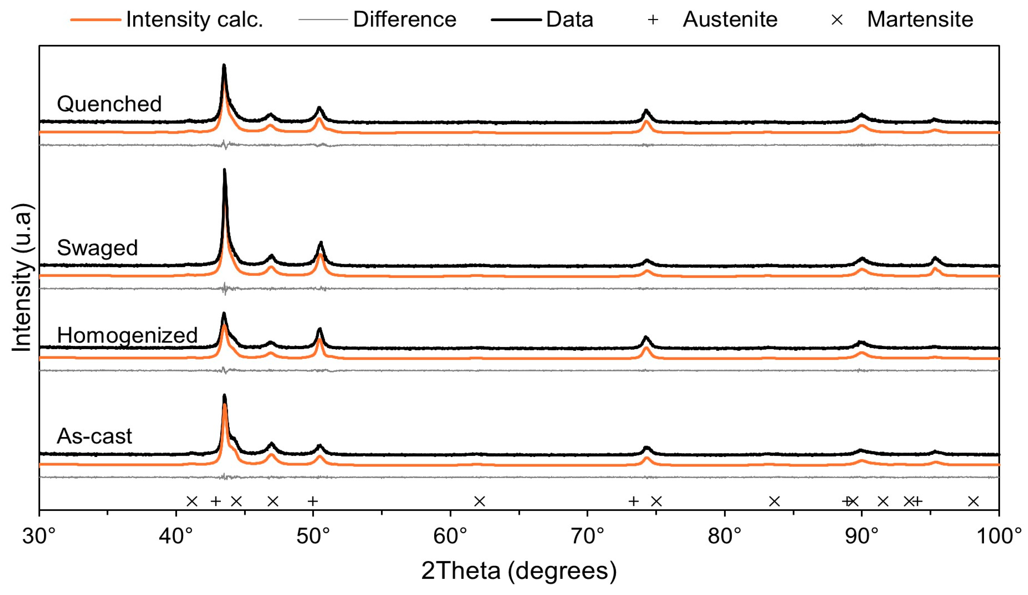
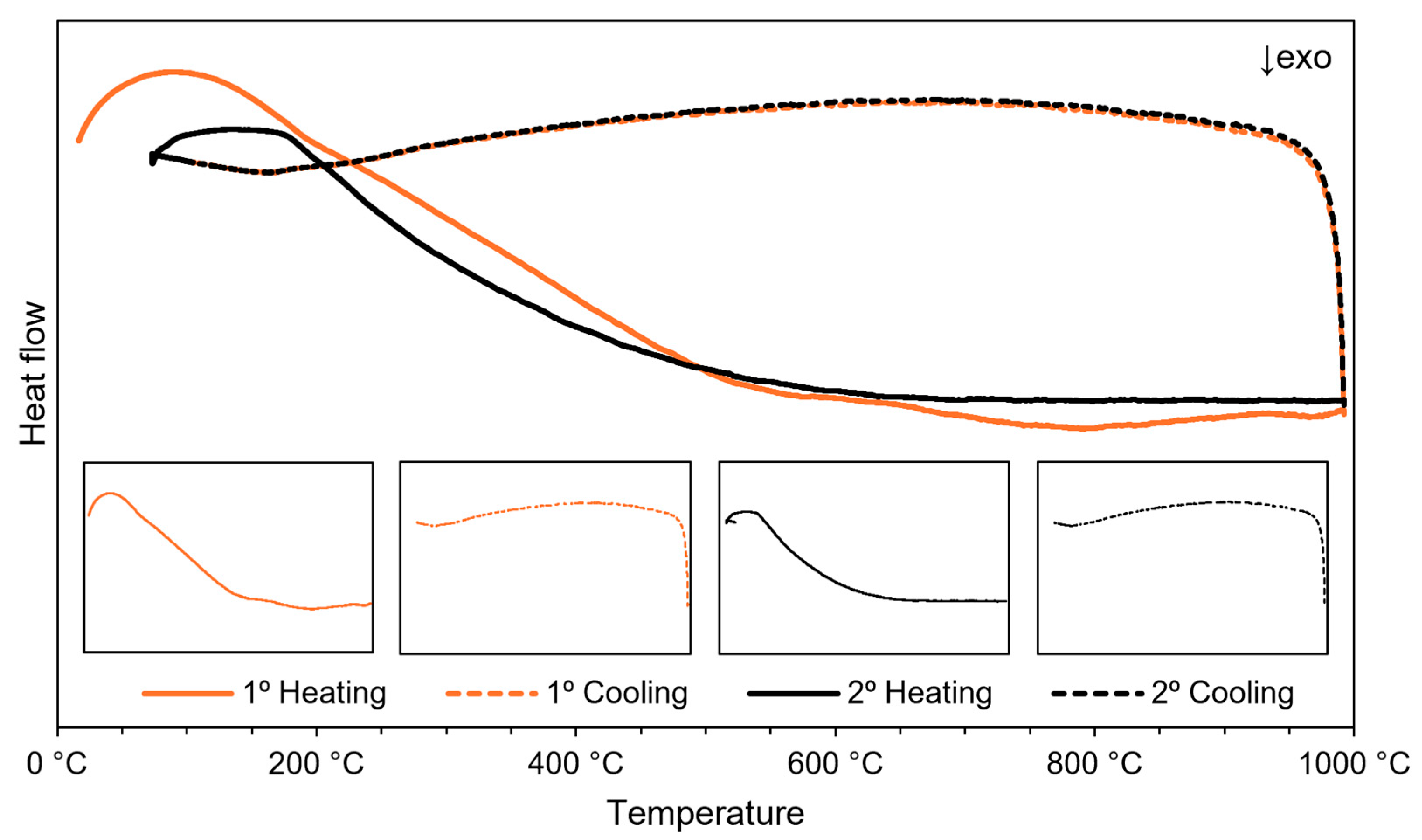
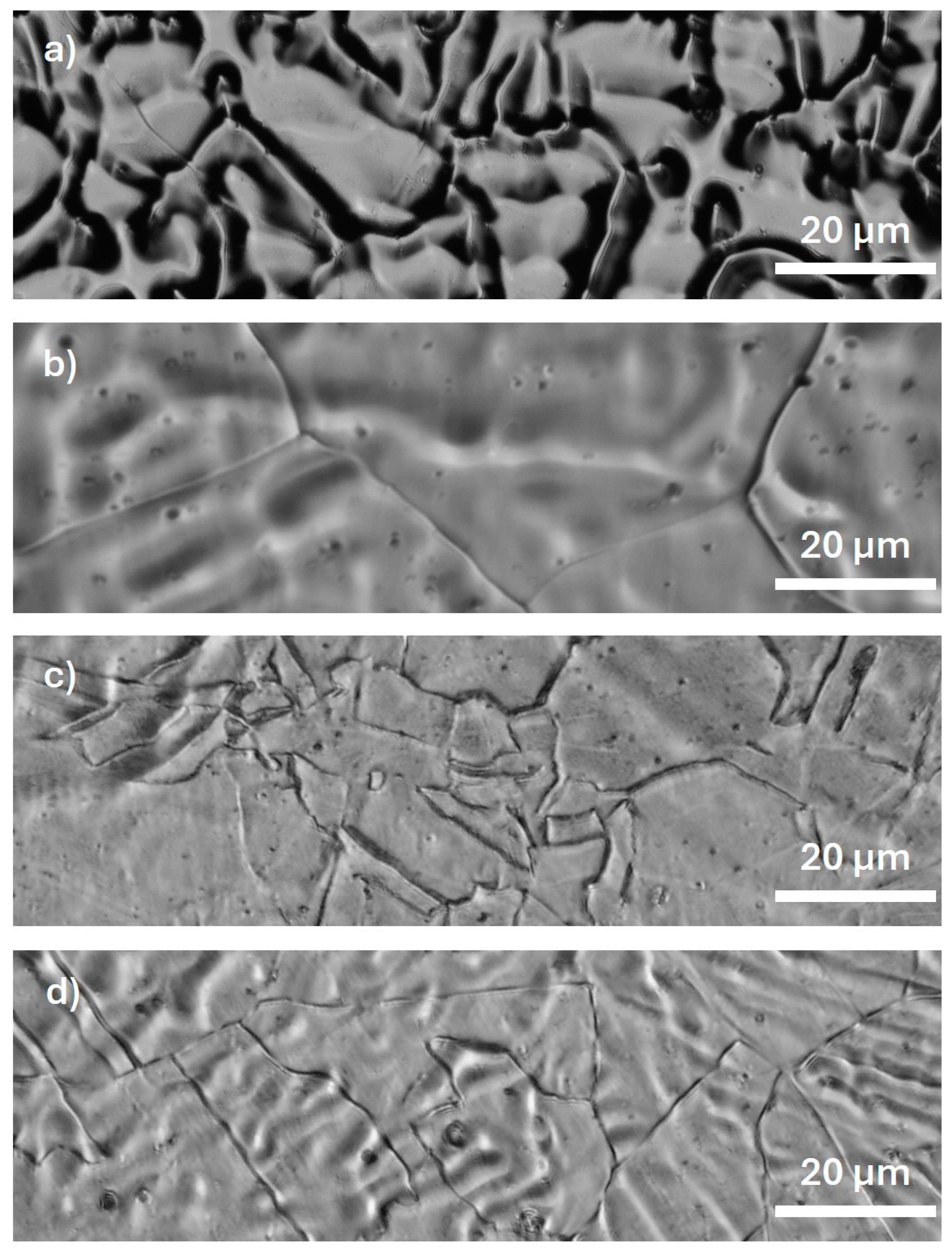
3.1. (Micro)Structural Characterization

XRD analysis was conducted to identify the phases present in each microstructure during alloy processing. Figure 1 illustrates the XRD patterns obtained after Rietveld refinement for as-cast, homogenized, swaged, and quenched samples. Typical peaks of CFC austenite (γ) and HCP martensite (ε) phases were observed in all analyzed samples. These phases were also described by Dargusch et al. [14] and Zhang and Cao [19].
Previous studies [1,16,19] reported that the increase in Mn content significantly expanded the austenite (γ) phase field in Fe-Mn system alloys due to its stabilizing effect. Contents above 29% form a fully austenitic phase composition. However, in this work, the presence of the martensite (ε) phase in the as-cast alloy condition can be attributed to the lack of local homogeneity in the composition. According to the literature [20,21], Mn is easily volatilized at high temperatures because of its high vapor pressure, resulting in areas of Mn depletion with lower concentrations. For this reason, a greater amount of Mn was added during alloy preparation. After homogenization, a decrease in peak intensity related to the martensite (ε) phase and an increase in those associated with the austenite (γ) phase were observed, indicating a transformation and improved homogeneity of the composition. With swaging, peaks of the austenite (γ) phase were reduced but became more intense again after quenching. The formation of the martensite (ε) phase in Fe-Mn alloys can also be influenced by plastic deformation. The influence and peak intensity decrease as the Mn content increases [17]. However, in this study, due to successive swaging passes, the presence of the martensite phase was detected even with 35 wt% Mn in the composition. The Rietveld method was employed to determine the unit cell parameters and to quantify the composition of the different phases. The results and refinement factors for all alloy conditions are listed in Table 2.
As shown in Table 2, in the as-cast condition, 25.8% of the martensite phase in the composition was observed, attributed to the previously mentioned Mn depletion. After homogenization heat treatment, the homogeneity of the composition was corrected, restoring the predominance of the austenite phase and 1.9% of the martensite phase. The swaging process, as a form of plastic deformation, favored the martensitic transformation, increasing the martensite phase content to 79.6%. Finally, the solubilization heat treatment again promoted the austenitization of the alloy, which, after quenching, consisted mainly of the austenite phase with only 3.6% of martensite.
It can also be observed from Table 2 that the lattice parameters and unit cell volume of the found phases remained generally constant, with only minor changes due to deformation effects on the crystalline lattice during alloy processing.
The thermal behavior of the Fe-35Mn alloy was determined using DSC analysis. The sample in the quenched condition, which retained the austenite (γ) phase at room temperature, was investigated. DSC curves and transformation temperatures are shown in Figure 2.
No additional phase changes were detected during both the heating and cooling cycles examined. According to the literature data [22], adding high manganese content to iron tends to lower the transformation temperature and broaden the stability range of the austenite phase (γ). Witusiewicz, Sommer, and Mittemeijer [22] investigated the variation in the enthalpy of formation of γ-Fe-Mn alloys as a function of Mn content. The authors concluded that the transformations correspond to endothermic changes with elevated energy levels. Analyzing the heating curve for the first cycle (solid black line), more distinct and pronounced exothermic transitions are observed between temperatures of 500 and 1000 °C, possibly indicating the relief of internal stresses accumulated at the end of processing. During cooling (dashed black line), no changes were noted. In the second cycle, the heating (solid gray line) and cooling (dashed gray line) curves also showed no evidence of thermal events.
The micrographs in Figure 3 illustrate the evolution of the microstructure in the Fe-35Mn alloy through the applied processing route. In the as-cast Fe-35Mn alloy, the morphology obtained was qualitatively similar to the dendritic form resulting from the alloy’s cooling inside a furnace equipped with a water-based refrigeration system (Figure 3a). With the homogenization heat treatment, the microstructure began to exhibit large, equiaxed grains (Figure 3b). The formation of twins and deformation bands can be observed after swaging and quenching (Figure 3c–d).
According to Hermawan, Dubé, and Mantovani [15], the Fe-Mn system can exhibit various microstructures based on the degree of plastic deformation and the Mn content present. The matrix featuring triangular structures suggests the presence of austenite (γ) and martensite (ε) phases, while their absence indicates only the austenite (γ) phase [15]. In Figure 3d, the typical microstructure of the Fe-35Mn alloy can be visualized, composed predominantly of austenitic grains with an average size of 102.38 μm (46.69 μm), uniformly distributed following the established processing route study.
The austenite (γ) phase, the main constituent of the Fe-35Mn alloy, exhibits relatively large grains, which could explain its superior ductility and low strength [15].

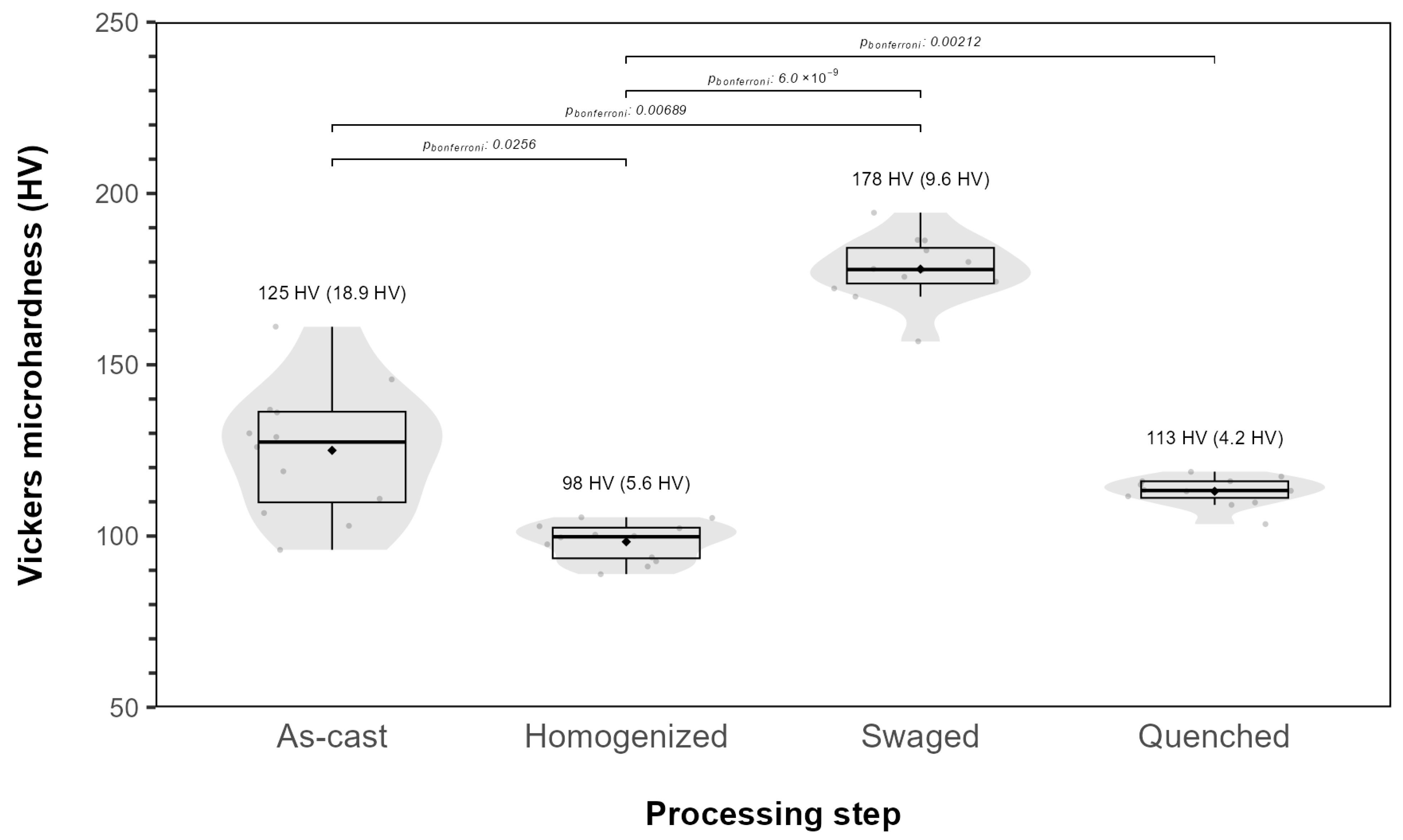
3.2. Mechanical Properties

The microhardness evaluation was performed after each processing step to verify the influence of the microstructural changes. The values found can be seen in Figure 4. The microhardness values showed variations during the alloy processing, however, they correspond with the changes introduced in the microstructure at each step. The martensite (ε) phase, which has an HCP structure, is harder and denser than the austenite (γ) phase with a CFC structure, contributing to the strengthening of the alloys in the Fe-Mn system [13,15]. Thus, after the melting and swaging steps, where there was an increase in the volume fraction of the martensite (ε) phase, the highest microhardness values are observed. Following the homogenization heat treatment and quenching, the microhardness values were relatively lower, with 113 HV being the microhardness obtained at the end of the alloy processing.
The Shapiro–Wilk test indicated that all groups followed a normal distribution (As-cast, p = 0.977; swaged, p = 0.870; homogenized, p = 0.444; quenched, p = 0.514). However, Levene’s test showed heterogeneity of variance among the groups (p = 5.52 × 10⁻⁵), and two outliers were observed: one in the swaged group (157 HV) and another in the quenched group (104 HV). To compare the groups, the non-parametric Kruskal–Wallis test was applied, revealing significant differences in Vickers microhardness across the groups (χ2(3) = 37.9; p = 2.99 × 10⁻⁸; η2ₕ = 0.793). Dunn’s test with a Bonferroni correction was performed as a post hoc analysis, demonstrating statistically significant differences between the groups: As-cast (median = 127 HV; IQR = 26.4) differed from swaged (median = 178 HV; IQR = 10.4 HV) (p = 2.56 × 10⁻²) and homogenized (median = 99.8 HV; IQR = 8.95 HV) (p = 6.89 × 10⁻³), and swaged differed from both homogenized (p = 6.0 × 10⁻⁹) and quenched (median = 113 HV; IQR = 4.85 HV) (p = 6.89 × 10⁻³ and p = 2.12 × 10⁻³).
Compared to the values obtained with annealed pure iron (119 HV), with 316 L stainless steel (195 HV)—considered reference standard in stent manufacturing—and with the same alloy composition obtained by powder metallurgy (143 HV), it is noted that there was a reduction in the microhardness value of the Fe-35Mn alloy processed in this research (113 HV) [23,24]. According to Nayak, Biswal, and Sahoo [23], this reduction indicates better performance for application in temporary implants.
The results obtained from the tensile test are presented in Table 3. This table shows the mechanical properties of the Fe-35Mn alloy after processing outlined in this study, including ultimate tensile strength, elongation to failure, yield strength, and Young’s modulus.
Although with an elongation to failure of 39% (6%) the experimental Fe-35Mn alloy processed in this study is similar to annealed pure iron (40%) and 316 L stainless steel (40%), the tensile test results indicate that the other analyzed mechanical properties are superior. In terms of ultimate tensile strength, the Fe-35Mn alloy achieved 533 MPa (129 MPa), while annealed pure iron supports 210 MPa, and 316 L stainless steel supports 490 MPa. The same is true for yield strength: the Fe-35Mn alloy has a yield strength of 297 MPa (157 MPa), compared to 150 MPa for annealed pure iron and 190 MPa for 316 L stainless steel. Regarding Young’s modulus, the reduction observed in the value for the Fe-35Mn alloy, which stands at 171 GPa (17 GPa), compared to annealed pure iron (200 GPa) and 316 L stainless steel (193 GPa), reinforces its potential for biomedical applications, as it is closer to the Young’s modulus of human bone (10–30 GPa) and helps avoid the stress shielding effect [10,13,25].
When compared to the alloy with the same nominal composition produced via powder metallurgy, an increase in mechanical resistance is also noted. Although this alloy exhibits properties comparable to 316 L stainless steel, which is currently used in most implanted stents, its values of ultimate tensile strength (428 MPa), yield strength (234 MPa), and elongation to failure (32%) are below those found for the Fe-35Mn alloy processed in this study [13,15]. The non-parametric one-sample Wilcoxon signed-rank test suggested that the mechanical properties of the Fe-35Mn alloy differed from those of pure iron, 316 L stainless steel, and the Fe-35Mn alloy processed by powder metallurgy.
The IET was used to confirm the value of Young’s modulus determined from the tensile test. After a statistical study of the measurements, the data are presented in Table 4.
An average value of 182 GPa (3 GPa) was obtained for the Young’s modulus of the experimental alloy Fe-35Mn. When compared with the value derived from the previously presented tensile test, it is evident that the values were similar but exhibited a lower degree of dispersion among the measurements. The non-parametric Wilcoxon rank-sum test revealed no statistically significant difference between the methods used to measure the Young’s modulus. The median values were 171 GPa (17 GPa) for the tensile test and 182 GPa (3 GPa) for the IET.

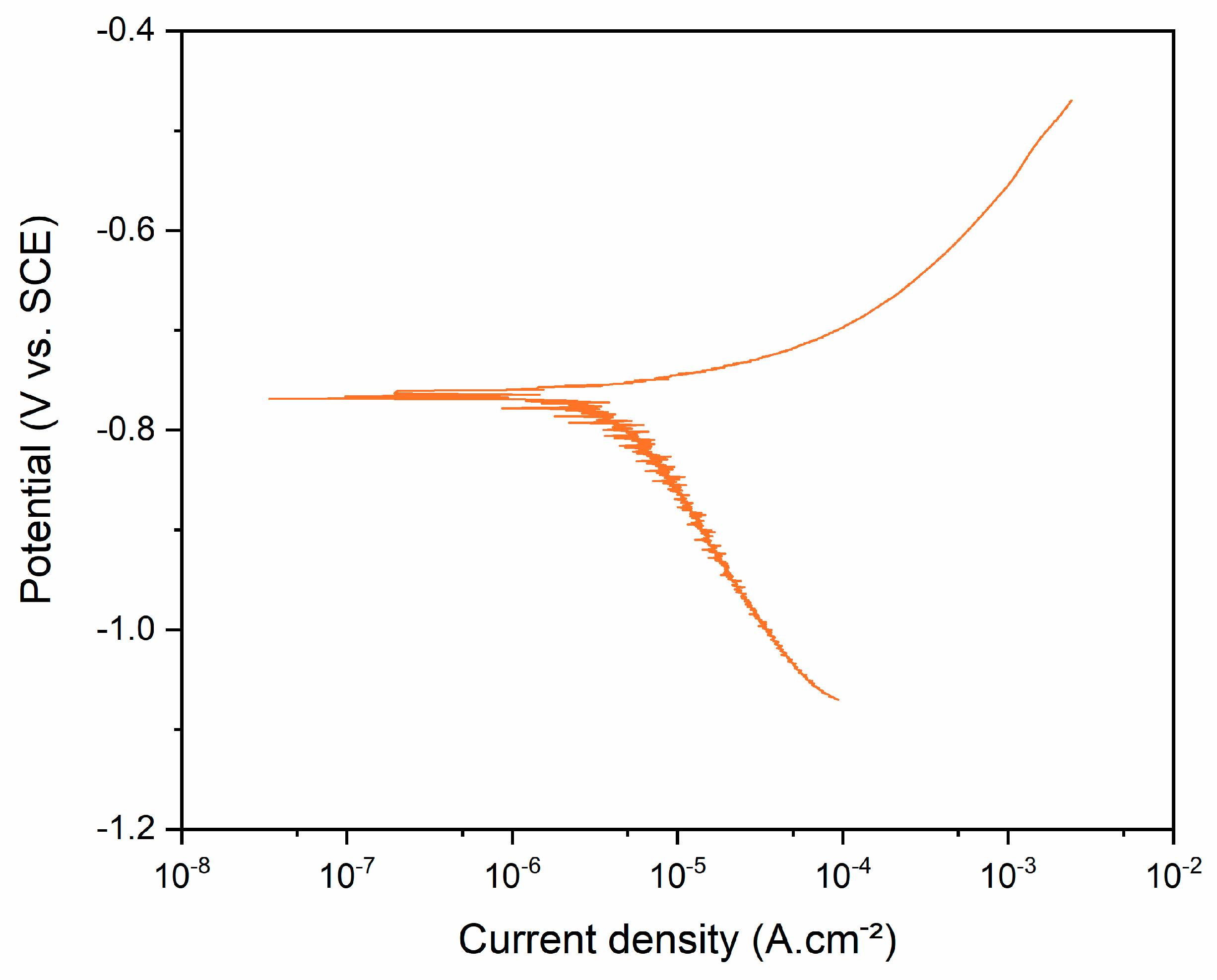
3.3. Corrosion Behavior

Open circuit potential measurements as a function of immersion time of the Fe-35Mn alloy samples were conducted in Hank’s solution. The samples showed a rapid increase in potential during the initial exposure period, followed by stabilization with some fluctuations. The negative average value of −0.785 V (±0.016 V) for the OCP indicates low chemical stability of the alloy. This behavior can be attributed to the addition of Mn, which reduces the standard electrode potential of the system and renders the spontaneous passive film more thermodynamically unstable [26].
The potentiodynamic polarization curves of the Fe-35Mn alloy in Hank’s solution are shown in Figure 5. The average corrosion-related parameters, such as OCP, corrosion potential, and current density determined by the Tafel extrapolation method, are displayed in Table 5.
According to Wang et al. [27], corrosion occurs based on the potential (Ecorr) in a thermodynamic system, where a higher potential corresponds to better corrosion resistance, while the corrosion rate is kinetically assessed by the current density (Icorr); the lower the current density, the lower the rate. Generally, a lower potential indicates a higher corrosion current density [26].
The curves demonstrated that all analyzed samples exhibited similar corrosion behavior, with no passivation occurring in the anodic region. The oxide films became unstable and discontinuous in the medium with increasing potential, rendering the material more susceptible to corrosion [28]. Dehestani et al. [17] reported that the addition of manganese to the structure of an iron-based material results in decreased corrosion resistance. As a less noble element with a lower electrochemical potential, manganese makes the iron matrix more prone to corrosion in physiological media. According to Liu et al. [29], galvanic corrosion mechanisms, in which one metal corrodes preferentially to another, can be introduced into the system due to the potential difference between the metallic elements, also increasing their corrosion rate.
The results obtained in this study, when compared to those indicated by Francis et al. [24] for 316 L steel and especially for as-cast pure iron, show that the addition of Mn led to reduced stability of the alloy in Hank’s solution, resulting in decreased corrosion resistance. The 316 L stainless steel exhibits a corrosion potential of −0.26 V and a current density of 0.454 μA.cm−2, while as-cast pure iron has a potential of −0.38 V and a current density of 0.652 μA.cm−2. In contrast, the experimental Fe-35Mn alloy processed in this study showed a more negative potential of −0.76 V and a high current density of 3.88 μA.cm−2. Previous studies by Shuai et al. [30] have already demonstrated that a lower corrosion potential indicates poor surface corrosion resistance, and an increased current density suggests an enhanced corrosion rate.
Compared to the corrosion parameters of the Fe–35Mn alloy produced via powder metallurgy, as reported by Dargusch et al. [1], a slight decrease in corrosion potential was observed, from −0.74 V to −0.76 V, indicating a slightly higher susceptibility to corrosion. However, the corrosion current density remained significantly lower at 3.88 μA·cm−2, in contrast to the 34 μA·cm−2 reported by the authors, suggesting an overall lower corrosion rate for the Fe–35Mn alloy processed in the present study.
The non-parametric one-sample Wilcoxon signed-rank test provided evidence that the corrosion properties of the Fe-35Mn alloy differ from those of as-cast pure iron, 316 L stainless steel, and the Fe-35Mn alloy processed by powder metallurgy.

4. Conclusions

In this study, a new processing route using an arc melting furnace was developed for the biodegradable Fe-35Mn alloy and its effects on the microstructure and properties, especially mechanical and corrosion, were evaluated. In addition, a statistical analysis was performed to check the correlation between variables and identify significant patterns in the data collected. Some conclusions are drawn as follows:
  • The experimental Fe-35Mn alloy was successfully fabricated, and its microstructural characteristics after processing are consistent with the existing literature, showing the formation of a structure predominantly composed of austenite phase, with large, uniformly distributed grains.
  • The determined mechanical properties, such as ultimate tensile strength, elongation to failure, yield strength, and Young’s modulus, were similar to or superior to those of annealed pure iron and 316 L stainless steel, which were used as reference standards for evaluating other alloys intended for biomedical applications.
  • The processed alloy proved to be more susceptible to corrosion. When compared to pure iron and 316 L stainless steel, the alloy exhibited a lower corrosion potential and an increased current density, suggesting, respectively, lower surface corrosion resistance and a higher corrosion rate. These results underscore the potential of the alloy for use in temporary biomedical devices, such as stents.
Although the results are promising, future studies could expand the characterization of the Fe–35Mn alloy processed via arc melting by conducting both in vitro and in vivo investigations, aiming to achieve a more comprehensive understanding of its behavior.

Author Contributions

Conceptualization, K.B.d.S., J.P.A.C. and A.P.R.A.; data curation, K.B.d.S., J.P.A.C., R.Z.N. and A.C.F.; funding acquisition, A.P.R.A.; methodology, K.B.d.S., R.Z.N., A.C.F. and A.P.R.A.; project administration, A.P.R.A.; resources, R.Z.N., A.C.F. and A.P.R.A.; supervision, A.P.R.A.; visualization, K.B.d.S., J.P.A.C. and R.Z.N.; writing—original draft, K.B.d.S. and J.P.A.C.; writing–review and editing, K.B.d.S., J.P.A.C., R.Z.N., A.C.F. and A.P.R.A. All authors have read and agreed to the published version of the manuscript.

Funding

This study was financed in part by the Coordenação de Aperfeiçoamento de Pessoal de Nível Superior—Brasil (CAPES)—Finance Code 001.

Data Availability Statement

The datasets presented in this article are not readily available because the data are part of an ongoing study. Requests to access the datasets should be directed to corresponding author.

Conflicts of Interest

The authors declare no conflict of interest.

References

  1. Dargusch, M.S.; Choudhury, D.; Saha, T.; Pramanik, A.; Tran, P.A.; O’Mullane, A.P.; Brandt, M. Exploring the Role of Manganese on the Microstructure, Mechanical Properties, Biodegradability, and Biocompatibility of Porous Iron-Based Scaffolds. ACS Biomater. Sci. Eng. 2019, 5, 1686–1702. [Google Scholar] [CrossRef] [PubMed]
  2. Li, H.; Zheng, Y.; Qin, L. Progress of Biodegradable Metals. Prog. Nat. Sci. Mater. Int. 2014, 24, 414–422. [Google Scholar] [CrossRef]
  3. Sun, H.; Chen, F.; Chen, Z.-K. Current Status and Outlook on the Clinical Translation of Biodegradable Metals. Mater. Today 2019, 23, 57–71. [Google Scholar] [CrossRef]
  4. Wang, Z.; Song, J.; Peng, Y. New Insights and Perspectives into Biodegradable Metals in Cardiovascular Stents: A Mini Review. J. Alloys Compd. 2024, 958, 175313. [Google Scholar] [CrossRef]
  5. Rangel, R.d.C.R.; Rangel, A.L.R.; da Silva, K.B.; Escada, A.L.D.A.; Chaves, J.A.M.; Maia, F.R.; Pina, S.; Reis, R.L.; Oliveira, J.M.; Alves, A.P.R. Characterization of Iron Oxide Nanotubes Obtained by Anodic Oxidation for Biomedical Applications—In Vitro Studies. Materials 2024, 17, 3627. [Google Scholar] [CrossRef]
  6. Rabeeh, V.P.M.; Hanas, T. Progress in Manufacturing and Processing of Degradable Fe-Based Implants: A Review. Prog. Biomater. 2022, 11, 163–191. [Google Scholar] [CrossRef]
  7. AlMangour, B.; Sivasankaran, S.; Ammar, H.R.; Almufadi, A.A.; Alsaiari, N. Synthesis, Microstructures, Mechanical, and Corrosion Behavior of FeMn(35−x)–x (Cu, W, and Co)-Based Biodegradable Alloys Prepared by Mechanical Alloying and Selective Laser Melting. Metall. Mater. Trans. A 2023, 54, 3767–3780. [Google Scholar] [CrossRef]
  8. Kiani, F.; Wen, C.; Li, Y. Prospects and Strategies for Magnesium Alloys as Biodegradable Implants from Crystalline to Bulk Metallic Glasses and Composites—A Review. Acta Biomater. 2020, 103, 1–23. [Google Scholar] [CrossRef]
  9. Eliaz, N. Corrosion of Metallic Biomaterials: A Review. Materials 2019, 12, 407. [Google Scholar] [CrossRef]
  10. Hu, T.; Yang, C.; Lin, S.; Yu, Q.; Wang, G. Biodegradable Stents for Coronary Artery Disease Treatment: Recent Advances and Future Perspectives. Mater. Sci. Eng. C 2018, 91, 163–178. [Google Scholar] [CrossRef]
  11. Zhang, M.; Yang, N.; Dehghan-Manshadi, A.; Venezuela, J.; Bermingham, M.J.; Dargusch, M.S. Fabrication and Properties of Biodegradable Akermanite-Reinforced Fe35Mn Alloys for Temporary Orthopedic Implant Applications. ACS Biomater. Sci. Eng. 2023, 9, 1261–1273. [Google Scholar] [CrossRef] [PubMed]
  12. Conti, M.C.; Aquilina, D.; Paternoster, C.; Vella, D.; Sinagra, E.; Mantovani, D.; Cassar, G.; Wismayer, P.S.; Buhagiar, J. Influence of Cold Rolling on In Vitro Cytotoxicity and Electrochemical Behaviour of an Fe-Mn-C Biodegradable Alloy in Physiological Solutions. Heliyon 2018, 4, e00926. [Google Scholar] [CrossRef] [PubMed]
  13. Hermawan, H.; Dubé, D.; Mantovani, D. Degradable Metallic Biomaterials: Design and Development of Fe–Mn Alloys for Stents. J. Biomed. Mater. Res. Part A 2010, 93, 1–11. [Google Scholar] [CrossRef]
  14. Dargusch, M.S.; Venezuela, J.; Dehghan-Manshadi, A.; Johnston, S.; Yang, N.; Mardon, K.; Lau, C.; Allavena, R. In Vivo Evaluation of Bioabsorbable Fe-35Mn-1Ag: First Reports on In Vivo Hydrogen Gas Evolution in Fe-Based Implants. Adv. Healthcare Mater. 2021, 10, 2000667. [Google Scholar] [CrossRef] [PubMed]
  15. Hermawan, H.; Dubé, D.; Mantovani, D. Developments in Metallic Biodegradable Stents. Acta Biomater. 2010, 6, 1693–1697. [Google Scholar] [CrossRef]
  16. Dehghan-Manshadi, A.; StJohn, D.H.; Dargusch, M.S. Tensile Properties and Fracture Behaviour of Biodegradable Iron–Manganese Scaffolds Produced by Powder Sintering. Materials 2019, 12, 1572. [Google Scholar] [CrossRef]
  17. Dehestani, M.; Trumble, K.; Wang, H.; Wang, H.; Stanciu, L.A. Effects of Microstructure and Heat Treatment on Mechanical Properties and Corrosion Behavior of Powder Metallurgy Derived Fe–30Mn Alloy. Mater. Sci. Eng. A 2017, 703, 214–226. [Google Scholar] [CrossRef]
  18. Mostaed, E.; Vedani, M.; Hashempour, M.; Bestetti, M. Influence of ECAP Process on Mechanical and Corrosion Properties of Pure Mg and ZK60 Magnesium Alloy for Biodegradable Stent Applications. Biomatter 2014, 4, e28283. [Google Scholar] [CrossRef]
  19. Zhang, Q.; Cao, P. Degradable Porous Fe-35wt.% Mn Produced via Powder Sintering from NH4HCO3 Porogen. Mater. Chem. Phys. 2015, 163, 394–401. [Google Scholar] [CrossRef]
  20. Liu, P.; Zhang, D.; Dai, Y.; Lin, J.; Li, Y.; Wen, C. Microstructure, Mechanical Properties, Degradation Behavior, and Biocompatibility of Porous Fe-Mn Alloys Fabricated by Sponge Impregnation and Sintering Techniques. Acta Biomater. 2020, 114, 485–496. [Google Scholar] [CrossRef]
  21. Xu, Z.; Hodgson, M.A.; Cao, P. Effect of Immersion in Simulated Body Fluid on the Mechanical Properties and Biocompatibility of Sintered Fe–Mn-Based Alloys. Metals 2016, 6, 309. [Google Scholar] [CrossRef]
  22. Witusiewicz, V.T.; Sommer, F.; Mittemeijer, E.J. Reevaluation of the Fe-Mn Phase Diagram. J. Phase Equilib. Diffus. 2004, 25, 346–354. [Google Scholar] [CrossRef]
  23. Nayak, P.; Biswal, A.K.; Sahoo, S.K. Processing and Characterization of Fe-35Mn Biodegradable Metallic Materials. Mater. Today Proc. 2020, 33, 5284–5289. [Google Scholar] [CrossRef]
  24. Francis, A.; Yang, Y.; Virtanen, S.; Boccaccini, A.R. Iron and Iron-Based Alloys for Temporary Cardiovascular Applications. J. Mater. Sci. Mater. Med. 2015, 26, 1–16. [Google Scholar] [CrossRef] [PubMed]
  25. da Silva, K.B.; Carobolante, J.P.A.; Rajan, S.S.; Júnior, C.B.; Sabino, R.M.; Seixas, M.R.; Nakazato, R.Z.; Popat, K.C.; Claro, A.P.R.A. Mechanical Properties, Corrosion Behavior, and In Vitro Cell Studies of the New Ti-25Ta-25Nb-5Sn Alloy. Materials 2023, 16, 1970. [Google Scholar] [CrossRef]
  26. Cheng, J.; Liu, B.; Wu, Y.H.; Zheng, Y.F. Comparative In Vitro Study on Pure Metals (Fe, Mn, Mg, Zn, and W) as Biodegradable Metals. J. Mater. Sci. Technol. 2013, 29, 619–627. [Google Scholar] [CrossRef]
  27. Wang, H.; Zheng, Y.; Liu, J.; Jiang, C.; Li, Y. In Vitro Corrosion Properties and Cytocompatibility of Fe-Ga Alloys as Potential Biodegradable Metallic Materials. Mater. Sci. Eng. C 2017, 71, 60–66. [Google Scholar] [CrossRef] [PubMed]
  28. Obayi, C.S.; Tolouei, R.; Mostavan, A.; Paternoster, C.; Turgeon, S.; Okorie, B.A.; Obikwelu, D.O.; Mantovani, D. Effect of Grain Sizes on Mechanical Properties and Biodegradation Behavior of Pure Iron for Cardiovascular Stent Application. Biomatter 2016, 6, e959874. [Google Scholar] [CrossRef]
  29. Liu, R.-Y.; He, R.-G.; Xu, L.-Q.; Guo, S.-F. Design of Fe–Mn–Ag Alloys as Potential Candidates for Biodegradable Metals. Acta Metall. Sin. 2018, 31, 584–590. [Google Scholar] [CrossRef]
  30. Shuai, C.; Yang, W.; Yang, Y.; Pan, H.; He, C.; Qi, F.; Xie, D.; Liang, H. Selective Laser Melted Fe-Mn Bone Scaffold: Microstructure, Corrosion Behavior, and Cell Response. Mater. Res. Express 2020, 7, 015404. [Google Scholar] [CrossRef]
Figure 1. XRD refined patterns of the Fe–35Mn alloy for conditions as-cast, homogenized, swaged and quenched. The black line represents the experimental XRD pattern. The orange line represents the pattern calculated by Rietveld refinement. The gray line at the bottom of the curve presents differences between the experimental and calculated intensities. Markers + and × indicate XRD pattern of the austenite (ICSD 631730) and martensite (ICSD 103519) phases, respectively.
Metals 15 00462 g001
Figure 2. Thermogram of the Fe–35Mn alloy sample (quenched condition) on heating and cooling, measured with DSC from room temperature to 1000 °C.
Metals 15 00462 g002
Figure 3. Typical optical micrographs of the Fe-35Mn alloy obtained during processing for conditions (a) as-cast, (b) homogeniszed, (c) swaged, and (d) quenched.
Metals 15 00462 g003
Figure 4. The Vickers microhardness values, Hv, of the Fe-35Mn alloy after each processing step—as-cast, homogenized, swaged, and quenched conditions. Boxplot with violin plot representation showing the distribution of the microhardness properties. The data points are represented by circles, while the diamond symbol indicates the mean value. Standard deviation in parentheses.
Metals 15 00462 g004
Figure 5. Average potentiodynamic polarization curve of Fe-35Mn alloy samples in Hank’s solution at 37 °C.
Metals 15 00462 g005
Table 1. Chemical composition of Fe-35Mn alloy by XRF expressed as a compound mass percentage, normalized to 100%.
Concentration (wt %)
AlloyMnSiAlSFe
Fe-35Mn35.180.150.080.07Balance
Table 2. Unit cell parameters and austenite and martensite phase contents in the as-cast, homogenized, swaged, and quenched Fe-35Mn alloy, calculated from Rietveld refinement of the XRD patterns.
SamplePhaseContent (wt %)Lattice Parameters (Å)Volume (A3)Refinement Factors
acRwpGOFRBragg
As-castγ74.23.55-44.9910.741.032.81
ε25.82.544.1023.091.32
Homogenizedγ98.13.56-45.2911.901.074.32
ε1.92.544.1022.961.97
Swagedγ20.43.58-46.209.941.030.953
ε79.62.444.2622.031.12
Quenchedγ96.43.57-45.8411.111.113.80
ε3.62.554.1023.181.97
Table 3. Mechanical properties of the Fe-35Mn alloy after processing. The data presented were obtained after performing statistical tests, indicating the significance of the variations in mechanical properties. The table shows the number of samples (n), mean, maximum value (Max), minimum value (Min), and standard deviation (SD) of each mechanical property evaluated.
PropertiesnMinMaxMeanSD
Young’s modulus 3154 GPa189 GPa171 GPa17 GPa
Yield strength3144 MPa457 MPa297 MPa157 MPa
Ultimate tensile strength3388 MPa636 MPa533 MPa129 MPa
Elongation to failure334%46%39%6%
Table 4. Values of the young’s modulus obtained by IET. The table presents the maximum and minimum values (Max and Min), mean, and standard deviation (SD). These parameters provide a comprehensive view of the distribution and variability of the data, allowing for a better interpretation of the results obtained in the statistical test.
VariablenMinMaxMeanSD
Young’s modulus5177 GPa186 GPa182 GPa3 GPa
Table 5. Corrosion average parameters of the Fe-35Mn alloy after route processing in arc melting furnace. The data presented were obtained after performing statistical tests, indicating the significance of the variations in the corrosion properties. The table shows the number of samples (n), mean, maximum value (Max), minimum value (Min), and standard deviation (SD) of each parameter evaluated.
VariablenMinMaxMeanSD
OCP3−0.804 V−0.775 V−0.785 V0.016 V
Ecorr3−0.770 V−0.760 V−0.763 V0.006 V
Icorr33.823 µA·cm−23.956 µA·cm−2 3.889 µA·cm−20.066 µA·cm−2
Disclaimer/Publisher’s Note: The statements, opinions and data contained in all publications are solely those of the individual author(s) and contributor(s) and not of MDPI and/or the editor(s). MDPI and/or the editor(s) disclaim responsibility for any injury to people or property resulting from any ideas, methods, instructions or products referred to in the content.

