Article

The Effect of Ti and Mo Microalloying on Hydrogen Embrittlement Resistance of Ultra-High Strength Medium Mn Steel

by
Pujunhuan Zhang
1,†,
Yang Zhao
1,2,†,
Jianglong Pan
1,2,
Weizhuo Hao
3,
Shuyi Wang
1,2 and
Minghui Cai
1,4,*
1
School of Materials Science and Engineering, Northeastern University, Shenyang 110819, China
2
Key Laboratory of Lightweight Structural Materials, Liaoning Province, Northeastern University, Shenyang 110819, China
3
School of Metallurgy, Northeastern University, Shenyang 110819, China
4
State Key Laboratory of Digital Steel, Northeastern University, Shenyang 110819, China
*
Author to whom correspondence should be addressed.
These authors contributed equally to this work.
Metals 2025, 15(4), 397; https://doi.org/10.3390/met15040397
Submission received: 15 February 2025 / Revised: 25 March 2025 / Accepted: 26 March 2025 / Published: 1 April 2025
(This article belongs to the Special Issue Recent Advances in High-Performance Steel)

Abstract

This study elucidated the effect of Ti–Mo microalloying on the hydrogen embrittlement (HE) resistance and fracture behavior of warm-rolled Fe-5.6Mn-0.16C-1Al (wt%) steel. After intercritical annealing, both steels, i.e., without and with Ti–Mo microalloying, showed ultrafine ferrite (α) and austenite (γR) duplex microstructure. The addition of Ti–Mo to 5.6Mn steel reduces the volume fraction of γR, facilitating the formation of (Ti, Mo)C carbides in α phase and further refining the final microstructure. The product of ultimate tensile strength (UTS) and total elongation (TEL) of 5.6MnTiMo can be as high as 35 GPa·% with an ultra-high yield strength of above 1.2 GPa. Furthermore, the addition of Ti–Mo also had a significant effect on the resistance to HE of medium Mn steels. Firstly, the limited (Ti, Mo)C carbides precipitated in γR could act as irreversibly trap sites to capture a considerable amount of H, effectively increasing the CH (Diffusible Hydrogen Content). Additionally, 5.6MnTiMo displayed higher γR stability, resulting in a reduced susceptibility to HE. The H-assisted microcracks mainly formed inside γ(α′) and extended along γ(α′) grain boundaries, leading to intergranular cracking and premature fracture.

1. Introduction

As one potential candidate of the third-generation advanced high strength steels (AHSSs) [1], medium Mn steels containing 3~10 wt% Mn commonly exhibit an ultrafine ferrite-based duplex microstructure, in which metastable austenite can easily transform to martensite by a transformation-induced plasticity (TRIP) effect upon loading, thus significantly improving the final strength and ductility [2,3]. Over the past decade, the research of medium Mn steels mainly focused on the optimization of chemical composition and annealing process to achieve the desired microstructure containing a large amount of retained austenite (γR) that gives the optimal strength–ductility balance [4,5,6].
As early as 1972, Miller [7] obtained ultrafine grained microstructure with ferrite (α) and γR after intercritical annealing of Fe-5.7Mn-0.11C (wt%), with tensile strength exceeding 850 MPa and elongation reaching 34%. However, the soft ferrite matrix usually causes the lower yield strength, which restricts the potential of medium Mn steel for application as structural reinforcement. To address this issue, microalloying elements such as Nb, V and Ti, etc., have been added to improve the matrix strength mainly through both grain refinement and precipitation strengthening [8,9,10,11]. For example, an addition of 0.11 wt% Ti and 0.19 wt% Mo was demonstrated to improve the yield strength of the cold-rolled 4.1 wt% Mn steel without loss of total elongation after intercritical annealing at 690 °C [10,11]. In our preliminary work, a superior balance of high yield strength (>1 GPa) and good elongation (>20%) was achieved in the cold-rolled 6.5 wt% Mn steel [5,10].
However, the ultra-high yield strength easily induces the hydrogen embrittlement (HE) problem and further affects the final service properties of medium Mn TRIP steels. In general, the hydrogen attached to the retained austenite will be inherited to the fresh martensite due to the TRIP effect during plastic deformation [12,13,14,15,16]. In view of a much higher diffusion coefficient in martensite than the retained austenite, the hydrogen-induced cracks more easily form at the interface, thereby causing the hydrogen-induced “brittle” fracture [14]. In addition, martensitic HE susceptibility significantly increases with increasing the strength level to above 1 GPa. As one example, Han et al. [12] reported that the ductility loss of 5.6 wt% Mn steel with tensile strength of ~1.4 GPa can reach as high as 87% when the hydrogen content (CH) was about 1.2 ppm. Therefore, reducing the HE sensitivity of super-high strength medium Mn steels while maintaining the continuous TRIP effect is a matter of great interest. Park et al. [11] attempted to improve the HE resistance by introducing MC carbides as H trapping sites in the Ti, Nb and V-added medium Mn steels. Li et al. [17] insisted that adding the Cu element to medium Mn steels can improve the interfacial compatibility between ferrite and austenite, thus improving the HE resistance of medium Mn steels.
Nevertheless, the synergistic effects of Ti–Mo multi-microalloying on hydrogen embrittlement (HE) resistance and fracture mechanisms in ultra-high yield strength (>1.5 GPa) medium-Mn steels have not been fully understood. In this study, therefore, Fe-5.6Mn-0.16C-1Al (wt%) steel was selected as the base material, while a counterpart steel with Ti–Mo addition was investigated for comparative purposes. The HE susceptibility and fracture behavior of both steels were evaluated through slow strain rate tensile (SSRT) tests. The underlying HE mechanisms were further elucidated via a detailed microstructural characterization.

2. Experimental Procedure

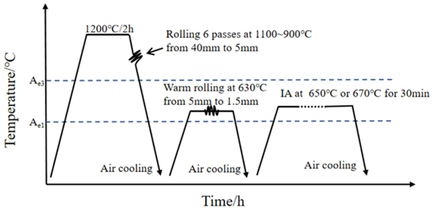
2.1. Experimental Materials

Two 50 kg experimental ingots with two different chemical compositions of Fe-0.16C-5.6Mn-1.0Al and Fe-0.16C-5.6Mn-1.0Al-0.08Ti-0.22Mo (wt%) were melted in a vacuum induction furnace, which referred to as 5.6Mn steel and 5.6MnTiMo steel, respectively. The ingots were homogenized at 1250 °C for 2 h to eliminate the segregation of alloying elements, and then hot rolled into 5 mm thick plates and air-cooled to room temperature. Subsequently, these hot-rolled plates were reheated to approximately 600~650 °C for 10 min, then further warm-rolled into 1.5 mm thick plates by five passes with total reduction of about 70%. Finally, the warm-rolled plates were subjected to intercritical annealing at 650 and 670 °C for 30 min, respectively, followed by air-cooling to room temperature, as illustrated in Figure 1.

2.2. Electrochemical Charging

Electrochemical hydrogen charging of the tensile samples was performed in accordance with ASTM G142 standard [18]. The electrochemically H-charging solution was 3% NaCl plus 0.3% NH4SCN aqueous solution with current densities of 0.2 mA/cm2, 1 mA/cm2 and 5 mA/cm2, respectively, and the charging time of 1 h. After electrochemical hydrogen charging, liquid nitrogen was used for cooling the specimens to avoid the effusion of hydrogen before analyzing the specimens.

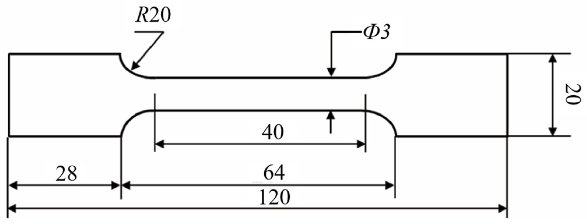
2.3. Mechanical Properties Measurements

Tensile test specimens were prepared according to ASTM standard (E 8M-04) [19] with the long axes parallel to the rolling direction (shown in Figure 2). Uniaxial quasi-static tensile tests were first conducted using a SUNS/UTE5305 testing machine (SUNS Technology Stock Co., Ltd., Shenzhen, China) at RT with a strain rate of 1 × 10⁻³ s−1 to evaluate the tensile properties of samples. The hardness was measured at room-temperature using a XP Nano-indenter (Kaysight Technologies, Böblingen, Germany) with a testing depth of 2000 nm and a dwell time of 10 s. Five hardness values were tested on each sample.

2.4. Slow-Strain Rate Tensile (SSRT) Tests

Before starting the slow-strain rate tensile (SSRT) tests, the specimens were precharged in the same solution, current density, and charging time mentioned in Section 2.2. The dwell time between the end of H pre-charging and the start of tensile testing was below 20 min. The hydrogen pre-charged SSRT tests were performed at the strain rate of 5.0 × 10⁻⁵ s−1. As a reference, an SSRT test with a similar strain rate was performed for the H-uncharged samples. Tensile tests were repeated twice to ensure the reliability of tensile data.

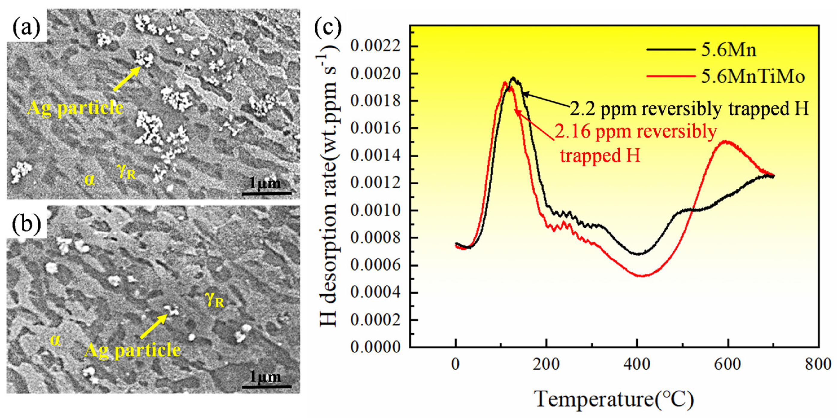
2.5. Hydrogen Content and Distribution Measurements

The diffusible hydrogen content of the specimens was measured using HTDS-003 thermal desorption spectroscopy (R-DEC Co., Ltd., Tsukuba, Ibaraki, Japan) with a quadrupole mass spectrometer at a heating rate of 200 °C/h from RT to 800 °C in vacuum. In order to characterize the spatial distribution of H in the specimens and visualize the distribution position of H in the material, the Ag decoration method was used [11,20,21]. The H-charged specimens were electrolytically polished and corroded for 3 s with 15% sodium bisulfite aqueous solution (NaHSO3), and then quickly placed in a supersaturated aqueous solution of 0.5 g AgBr and 40 mL 5 wt% NaNO2 for 5 min. Finally, the distribution of Ag particles on the surface of the specimens was observed by SEM.

2.6. Microstructural Characterization

The microstructures of the specimens were characterized using JSM-7000F field emission scanning electron microscopy (JEOL, Tokyo, Japan) and NordlysNano electron backscattered diffraction equipment (Oxford Instruments, Abingdon, UK). The EBSD and SEM specimens were first mechanically polished and then electrolytically polished for 30 s at 30 V with an electrolytic polishing solution consisting of perchloric acid (HClO4) and alcohol (C2H5OH) (1:15). Also, the SEM specimens were further etched with 15% sodium bisulfite aqueous solution (NaHSO3). Additionally, SEM analysis was used to investigate the features of the fracture surface and hydrogen microprint specimen.
JEM 2100F Transmission electron microscopy (JEOL, Tokyo, Japan) was used to determine the size and volume fraction of nano-precipitates [22]. TEM specimens were electro–polished in a mixture of 5% perchloric acid (HClO4) and 95% glacial acetic acid (CH3COOH) at −20 °C using a Tenupol-5 Twin–Jet polisher (Struers, Copenhagen, Denmark). Mn concentration distribution (wt%) was quantitatively measured by TEM-EDXS. EBSD data and TEM images were processed using Channel 5 and Digital Micrograph software 3.5, respectively. Grain size characterization was conducted via AZtecCrystal v3.3 software (Oxford Instruments, Abingdon, UK) with optimized band detection mode, employing a 50 nm step size and 95% confidence index threshold for EBSD data acquisition and post-processing.
The volume fraction of γR were measured by D/MAX-2200 X-ray Diffraction (RIGAKU, Tokyo, Japan) using Cu Kα radiation with a scanning speed of 6°min−1 and a 2θ range of 40° to 100°. The γR volume fraction was calculated using the integrated intensities of both ferrite and austenite diffraction peaks [23].

3. Experimental Results

3.1. Mechanical Properties After Intercritical Annealing

Engineering stress–strain curves and hardness values of the warm-rolled steels after intercritical annealing at both 650 and 670 °C for 30 min are displayed in Figure 3. All tensile curves revealed marked Lüders strain, and the jagged flow feature was observed, indicating dynamic strain aging and strong local deformation occurred during tensile deformation [24]. Compared with 5.6Mn steel, the addition of Ti–Mo increased the yield strength (YS) by 236~241 MPa and ultimate tensile strength (UTS) by 183~280 MPa under both annealing conditions, see Figure 3a,b. This indicates that the addition of Ti–Mo microalloying elements dramatically improved both YS and UTS values of medium Mn steels while sacrificing the value of TEL. The product of UTS and TEL values of 5.6MnTiMo treated at 670 °C for 30 min can be reached 35 GPa%. We compared the YS and TEL values of current medium Mn steels with the reported values in the literature [10,22,23,24,25,26,27,28,29,30,31,32,33,34,35,36,37,38,39,40], see Figure 3, implying that the Ti–Mo microalloyed medium Mn steels produced by warm rolling exhibits an excellent balance of YS and TEL, e.g., the maximal YS value can reach approximately 1200 MPa, with the TEL value being 26%. Furthermore, the addition of Ti–Mo can also increase the hardness of medium Mn steels, for example, the hardness increased by 58~76 HV under both annealing conditions.

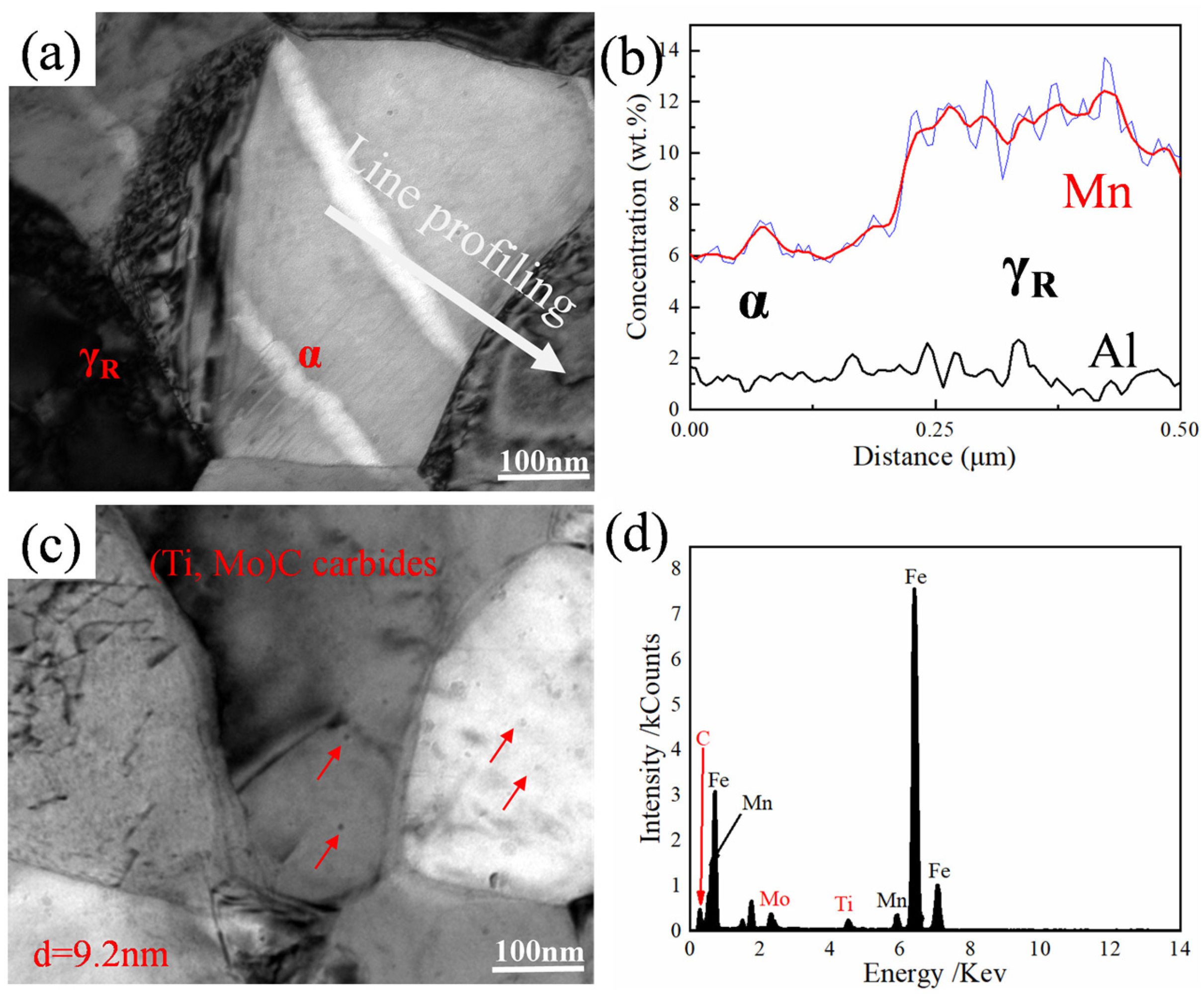
3.2. Microstructure After Intercritical Annealing

To clarify the influence of Ti–Mo addition on the microstructure characteristics of medium Mn steels, EBSD analysis was conducted on the specimens annealed at both 650 and 670 °C for 30 min, and the results are displayed in Figure 4. After warm rolling and intercritical annealing, all specimens basically showed the ultrafine α and γR duplex microstructure, which is characterized by a mix of equiaxed and lamellar morphology. Typically, austenite grains nucleated preferentially at grain boundaries or grain boundary triple junctions of ferrite grains.
To reveal the recrystallization behavior of the deformed martensite and its dependence on Ti–Mo addition, both grain aspect ratio and grain boundaries distribution of both 5.6Mn and 5.6MnTiMo steels were quantitatively analyzed, see Figure 5. The 5.6MnTiMo steel exhibited the higher fraction of α grains with grain aspect ratio < 2 and low-angle grain boundaries (2°~15°). Moreover, the 5.6MnTiMo steel presented smaller grain size relative to 5.6Mn steel, especially for α grains. The average grain size calculated by channel 5 is detailed in Table 1. These above results indicate that the addition of Ti–Mo facilitated the formation of equiaxed grains, and specifically, both α and γR grains in 5.6MnTiMo steels were not sufficiently recovered and recrystallized due to the pinning effect of nano-particles at the grain boundaries by the addition of Ti–Mo, which was also supported by the kernel averaged misorientation (KAM) analysis.
X-ray diffraction (XRD) patterns in Figure 7 demonstrate that both 5.6Mn and 5.6MnTiMo steels exhibit the mixing structures of face-centered cubic (FCC) γR and body-centered cubic (BCC) α phases, which is consistent with the above-mentioned EBSD results in Figure 4. According to Thermo-Calc calculation (Figure 6), the C content in γR was found to decrease with the addition of Ti–Mo in medium Mn steels due to the formation of (Ti, Mo)C carbides.
Thus, the addition of Ti–Mo slightly decreased the volume fraction of γR, regardless of intercritical annealing temperature (Figure 7a). After tensile tests, the peaks of all fcc-structured γR disappeared (Figure 7b), demonstrating that the retained austenite fully transformed to martensite, i.e., TRIP phenomenon occurred upon tensile loading.
The elemental partitioning and nano-precipitation behavior were further demonstrated using TEM analysis for both 5.6Mn and 5.6MnTiMo steels annealed at 670 °C for 30 min. Figure 8a revealed that no precipitates were formed in α and γR for 5.6Mn. The Mn concentration in γR measured by EDXS line profile was about 12 wt%, which is much higher than about 6 wt% in α (Figure 8b). For the 5.6MnTiMo sample, spheroidal MC carbides with an average size of about 9.2 nm were mainly precipitated in the α matrix, with some amount of nano-precipitates at or near the grain boundaries. The MC carbides were further verified as (Ti, Mo)C carbide by EDXS spot analysis (Figure 8d). Therefore, the formation of (Ti,Mo)C carbides was considered the main reason for the decrease in average grain size of both α and γR and the increase in KAM value in 5.6MnTiMo samples.

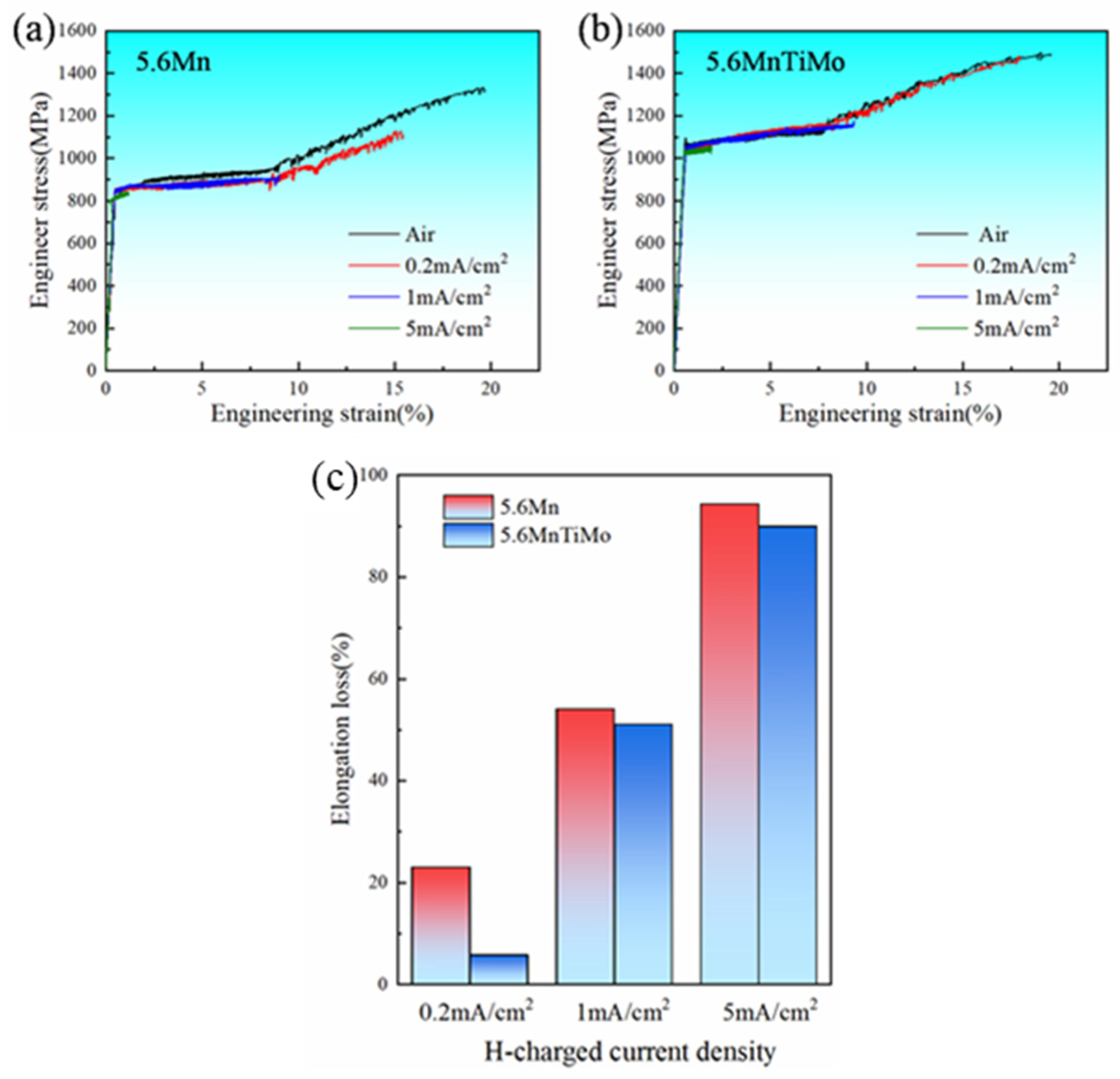
3.3. Hydrogen Embrittlement (HE) Resistance

To evaluate the HE resistance of both 5.6Mn and 5.6MnTiMo steels, slow-strain rate tensile (SSRT) tests were carried out on the H-uncharged and H-charged samples after intercritical annealing at 670 °C for 30 min. In comparison with Figure 9a,b, all H-uncharged and H-charged specimens exhibited similar yield strength and strain hardening behavior, but the addition of Ti–Mo significantly improved the yield and tensile strengths. The H-charged specimens showed premature fracture, and smaller elongation with increasing the current density.
The hydrogen embrittlement susceptibility index of each specimen was evaluated by an elongation loss parameter (Iδ) [22], defined as:
I δ = δ A ir δ H / δ A ir × 100 %
where δAir is the total elongation of an H-uncharged specimen, and δH is the total elongation of an H-charged specimen.
Figure 9c illustrates the variation in HE susceptibility with current density for both 5.6Mn and 5.6MnTiMo steels. The HE susceptibility of both steels gradually increased with increasing the current density. At identical current densities, however, all 5.6MnTiMo samples exhibited the lower HE susceptibility relative to 5.6Mn counterparts. Notably, as the current density was 0.2 mA/cm2, the Iδ value for 5.6MnTiMo steel was only 1.94%. These results demonstrate that the addition of Ti–Mo not only improved the strength levels, but also enhanced the HE resistance of ultra-high-strength medium Mn steels.

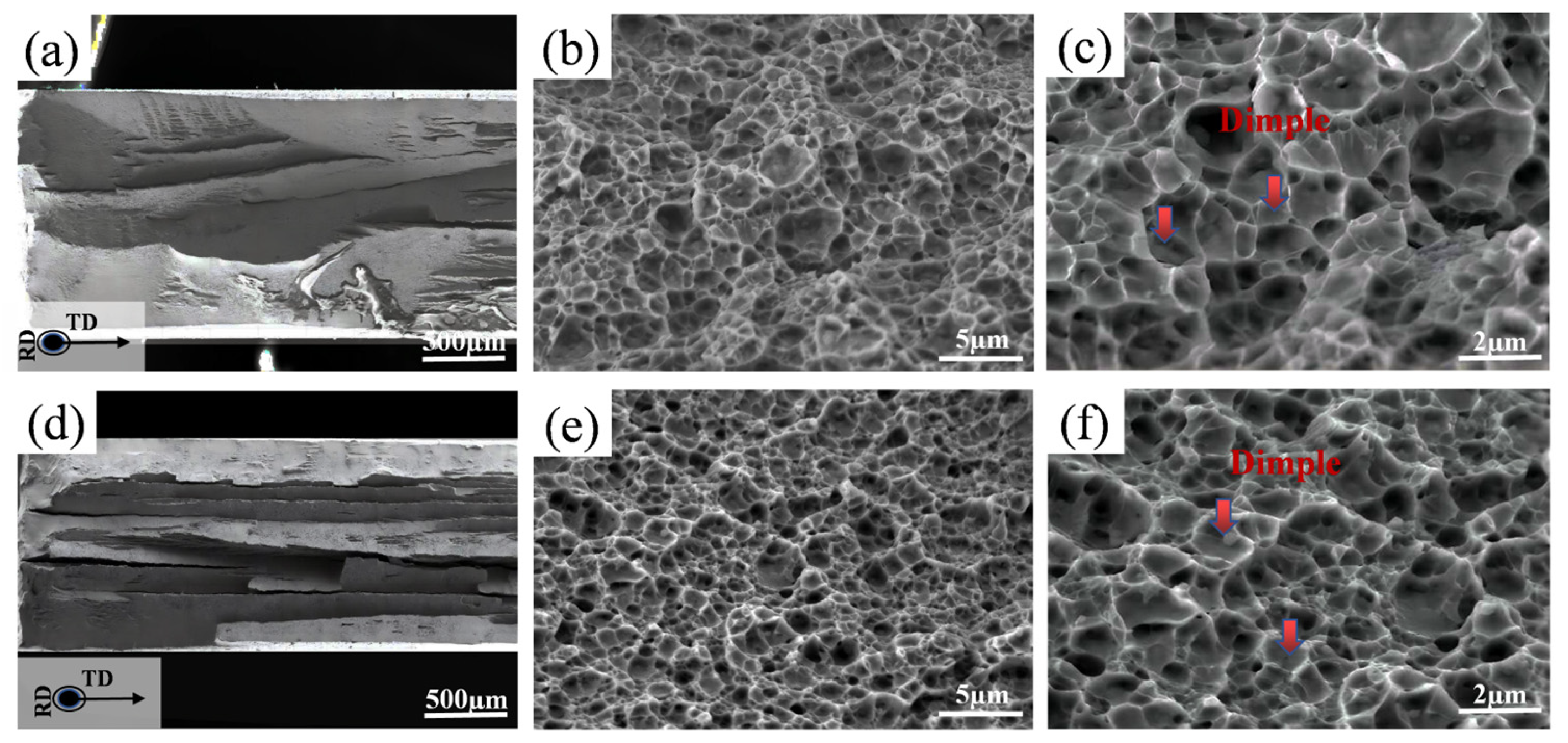
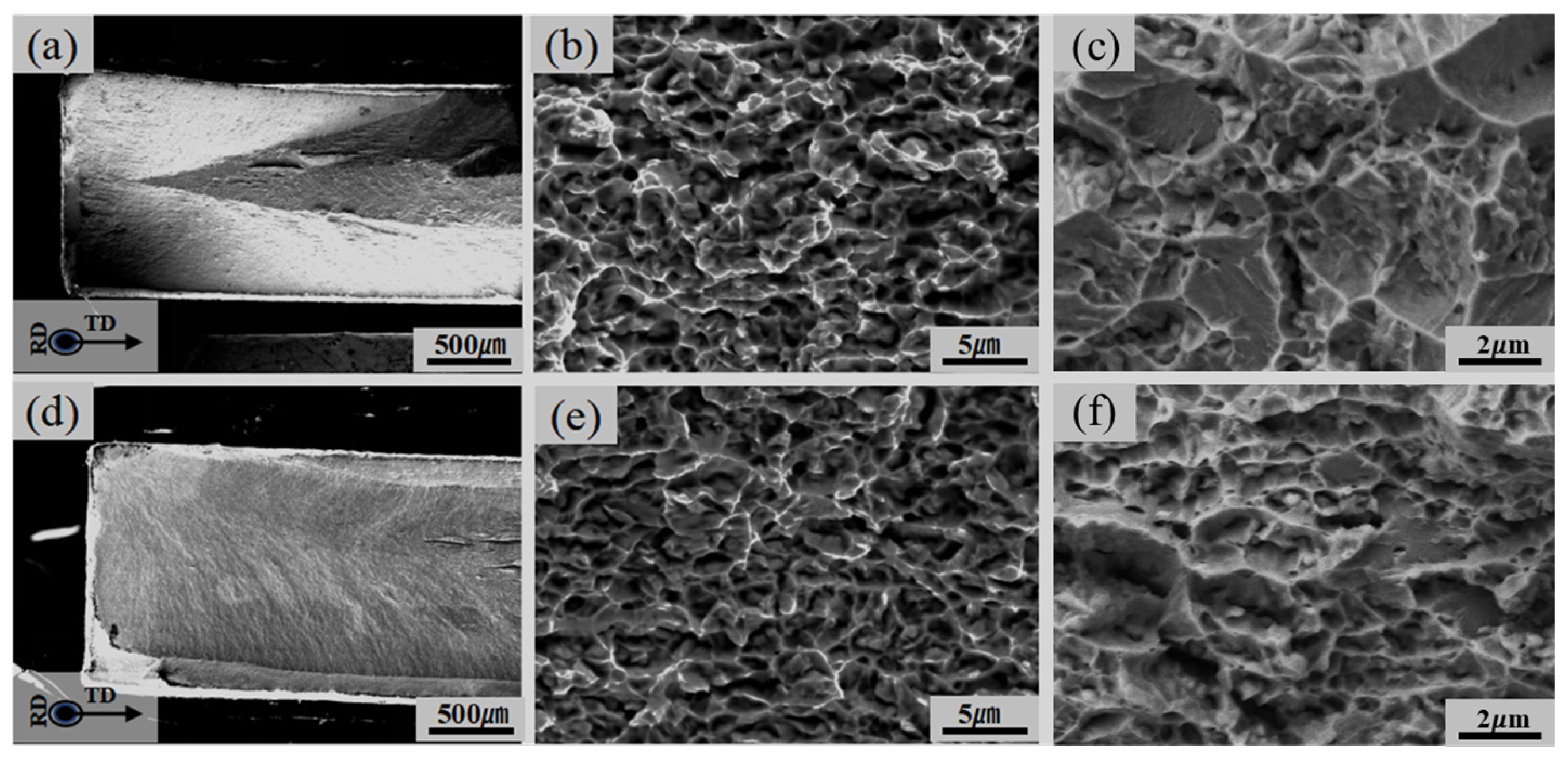
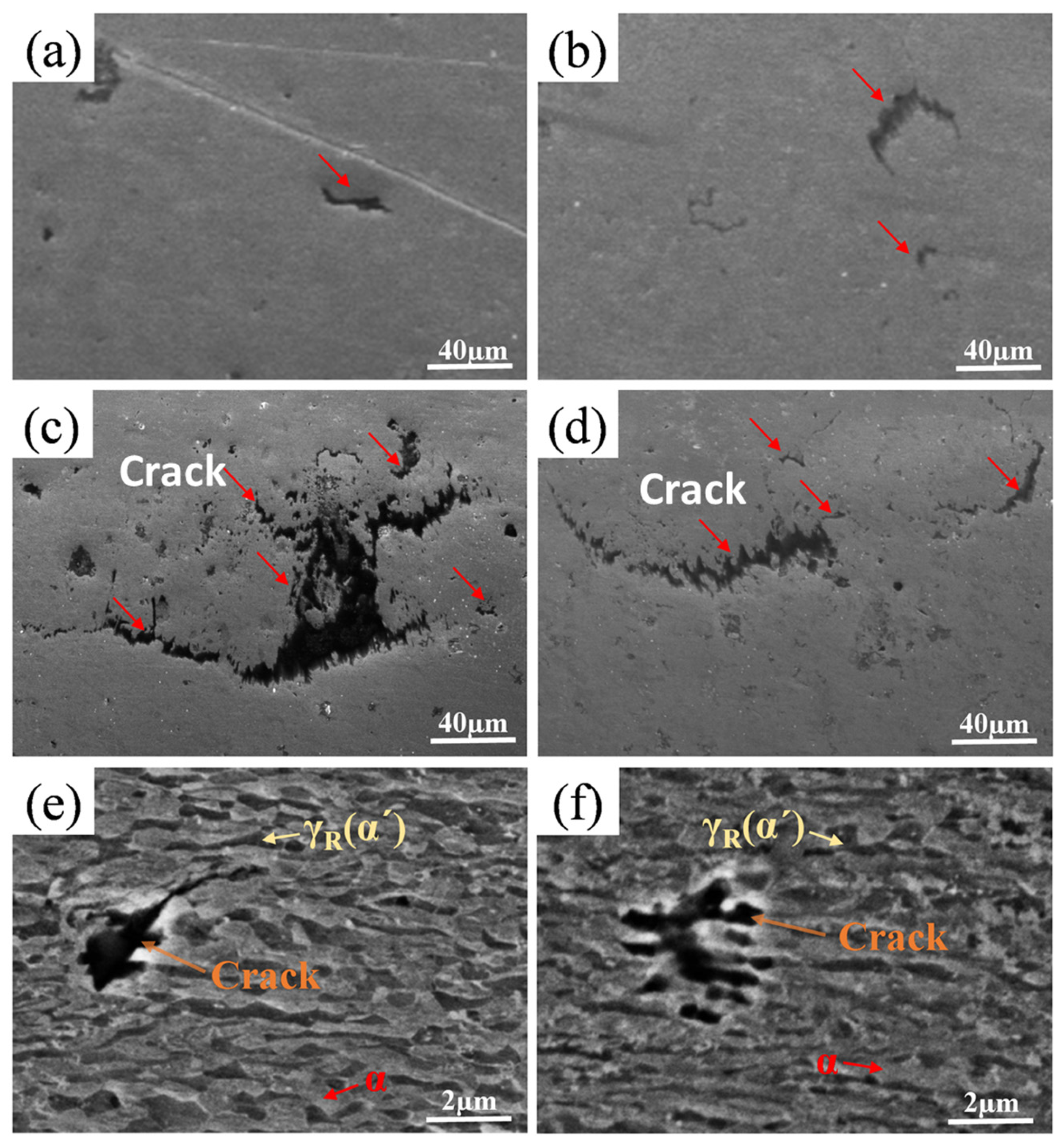
3.4. Fracture Behavior

Figure 10 and Figure 11 show the SEM cross-section fractography from the tensile fracture surface in the H-uncharged and H-charged specimens with a current density of 5 mA/cm2, respectively. All the macro-graphs had obvious delamination phenomena and necking shrinkage (Figure 10a,d). Furthermore, the fracture morphologies were found to be rather heterogeneous with numerous fine and deep dimples, which is a typical ductile fracture mode. In contrast, the addition of Ti–Mo promoted the formation of finer and deeper dimples, due to the finer α and γR duplex microstructure in 5.6MnTiMo steel relative to 5.6Mn steel.
As the H-charging current density of 5 mA/cm2 was used for 5.6Mn and 5.6MnTiMo samples, the macro-graphs in Figure 11a,d exhibited a herringbone pattern, which is totally different from the H-uncharged samples in Figure 10. Furthermore, the fractography of all H-charged specimens was characterized by a representative “hydrogen-induced” brittle fracture. Whereas the 5.6Mn samples revealed ductile fracture featured by fine dimples (Figure 9c,f), the 5.6MnTiMo samples exhibited both flat fractured surfaces (Figure 10c,f) and cracks propagating along the boundaries.
To clarify the influence of hydrogen on the formation and propagation of cracks, the above specimens were further observed using SEM parallel to the tensile axis, and the results are displayed in Figure 12. It can be observed that the number of H-assisted microcracks (HICs) was greater for the H-charged samples (Figure 12a,b), whereas only several microcracks were observed in the H-charged samples (Figure 12a,b). This is especially clear for the H-charged specimens where more HICs formed upon tensile loading. The number of internal microcracks was assumed to coincide with the damage parameter of materials. High magnified images in Figure 12e,f demonstrated that HICs formed inside γ(α′) and propagated along the γ(α′) grain boundaries. Consequently, it could be concluded that increased hydrogen content in medium Mn steels more easily caused the nucleation and propagation of microcracks. This accelerates damage accumulation at low strain levels, leading to premature fracture.

4. Discussion

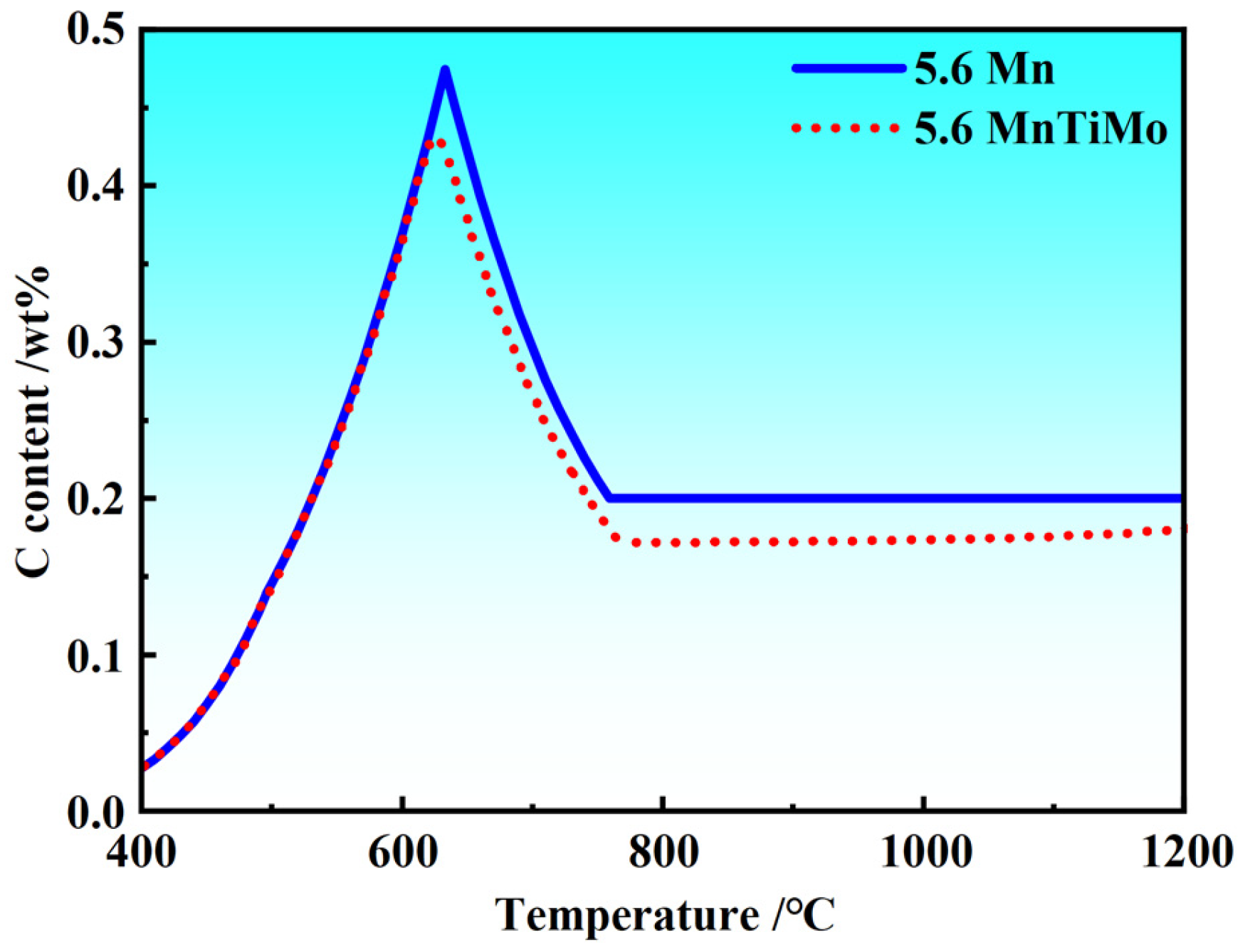
4.1. The Effect of γR Volume Fraction and Ti–Mo Addition on CH

Medium Mn steels are renowned for their ultrafine α and γR duplex microstructure. The γR phase exhibits approximately ten times higher hydrogen solubility and 3~4 orders of magnitude lower hydrogen diffusivity compared to α-ferrite [41,42,43], enabling γR to act as an effective hydrogen reservoir in hydrogen-charged environments [12,13]. To validate this, Ag decoration experiments [11,23,24] were employed to map hydrogen distribution in the α and γR phases of 5.6Mn and 5.6MnTiMo steels (Figure 13a,b). Ag particles preferentially adhered to γR, confirming that >90% of hydrogen atoms dissolved in γR, with hydrogen concentration (CHCH) scaling linearly with γR volume fraction.
TDS analysis (Figure 13c) revealed hydrogen release profiles for both steels after charging at 1 mA/cm2 for 1 h. A diffusible hydrogen desorption peak emerged near 150 °C for both alloys, corresponding to CHCH values of 2.2 ppm (5.6Mn) and 2.16 ppm (5.6MnTiMo). Diffusible hydrogen content is strongly influenced by γR, grain size, and grain boundary characteristics [44,45]. Ti–Mo alloying reduced the γR, which would nominally decrease diffusible hydrogen. However, it simultaneously increased the density of low-angle grain boundaries and refined both α and γR grains, counteracting this trend. This interplay resulted in only minor differences in reversible hydrogen content between the two steels.
To further identify the hydrogen trapping behavior of medium Mn steels, the binding energy of hydrogen was evaluated using Equation (2) [46], together with hydrogen charging conditions and the corresponding hydrogen content (Peak 1) in Figure 13c.
ln ϕ T c 2 1 T c = E a T R
where ϕ is heating rate, T c is peak temperature, E a T is trap activation energy, and R is gas constant.
The calculated binding energies were 51.2, 23.9, and 28.9 kJ/mol for high-angle grain boundaries, dislocations, and nanosized (Ti, Mo)C precipitates, respectively. A comparison with literature values [47,48] (Table 2) confirms that the second desorption peak in 5.6MnTiMo steel corresponds to hydrogen release from nanoscale (Ti, Mo)C particles, acting as effective hydrogen traps [48].
Additionally, the α/γ(α′) interface is the primary site for hydrogen-induced crack initiation [49]. In medium Mn steels, ferrite acts as the soft phase, while γ/α′ serves as the hard phase, resulting in pronounced strain in compatibility between various phases during tensile deformation. In the 5.6MnTiMo sample, the high density of nano-scale precipitates within ferrite enhances its hardness of ferrite (as evidenced by the >200 MPa increase in yield strength shown in Figure 9a,b), thereby reducing strain incompatibility with the γ(α′) phase. Consequently, the 5.6MnTiMo alloy exhibits better resistance to hydrogen embrittlement compared to the 5.6Mn alloy.

4.2. Effect of Ti–Mo Addition on Hydrogen Embrittlement (HE) Susceptibility

It is widely accepted that high-strength steels are particularly prone to hydrogen embrittlement, especially for the strength level exceeding 1000 MPa [50]. Our ultra-high-strength steel (strength level 1.0~1.2 GPa) contained ultrafine α and γR and showed the TRIP effect, characterized by the deformation-driven transformation from fcc-structured γR into body-centered tetragonal α′-martensite. The newly formed α′-martensite and the associated hetero-interfaces were strongly prone to H-induced cracking, which is considered to be the major reason for the premature failure of medium Mn steels when exposed to a hydrogen environment [51].
In the current experiments, it was observed that the HE susceptibility of 5.6MnTiMo was lower than that of 5.6Mn. For this case, Zhang et al. [52] and Shao et al. [53] found that the HE susceptibility of medium Mn steel decreased with the increased γR stability. Therefore, the mechanical stability of γR had a crucial effect on the HE susceptibility of experimental steels. Figure 13a represents the γR volume fractions of undeformed specimens, H-charged tensile fracture specimens with different current densities, and H-uncharged tensile fracture specimens, respectively. The mechanical stability of γR can be assessed by k, which is inversely proportional to the γR stability as below [30]:
k = ln ( V R A / V R A 0 ) / ε
where VRA0 and VRA are the initial austenite fraction and the austenite fraction at true strain ε , k is the mechanical stability of γR, respectively. The higher k value corresponds to the lower stability of retained austenite. The calculated k values of the two experimental steels were shown in Table 3.
Table 2 demonstrated that the mechanical stability of γR of 5.6MnTiMo was higher than that of 5.6Mn, implying that the addition of Ti–Mo improved the mechanical stability of γR in medium Mn steels. As mentioned above, the martensitic transformation was closely related to the plastic instability. From Figure 14b, the sawtooth amplitude of 5.6MnTiMo was less than that of 5.6Mn, which means that the TRIP phenomenon was weakened [54,55]. This corresponds to the theoretically calculated k value, which corresponds well with our preliminary studies [10,56,57].
The aforementioned results demonstrate that Ti–Mo microalloying significantly enhances the mechanical stability of the retained austenite (γR) phase. As illustrated in Figure 15, which illustrates hydrogen-induced cracking propagation, the stabilized γR phase undergoes a slower strain-driven transformation to α′-martensite under tensile loading. This delayed phase transformation kinetics correlates with retarded hydrogen diffusion within the microstructure, as the metastable γR phase temporarily retains hydrogen atoms within its lattice structure. Consequently, the delayed hydrogen redistribution mitigates localized hydrogen accumulation at crack tips, resulting in postponed crack initiation and propagation in the 5.6MnTiMo steel compared to the 5.6Mn counterpart.

5. Conclusions

The effect of Ti–Mo on the HE resistance and fracture behavior of warm-rolled medium Mn steels was investigated using slow strain rate tensile tests, together with the underlying HE mechanisms by the detailed microstructural characterization. The main conclusions are summarized as follows:
(1)
The 5.6MnTiMo exhibits smaller grain sizes relative to the 5.6Mn, especially in the α phase grain size. The (Ti, Mo)C carbides were mostly precipitated in the α phase and rarely in the γR phase.
(2)
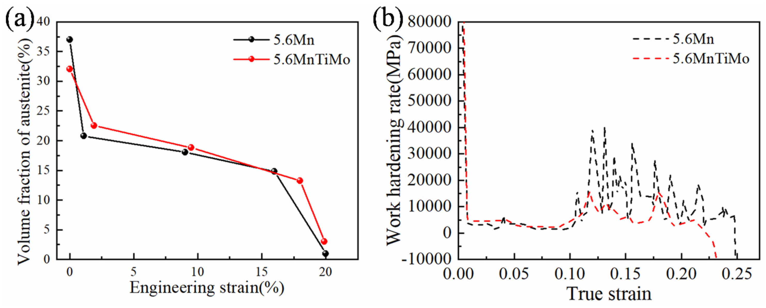
The addition of Ti–Mo increased both yield strength and ultimate tensile strength without sacrificing total elongation due to the synergistic effects of precipitation hardening and grain refinement. The 5.6MnTiMo steel exhibits a strength–ductility product of approximately 35 GPa·% combined with yield strength exceeding 1.2 GPa.
(3)
The enhanced hydrogen embrittlement resistance of the 5.6MnTiMo alloy over the 5.6Mn alloy may be associated with the deep hydrogen traps and the improved strain compatibility at α/γ(α′) phase boundaries due to the addition of Ti and Mo.
(4)
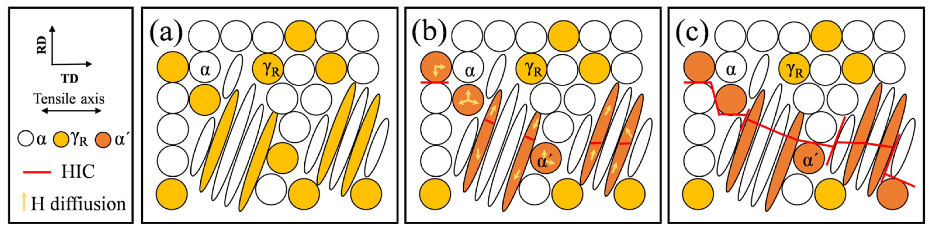
Upon tensile loading, H-assisted microcracks mainly formed within γ(α′) and propagated along the γ(α′) grain boundaries, causing the intergranular cracking and premature fracture.

Author Contributions

Conceptualization, M.C.; methodology, P.Z., J.P. and Y.Z.; formal analysis, J.P., W.H. and S.W.; investigation, W.H. and Y.Z.; data curation, P.Z. and J.P.; writing—original draft preparation, P.Z. and Y.Z.; writing—review and editing, M.C.; supervision, M.C.; project administration, M.C. All authors have read and agreed to the published version of the manuscript.

Funding

This research and APC were funded by National Natural Science Foundation of China (51975111 and 52274379), and Fundamental Research Funding of the Central Universities (2023GFZD14).

Data Availability Statement

The original contributions presented in this study are included in the article. Further inquiries can be directed to the corresponding author.

Conflicts of Interest

All authors state that there is no conflict of interest.

Abbreviations

SymbolsFull writings
HEHydrogen Embrittlement
TRIPTransformation Induced Plasticity
AHSSsAdvanced High-Strength Steels
SSRTSlow-Strain Rate Tensile Test
UTSUltimate Tensile Strength
TELTotal Elongation
YSYield Strength
CHDiffusible Hydrogen Content
RTRoom Temperature
HICHydrogen Induced Cracks
MCMC carbides
TDSThermal Desorption Spectroscopy
kMechanical Stability Factor of Retained Austenite
EDXSEnergy-Dispersive X-ray Spectroscopy
EBSDElectron Backscatter Diffraction
TEMTransmission Electron Microscopy
SEMScanning Electron Microscopy
XRDX-Ray Diffraction
FCCFace-Centered Cubic
BCCBody-Centered Cubic

References

  1. Cai, Z.H.; Cai, B.; Ding, H.; Chen, Y.; Misra, R.D.K. Microstructure and deformation behavior of the hot-rolled medium manganese steels with varying aluminum-content. Mater. Sci. Eng. A 2016, 676, 263–270. [Google Scholar] [CrossRef]
  2. Xu, Y.-B.; Hu, Z.-P.; Zou, Y.; Tan, X.-D.; Han, D.-T.; Chen, S.-Q.; Ma, D.-G.; Misra, R.D.K. Effect of two-step intercritical annealing on microstructure and mechanical properties of hot-rolled medium manganese TRIP steel containing δ-ferrite. Mater. Sci. Eng. A 2017, 688, 40–55. [Google Scholar] [CrossRef]
  3. Sarkar, A.; Sanyal, S.; Bandyopadhyay, T.K.; Mandal, S. Influence of annealing parameters on phase evolution and recrystallization kinetics of a Mn-Al-Si alloyed duplex steel. Mater. Charact. 2017, 134, 213–224. [Google Scholar] [CrossRef]
  4. Lee, Y.K.; Han, J. Current opinion in medium manganese steel. Mater. Sci. Technol. 2014, 31, 843–856. [Google Scholar] [CrossRef]
  5. Cai, M.H.; Zhu, W.J.; Stanford, N.; Pan, L.B.; Chao, Q.; Hodgson, P.D. Dependence of deformation behavior on grain size and strain rate in an ultrahigh strength-ductile Mn-based TRIP alloy. Mater. Sci. Eng. A 2016, 653, 35–42. [Google Scholar] [CrossRef]
  6. Wang, C.; Cao, W.; Shi, J.; Huang, C.; Dong, H. Deformation microstructures and strengthening mechanisms of an ultrafine grained duplex medium-Mn steel. Mater. Sci. Eng. 2013, 562, 89–95. [Google Scholar] [CrossRef]
  7. Miller, R.L. Ultrafine-grained microstructures and mechanical properties of alloy steels. Metall. Mater. Trans. B 1972, 3, 905–912. [Google Scholar] [CrossRef]
  8. Han, Y.; Shi, J.; Xu, L.; Cao, W.Q.; Dong, H. TiC precipitation induced effect on microstructure and mechanical properties in low carbon medium manganese steel. Mater. Sci. Eng. A 2011, 530, 643–651. [Google Scholar] [CrossRef]
  9. Lee, S.; Estrin, Y.; De Cooman, B.C. Constitutive modeling of the mechanical properties of V-added medium manganese TRIP steel. Metall. Mater. Trans. A 2013, 44, 3136–3146. [Google Scholar] [CrossRef]
  10. Pan, H.; Ding, H.; Cai, M. Microstructural evolution and precipitation behavior of the warm-rolled medium Mn steels containing Nb or Nb-Mo during intercritical annealing. Mater. Sci. Eng. A 2018, 736, 375–382. [Google Scholar] [CrossRef]
  11. Park, T.M.; Kim, H.-J.; Um, H.Y.; Goo, N.H.; Han, J. The possibility of enhanced hydrogen embrittlement resistance of medium-Mn steels by addition of micro-alloying elements. Mater. Charact. 2020, 165, 110386. [Google Scholar]
  12. Han, J.; Nam, J.-H.; Lee, Y.-K. The mechanism of hydrogen embrittlement in intercritically annealed medium Mn TRIP steel. Acta Mater. 2016, 113, 1–10. [Google Scholar]
  13. Ryu, J.H.; Chun, Y.S.; Lee, C.S.; Bhadeshia, H.K.D.H.; Suh, D.W. Effect of deformation on hydrogen trapping and effusion in TRIP-assisted steel. Acta Mater. 2012, 60, 4085–4092. [Google Scholar] [CrossRef]
  14. Zhu, X.; Li, W.; Hsu, T.Y.; Zhou, S.; Wang, L.; Jin, X. Improved resistance to hydrogen embrittlement in a high-strength steel by quenching–partitioning–tempering treatment. Scr. Mater. 2015, 97, 21–24. [Google Scholar] [CrossRef]
  15. Bhadeshia, H.K.D.H. Prevention of hydrogen embrittlement in steels. ISIJ Int. 2016, 56, 24–36. [Google Scholar]
  16. Liu, Y.; Cao, Z.; Huang, C.; Hu, C.; Huang, M. Enhancing hydrogen embrittlement resistance of TRIP-rich medium Mn steel by morphology optimization. Sci. China Mater. 2023, 66, 4258–4266. [Google Scholar]
  17. Li, Y.; Li, W.; Min, N.; Liu, H.; Jin, X. Homogeneous elasto-plastic deformation and improved strain compatibility between austenite and ferrite in a co-precipitation hardened medium Mn steel with enhanced hydrogen embrittlement resistance. Int. J. Plast. 2020, 133, 102805. [Google Scholar]
  18. ASTM G142-98(2013); Standard Test Method for Determination of Susceptibility of Metallic Materials to Hydrogen Embrittlement (HE). ASTM International: West Conshohocken, PA, USA, 2013.
  19. ASTM E8M-04; Standard Test Methods for Tension Testing of Metallic Materials [Metric]. ASTM International: West Conshohocken, PA, USA, 2004.
  20. Sun, X.Y.; Zhang, B.; Lin, H.Q.; Zhou, Y.; Sun, L.; Wang, J.Q.; Han, E.H.; Ke, W. Correlations between stress corrosion cracking susceptibility and grain boundary microstructures for an Al–Zn–Mg alloy. Corros. Sci. 2013, 77, 103–112. [Google Scholar]
  21. Vandijk, N.; Butt, A.; Zhao, L.; Sietsma, J.; Offerman, S.; Wright, J.; Vanderzwaag, S. Thermal stability of retained austenite in TRIP steels studied by synchrotron X-ray diffraction during cooling. Acta Mater. 2005, 53, 5439–5447. [Google Scholar]
  22. Wang, G.; Yan, Y.; Li, J.; Huang, J.; Qiao, L.; Volinsky, A.A. Microstructure effect on hydrogen-induced cracking in TM210 maraging steel. Mater. Sci. Eng. A 2013, 586, 142–148. [Google Scholar] [CrossRef]
  23. Ryu, J.H.; Kim, S.K.; Lee, C.S.; Suh, D.-W.; Bhadeshia, H.K.D.H. Effect of aluminium on hydrogen-induced fracture behaviour in austenitic Fe–Mn–C steel. Proc. R. Soc. A Math. Phys. Eng. Sci. 2013, 469, 2149. [Google Scholar] [CrossRef]
  24. Tarzimoghadam, Z.; Rohwerder, M.; Merzlikin, S.V.; Bashir, A.; Yedra, L.; Eswara, S.; Ponge, D.; Raabe, D. Multi-scale and spatially resolved hydrogen mapping in a Ni–Nb model alloy reveals the role of the δ phase in hydrogen embrittlement of alloy 718. Acta Mater. 2016, 109, 69–81. [Google Scholar]
  25. Tsuchida, N.; Tomota, Y.; Nagai, K.; Fukaura, K. A simple relationship between Lüders elongation and work-hardening rate at lower yield stress. Scr. Mater. 2006, 54, 57–60. [Google Scholar]
  26. Shi, J.; Sun, X.; Wang, M.; Hui, W.; Dong, H.; Cao, W. Enhanced work-hardening behavior and mechanical properties in ultrafine-grained steels with large-fractioned metastable austenite. Scr. Mater. 2010, 63, 815–818. [Google Scholar]
  27. Gibbs, P.J.; De Moor, E.; Merwin, M.J.; Clausen, B.; Speer, J.G.; Matlock, D.K. Austenite stability effects on tensile behavior of manganese-enriched-austenite transformation-induced plasticity steel. Metall. Mater. Trans. A 2011, 42, 3691–3702. [Google Scholar]
  28. Heo, Y.U.; Suh, D.W.; Lee, H.C. Fabrication of an ultrafine-grained structure by a compositional pinning technique. Acta Mater. 2014, 77, 236–247. [Google Scholar] [CrossRef]
  29. Suh, D.W.; Park, S.J.; Lee, T.H.; Oh, C.S.; Kim, S.J. Influence of Al on the microstructural evolution and mechanical behavior of low-carbon, manganese transformation-induced-plasticity steel. Metall. Mater. Trans. A 2009, 41, 397–408. [Google Scholar] [CrossRef]
  30. Cai, Z.H.; Ding, H.; Misra, R.D.K.; Ying, Z.Y. Austenite stability and deformation behavior in a cold-rolled transformation-induced plasticity steel with medium manganese content. Acta Mater. 2015, 84, 229–236. [Google Scholar]
  31. Lee, C.Y.; Jeong, J.; Han, J.; Lee, S.J.; Lee, S.; Lee, Y.K. Coupled strengthening in a medium manganese lightweight steel with an inhomogeneously grained structure of austenite. Acta Mater. 2015, 84, 1–8. [Google Scholar] [CrossRef]
  32. Jeong, M.S.; Park, T.M.; Choi, S.; Lee, S.J.; Han, J. Recovering the ductility of medium-Mn steel by restoring the original microstructure. Scr. Mater. 2021, 190, 16–21. [Google Scholar]
  33. Park, S.J.; Hwang, B.; Lee, K.H.; Lee, T.H.; Suh, D.W.; Han, H.N. Microstructure and tensile behavior of duplex low-density steel containing 5mass% aluminum. Scr. Mater. 2013, 68, 365–369. [Google Scholar] [CrossRef]
  34. Lee, S.; De Cooman, B.C. Tensile behavior of intercritically annealed 10 pct Mn multi-phase steel. Metall. Mater. Trans. A 2013, 45, 709–716. [Google Scholar] [CrossRef]
  35. Wang, T.; Hu, J.; Misra, R.D.K. Microstructure evolution and strain behavior of a medium Mn TRIP/TWIP steel for excellent combination of strength and ductility. Mater. Sci. Eng. A 2019, 753, 99–108. [Google Scholar] [CrossRef]
  36. Ma, Y.; Sun, B.; Schökel, A.; Song, W.; Ponge, D.; Raabe, D.; Bleck, W. Phase boundary segregation-induced strengthening and discontinuous yielding in ultrafine-grained duplex medium-Mn steels. Acta Mater. 2020, 200, 389–403. [Google Scholar] [CrossRef]
  37. Sun, S.; Cai, M.; Ding, H.; Huang, H.; Pan, H. Deformation mechanisms of a novel Mn-based 1 GPa TRIP/TWIP assisted lightweight steel with 63% ductility. Mater. Sci. Eng. A 2021, 802, 140658. [Google Scholar] [CrossRef]
  38. Lee, S.; De Cooman, B.C. Tensile behavior of intercritically annealed ultra-fine grained 8% Mn multi-phase steel. Steel Res. Int. 2015, 86, 1170–1178. [Google Scholar] [CrossRef]
  39. Lei, M.; Hui, W.; Wang, J.; Zhang, Y.; Zhao, X. Microstructure and mechanical properties of hot-rolled V-microalloyed Al-containing medium-Mn steel. J. Iron Steel Res. Int. 2020, 27, 537–548. [Google Scholar] [CrossRef]
  40. Cai, M.; Li, Z.; Chao, Q.; Hodgson, P.D. A novel Mo and Nb microalloyed medium Mn TRIP steel with maximal ultimate strength and moderate ductility. Metall. Mater. Trans. A 2014, 45, 5624–5634. [Google Scholar] [CrossRef]
  41. Zhu, X.; Zhang, K.; Li, W.; Jin, X. Effect of retained austenite stability and morphology on the hydrogen embrittlement susceptibility in quenching and partitioning treated steels. Mater. Sci. Eng. A 2016, 658, 400–408. [Google Scholar] [CrossRef]
  42. Gao, Z.H.; Xue, Y.F.; Li, J.X.; Xu, L.N.; Qiao, L.J. The mechanism of the high resistance to hydrogen-induced strength loss in ultra-high strength high-entropy alloy. Metals 2022, 12, 971. [Google Scholar] [CrossRef]
  43. Takai, K.; Watanuki, R. Hydrogen in trapping states innocuous to environmental degradation of high-strength steels. ISIJ Int. 2003, 43, 520–526. [Google Scholar]
  44. Nagao, A.; Hayashi, K.; Ol, K.; Mitao, S. Effect of uniform distribution of fine cementite on hydrogen embrittlement of low carbon martensitic steel plates. ISIJ Int. 2012, 52, 213–221. [Google Scholar]
  45. Luo, Y.; Lu, H.; Min, N.; Li, W.; Jin, X. Effect of Mo and Nb on mechanical properties and hydrogen embrittlement of hot-rolled medium-Mn steels. Mater. Sci. Eng. A 2022, 844, 143108. [Google Scholar]
  46. Itakura, M.; Kaburaki, H.; Yamaguchi, M.; Okita, T. The effect of hydrogen atoms on the screw dislocation mobility in bcc: Iron: A first-principles study. Acta Mater. 2013, 61, 6857–6867. [Google Scholar]
  47. Abe, A.; Suzaki, H.; Takai, K.; Ishikawa, N.; Sueyoshi, H. Identification of hydrogen trapping sites, binding energies, and occupation ratios at vacancies, dislocations and grain boundaries in iron of varying carbon content. Mater. Sci. Technol. Conf. Exhib. 2011, 2011, 1277–1284. [Google Scholar]
  48. Lee, H.G.; Lee, J.Y. Hydrogen trapping by TiC particles in iron. Acta Metall. 1984, 32, 131–136. [Google Scholar] [CrossRef]
  49. Zhou, Y.; Wang, Z.; Li, W.G.; Li, J.X. Unveiling the inconsistent improvement of hydrogen embrittlement resistance by Nb precipitates in medium Mn steels: Impact of hydrogen distribution. Mater. Sci. Eng. A 2025, 922, 147629. [Google Scholar]
  50. Matsuoka, S.; Homma, N.; Tanaka, H.; Fukushima, Y.; Murakami, Y. Effect of hydrogen on the tensile properties of 900-MPa-class JIS-SCM435 low-alloy-steel for use in storage cylinder of hydrogen station. J. Jpn. Inst. Met. Mater. 2006, 70, 1002–1011. [Google Scholar]
  51. Sun, B.H.; Lu, W.J.; Gault, B.; Ding, R.; Makineni, S.K.; Wan, D.; Wu, C.H.; Chen, H.; Ponge, D.; Raabe, D. Chemical heterogeneity enhances hydrogen resistance in high-strength steels. Nat. Mater. 2021, 20, 1629–1634. [Google Scholar] [PubMed]
  52. Zhang, Y.; Hui, W.; Zhao, X.; Wang, C.; Cao, W.; Dong, H. Effect of reverted austenite fraction on hydrogen embrittlement of TRIP-aided medium Mn steel (0.1C-5Mn). Eng. Fail. Anal. 2019, 97, 605–616. [Google Scholar]
  53. Shao, C.; Hui, W.; Zhang, Y.; Zhao, X.; Weng, Y. Effect of intercritical annealing time on hydrogen embrittlement of warm-rolled medium Mn steel. Mater. Sci. Eng. A 2018, 726, 320–331. [Google Scholar] [CrossRef]
  54. Sun, B.; Vanderesse, N.; Fazeli, F.; Scott, C.; Chen, J.; Bocher, P.; Jahazi, M.; Yue, S. Discontinuous strain-induced martensite transformation related to the Portevin-Le Chatelier effect in a medium manganese steel. Scr. Mater. 2017, 133, 9–13. [Google Scholar] [CrossRef]
  55. Xu, J.; Wang, Z.; Yan, Y.; Li, J.; Wu, M. Effect of hot/warm rolling on the microstructures and mechanical properties of medium-Mn steels. Mater. Charact. 2020, 170, 110682. [Google Scholar] [CrossRef]
  56. Pan, H.J.; Ding, H.; Cai, M.H.; Kibaroglu, D.; Ma, Y.; Song, W.W. Precipitation behavior and austenite stability of Nb or Nb–Mo micro-alloyed warm-rolled medium-Mn steels. Mater. Sci. Eng. A 2019, 766, 138371. [Google Scholar] [CrossRef]
  57. Wang, S.Y.; Wang, C.Z.; Li, H.Y.; Xu, D.F.; Zhang, H.T.; Xiao, N.; Cai, M.H.; Lee, Y.K. Ti-Mo microalloyed medium Mn steels: Precipitation and strengthening mechanism. Mater. Sci. Eng. A 2024, 909, 146866. [Google Scholar] [CrossRef]
Figure 1. Schematic illustration of thermo-mechanical process comprising warm rolling and intercritical annealing (IA).
Metals 15 00397 g001
Figure 2. Geometry of uniaxial quasi-static tensile tests and slow strain rate tensile tests (SSRT) specimens, all dimensions in millimeters.
Metals 15 00397 g002
Figure 3. Engineering stress–strain curves of warm-rolled 5.6Mn and 5.6MnTiMo steels after intercritical annealing at 650 °C (a) and 670 °C (b); (c) Plot between yield strength (YS) and total elongation (TEL) in various medium Mn steels; (d) Average hardness values of experimental steels under both annealing conditions. Note: Data points in (c) were obtained from various medium Mn steels in literature [10,25,26,27,28,29,30,31,32,33,34,35,36,37,38,39].
Metals 15 00397 g003
Figure 4. EBSD IQ-phase and KAM maps of 5.6Mn (a,c) and 5.6MnTiMo (b,d) samples after intercritical annealing at 650 °C (a,b) and 670 °C (c,d). The blue and red colors represent α and γR, respectively, and the white and black lines indicate low-angle (2°~15°) and high-angle (>15°) grain boundaries, respectively.
Metals 15 00397 g004
Figure 5. Grain aspect ratio and grain boundaries distribution plots in the ferrite matrix of warm-rolled 5.6Mn (a,b,e,f) and 5.6MnTiMo (c,d,g,h) samples after intercritical annealing at 650 °C (ad) and 670 °C (eh).
Metals 15 00397 g005
Figure 6. The calculated fraction of carbon concentration in austenite as a function of temperature.
Metals 15 00397 g006
Figure 7. X-ray diffraction (XRD) patterns of warm-rolled 5.6Mn and 5.6MnTiMo samples after intercritical annealing at 650 °C and 670 °C before (a) and after (b) tensile tests.
Metals 15 00397 g007
Figure 8. TEM micrograph of warm-rolled 5.6Mn (a) and 5.6MnTiMo (c) samples after intercritical annealing at 670 °C for 30 min; (b) EDXS line profile of 5.6Mn sample showing the representative change in Mn and Al concentration nearby the ferrite and austenite interface; (d) EDXS spot analysis of [Ti, Mo]C precipitates in the 5.6MnTiMo sample, as marked by red arrows.
Metals 15 00397 g008
Figure 9. Slow-strain rate tensile (SSRT) testing results of 5.6Mn (a) and (b) 5.6MnTiMo steels after warm rolling and intercritical annealing at 670 °C for 30 min; (c) show the change in elongation loss of H-charged samples as a function of current density.
Metals 15 00397 g009
Figure 10. SEM fractography of H-uncharged of (ac) 5.6Mn, and (df) 5.6MnTiMo after warm rolling and intercritical annealing at 670 °C for 30 min.
Metals 15 00397 g010
Figure 11. SEM fractography of H-charged of (ac) 5.6Mn, and (df) 5.6MnTiMo after warm rolling and intercritical annealing at 670 °C for 30 min.
Metals 15 00397 g011
Figure 12. SEM microstructure of (a,b) H-uncharged specimens, and (cf) H-charged specimens at the normal direction. (a,c,e) 5.6Mn steel and (b,d,f) 5.6MnTiMo steel.
Metals 15 00397 g012
Figure 13. SEM fractography of (a) 5.6Mn, and (b) 5.6MnTiMo after immersion in Ag decoration solution and the corresponding hydrogen desorption rate curves (c).
Metals 15 00397 g013
Figure 14. (a) Changes in the volume fraction of γR with engineering strain in 5.6Mn and 5.6MnTiMo steels; (b) Work hardening rate curves of H-uncharged 5.6Mn and 5.6MnTiMo specimens.
Metals 15 00397 g014
Figure 15. Schematic diagrams showing the HIC propagation. (a) Initial state; (b) crack nucleation; (c) crack propagation.
Metals 15 00397 g015
Table 1. Average grain size of both ferrite and austenite in 5.6Mn and 5.6MnTiMo samples.
SteelsPhaseAverage Grain Size (μm)Volume Fraction of γR
650 °C670 °C650 °C670 °C
5.6Mnα1.33 ± 0.321.45 ± 0.2933%37%
γR0.38 ± 0.130.52 ± 0.18
5.6MnTiMoα1.12 ± 0.261.21 ± 0.2829%32%
γR0.36 ± 0.120.48 ± 0.15
Table 2. A comparison between the calculated hydrogen trap binding energies and those reported in the literature.
Hydrogen Trap StateHydrogen Trap Binding Energy Calculated in This Study (kJ/mol)Hydrogen Trap Binding Energy Reported in the Literature (kJ/mol) [45,46]
High-angle grain boundary51.247.47
Dislocation23.924.70
Nanosized (Ti, Mo)C precipitate28.928.17
Table 3. The k value of both 5.6Mn and 5.6MnTiMo steels.
SteelsVRA0 (%)VRA (%)k
5.6Mn36118.8
5.6MnTiMo33312.6
Disclaimer/Publisher’s Note: The statements, opinions and data contained in all publications are solely those of the individual author(s) and contributor(s) and not of MDPI and/or the editor(s). MDPI and/or the editor(s) disclaim responsibility for any injury to people or property resulting from any ideas, methods, instructions or products referred to in the content.

