Article

Influence of Nd:YAG Laser Melting on an Investment-Casting Co-Cr-Mo Alloy

by
Francisco Cepeda Rodríguez
1,2,*,
Carlos Rodrigo Muñiz Valdez
1,
Juan Carlos Ortiz Cuellar
1,
Jesús Fernando Martínez Villafañe
1,
Jesús Salvador Galindo Valdés
1 and
Gladys Yerania Pérez Medina
2
1
Facultad de Ingeniería, Universidad Autónoma de Coahuila, Fundadores Km. 13, Las Glorias, Ciudad Universitaria, Arteaga 25350, Coahuila, Mexico
2
Corporación Mexicana de Investigación en Materiales S.A. de C.V., C. Ciencia y Tecnología 790, Saltillo 400, Saltillo 25290, Coahuila, Mexico
*
Author to whom correspondence should be addressed.
Metals 2025, 15(4), 385; https://doi.org/10.3390/met15040385
Submission received: 22 February 2025 / Revised: 18 March 2025 / Accepted: 25 March 2025 / Published: 29 March 2025

Abstract

The investment casting process, also known as lost-wax casting, is widely used for producing ferrous and non-ferrous metal parts due to its excellent surface finish and dimensional accuracy. In recent years, the use of Co-Cr-Mo alloy has increased due to its high corrosion resistance, good biocompatibility, and relatively high wear resistance. Laser melting of materials has been demonstrated to refine the surface grain structure, reduce surface roughness, and improve both wear and corrosion resistance. The ability to fine-tune parameters such as laser power density and scanning speed facilitates the optimization of the treated layers’ thickness and homogeneity, thereby addressing many of the shortcomings inherent in conventional methods. This study investigates the microstructural, mechanical wear and bioactive behavior of investment-cast Co-Cr-Mo parts subjected to a Nd:YAG laser surface treatment. The effects of different processing parameters were analyzed quantitatively and comprehensively. The specimens were characterized using metallographic techniques, bioactivity evaluation, scanning electron microscopy (SEM), energy-dispersive X-ray spectroscopy (EDX), wear testing (Pin-on-Disk), and hardness testing. Our results demonstrate that Nd:YAG laser melting significantly enhances the surface properties and maintains the dimensional accuracy of complex Co-Cr-Mo biomedical components, through microstructural refinement, increased hardness, improved wear resistance, and preserved biocompatibility. The specific combination of investment casting with precisely controlled laser surface modification represents a significant advancement for improving the longevity and performance of biomedical implants.

1. Introduction

In today’s era of advanced biomedical engineering, the quest for improved materials in orthopedic implants is paramount. Modern prosthetic devices must not only exhibit optimal mechanical strength and corrosion resistance, but also present superior surface properties that enhance osseointegration and minimize adverse biological reactions [1]. Critical factors influencing implant success include precise design, excellent initial surface finish, robust mechanical behavior, and effective chemical interaction with the surrounding bone tissue.
Cobalt chromium molybdenum (Co-Cr-Mo) alloys, particularly those complying with ASTM F75, are widely used in hip and knee prostheses due to their wear and corrosion resistance, as well as their proven biocompatibility [2]. Investment casting has emerged as a preferred manufacturing method for complex components. This technique provides superior dimensional accuracy and precise control over the alloy’s surface characteristics. It integrates mechanical robustness with favorable biological interactions while delivering an exceptional surface finish compared to conventional machining methods. Additionally, investment casting enables the production of intricate geometries that would otherwise be difficult or prohibitively expensive to achieve through traditional subtractive techniques [3,4]. Although investment casting offers superior dimensional accuracy for complex components, the as-cast surface often exhibits microstructural heterogeneities, dendritic structures, and potential casting defects that can lead to reduced wear resistance and biocompatibility.
Despite the advantages of investment casting, implant failure still occurs, often due to the loosening of the prosthesis and the generation of wear debris [3]. The release of metal ions and particles from the implant surface can provoke an inflammatory response, leading to fibrous tissue formation (osteolysis) that ultimately compromises the bone–implant interface and necessitates revision surgeries [5,6]. Considering these challenges, extensive research has focused on improving the surface characteristics of Co-Cr-Mo alloys to enhance their wear resistance and bioactivity. Moreover, ensuring long-term implant stability not only improves patient outcomes, but also reduces the economic burden associated with revision surgeries.
Among the various surface modification techniques, laser surface treatment, particularly using Nd:YAG lasers, has shown considerable promise. This non-contact process offers a chemically clean environment, precise control over the temperature profile and penetration depth, and the ability to modify the microstructure without introducing extraneous contaminants [7,8]. Unlike the inhomogeneous and porous layers resulting from additive manufacturing [9] or plasma-sprayed coating [10], the laser remelting of investment-cast components offers the potential for dense, homogeneous surface modification through the controlled melting and rapid solidification of the existing substrate material. Our approach specifically targets the elimination of as-cast inhomogeneities rather than building new material layers, which fundamentally differentiates our expected outcomes from those reported in previous studies [9,10]. Furthermore, these microstructural changes are associated with enhanced bioactivity and wear resistance, which is crucial for promoting stable osseointegration and long-term implant success [11,12]. The ability to tailor the surface at a microscale level opens up new avenues for the integration of implants with native bone tissue, potentially mitigating complications such as implant loosening and chronic inflammation [1].
The success of laser surface modification, however, relies on the careful optimization of processing parameters such as laser power, pulse duration, and scanning speed. These parameters govern the depth and uniformity of the remelted layer, which in turn influence the mechanical performance and biological response of the treated implant surface [13,14,15]. Recent studies have emphasized that optimizing these factors can significantly improve the overall performance of Co-Cr-Mo components, making them even more suitable for demanding biomedical applications [2,12]. In addition, the integration of simulation techniques with experimental studies is beginning to offer deeper insights into the thermal and metallurgical phenomena occurring during laser treatment, thereby facilitating a more rational design of processing protocols.
Traditional surface modification methods, such as plasma spraying and chemical treatments, have been widely employed to enhance implant surfaces. However, these techniques often suffer from issues related to coating uniformity, limited control over layer thickness, and the risk of introducing contaminants that can adversely affect cell–implant interactions [1]. In contrast, laser treatment enables localized and precisely controlled surface melting, resulting in a refined microstructure that enhances wear resistance [16,17,18,19]. Likewise, unlike conventional heat treatment that gradually applies uniform heat to the bulk samples, surface laser treatment provides localized heating of the surface and rapid cooling, exerting a thermal influence on the surrounding area. Hedberg et al. [20] conducted a comprehensive study on Co-Cr-Mo dental alloys fabricated using selective laser melting, demonstrating up to a 38% improvement in corrosion resistance and a 27% enhancement in biocompatibility compared to conventionally manufactured counterparts. Their investigation of metal ion release showed a significant reduction of 42% in Co ions and 33% in Cr ions from laser-processed samples in simulated body fluids. Bartolomeu et al. [21] reported that optimized laser parameters can significantly reduce the friction coefficient of Co-Cr-Mo surfaces by 25–30% (from 0.52 to 0.38) under simulated physiological conditions. Their electrochemical analysis demonstrated enhanced corrosion resistance, with a 65% increase in polarization resistance and a 40% reduction in passive current density. Surface roughness measurements showed a decrease from Ra = 3.2 μm to Ra = 0.8 μm after laser treatment, creating a more favorable interface for biological interactions. These improvements were achieved through carefully controlled laser parameters with power densities of 5–7 kW/cm2 and pulse durations of 10–15 ms.
The intense surface heating and swift cooling promote martensitic transformations and increase the dislocation density, thereby improving hardness and wear resistance. As the depth from the treated surface increases, the thermal effect diminishes, leading to a reduction in grain size [22]. Additionally, applying laser scanning to alloys containing precipitates can further enhance mechanical performance [14]. Moreover, the ability to fine-tune parameters such as laser power density and scanning speed facilitates the optimization of the treated layers’ thickness and homogeneity, thereby addressing many of the shortcomings inherent in conventional methods. These findings establish a clear precedent for the beneficial effects of laser processing on Co-Cr-Mo alloys, though their focus on additive manufacturing differs from the post-processing approach investigated in our study.
According to industry reports [23], investment casting accounts for approximately 65% of all Co-Cr-Mo implant production, with an estimated market value of $2.3 billion in 2024, making improvements in this manufacturing pathway particularly impactful. The specific application of Nd:YAG laser melting to investment-cast components represents a novel approach. Investment-cast components exhibit fundamentally different initial microstructures and surface characteristics compared to additively manufactured or coated components, with casting typically resulting in larger grain sizes (50–100 μm) and distinct dendritic structures. Mantrala et al. [24] demonstrated that these microstructural differences significantly affect the thermal response and subsequent phase transformations during laser processing. Our research addresses this gap by systematically investigating the effects of laser melting on investment-cast Co-Cr-Mo components, providing much-needed insights into this commercially relevant manufacturing pathway.
By merging advanced casting techniques with optimized laser surface treatments, this study seeks to contribute to the development of more durable and biocompatible orthopedic implants, ultimately enhancing implant longevity and patient quality of life. The improved wear resistance translates to reduced debris generation, potentially decreasing implant failure rates and the need for revision surgeries, which benefits both patients and healthcare providers. Enhanced biocompatibility and surface properties may lead to faster osseointegration and improved long-term clinical outcomes, which is particularly valuable for younger and more active patients. The primary end-users of this research include orthopedic implant manufacturers who utilize investment casting for producing Co-Cr-Mo components, biomedical engineers developing next-generation implant designs, and clinicians who select implant materials for patients with specific needs. Therefore, the primary objective of this study is to systematically examine the microstructural characteristics, mechanical wear resistance, and bioactive behavior of investment-cast Co-Cr-Mo alloy components subjected to surface melting using an Nd:YAG laser.

2. Materials and Methods

2.1. Specimen Preparation

The cobalt-based alloy was produced via investment casting in a high-induction furnace under an argon inert atmosphere. The alloy was melted and maintained at 1600 °C before being discharged into investment molds preheated to 900 °C. Following casting, the as-cast alloy was machined and precisely cut in a rectangular plate using an abrasive waterjet (AWJ) system. Table 1 shows that the resulting chemical composition of the casting alloy complied with ASTM F-75/2023 standards [25]. The chemical composition of the Co-Cr-Mo alloy was determined using optical emission spectroscopy with a Bruker Q4 TASMAN spectrometer.

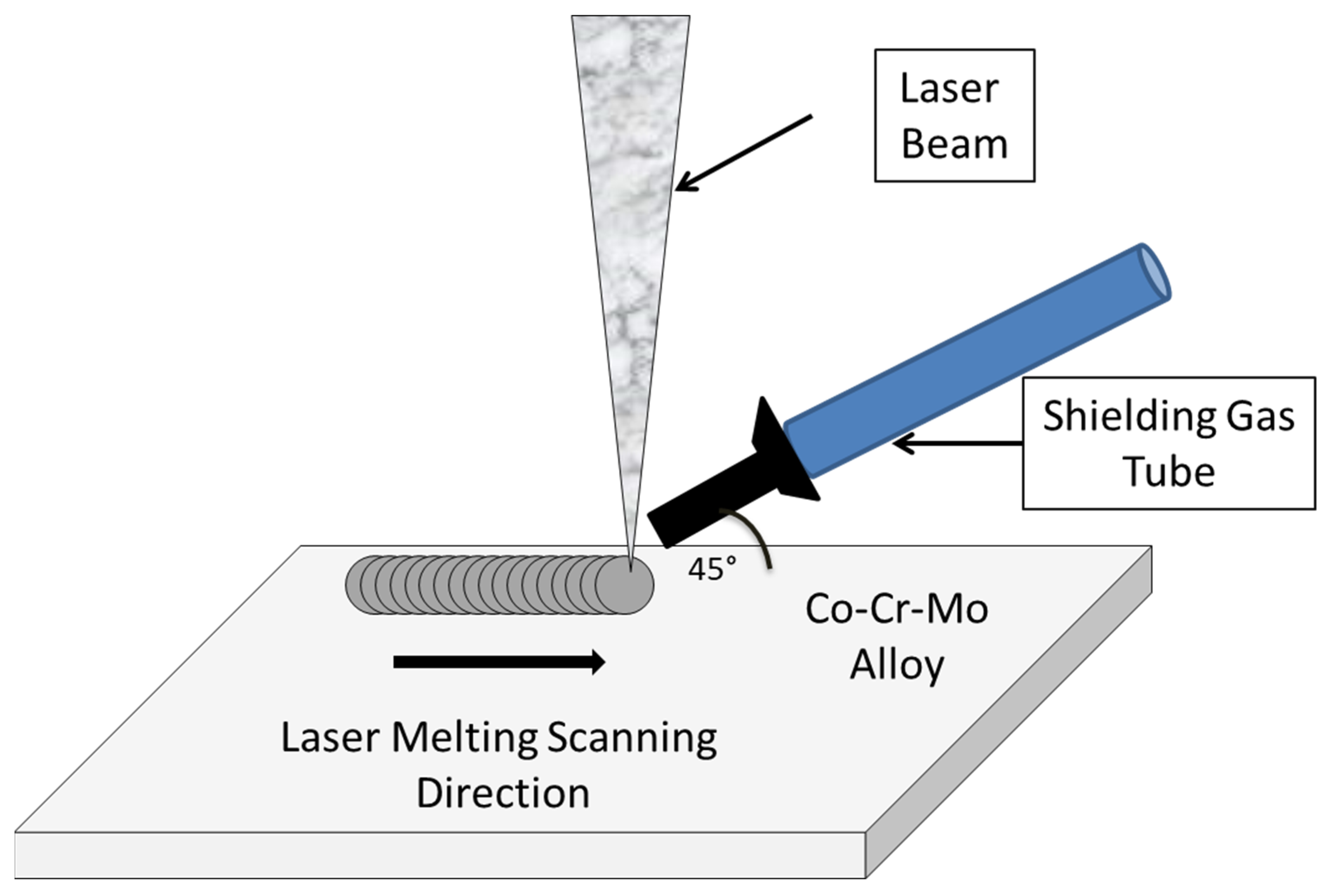
2.2. Nd:YAG Laser Melting Treatment

Prior to laser melting (LM), the specimens were polished using 1200-grit Si-C paper to achieve a uniform and smooth surface, and subsequently cleaned with acetone to remove any residual oxides and contaminants. LM was accomplished using a Nd:YAG HTS LS pulsed laser apparatus, which is capable of operating at a maximum output peak power of 7500 W. The Nd:YAG laser used in this study generated pulses with a near-Gaussian spatial profile (M2 ≈ 5). Table 2 shows the three parameter configurations selected for the laser treatments. Shielding argon gas was used to protect the contaminants from the melting atmosphere. Likewise, the melting scanning speed (0.5 mm/seg), pulse frequency (8 Hz), and laser spot diameter (0.8 mm) were maintained at a constant during the experimental procedures. The samples were processed under these conditions, and their results were compared with those of an untreated Control Sample (CS).
The pulse energy is a function of both pulse width and laser peak power, and is obtained using the following governing equation:
P u l s e   E n e r g y = P p e a k × P u l s e W i d t h
Figure 1 illustrates the schematic representation of the LM process applied to the samples. Disks measuring 39 mm in diameter and 7 mm in thickness for Pin-On-Disk wear tests were prepared. The samples were completely laser melted using the specified parameter configurations (see Table 2). The laser-treated area was achieved by overlapping adjacent laser tracks with a 50% overlap ratio in both the X and Y directions. The scanning pattern was designed in a meander strategy with alternating directions to minimize distortion and ensure uniform treatment. After laser treatment, the surfaces were lightly polished with 1000-grit SiC paper to remove any surface irregularities resulting from the overlapping tracks, producing a uniform surface suitable for wear testing. Following the same procedure, square plates of 7 mm × 7 mm × 5 mm (length × width × thickness) were prepared for in vitro bioactivity assessments.

2.3. Pin-on-Disk Wear Test

Wear tests were conducted in accordance with ASTM G99 [26]. All samples were polished to standardize the surface roughness prior to testing. Four specimens were evaluated: three laser-treated samples (each corresponding to one of the parameter configurations) and one untreated sample serving as the CS. Tests were performed under dry sliding conditions for 40 min at a constant sliding velocity of 251.32 cm/min, with an applied deadweight load of 61 N. The applied load of 61 N corresponds to approximately 15–20% of the typical joint reaction forces experienced during normal walking in hip implants, which has been established as an appropriate scaling factor for accelerated wear testing [27]. The sliding velocity of 251.32 cm/min was chosen to represent the average relative motion between articulating surfaces during gait cycles, while maintaining the testing parameters within the ASTM F732 standard recommendations for the wear testing of polymeric materials used in total joint prostheses [27]. The test duration of 40 min provided sufficient time to establish the steady-state wear conditions while enabling the assessment of both the running-in and steady-state wear behavior, which are critical factors in predicting the long-term performance of biomedical implants in vivo. Wear tests were performed in duplicate (n = 2) under identical conditions to ensure statistical reliability, finding a coefficient of variation of less than 2% between measurements. These conditions were selected to simulate the tribological environment encountered in orthopedic implants. The contra-body (pin) used in the wear resistance test was a 6 mm diameter Al2O3 ceramic ball with a hardness of 1600 HV and a surface roughness (Ra) of 0.02 μm. This material was selected to simulate a hard counterface that might be encountered in biomedical applications and to ensure that the wear predominantly occurred on the test sample rather than on the pin.

2.4. In Vitro Bioactivity Assessment

To evaluate bioactivity, simulated body fluid (SBF) was prepared with ionic concentrations similar to those of human blood plasma. The SBF preparation was carried out by first adding 3.399 g of NaCl to 350 mL of deionized water at 37 °C, with constant stirring until completely dissolved. Subsequently, 0.175 g of NaHCO3 was added and stirred until dissolution, followed by 0.112 g of KCl, which was also dissolved completely. After this, 0.114 g of K2HPO4·3H2O was added and stirred until dissolution. Then, 17.5 mL of 1N HCl (pH = 2.31) was gradually added to the solution. Next, 0.184 g of CaCl2·H2O was added and dissolved completely, followed by 0.0355 g of Na2SO4 and 0.152 g of MgCl2·6H2O, each added sequentially after the complete dissolution of the previous component. TRIS (tris-hydroxymethyl aminomethane) was then added until dissolution, resulting in a pH of approximately 8.20. The solution was diluted to a final volume of 500 mL with deionized water, and the pH was adjusted to 7.4 ± 0.02 using 1N HCl at 36.5 °C. The prepared SBF was stored in a clean polypropylene container at 4 °C. Each specimen was first polished and then immersed in 50 mL of SBF at 37 °C for 15 days, with the solution being renewed every 7 days to simulate physiological conditions. At the end of the total duration of 15 days, the sample was removed, rinsed with 99.9% anhydrous ethanol, and allowed to dry at room temperature. Subsequently, the samples were characterized using scanning electron microscopy (SEM) and attenuated total reflectance infrared spectroscopy (ATR-IR).

2.5. Characterization Methods

The laser-treated samples and the CS were thoroughly characterized using an optical microscope (OLYMPUS PMG3 with Image Pro Plus 6.0 software) for microstructural observations, while scanning electron microscopy (Acronym: SEM, Manufacturer: JEOL, Location: Peabody, MA, USA, Model: JSM-6490LV) provided detailed surface morphology. Elemental analysis was performed using energy-dispersive X-ray (EDX) analysis with INCA 5.1 software, and microhardness measurements were obtained with a FUTURE-TECH FM7 microhardness tester following the ASTM E34 standards [28]. In addition, surface roughness was measured using a Surftest VS-3000 instrument with a resolution of 0.005 μm. These combined analyses ensured a comprehensive understanding of the effects of the Nd:YAG LM on the material properties.

3. Results and Discussion

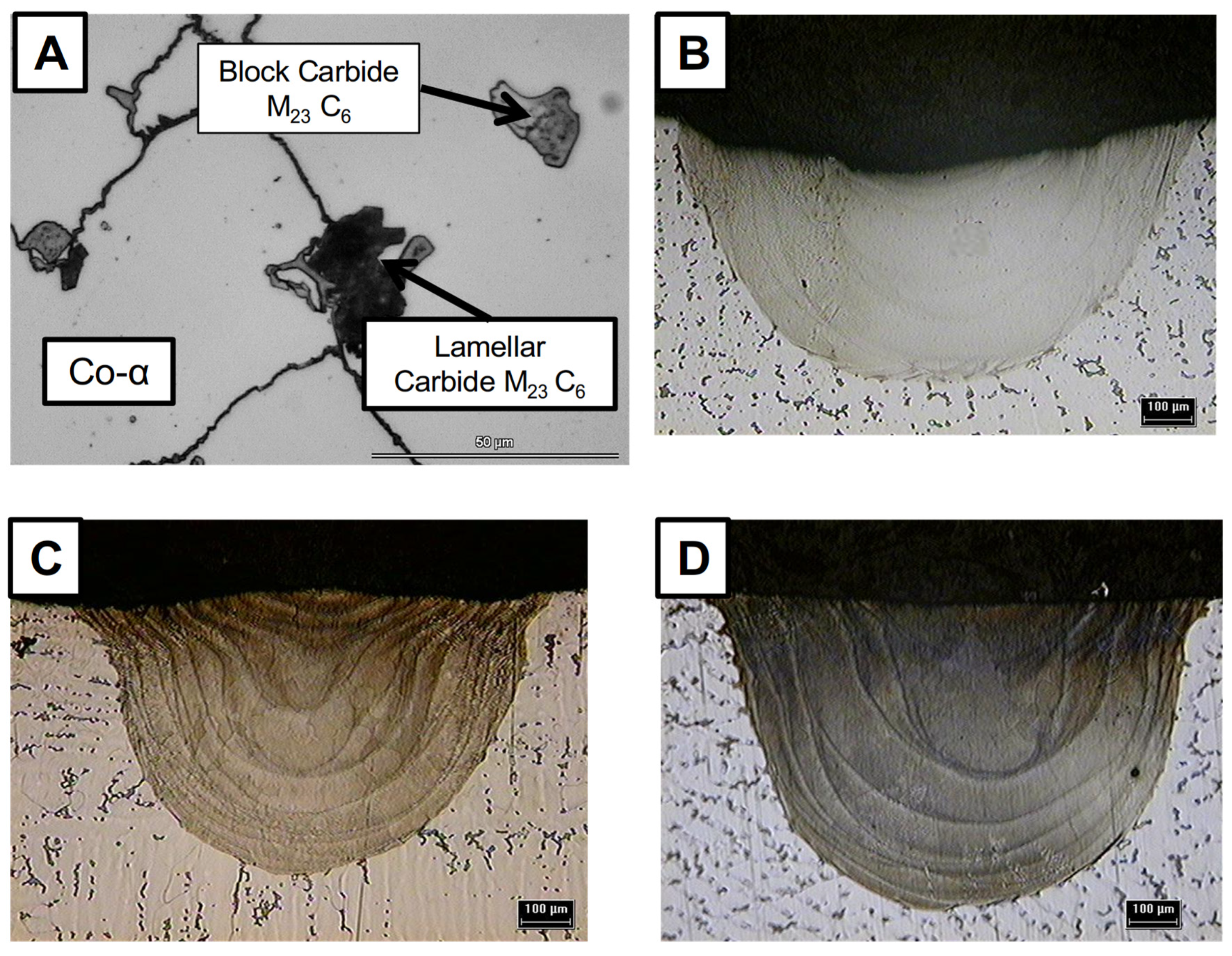
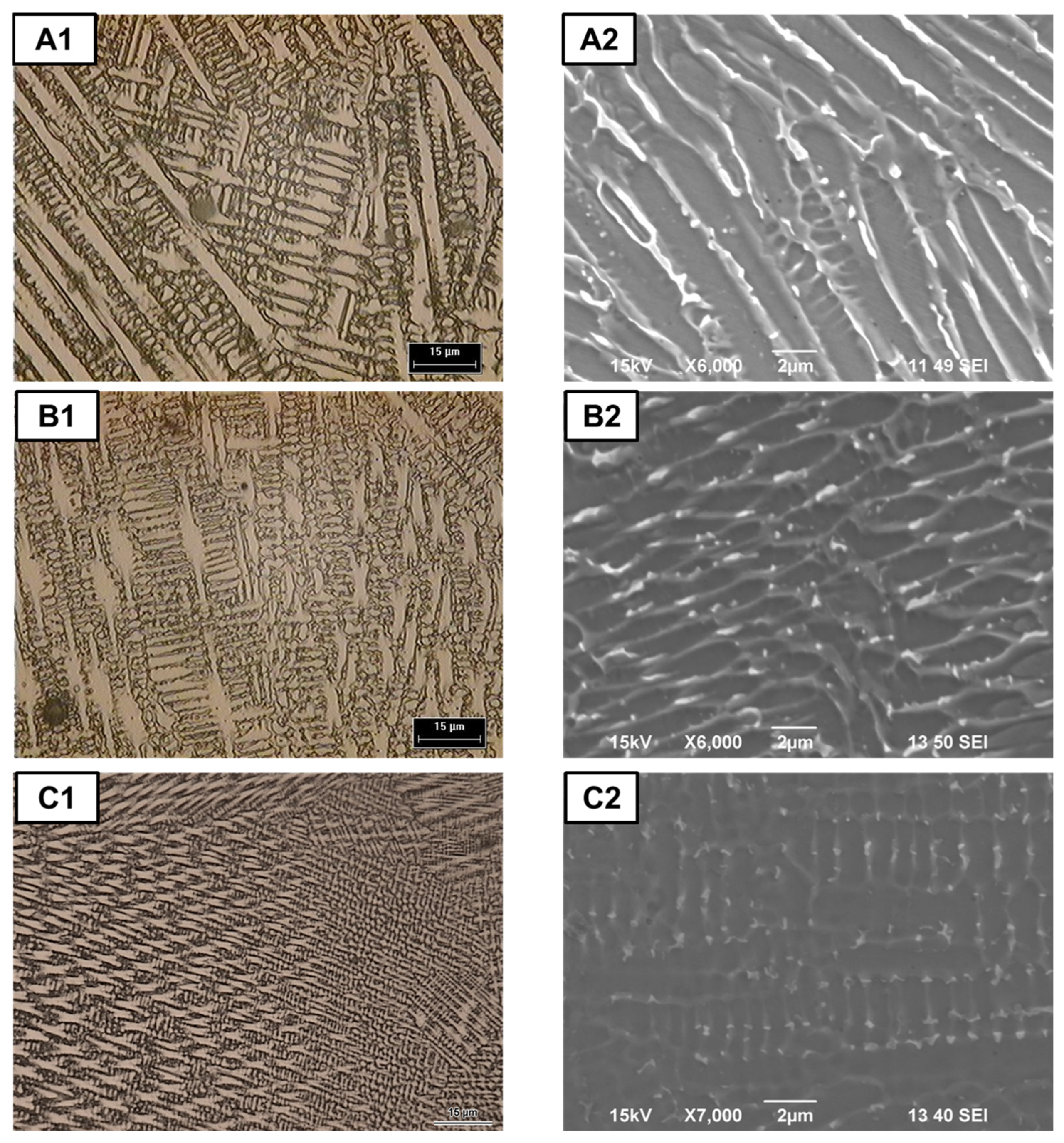
3.1. Macro and Microstructural Analysis

The macrostructure of the transverse cross-sections of the laser-surface-melted samples was examined to quantify the geometric characteristics and detect any defects within the fusion zone. Figure 2 shows the microstructure of the CS (investment-cast alloy) at 500× magnification. Figure 2A shows the microstructure of the CS that consists of the cobalt alpha (Co-α) matrix phase, which exhibits a face-centered cubic (FCC) structure and appears as a bright phase. Within this matrix, two distinct forms of primary carbides M23C6 are present in both block and laminar morphologies. These carbide sizes range between 15 and 25 µm.
The macrostructural analysis of the transverse cross-sections of the laser-surface-melted samples was conducted to quantify their geometric characteristics and identify any potential defects within the fusion zone. Figure 2 shows the macrostructure of (B) Sample 1 (S1), (C) Sample 2 (S2), and (D) Sample 3 (S3) at 50× magnification. The samples appear free of discernible defects and discontinuities; however, notable differences in the penetration depth (P), fusion zone width (W), and penetration–width aspect ratio (P:W) are evident. The P:W ratio can be obtained using the following equation:
P : W = P W
The P:W ratios for the samples were: S1 = 0.55, S2 = 0.66, and S3 = 0.89. The P:W ratios for the samples were: S1 = 0.55, S2 = 0.66, and S3 = 0.89. While these variations correlate with the applied pulse energy levels, the relationship is more complex. The literature indicates [29,30,31,32] that the pulse energy significantly influences both the penetration depth and the bead width. As the pulse energy increases, the energy density applied to the material rises, leading to deeper penetration. This increase in energy can also cause a broader bead width; however, the effect on penetration depth is typically more pronounced than that on bead width. Consequently, elevating the pulse energy tends to enhance the penetration–width ratio, favoring a greater depth relative to width, so that the penetration depth increases primarily with the pulse energy, while width is more strongly influenced by the laser spot diameter [29,30,31,32]. According to classical thermal conduction theory on pulsed laser applications [33], an increase in the pulse energy enhances the heat transfer into the material, resulting in a larger fusion zone. Beyond classical thermal conduction, our microstructural investigation reveals specific relationships between laser parameters and LM zone development. Li, Chen, and Zhang’s theoretical model [34] demonstrates how pulse energy and duration collectively influence the heat accumulation patterns, explaining the asymmetric thermal distribution observed across samples. This asymmetry particularly affects the fusion boundary morphology and explains why the increased energy created not just larger, but differently structured fusion zones. Similarly, Chen, Gu, and Bi’s numerical analysis [35] found that variations in pulse parameters create distinct temperature gradients that directly influence the amount of heat conduction within the fusion zone. These findings reinforce that a higher pulse energy results in enhanced heat transfer and accumulation, ultimately producing a larger fusion zone.
The increased fusion zone dimensions imply a more extensive area undergoing rapid solidification, which is expected to produce a finer grain structure and potentially improve wear resistance. This observation sets the foundation for understanding how the processing parameters influence the overall performance of the alloy in biomedical applications.
Figure 3 shows a micrograph of the laser-melted samples at different magnifications and the parameter configurations resulting in heterogeneous layered microstructures with diverse carbide precipitation behavior on dendritic arms and grain morphology. It can be seen in Figure 3 that following the implementation of the LM process, the microstructures change markedly regarding the CS microstructure (see Figure 2A). The microstructures from Figure 3A1–C1 at 50×, owing to the rapid melting and subsequent solidification, show that the LM region is characterized by a predominantly fine dendritic microstructure. Furthermore, there is a clear tendency toward reduced dendritic growth due to the rapid solidification inherent in the treatment. In addition, as the pulse energy increases, as observed in Sample 3, the thermal gradient between the molten zone and the surrounding material becomes significantly higher, decreasing the solidification period and therefore the growth of dendritic arms. This results in an even more refined microstructure, where dendritic growth is notably lower in Sample 3 (Figure 3A1) compared to Samples 1 and 2 (Figure 3B1,C1).
The superior microstructural refinement observed in Sample 3 (Figure 3C1,C2) directly correlates with the highest pulse energy applied (39.37 J), which significantly intensifies the thermal gradient between the molten zone and the surrounding material. This enhanced thermal gradient accelerates the solidification rate, resulting in markedly reduced dendritic arm spacing and substantially finer carbide precipitation compared to Samples 1 and 2. Quantitative analysis reveals that the average carbide size in Sample 3 is less than 0.5 µm (±0.1 µm), representing a reduction of over 75% compared to Sample 1 (1.7 ± 0.3 µm). This dramatic refinement occurs because the higher energy input creates a deeper and wider molten pool with a more homogeneous temperature distribution, while simultaneously promoting faster cooling rates upon solidification. The rapid quenching effect prevents the coarsening of the microstructural features and results in the formation of ultrafine carbides uniformly distributed throughout the interdendritic regions.
Likewise, Figure 3A2–C2 at 6000X clearly illustrates a dendritic structure with prominent branching. Additionally, a continuous network of fine carbide precipitates develops along the interdendritic boundaries (bright phase). Energy-dispersive X-ray spectroscopy (EDS) analyses of both the matrix and the interdendritic regions indicate that the β-phase is rich in carbon, suggesting that it is predominantly a carbide phase—most likely of the M23C6 type, as reported by Taylor and Waterhouse [36]. Essentially, the LM process promotes the dissolution of coarse carbides and their subsequent reprecipitation as a fine carbide network during rapid solidification. The pulse energy directly affects both the solidification rate and thermal gradient. As the pulse energy increases, these changes further refine the size and distribution of carbides within the solidified metal pool [37,38,39,40].

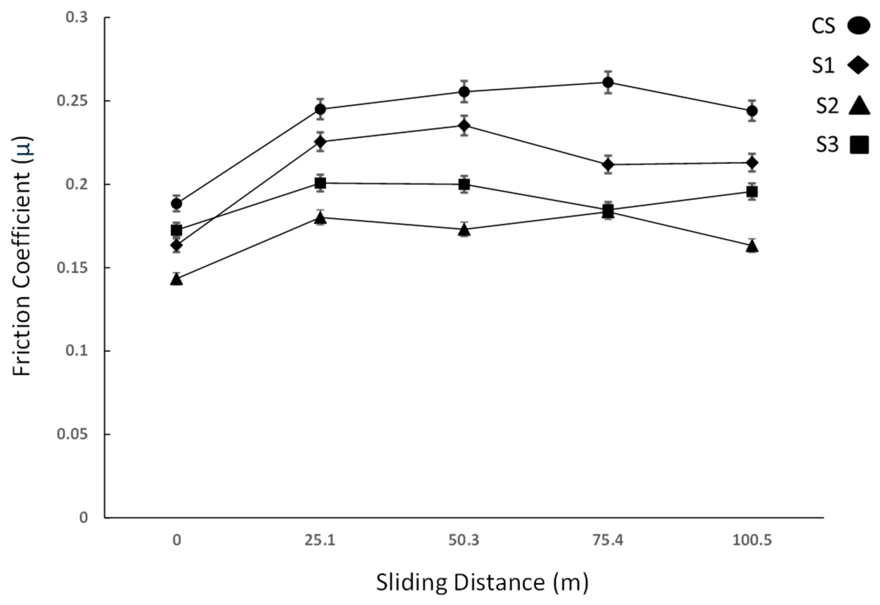
3.2. Microhardness and Wear Test

The microhardness evaluation yields an average value of 325 Vickers for the CS, which complies with the microhardness requirements of the ASTM F75 standards, which specify a range between 266 and 345 Vickers [25]. Samples 1–3 show an increment in microhardness regarding the CS (Table 3). Table 3 indicates that Sample S3, subjected to a pulse energy of 39.37 joules, exhibits an increase of over 120 Vickers in microhardness within the melted zone, corresponding to a 27% enhancement.
The increase in the microhardness of the samples is predominantly due to grain refinement and the reduction in the secondary dendritic arm size, which are directly proportional to the rapid solidification process inherent in laser material processing [33,41]. Similar results were reported by DebRoy et al. (2018), who discussed how the rapid solidification inherent in laser-based processes leads to a significantly refined microstructure. This refinement results in enhanced mechanical properties, such as increased strength and hardness, primarily due to reduced grain sizes and minimized microsegregation. Their findings emphasize the critical role of high cooling rates in achieving superior mechanical performance [42]. Likewise, elevated cooling rates during laser processing are instrumental in producing finer microstructures with fewer defects, thereby improving the overall mechanical properties and performance of the material [43].
Preceding the wear test, the disk surfaces of both the CS and the LM sample were polished to a mirror finish to reach a similar alloy roughness. Five measurements were taken for each sample. The untreated CS had an average roughness of 0.150 mm ± 0.03 mm. After laser treatment, the surface roughness values were 0.149 ± 0.002 mm, 0.145 ± 0.002 mm, and 0.143 ± 0.003 μm for samples S1, S2, and S3, respectively.
Figure 4 shows a comparative plot of the friction coefficient vs. the sliding distance between the CS and the LM sample during the Pin-On-Disk wear test. The coefficient of friction (μ) was calculated using the following equation:
µ = T P R
where T is the frictional torque, P is the applied load, and R is the main pin radius.
It can be seen in the plot of Figure 4 that an initial increase in the friction coefficient (µ) was observed until a plateau (stable steady state) was reached approximately at 25.3 m of sliding distance. This behavior is consistent with prior reports on Co-Cr-Mo alloys, where friction coefficients of less than 0.25 are common under dry conditions for HCP matrix alloys, whereas FCC matrices typically exhibit μ values between 0.25 and 0.30 [37,38,44]. Hence, from the results of this work, it is apparent that the microstructural modifications caused by LM can reduce the μ to values below 0.25 on Co-Cr-Mo steel wear pairs, mainly in S2 and S3, which reached stable steady states in relatively short sliding distances and then showed a μ below 0.20. Apparently, this can be attributed to the development of dendritic growth combined with an improved distribution of carbide microsegregation (see Figure 3B2,C2).
These quantitative results indicate that microstructural modifications induced by laser melting (LM) can effectively reduce the friction coefficients within Co alloy and steel wear pairs. These observations highlight the critical influence of microstructural integrity on frictional behavior.
Figure 5 shows SEM micrographs of the worn surfaces for the investment-cast CS (Figure 5A1) and for the LM sample along the wear path. In SEM analysis, similar wear mechanisms are achieved on the S1–S3 samples.
It can be seen in Figure 5 that in the CS and LM sample, the predominant wear mechanisms are abrasion and delamination. In addition, in the CS (Figure 5A1,A2), the detachment of coarse carbide phases is observed. In general, it is found that the severity of delamination and abrasion tends to decrease LM samples. In turn, this indicates that an increase in energy input enhances the resistance to abrasion and delamination wear mechanisms. Apparently, the differences in wear performance of the LM microstructures are closely related to the size and improvement of interdendritic carbide distribution, as well as the coarser dendritic arms (Figure 3).
The wear behavior of each sample was evaluated by measuring the amount of wear debris produced. Table 4 presents the wear rates for the LM samples, including the CS. Notably, the LM samples exhibited minimal wear rates. The sample subjected to the highest pulse energy (S3, Pulse Energy = 39.37 J) demonstrated superior performance, with a wear rate less than 50% of that observed in the untreated sample. This improvement aligns with the lowest coefficient of friction (μ) recorded during the wear test, as depicted in Figure 4. Similar findings have been reported in the literature. These observations underscore the significant role of laser treatment parameters, particularly pulse energy, in enhancing wear resistance and reducing friction in materials.
Thus, from the outcome of this work, a viable alternative to enhancing the wear properties of Co-Cr-Mo alloys with an almost fully FCC matrix is through the surface modification induced by LM processing. Although Co-Cr-Mo with an HCP matrix has thus far shown to exhibit the best wear properties [36,37,38], Co-Cr-Mo alloys with fully FCC matrices through the surface modification induced by LM processing can give rise to a new generation of biocompatible implants. The main advantages of an FCC-Co-Cr-Mo matrix are improved fatigue and corrosion resistance when compared with those reported for Co-Cr-Mo alloys with a predominant HCP matrix [39]. Thus, the outcome of the present work indicates that it is possible to modify the as-cast structure of high-carbon Co-Cr-Mo alloys via LM to develop low-wear, low-friction, and potentially high-fatigue-strength biomedical metallic components.
The LM process significantly improved microhardness from 325 HV to 445 HV (approximately 27% increase), which aligns with the findings reported by Takaichi et al. (2013) [45], who observed similar improvements, with hardness values increasing from 330 to 340 HV to 430–460 HV after the selective laser melting of Co-Cr-Mo alloys [45]. The microstructural refinement achieved through our laser melting procedure, particularly the dissolution of coarse carbides and their reprecipitation as a fine network, correlates with the enhanced mechanical properties and corresponds to the microstructural evolution reported by Bartolomeu et al. (2017) [46]. Their study demonstrated that selective laser melting reduced the coefficient of friction from 0.45 to 0.31, while increasing hardness to 395–460 HV [46]. These comparisons validate that improved laser melting parameters can effectively address the mechanical limitations of conventionally cast Co-Cr-Mo alloys while preserving their essential bioactive properties.
Although no wear testing was carried out under a lubricant that resembles human fluids, the exhibited low friction and high wear resistance found in the LM surfaces provide an essential insight into the potential development of novel prostheses with enhanced fatigue properties.

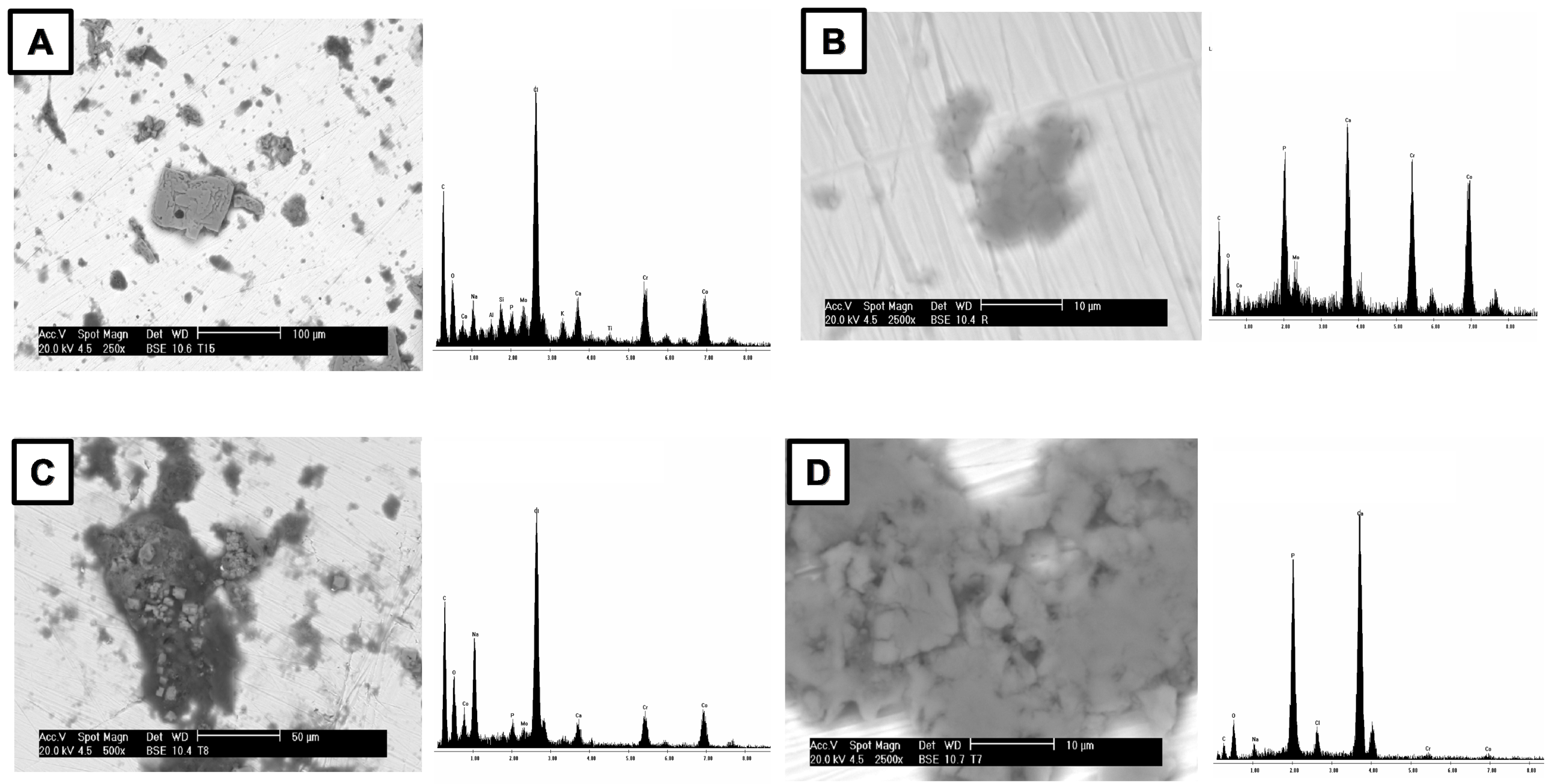
3.3. In Vitro Bioactivity

FTIR–ATR analysis of the samples following immersion in physiological solution confirms the formation of an apatite layer on the metallic substrates. In Figure 6, the IR spectrum of the CS clearly displays the characteristic P-O stretching vibration at 566, 598, and 600 cm⁻1, which are indicative of apatite deposition [47,48]. These bands serve as a qualitative marker of bioactivity, demonstrating that the surface modification process has successfully induced the formation of a bioactive apatite phase on the metal surfaces [47]. The LM sample spectra (Figure 6, S1 to S3) present the same absorption bands as the CS—at 566, 598, and 600 cm⁻1. Likewise, S3 clearly displays the characteristic P-O stretching vibration at 1054 cm−1, which is another indication of induced apatite formation [49,50].
The observed differences in the intensity of the P-O stretching bands likely reflect variations in the amount of apatite formed on each sample. S3 was the one that exhibited all the characteristic absorption bands indicative of apatite formation. Overall, the FTIR–ATR results strongly suggest that all the samples are bioactive, although the extent of apatite formation varies among them.
Figure 7A–D shows micrographs and EDS analyses of the CS, S1−S3, respectively. The EDX analysis revealed the presence of calcium and phosphorus elements in the CS and the LM sample, indicating the formation of apatite. It is important to note that the structural and morphological characterization of the metal substrate surfaces treated in SBF reveals the formation of spherical phases, which, when analyzed using EDX, also exhibit the presence of calcium and phosphorus. Quantitative analysis of apatite layer thickness, measured at 10 locations per sample, averaged 2.1 ± 0.7 μm for the CS, 3.8 ± 0.6 μm for S1, 5.2 ± 0.5 μm for S2, and 6.7 ± 0.4 μm for S3, demonstrating a clear correlation between laser treatment parameters and apatite formation capability. Additionally, the average diameter of the spherical apatite formations increased from 8 ± 3 μm (CS) to 25 ± 4 μm (S3), indicating more substantial mineral deposition on laser-treated surfaces. These findings are consistent in that the surface of the treated substrates has become bioactive and formed an apatite layer, enhancing their bioactivity and potential for use in biomedical applications. The data provided through these detailed analyses underscore the transformation of the metallic substrates when exposed to SBF. The presence of calcium and phosphorus, detected through both microstructural observation and EDX analysis, suggests that the SBF treatment induces the deposition of apatite, a key biomolecule involved in the formation of bone tissue. The uniformity of the spherical apatite phase further indicates a controlled and homogeneous surface modification, which is beneficial for the long-term stability and performance of metallic implants in physiological environments. This apatite formation is a positive indicator of the bioactive potential of the substrates, essential for promoting osteointegration once implanted in biological systems. The imaging and spectroscopic data overall provide robust evidence for the successful surface modification of these alloys, showing promise for future biomedical applications, particularly in bone replacement.

4. Conclusions

This study investigates the influence of Nd:YAG laser melting on the microstructure, microhardness, wear, and biocompatibility properties of a Co-Cr-Mo ASTM F-75 alloy, and the main findings are summarized below:
  • Nd:YAG laser melting significantly enhances the mechanical properties and preserves the bioactive properties of investment-cast Co-Cr-Mo alloys. The process achieves a refined microstructure, eliminating microstructural defects that serve as stress concentrators, increasing hardness, improving wear resistance, and preserving bioactivity, offering a promising approach for the development of more durable biomedical implants;
  • The LM process promotes the dissolution of coarse carbides and their subsequent reprecipitation as a fine carbide network during rapid solidification. The energy directly influences the solidification rate and thermal gradient, thereby further refining both the carbide size and distribution within the solidified metal pool as the pulse energy increases;
  • Using an adequate set up of laser parameters such as peak power and pulse duration led to an increase in the microhardness from 325 Vickers to 445 Vickers (approx. 25%), which is correlated to the grain size reduction of 25–35% compared to the untreated sample;
  • The exhibited low friction and high wear resistance found in the LM surfaces provides an essential insight into the potential development of novel prostheses with enhanced fatigue properties;
  • Laser melting at high energy densities promoted the formation of uniform phase distributions and a homogenized microstructure, which significantly enhanced the wear resistance of the alloy. The process also reduced the wear volume loss by nearly 50% due to the effective microstructure improvement and the elimination of porosities and inhomogeneities. It is important to exercise precise control over variables to achieve the desirable microstructural modifications that enhance both the mechanical properties and the longevity of biomedical implants;
  • TIR-ATR analysis of SBF-treated samples revealed P-O absorption bands at 1054, 566, 598, and 600 cm−1, confirming apatite formation and bioactivity. SEM-EDS and calcium consumption analyses further support the hypothesis that all samples are bioactive. Importantly, the laser surface treatment did not compromise their bioactivity. These results demonstrate that surface modification preserves the essential biological properties of the substrates;
  • Based on our findings, we recommend the following optimal processing parameters for the Nd:YAG laser melting of Co-Cr-Mo alloys: pulse energy ≈ 37 J, a pulse duration of 7 ms, and a peak power of ≈ 5600 W. These parameters provide the best balance between microstructural refinement, mechanical property enhancement, and the preservation of bioactivity;
  • Despite these promising results, the investigation was conducted under controlled laboratory conditions, which may differ from clinical environments. Future research should focus on optimizing laser processing parameters for specific implant geometries, conducting long-term in vivo performance studies to validate biocompatibility and wear resistance, and exploring additional laser surface modification techniques.

Author Contributions

Conceptualization, F.C.R. and C.R.M.V.; methodology, F.C.R., J.C.O.C. and G.Y.P.M.; validation, J.F.M.V.; formal analysis, F.C.R. and G.Y.P.M.; investigation, J.C.O.C., J.F.M.V. and J.S.G.V.; data curation, C.R.M.V. and J.S.G.V.; writing—original draft preparation, F.C.R.; writing—review and editing, F.C.R. and G.Y.P.M.; supervision, G.Y.P.M.; project administration, G.Y.P.M. All authors have read and agreed to the published version of the manuscript.

Funding

This research received no external funding.

Data Availability Statement

The original contributions presented in this study are included in the article. Further inquiries can be directed to the corresponding author.

Conflicts of Interest

Author Francisco Cepeda Rodríguez and Gladys Yerania Pérez Medina were employed by the company Corporación Mexicana de Investigación en Materiales S.A. de C.V. The remaining authors declare that the research was conducted in the absence of any commercial or financial relationships that could be construed as a potential conflict of interest.

Abbreviations

The following abbreviations are used in this manuscript:
CSControl Sample
SEMScanning electron microscopy
EDXEnergy-dispersive X-ray
ATR-IRAttenuated total reflectance infrared spectroscopy
LMLaser melting
S1Sample 1
S2Sample 2
S3Sample 3
Nd:YAGNeodymium-doped Yttrium Aluminum Garnet
PPenetration depth
WFusion zone width
P:WPenetration–width aspect ratio
µFriction coefficient

References

  1. Gittens, R.A.; Schwartz, Z.; Smith, T.J.; Boyan, B.D. Implant osseointegration and the role of microroughness and nanostructures: Lessons for spine implants. Acta Biomater. 2014, 10, 2907–2921. [Google Scholar] [CrossRef] [PubMed]
  2. Vidyasagar, K.E.C.; Kalyanasundaram, D. Enhanced corrosion resistance of CoCrMo by laser-based surface modification. Surf. Eng. 2024, 40, 100–111. [Google Scholar] [CrossRef]
  3. Yıldırım, M.; Keleş, A. Production of Co–Cr–Mo biomedical alloys via investment casting technique. Turk. J. Electromech. Energy 2018, 3, 87–94. [Google Scholar]
  4. Pattnaik, S.; Karunakar, D.B.; Jha, P.K. Developments in investment casting process—A review. J. Mater. Process. Technol. 2012, 212, 2332–2348. [Google Scholar] [CrossRef]
  5. Connors, J.P.; Stelzer, J.W.; Garvin, P.M.; Wellington, I.J.; Solovyova, O. The Role of the Innate Immune System in Wear Debris-Induced Inflammatory Peri-Implant Osteolysis in Total Joint Arthroplasty. Bioengineering 2022, 9, 764. [Google Scholar] [CrossRef]
  6. Öztürk, O.; Çolak, İ.; Eroglu, A.E. Metal ion release from nitrogen ion implanted CoCrMo orthopedic implant material. Mater. Sci. Eng. C 2014, 45, 1–7. [Google Scholar] [CrossRef]
  7. Ganjali, M.; Yazdanpanah, A.; Mozafari, M. Laser Deposition of Nano Coatings on Biomedical Implants. In Emerging Applications of Nanoparticles and Architecture Nanostructures; Elsevier: Amsterdam, The Netherlands, 2018; pp. 235–254. [Google Scholar] [CrossRef]
  8. Ghayad, I.M.; Girgis, N.N.; Ghanem, W.A. Laser surface treatment of metal implants: A review article. J. Metall. Eng. 2015, 4, 48–56. [Google Scholar] [CrossRef]
  9. Wang, D.; Wu, S.; Yang, Y.; Huang, S.; Luo, Z. Effects of laser polishing on surface microstructure and corrosion resistance of additive manufactured CoCr alloys. Appl. Surf. Sci. 2018, 443, 167–175. [Google Scholar] [CrossRef]
  10. Liu, D.; Kong, D. Effects of Laser Remelting on Microstructure and Tribological Properties of Plasma–Sprayed Fe45Mn35Co10Cr10 High–Entropy Alloy Coating. Surf. Coat. Technol. 2023, 474, 129202. [Google Scholar] [CrossRef]
  11. Lohberger, B.; Stuendl, N.; Glaenzer, D.; Rinner, B.; Donohue, N.; Lichtenegger, H.C.; Ploszczanski, L.; Leithner, A. CoCrMo surface modifications affect biocompatibility, adhesion, and inflammation in human osteoblasts. Sci. Rep. 2020, 10, 1682. [Google Scholar] [CrossRef]
  12. Walker, J.C.; Cook, R.B.; Murray, J.W.; Clare, A.T. Pulsed electron beam surface melting of CoCrMo alloy for biomedical applications. Wear 2013, 301, 250–256. [Google Scholar] [CrossRef]
  13. Ju, J.; Zhou, Y.; Kang, M.; Wang, J. Optimization of process parameters, microstructure, and properties of laser cladding Fe-based alloy on 42CrMo steel roller. Materials 2018, 11, 2061. [Google Scholar] [CrossRef] [PubMed]
  14. Jung, C.W.; Lee, J.H.; Joo, S.-H.; Kim, R.E.; Kim, H.S.; Moon, J. Effect of laser surface treatment on microstructural evolution and mechanical properties of a Co–Cr–Fe–Ni–Mo medium–entropy alloy. Mater. Sci. Eng. A 2025, 922, 147617. [Google Scholar] [CrossRef]
  15. Li, D.; Zhou, Y.; Chen, P.; Li, C.; Zhang, J.; Li, Z.; Zhou, Z.; Li, M.; Xu, Y. Polylactic Acid/Calcium Silicate Composite Scaffold Fabricated by Selective Laser Sintering with Coordinated Regulation of Bioactivity Induction, Degradation, and Mechanical Enhancement. J. Polym. Environ. 2025, 33, 1778–1791. [Google Scholar] [CrossRef]
  16. Pakieła, W.; Tański, T.; Brytan, Z.; Chladek, G.; Pakieła, K. The impact of laser surface treatment on the microstructure, wear resistance and hardness of the AlMg5 aluminum alloy. Appl. Phys. A 2020, 126, 231. [Google Scholar] [CrossRef]
  17. Furlani, M.R.; de Carvalho, S.M.; de Lima, M.S.F. Effect of laser surface hardening on a low carbon steel substrate wear and hardness. arXiv 2021, arXiv:2111.13183. [Google Scholar] [CrossRef]
  18. Rapheal, G.; Kumar, S.; Blawert, C.; Dahotre, N.B. Wear-resistant thin films of MRI-230D-Mg alloy using plasma-driven electrolytic oxidation. arXiv 2017, arXiv:1705.00116. [Google Scholar] [CrossRef]
  19. Acharya, S.; Soni, R.; Suwas, S.; Chatterjee, K. Additive manufacturing of Co–Cr alloys for biomedical applications: A concise review. J. Mater. Res. 2021, 36, 3746–3760. [Google Scholar] [CrossRef]
  20. Hedberg, Y.S.; Qian, B.; Shen, Z.; Virtanen, S.; Wallinder, I.O. In vitro biocompatibility of CoCrMo dental alloys fabricated by selective laser melting. Dent. Mater. 2014, 30, 525–534. [Google Scholar] [CrossRef]
  21. Bartolomeu, F.; Buciumeanu, M.; Costa, E.; Alves, N.; Gasik, M.; Silva, F.S.; Miranda, G. Multi-material Ti6Al4V & PEEK cellular structures produced by selective laser melting and hot pressing: A tribocorrosion study targeting orthopedic applications. J. Mech. Behav. Biomed. Mater. 2019, 89, 54–64. [Google Scholar] [CrossRef]
  22. Rezayat, M.; Besharatloo, H.; Mateo, A. Investigating the Effect of Nanosecond Laser Surface Texturing on Microstructure and Mechanical Properties of AISI 301LN. Metals 2023, 13, 2021. [Google Scholar] [CrossRef]
  23. Research and Markets. Microelectronic Medical Implants Market Report: Trends, Forecast and Competitive Analysis to 2030; ID: 6007972; Lucintel: Dallas, TX, USA, 2024. [Google Scholar]
  24. Mantrala, K.M.; Das, M.; Balla, V.K.; Rao, C.S.; Rao, V.V.S. Laser-deposited CoCrMo alloy: Microstructure, wear, and electrochemical properties. J. Mater. Res. 2014, 29, 2021–2027. [Google Scholar] [CrossRef]
  25. ASTM F0075-23; Standard Specification for Castings, Wear-Resistant, Chromium Alloy (Class I and Class II). ASTM International: West Conshohocken, PA, USA, 2023.
  26. ASTM G99-23; Standard Test Method for Wear and Friction Testing with a Pin-on-Disk or Ball-on-Disk Apparatus. ASTM International: West Conshohocken, PA, USA, 2023.
  27. ASTM F732-17; Standard Test Method for Wear Testing of Polymeric Materials Used in Total Joint Prostheses. ASTM International: West Conshohocken, PA, USA, 2017.
  28. ASTM E384-22; Standard Test Method for Microindentation Hardness of Materials. ASTM International: West Conshohocken, PA, USA, 2022.
  29. Engel, S.L. Laser Welding Technology Learning Manual; HDE Technologies, Inc.: Elk Grove, CA, USA, 2016; ISBN 9780997766394. [Google Scholar]
  30. Majumdar, J.D.; Manna, I. Laser-Assisted Fabrication of Materials; Springer: Berlin/Heidelberg, Germany, 2013; ISBN 978-3642280290. [Google Scholar]
  31. Steen, W.M.; Mazumder, J. Laser Material Processing, 4th ed.; Springer: London, UK, 2010; ISBN 978-1-84996-062-5. [Google Scholar] [CrossRef]
  32. Ion, J.C. Laser Processing of Engineering Materials: Principles, Procedure and Industrial Application; Butterworth-Heinemann: Oxford, UK, 2005; ISBN 978-0750660794. [Google Scholar]
  33. Ready, J.F. LIA Handbook of Laser Materials Processing; Laser Institute of America, Magnolia Publishing: Orlando, FL, USA, 2001. [Google Scholar]
  34. Li, B.; Chen, J.; Zhang, Y. A Universal Theoretical Model for Thermal Accumulation in Materials Induced by Repetitive Pulsed Laser Irradiation. Int. J. Laser Sci. 2019, 1, 53–71. [Google Scholar] [CrossRef]
  35. Chen, G.; Gu, X.; Bi, J. Numerical Analysis of Thermal Effect in Aluminum Alloy by Repetition Frequency Pulsed Laser. Optik 2016, 127, 6371–6378. [Google Scholar] [CrossRef]
  36. Taylor, R.N.J.; Waterhouse, R.B. A Study of the Ageing Behavior of a Cobalt Based Implant Alloy. J. Mater. Sci. 1983, 18, 3265–3280. [Google Scholar] [CrossRef]
  37. Hsu, H.C.; Lian, S.S. Wear Properties of Co–Cr–Mo–N Plasma-Melted Surgical Implant Alloy. J. Mater. Process. Technol. 2003, 138, 231–235. [Google Scholar] [CrossRef]
  38. Saldivar-García, A.J.; López, H.F. Microstructural Effects on the Wear Resistance of Wrought and As-Cast Co-Cr-Mo-C Implant Alloys. J. Biomed. Mater. Res. A 2005, 74A, 269–274. [Google Scholar] [CrossRef]
  39. Huang, P.; López, H.F. Tribological Behavior of Cast and Wrought Co-Cr-Mo Implant Alloys. Mater. Sci. Technol. 1999, 15, 1324–1329. [Google Scholar] [CrossRef]
  40. Montero-Ocampo, C.; Talavera, M.; López, H.F. Effect of Alloy Preheating on the Mechanical Properties of As-Cast Co-Cr-Mo-C Alloys. Metall. Mater. Trans. A 1999, 29A, 611–620. [Google Scholar] [CrossRef]
  41. King, W.E.; Anderson, A.T.; Ferencz, R.M.; Hodge, N.E.; Kamath, C.; Khairallah, S.A. Laser Powder Bed Fusion Additive Manufacturing of Metals; Physics, Computational, and Materials Challenges. Appl. Phys. Rev. 2015, 2, 041304. [Google Scholar] [CrossRef]
  42. DebRoy, T.; Wei, H.L.; Zuback, J.S.; Mukherjee, T.; Elmer, J.W.; Milewski, J.O.; Beese, A.M.; Wilson-Heid, A.; De, A.; Zhang, W. Additive Manufacturing of Metallic Components—Process, Structure and Properties. Prog. Mater. Sci. 2018, 92, 112–224. [Google Scholar] [CrossRef]
  43. Han, T.; Chen, J.; Wei, Z.; Qu, N.; Liu, Y.; Yang, D.; Zhao, S.; Lai, Z.; Jiang, M.; Zhu, J. Effect of cooling rate on microstructure and mechanical properties of AlCrFe₂Ni₂ medium entropy alloy fabricated by laser powder bed fusion. J. Mater. Res. Technol. 2023, 25, 4063–4073. [Google Scholar] [CrossRef]
  44. Zhao, C.; Jia, X.; Zhao, Q.; Ma, H.; Yu, Z.; Zhang, Z. Research Progress on the Effect of Surface Texture on the Friction Properties of CoCrMo Alloys. Compos. Interfaces 2024, 31, 1269–1290. [Google Scholar] [CrossRef]
  45. Takaichi, A.; Suyalatu, T.; Nakamoto, N.; Joko, N.; Nomura, N.; Tsutsumi, Y.; Migita, S.; Doi, H.; Kurosu, S.; Chiba, A.; et al. Microstructures and mechanical properties of Co-29Cr-6Mo alloy fabricated by selective laser melting process for dental applications. J. Mech. Behav. Biomed. Mater. 2013, 21, 67–76. [Google Scholar] [CrossRef]
  46. Bartolomeu, F.; Sampaio, M.; Carvalho, O.; Pinto, E.; Alves, N.; Silva, F.S. Tribological behavior of Ti6Al4V cellular structures produced by Selective Laser Melting. J. Mech. Behav. Biomed. Mater. 2017, 69, 128–134. [Google Scholar] [CrossRef]
  47. Redey, S.A.; Razzou, S.; Rey, C.; Bernache-Assollant, D.; Leroy, G.; Nordin, M.; Cournot, G. Osteoclast adhesion and activity on synthetic hydroxyapatite, carbonated hydroxyapatite and calcium carbonate: Relationship to surface energies. J. Biomed. Mater. Res. 1990, 24, 163–177. [Google Scholar] [CrossRef]
  48. Dorozhkin, S.V. Calcium Orthophosphates in Nature, Biology and Medicine. Materials 2009, 3, 3510–3606. [Google Scholar] [CrossRef]
  49. Román, J.; Padilla, S.; Vallet-Regi, M. Sol-gel glasses as precursors of bioactive glass ceramics. Chem. Mater. 2003, 15, 798–806. [Google Scholar] [CrossRef]
  50. Habraken, W.; Habibovic, P.; Epple, M.; Bohner, M. Calcium Phosphates in Biomedical Applications: Materials for the Future? Mater. Today 2016, 19, 69–87. [Google Scholar] [CrossRef]
Figure 1. Schema for LM of samples.
Metals 15 00385 g001
Figure 2. (A) Microstructure of CS, (B) transverse cross-section of Sample 1, (C) transverse cross-section of Sample 2, and (D) transverse cross-section of Sample 3.
Metals 15 00385 g002
Figure 3. Sample 1 microstructure (A1) 50× and (A2) 6000×, Sample 2 microstructure (B1) 50× and (B2) 6000×, and Sample 3 microstructure (C1) 50× and (C2) 6000×.
Metals 15 00385 g003
Figure 4. Comparative plot of exhibited friction coefficients μ.
Metals 15 00385 g004
Figure 5. Wear surfaces of Control Sample (A1) 50× and (A2) 1000× and laser-melted sample (B1) S1 at 50× and (B2) S3 at 1000×.
Metals 15 00385 g005
Figure 6. FTIR−ATR spectra of the samples.
Metals 15 00385 g006
Figure 7. SEM and EDX analysis (A) CS, (B) S1, (C) S2, and (D) S3.
Metals 15 00385 g007
Table 1. Chemical composition (wt.%) of the investment casting samples vs. ASTM F-75/2023 Adapt from ref. [21].
CSMnSiPCrNiMoWFeCo
ASTM F75/2023 standard0.350.0041.001.000.00527.00–30.001.005.00–7.000.040.75Bal
max.max.max.max.max. max. max.max.
Cast samples0.230.0020.210.660.001429.180.435.420.020.38Bal
Table 2. Parameter configuration of LM process.
Peak Power (watts)Pulse Width (ms)Pulse Energy (J)
Sample 14875524.37
Sample 25250631.5
Sample 35650739.37
Table 3. Microhardness average of the LM samples.
Microhardness (Vickers)
CS325.2
Sample 1416.5
Sample 2435.8
Sample 3445.6
Table 4. Wear rate.
Table 4. Wear rate.
SampleWear Rate × 10−4 (mm³/Nm)
CS1.77
S11.40
S21.02
S30.96
Disclaimer/Publisher’s Note: The statements, opinions and data contained in all publications are solely those of the individual author(s) and contributor(s) and not of MDPI and/or the editor(s). MDPI and/or the editor(s) disclaim responsibility for any injury to people or property resulting from any ideas, methods, instructions or products referred to in the content.

