Article

Corrosion Inhibition and Rust Conversion of Catechin on Archaeological Iron of Nanhai I

1
School of Archaeology and Museology, Peking University, Beijing 100871, China
2
National Centre for Archaeology, Beijing 100013, China
3
Guangdong Provincial Institute of Cultural Relics and Archaeology, Guangzhou 510075, China
*
Author to whom correspondence should be addressed.
Metals 2022, 12(5), 714; https://doi.org/10.3390/met12050714
Submission received: 24 March 2022 / Revised: 15 April 2022 / Accepted: 20 April 2022 / Published: 22 April 2022
(This article belongs to the Special Issue Metals for Art and Cultural Heritage)

Abstract

:
This work took the iron objects from the Nanhai No. 1 shipwreck in the Southern Song Dynasty of China as the sample to test and analyze the application potential of catechin, an environmentally friendly corrosion inhibitor and rust converter. The article used metallographic microscopy to clarify that the structure of the iron artifact was hypereutectic white iron. By means of micro-Raman, Fourier transform infrared spectroscopy (FTIR), X-ray diffraction (XRD), scanning electron microscope (SEM), potentiodynamic polarization and electrochemical impedance spectroscopy, catechin had the ability to react with iron oxyhydroxides such as goethite, akaganeite and lepidocrocite in the rust, forming an amorphous substance with a marked signal about 1380 cm−1 as phenolic-Fe in infrared properties. The new products could make the original rust layer form a laminated dense structure. After the archaeological iron was soaked in 3.0 g/L catechin, the corrosion current density decreased by 37.13% and the corrosion potential shifted positively by 32.67 mV. The anode reaction was more inhibited than the cathode in the polarization curve. The rust resistance in electrochemical impedance increased to 3.75 times and the ion diffusion resistance increased to 6.33 times. The corrosion inhibition efficiency was 21.75% and the rust conversion efficiency was 73.26%. After 36 h of accelerated corrosion, the protection effect of the newly transformed rust layer was still better than that of the original state. Catechin was a mild protection material which showed satisfactory performance for archaeological iron and has a good application prospect.

1. Introduction

Archaeological iron artifacts are an important carrier of the development of human civilization, as well as a physical witness of the evolution of ancient industries, so the protection of iron cultural heritage has attracted more and more attention [1,2,3]. Due to metal activity, archaeological iron is often generally more fragile and corroded than other metal artifacts unearthed in the same environment [4,5]. To slow down the continuous corrosion and deterioration, it is necessary to reasonably select a corrosion inhibitor based on the common protection process, which is one of the effective means that can be achieved at present.
Nanhai I is a wooden merchant ship of the Southern Song Dynasty (1127–1279 A.D.) that was excavated in Guangdong Province and is now displayed in the Maritime Silk Road Museum of Guangdong [6]. Judging from the hull structure, it could be called as “Fu Ship (Fujian-style Freighter)” which belonged to a typical type in ancient China. It is the oldest, largest, and best-preserved one among the sunken ships which have been found so far. The wreck may be defined in the era of the mid-13th century due to the overloaded goods on board [7]. It provides numerous historical information about the Maritime Silk Road and extremely rare materials for the study on ancient overseas trade.
In ancient trade, metal raw materials and products dominated the international market, which could be used to make weapons and labor tools. A huge number of daily necessities and commercial products which made of iron or steel, such as pots, nails, and long slabs, were excavated from the Nanhai I. How to stabilize them under favorable conditions against further corrosion has been a serious problem. Further corrosion of the iron objects often occurs after extraction from marine condition that tended to transform the metal core into corrosion products [8,9].
The concept of conservation is to preserve the authenticity of information as much as possible. The protection of metal cultural heritage cannot be compared to an operation such as industrial cleaning. It is more inclined to preserve the metal core and rust together, because the rust layer is still a trace that can reflect historical changes. Therefore, metal artifacts required careful treatments and mild reagents for corrosion inhibition. That is, the more active rust should be treated with inhibition reagents and converted into harmless components [10,11].
Several articles have focused on the structure and composition of the rust layers of iron or steel [12,13,14]. In the marine environment, the long-term immersion of iron is prone to form thick rust with many different phases: oxides, oxyhydroxides, sulphides, sulphates, chlorides, etc. Furthermore, the prior phases of rusts are usually a complex mixed with goethite (α-FeOOH), akaganeite (β-FeOOH), lepidocrocite (γ-FeOOH), and magnetite (Fe3O4) [15]. The rust composition and distribution of archaeological irons are always more complicated than the simulated in the laboratory, especially under natural marine corrosion for more than 800 years. The rust layer is usually not protective because the metal corrosion can be accelerated by a galvanic coupling effect between the iron substrate and the rust [16].
Green corrosion inhibitors have the advantages of being biodegradable, inexpensive and non-toxic. These advantages have fascinated numerous and intensive studies on the application of natural plants or their extracts for the corrosion inhibition of the metals [17,18]. Natural reagents are abundant organic compounds obtained from our life which have electronegative functional groups and π-electrons in triple or conjugated double bonds [19]. The aromatic rings and heteroatoms can donate electrons to vacant d-orbitals of iron atoms, to form covalent bonds and promote the adsorption of organic molecules at the iron interface [20]. The key components with good corrosion inhibition on metals are primarily flavonoids, glycosides, phenolic acids, terpenoids, alkaloids, volatile oils, etc. Most of these molecules have centers for π-electrons and functional groups (such as –C=C–, –OH, –COOH, and –NH2) [17,21]. The adsorption can lead natural reagents to get a mixed-type behavior of forming a protective layer to inhibit corrosion simultaneously, which can be regarded as anodic reaction of iron oxidation and cathodic reaction of oxygen reduction [22]. Besides, the anti-corrosion behavior of green inhibitors is also carried out by the rust conversion, which is able to effectively convert the iron rust into a more stable layer, serving as a barrier to block corrosive media [10].
In many studies on plant extracts, tea leaves contain various components which have high application affinity to metals and satisfied behavior of corrosion inhibition, such as polysaccharides, alkaloids (e.g., caffeine) and polyphenols (catechins and flavonoids) [23,24,25]. Catechin derivatives are a series of phenolic active compounds which are the major substances of green tea, constituting approximately 70% of total polyphenols in green tea and 12–25% dried weight [26,27]. In recent years, more emphasis has been focused on the corrosion inhibition effect of plant extracts rich in catechins [28,29,30,31]. Considering that the catechin inhibitors mentioned above still have a long way to go before they are put into use in industry, we can positively test pure catechin in practice in a conservation study. The application to metal cultural heritage is a potential field, which fits well with the concept of mild and eco-friendly treatment.
Therefore, this work aimed to investigate the protective behavior of catechin as a corrosion inhibitor by using real iron artifacts, for preventing further degradation and ensuring their preservation. The sample was collected from Nanhai No. 1 in the Southern Song Dynasty of China which is about 800 years old. The iron core composition and original rust of the iron sample were judged by metallographic microscope and Raman spectroscopy. The morphology and phase transformation of the catechin-modified rust were confirmed by SEM and XRD. Different iron oxyhydroxides modified by catechin were compared with the original state by infrared spectroscopy. Polarization curves and electrochemical impedance spectroscopy were used together with the above techniques to analyze the mechanism of corrosion inhibition of catechin on iron cultural heritage.

2. Materials and Methods

2.1. Archaeological Iron

The iron samples were all collected from the Nanhai I ship. The initial states of these archaeological iron were multiple stacked and completely covered by rusts and muds. Most of them have been completely corroded and shattered. During initial cleaning with deionized water to remove the silt and mud, the rust layers were gradually exposed. They were placed in a pH = 9 sodium hydroxide solution in archaeological laboratory for dechlorination. The concentration of Cl, NO3, SO42− was about 26.93, 4.17, 4.45 mg/L by ion chromatography, respectively. The surface appearance and the approximate dimension are shown in Figure 1.

2.2. Samples Preparation

The archaeological iron samples were immersed in either 1.0 or 3.0 g/L catechin solution with distilled water for 12 h. Then, half of them were subjected to alternating salt spraying for accelerated corrosion for 3 cycles. One cycle of spraying treatment included the relative humidity of 95% in the spraying stage for 6 h and the relative humidity of 40% in the drying stage for 6 h. After those process, samples were set at room condition for 24 h to complete the experimental pretreatment. Catechin was obtained from MREDA, USA.
Rust powder: Powder samples included the original rust and catechin-treated rust.
① original rust: The rust powder was scraped from the iron object with a razor and a brush, and then the rust was milled in an agate mortar for a uniform and fine size.
② pure FeOOH: The α-FeOOH was prepared by solution containing 5 g of FeSO4 and 1 g of NaOH per 100 mL of deionized water. The temperature was adjusted to 60 °C and pH to 13. The solution was fluxed with oxygen for 12 h. The precipitates were washed with DI water until the pH was neutral before being dried at 85 °C. The β-FeOOH was prepared using a 5 g of FeCl3 solution in 60 °C for 12 h. 0.5 g of EDTA and 5 mL of ammonia were added to form precipitates. The mixture was washed and dried at 85 °C. The γ-FeOOH was prepared by solution made of 5 g of FeCl2⋅4H2O, 5 g of urotropine and 2 g of NaNO2 per 100 mL of deionized water. The mixture was heated to 60 °C under constant stir for 12 h. The precipitates were washed and dried at 85 °C.
③ catechin-modified FeOOH: For test the interaction between FeOOH and catechin, 2 g of α, β, γ-FeOOH was added into 3.0 g/L of catechin solution in plastic tubes, respectively. The tubes were sealed and shaken to ensure complete reaction. At the end, the products were washed with DI water and dried at 85 °C for forming powder samples.
Cross-section of iron core: The sample was cut from the edge of the archaeological iron and then dried with a dryer at room temperature to remove free water on the surface. The object was embedded in epoxy resin, and then mechanically grinding using SiC paper (grade 80–2000) under absolute ethanol. Finally, this sample was polished with diamond paste to obtain a smooth cross-section.
Working electrode: Cutting along the above cutting edge to ensure the continuity and similarity of the rust layers and the iron core. Samples were cut into a small piece of 10 mm × 10 mm. It was carefully embedded in epoxy resin to mask and protect the cut edges for potentiodynamic polarization and electrochemical impedance test.

2.3. Characterization

Observation of the original sample: optical stereomicroscope (Eclipse LV100ND, Nikon, Tokyo, Japan) with digital camera (DS-Ri2, Nikon, Tokyo, Japan) was used to observe the metallographic structure of the iron core and the appearance of the original rust layers of the cross-section.
Original rust component: Different rust components were defined by micro-Raman spectroscopy performed on the cross-section sample using micro-Raman spectroscope (Thermo Scientific DXRxi, Thermo Fisher Scientific, Waltham, MA, USA) with a microscope (OLYMPUS BX51, Olympus Corporation, Tokyo, Japan). Raman excitation was provided by a frequency doubled Nd: YAG laser operating at 532 nm with a power of about 0.2 mW and with a probe diameter of about 1 mm. All spectra were calibrated using the 519.5 cm−1 line of a silicon wafer.
Micro-morphological comparison: scanning electron microscope (Quanta 200F, FEI Company, Hillsboro, OR, USA) was used to show the morphology of the original and modified sample surface in detail under an accelerating voltage of 10 kV.
Crystalline-amorphous transition: To determine the crystalline structure of the original and modified rust, X-ray diffraction patterns were recorded by a diffractometer (X’pert-3 Power, PANalytical, Almelo, Netherlands) with Cu anode. The generator voltage was 40 kV and tube current was 40 mA. Angular scanning was performed from 5° to 80° with a scan step size of 0.013°. The rust was previously powdered and screened to a particle size of less than 125 µm.
Chemical bonding comparison: KBr-matrix pellets were prepared to define chemical structures of original and modified rust using FTIR transmittance analysis (Tensor 27, Bruker, Karlsruhe, Germany) in the ranges from 400 to 4000 cm−1 at 4 cm−1 resolution. A total of 0.2 mg of powder sample was mixed with 200 mg of dry KBr (>99% FTIR grade, Sigma-Aldrich, Burlington, MA, USA), then milled in an agate mortar and pressed to be pellets.
Electrochemical state of corrosion: The polarization and electrochemical impedance spectroscopy were conducted with an electrochemical workstation (CS-350, CorrTestTM, Wuhan, China). Measurements were carried out in a three-electrode cell with 1.5 wt.% NaCl and 1.5 wt.% Na2SO4 mixture as an electrolyte. The three-electrode cell included a saturated calomel reference electrode filled with saturated KCl solution which served as a reference electrode, a platinum auxiliary electrode with an exposure surface of 10 mm × 10 mm as a counter electrode, and the iron objects with an exposure surface of 1 cm2 as working electrode as described above. Prior to impedance tests, the electrodes were kept in the solution for 30 min to stabilize the free corrosion potential. Potentiodynamic polarization curves were obtained with a scan rate of 0.5 mV s−1 in the potential range from −150 to +150 mV relative to the corrosion potential. Impedance test was performed at open circuit potential in an applied frequency ranged from 100 kHz to 10 mHz, and the sinusoidal perturbation signal with 10 mV amplitude was used. The obtained data were interpreted based on equivalent circuit using Cview and Zview (Cview2/ Zview2, Scribner Associates Inc., Southern Pines, NC, USA) to obtain the fitting parameters.

3. Results and Discussion

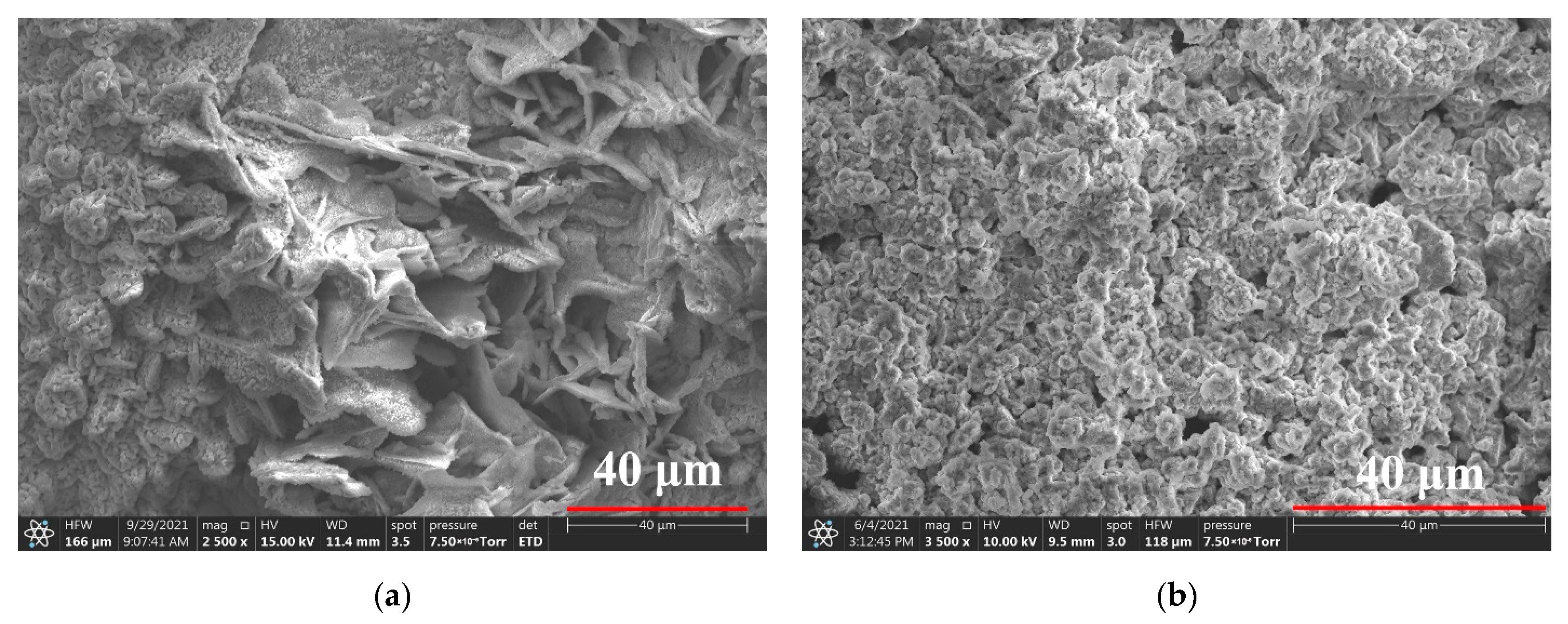
3.1. Metallographic Structure

The cross-section of the iron was silvery white in Figure 2. The white and long strips were primary cementite, and the matrix was eutectic ledeburite. The archaeological sample was a typical hypereutectic white cast iron with high carbon content in the range of 4.3–6.69%. When the hypereutectic molten iron was cooled, the wide and coarse primary cementite was produced first. Just because it could grow freely in the liquid, it was shaped as strips or flakes. Then, the temperature of the metal liquid continued to drop, and the eutectic phase occurred to be eutectic ledeburite. Due to the local cooling, primary cementite and eutectic ledeburite were also arranged in different cooling directions [32].
The elemental analysis was further tested by EDS, and the composition was 5.31% C, 0.11% Si, 0.15% Mn, 0.08% P, 0.05% S and 94.3% Fe, which agreed with the metallographic data.

3.2. Original Rust Composition

Figure 3 showed the rust on the surface of the archaeological iron was mainly divided into two major rust layers: the outer layer was yellow and presented a loose and porous appearance, while the inner one close to the iron core was black and dense. Many micro-cracks penetrated the whole rust layer, even breaking into the iron core and cutting the dense black layer to be peeled off easily. The oxides and hydroxides of iron compounds were identified by micro-Raman according to the literature values [15,33]. The components obtained in yellow rust were mixed and diverse, and there was no clear boundary. So the data were shown in the Figure 3 just because the signals at these points were intense, which did not mean that there was only one kind of rust near these point.
Raman spectra showed that the outermost rust was mainly formed by hematite (α-Fe2O3) and maghemite (γ-Fe2O3), with characteristic peaks at 215, 278 and 390 cm−1 to α-Fe2O3 and the relatively weak peak at 1591 cm−1 to γ-Fe2O3. The dots in the middle showed a strong peak at 393 cm−1 with less intense peaks at 299, 683 and 1306 cm−1 were attributed to the existence of goethite (α-FeOOH). A strongest peak closed to 251 cm−1 and a second peak in the Raman shift of 382 cm−1 was related to the lepidocrocite phase (γ-FeOOH) which coexisted in the middle part of the yellow rust layer with α-FeOOH phase. Akaganeite (β-FeOOH) was often regarded as the most dangerous compound in rust due to its hygroscopicity in post-excavation corrosion and easy transformation into other oxides, especially the magnetite (Fe3O4) [34]. In this object, akaganeite had the bands at 140, 305 and 392 cm−1, while magnetite was assigned the Raman band at 349, 461, 677 and 1413 cm−1 near the black dense layer. Therefore, maghemite, goethite, lepidocrocite and akaganeite were the dominant phases in the yellow loose rust layer, while the black dense layer was mainly composed of akaganeite and magnetite. The loose yellow rust had a tendency of exfoliation and the dense black rust had a lot of cracks, so that the inner iron core still had channels in contact with the external environment which was hard to stabilize.

3.3. Rust Conversion

XRD was also used to analyze the composition of rust powder and the data was compared respectively by Jade software (Jade 9.0, MDI, Livermore, CA, USA) and PDF-card references (α-FeOOH: #00–029–0713; β-FeOOH: #00–042–1315; γ-FeOOH: #00–008–0098; γ-Fe2O3: #00–039–1346). There was a good correspondence between the peaks of the original rust and the standard spectrum of β-FeOOH, which could be regarded as the main phase of rust in Figure 4a. Meanwhile, a significant signal of pure iron appeared, proving that there were some tiny iron particles that peeled off with rust due to corrosion. The peaks intensity of other rust components were weak, but a small amount of α-FeOOH and γ-FeOOH could be confirmed according to the PDF reference. Most iron-based compounds involved in corrosion reactions were prone to transform into β-FeOOH and γ-FeOOH in marine, resulting in the little amount of α-FeOOH in rust. Ferric oxyhydroxides has various types, where goethite was an electrochemically stable phase, whereas others were all active phase [35]. Therefore, the composition in the rust powder preliminarily indicated that the rust phase of these archaeological irons had not yet reached stability, and the subsequent stabilization treatment was necessarily required.
After catechin treatment, the XRD signals of archaeological rust were basically transformed into γ-Fe2O3, while the signals of β-FeOOH and γ-FeOOH were extremely low and almost undetected in Figure 4b. In order to explain this phenomenon, the diffraction patterns of three types of iron oxyhydroxides before and after catechin treatment were compared in Figure 4c–e, respectively. It was proved that FeOOH would chemically react with catechin, and the new products basically transformed the original crystalline state into amorphous compounds without obvious diffraction peaks. Therefore, the reactive α, β and γ-FeOOH in the original rust could be consumed by the catechin reaction to transform into an amorphous solid substance and deposited in the rust layer, while the relatively stable rust product Fe2O3 did not undergo significant conversion and still remained in the rust layer which could be detected by XRD.
All infrared peaks and corresponding chemical bonds were listed in Table 1. The infrared spectrum of catechin was shown in Figure 5a. The peak of 3385 cm−1 was the stretching vibration of −OH, indicating that there were a lot of hydroxyl groups on this molecule. Multiple groups of peaks within the wavenumber of 1000~2000 cm−1 indicated that the structure has obvious aromatic properties: 1456, 1519 and 1693 cm−1 were characteristic of aromatic ring C=C bond structure; 1320 cm−1 could be assigned to the bending vibration of the C−O bond connected to the hydroxyl group on phenolic substances; 1236 cm−1 was the bending vibration of the C−O bond; 1141 cm−1 and 1033 cm−1 was the stretching vibration of the phenolic C−O−C bond. Further, 823 cm−1 was the bending vibration of =C−H bond on the out-of-plane benzene ring [36]. The above functional groups were the basic parameters for identifying the presence of phenolic material. The phenolic hydroxyl groups were easy to coordinate with metal cations to form a stable deposited structure. The interaction between Fe3+ and the phenolic hydroxyl group has effect on the O−C bond stretching vibration, in which the band shifted from 1320 cm−1 in the spectra of the catechin to about 1380 cm−1 in the pure catechin-Fe composite in Figure 5b. This phenomenon can be explained by the chelation and redox reaction of Fe3+ with phenolic hydroxyl [37].
The composition of the original rust was also identified by FTIR. The absorption peaks at 833 cm−1 and 1627 cm−1 were corresponded to the bending of O−H bonds and the vibration of Fe−O bonds in β-FeOOH, respectively [38]. The peak at 1026 cm−1 was due to the O−H bending as a characteristic point in γ-FeOOH [39]. In addition, the characteristic peak of γ-Fe2O3 and β-FeOOH could be overlapped at 648 cm−1. As for the peak of 3377 cm−1, it was a broad adsorption of −OH group [40]. Therefore, Figure 5c showed the main component of the powdered rust were mainly akaganeite (β-FeOOH), lepidocrocite (γ-FeOOH) with a small amount of maghemite (γ-Fe2O3), which was basically consistent with the results obtained from the XRD above. After soaking with catechin, the characteristic positions of β-FeOOH and γ-FeOOH were significantly weakened, the signal of effective binding of catechin−Fe appeared at position 1380 cm−1, and the absorption of iron oxides appeared at position 553 cm−1. Combined with the results of XRD, this phenomenon could be explained that iron oxyhydroxides have successfully reacted with catechin to form catechin−FeOOH, and the iron oxides did not undergo obvious transformation, so that the infrared signal could be displayed.
The three main iron oxyhydroxides provided infrared images of the pure material before and after catechin treatment in Figure 5d–f. At the absorption positions of 1365–1372 cm−1, the reliable evidence of effective binding to catechin molecules appeared, and the aromatic peaks of catechin also showed a new signal on the original FeOOH structure.
Therefore, through the combined analysis of XRD and FTIR, it can basically be considered that catechin was chemically reacted with the oxyhydroxides on the surface of the archaeological iron, and the active α, β, γ-FeOOH could be consumed and transformed into an amorphous solid substance as catechin−FeOOH and deposited in the rust layer, while the original relatively stable iron oxide did not undergo obvious phase transformation and still remained.

3.4. Surface Modification

Scanning electron microscopy was used to compare the morphology of the rusted surface of archaeological iron before and after catechin treatment, and the results were shown in Figure 6. The original rust layer in Figure 6a has a loose and rough surface forming many pores that could not obstruct external corrosive media. The particle sizes varied greatly and a variety of rust products agglomerated irregularly.
The catechin treatment could successfully convert the active rust into new products and remained on the surface of archaeological iron. It gradually transformed the rust with different sizes and shapes into a multi-layer laminated structure, which made the rust layer more compact and denser in Figure 6b. Therefore, surface of the iron objects could be covered more uniformly, which was beneficial to delay the further corrosion of the internal iron core from the corrosive factors.

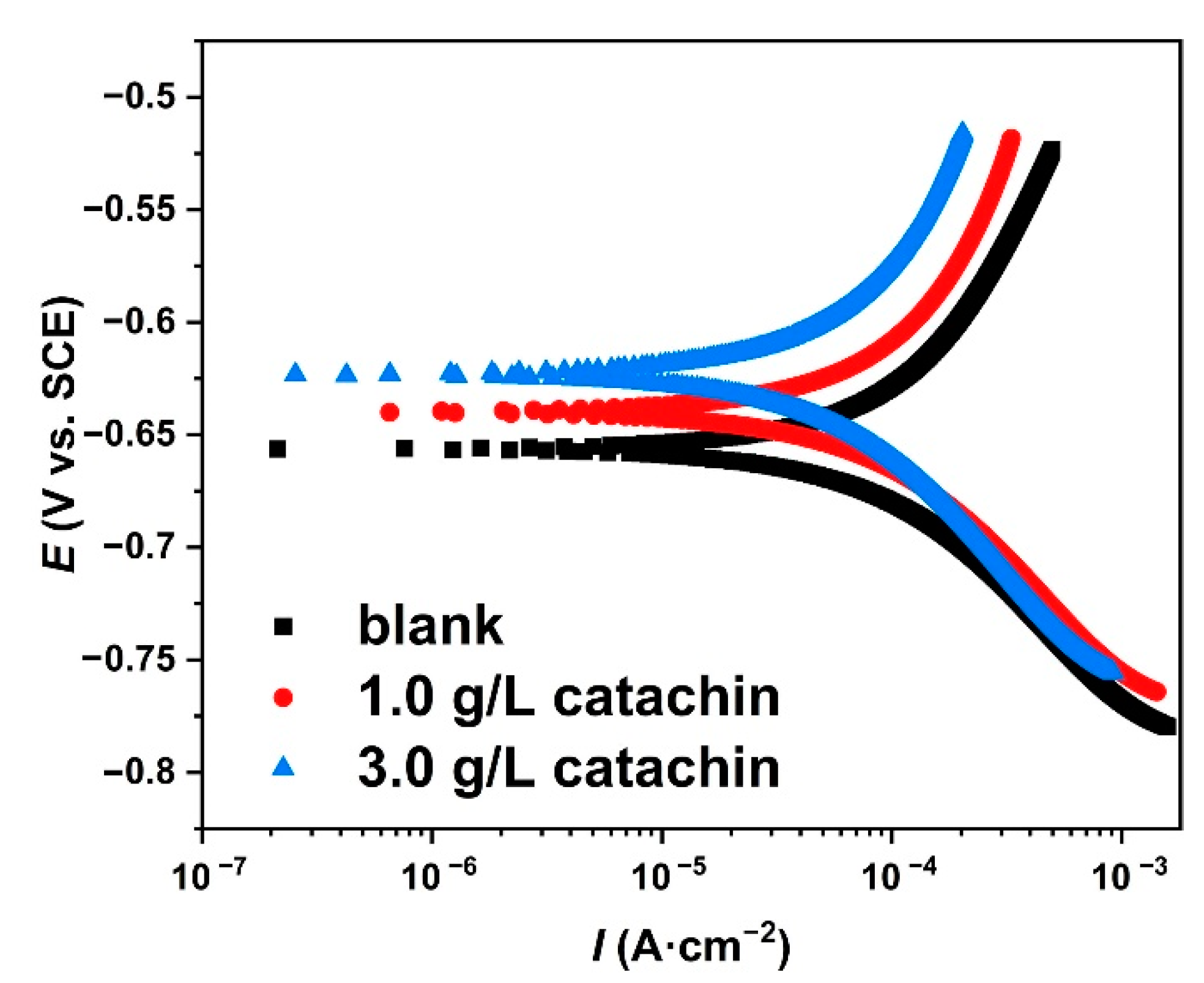
3.5. Potentiodynamic Polarization

The polarization curves of archaeological iron in mixed electrolyte (1.5 wt.% NaCl and 1.5 wt.% Na2SO4) at room temperature were shown in Figure 7. Table 2 presented the corrosion potential (Ecorr), corrosion current density (Icorr), Tafel slope (βa, βc), corrosion rate (vcorr) and chi-square (χ2) according to the data fitted by Cview. The shape of all curves was similar, indicating that the catechin pretreatment could delay the reaction by modifing rusted surface and obstructing the corrosion over metal, without causing marked changes in the electrochemical corrosion mechanism [41]. The curves shifted to lower current density with increasing concentration of catechin. The Icorr of the 3.0 g/L sample was about 98.64 μA·cm−2 which was almost half than that of the blank one. Indeed, it had the largest positive shift of Ecorr for catechin was 32.67 mV. A corrosion inhibitor was clearly judged to be a cathode or anode inhibitor when the shift of the corrosion potential was >85 mV [25]. Therefore, catechin could be regarded as a mixed-type inhibitor in this study. The slopes of both anodic and cathodic curves were not changed significantly with the catechin, although the anodic curve parallelly shifted more obviously than that of the cathode. The corrosion rate of archaeological iron decreased gradually after pretreatment with catechin. Under the addition of 3.0 g/L, the corrosion rate dropped to about 39% of the original state. Therefore, the behavior of catechin was focused on inhibiting the dissolution and diffusion of metals through surface modification of rusted cast iron. It could act as a qualified and mild green inhibitor to protect archaeological iron heritage.

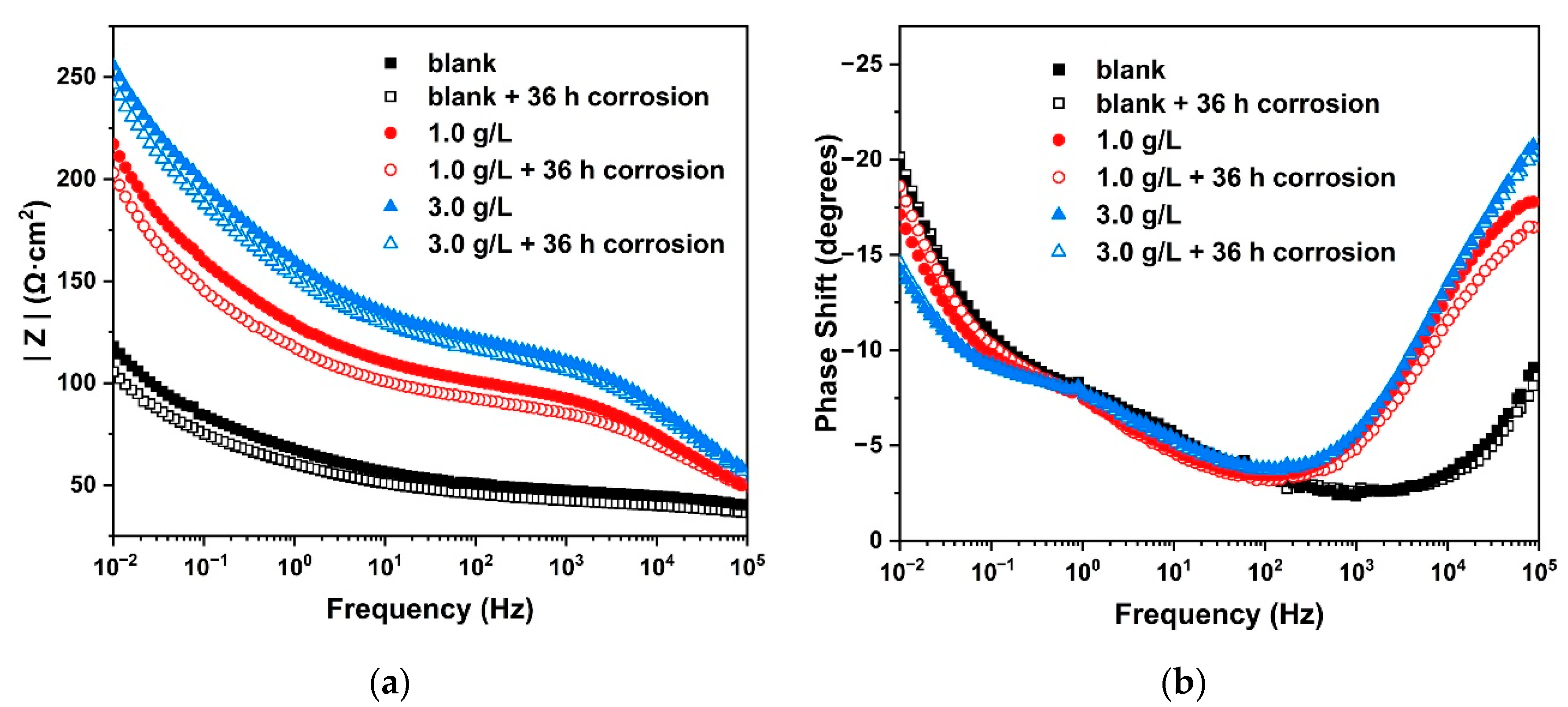
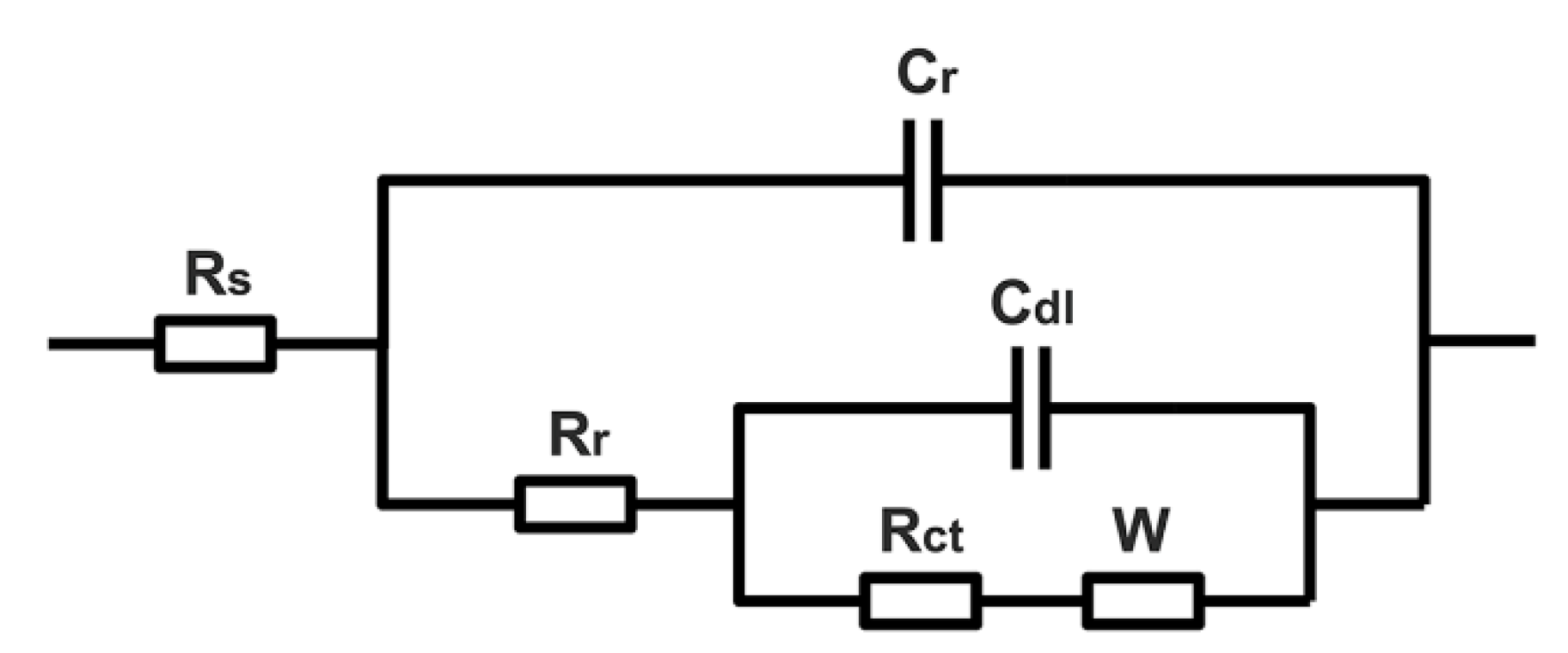
3.6. Electrochemical Impedance

Electrochemical impedance spectroscopy (EIS) is an effective tool to support potentiodynamic polarization for obtaining valuable information about the corrosion inhibition of catechin on the archaeological iron.
Figure 8 and Figure 9 showed the Nyquist and Bode plots of rusted and catechin-treated samples. To extract the electrochemical parameters, experimental data were fitted by the electrical equivalent circuits shown in Figure 10. In Figure 9a,b, Rs represented the electrolyte resistance, Cr the rust capacitance, Rr the rust resistance, Cdl the double-layer capacitance, Rct the charge transfer resistance, W the Warburg resistance or barrier diffusion impedance which was associated with the diffusion of corrosive electrolyte into the iron surface through pores in the rust layer that acted as a barrier. Due to the surface roughness and inhomogeneity, a constant phase angle element (CPE) was introduced to describe the Cdl and Cr in the fitting circuits [42,43].
For the rusted surface, the Nyquist plot was composed of a depressed capacitive semi-arc which was assigned to the resistance of rust and electrolyte in the high frequency, and a long tail in the low frequency region representing typical Warburg impedance. At medium frequency, there was a semicircular transition region representing the charge transfer resistance [44]. After the archaeological iron soaked with catechin, the radius of the semi-arc in the high-frequency region become significantly larger, which indicated that catechin effectively increased the resistance value of the rust layer. 1.0 g/L of catechin could increase the Rr of the original rust by 3 times, while 3.0 g/L of catechin could increase the Rr by 3.75 times. It was also proved that the modified rust by catechin was more inert and less conductive in corrosive media. The capacitance value Cr of the rust rose with the increase of the concentration of catechin in the high frequency, which could be considered as due to the weakly acidic catechin having reacted with the original rust to generate new compounds. Meanwhile, the tough surface of the original rust has turned out to be even and uniform, which was closer to the state of flat capacitors [45]. Then, the radius of the capacitive arc in the medium frequency was also broadened due to the catechin. This was because the catechin pretreatment could increase the charge transfer resistance of rust, indicating that catechin could modify rust to be denser than the original. The modified rust layer had the ability to effectively block the charge and ion transfer between the rusted cast iron and the corrosive solution, for improving the protection of the internal metal. The Warburg impedance in the low frequency region was also significantly improved, which also showed that the new layer could effectively block the diffusion of the external corrosive medium into the iron core compared with the original rust layer. After 36 h of accelerated corrosion, all three groups of samples showed a decreasing trend of impedance value, but the samples soaked with catechin could still guarantee the improvement of charge transfer resistance and rust resistance of archaeological iron.
The inhibition efficiency of the catechin was calculated from the charge transfer resistance (Rct) values according to the following Equation (1) [46]. Then, another equation could be constructed by the rust resistance (Rr) to analyze the efficiency of rust conversion using Equation (2). Rct(inh) and Rct(blank) were the charge transfer resistance values with and without inhibitor. Rr(inh) and Rr(blank) were the rust resistance values with and without the inhibitor, respectively. The corrosion inhibition efficiency of 3.0 g/L of catechin was 21.75% and the rust conversion efficiency was 73.26%. All fitted data are listed in Table 3.
η ct = Inhibition   efficiency   ( % ) = R ct ( inh )     R ct ( blank ) R ct ( inh ) × 100 %
η r = Conversion   efficiency   ( % ) = R r ( inh )     R r ( blank ) R r ( inh ) × 100 %

4. Conclusions

The surface rust of Nanhai No. 1 archaeological iron was mainly akaganeite, lepidocrocite and goethite with a small amount of maghemite by Raman spectroscopy and XRD analysis. After the archaeological iron soaked in 3.0 g/L of catechin for 12 h, there was only a strong signal of maghemite in XRD, while the signal of iron oxyhydroxides almost completely disappeared. The reaction between pure FeOOH and catechin could prove that the catechin has the ability to react with goethite, akaganeite and lepidocrocite in the rust, forming catechin−FeOOH with a marked signal about 1380 cm−1 as phenolic-Fe in infrared properties. The original crystalline state of FeOOH was converted into an amorphous form by catechin, so that the significant signal was lost in XRD. The relatively stable iron oxide did not undergo transformation reaction and could remain. The modified rust could transform the rough and porous rust layer into a laminated dense structure.
After the archaeological iron soaked in 3.0 g/L catechin, the corrosion current density decreased by 37.13% and the corrosion potential shifted positively by 32.67 mV. The anode reaction was more inhibited than the cathode in the polarization curve. The rust resistance in electrochemical impedance increased to 3.75 times and the ion diffusion resistance increased to 6.33 times. The corrosion inhibition efficiency was 21.75% and the rust conversion efficiency was 73.26%. After 36 h of accelerated corrosion, the protection effect of the newly transformed rust layer was still better than that of the original state.
Catechin could promote the stabilization and transformation of the rust layer, and effectively inhibit the further corrosion of the iron core. It has a good application prospect as an environment-friendly protection material for ancient iron cultural heritage.

Author Contributions

Conceptualization, M.J. and G.H.; methodology, P.H.; validation, M.J. and P.H.; investigation, Z.G.; resources, J.S. and Y.C.; writing—original draft preparation, M.J.; supervision, M.J. and G.H.; project administration, D.H., J.S. and Y.C.; funding acquisition, G.H. All authors have read and agreed to the published version of the manuscript.

Funding

This research was funded by the National Key Research and Development Program of China (State assignment No. 2020YFC1522100).

Institutional Review Board Statement

Not applicable.

Informed Consent Statement

Not applicable.

Data Availability Statement

The data presented in this study are available in the article.

Acknowledgments

Thanks to Li Chen in School of Physics, Peking University, for her support of SEM and EDS in this work.

Conflicts of Interest

The authors declare no conflict of interest.

References

  1. Batis, G.; Zacharopoulou, A.; Zacharopoulou, E.; Siova, H.; Argyropoulos, V. Dechlorination of large marine iron artifact using a novel technique involving impressed current. Anti-Corros. Methods Mater. 2015, 62, 259–269. [Google Scholar] [CrossRef]
  2. Parra, R.; Covelo, A.; Ramirez, R.J.; Tejeda, A.; Ortega, A.; Hernandez, M. Characterization of superficial modification of ferrous rusted substrates subjected to dechlorination-electrochemical process. J. Adhes. Sci. Technol. 2018, 32, 1341–1358. [Google Scholar] [CrossRef]
  3. Blahova, L.; Horak, J.; Prikryl, R.; Pekarek, J.; Tkacz, J.; Mencik, P.; Krcma, F. A novel technology for the corrosion protection of iron archaeological artefacts using parylene base removable bilayer. J. Cult. Herit. 2020, 42, 28–35. [Google Scholar] [CrossRef]
  4. Armetta, F.; Saladino, M.L.; Scherillo, A.; Caponetti, E. Microstructure and phase composition of bronze Montefortino helmets discovered Mediterranean seabed to explain an unusual corrosion. Sci. Rep. 2021, 11, 23022. [Google Scholar] [CrossRef]
  5. Bernabale, M.; Nigro, L.; Vaccaro, C.; Nicoli, M.; Montanari, D.; Bigini, P.; De Vito, C. Micro-Raman spectroscopy and complementary techniques for the study of iron weapons from Motya and Lilybaeum (Sicily, Italy): Corrosion patterns in lagoon-like and calcarenitic hypogea environments. J. Raman Spectrosc. 2022, 53, 272–287. [Google Scholar] [CrossRef]
  6. Hao, X.L.; Zhu, T.Q.; Xu, J.J.; Wang, Y.R.; Zhang, X.W. Microscopic study on the concretion of ceramics in the “Nanhai I” shipwreck of China, Southern Song Dynasty (1,127-1,279 AD). Microsc. Res. Techniq. 2018, 81, 486–493. [Google Scholar] [CrossRef]
  7. Wang, Y.; Xiao, D. The excavation of the “Nanhai No. 1” shipwreck of the Song Dynasty in 2014. Archaeology 2016, 12, 56–83. [Google Scholar]
  8. Remazeilles, C.; Leveque, F.; Conforto, E.; Refait, P. Long-term alteration processes of iron fasteners extracted from archaeological shipwrecks aged in biologically active waterlogged media. Corros. Sci. 2021, 181, 109231. [Google Scholar] [CrossRef]
  9. Simon, H.J.; Cibin, G.; Reinhard, C.; Liu, Y.; Schofield, E.; Freestone, I.C. Influence of microstructure on the corrosion of archaeological iron observed using 3D synchrotron micro-tomography. Corros. Sci. 2019, 159, 108132. [Google Scholar] [CrossRef]
  10. Zhao, X.D.; Cheng, Y.F.; Fan, W.; Vladimir, C.; Volha, V.; Alla, T. Inhibitive Performance of a Rust Converter on Corrosion of Mild Steel. J. Mater. Eng. Perform. 2014, 23, 4102–4108. [Google Scholar] [CrossRef]
  11. Flores Merino, S.; Jose Caprari, J.; Vasquez Torres, L. Inhibitive action of tara tannin in rust converter formulation. Anti-Corros. Methods Mater. 2017, 64, 136–147. [Google Scholar] [CrossRef]
  12. Remazeilles, C.; Neff, D.; Bourdoiseau, J.A.; Sabot, R.; Jeannin, M.; Refait, P. Role of previously formed corrosion product layers on sulfide-assisted corrosion of iron archaeological artefacts in soil. Corros. Sci. 2017, 129, 169–178. [Google Scholar] [CrossRef]
  13. Gao, X.L.; Han, Y.; Fu, G.Q.; Zhu, M.Y.; Zhang, X.Z. Evolution of the Rust Layers Formed on Carbon and Weathering Steels in Environment Containing Chloride Ions. Acta Metall. Sin-Engl. 2016, 29, 1025–1036. [Google Scholar] [CrossRef] [Green Version]
  14. Kamimura, T.; Hara, S.; Miyuki, H.; Yamashita, M.; Uchida, H. Composition and protective ability of rust layer formed on weathering steel exposed to various environments. Corros. Sci. 2006, 48, 2799–2812. [Google Scholar] [CrossRef]
  15. Saleh, S.A. Corrosion Mechanism of Iron Objects in Marine Environment an Analytical Investigation Study by Raman Spectrometry. Eur. J. Sci. Theol. 2017, 13, 185–206. [Google Scholar]
  16. Lair, V.; Antony, H.; Legrand, L.; Chausse, A. Electrochemical reduction of ferric corrosion products and evaluation of galvanic coupling with iron. Corros. Sci. 2006, 48, 2050–2063. [Google Scholar] [CrossRef]
  17. Alsabagh, A.M.; Migahed, M.A.; Abdelraouf, M.; Khamis, E.A. Utilization of Green Tea as Environmentally Friendly Corrosion Inhibitor for Carbon Steel in acidic media. Inter. J. Electrochem. Sci. 2015, 10, 1855–1872. [Google Scholar]
  18. Aourabi, S.; Driouch, M.; Sfaira, M.; Mahjoubi, F.; Hammouti, B.; Verma, C.; Ebenso, E.E.; Guo, L. Phenolic fraction of Ammi visnaga extract as environmentally friendly antioxidant and corrosion inhibitor for mild steel in acidic medium. J. Mol. Liq. 2021, 323, 114950. [Google Scholar] [CrossRef]
  19. Pradipta, I.; Kong, D.; Tan, J.B.L. Natural organic antioxidants from green tea inhibit corrosion of steel reinforcing bars embedded in mortar. Constr. Build. Mater. 2019, 227, 117058. [Google Scholar] [CrossRef]
  20. Abdallah, M. Rhodanine azosulpha drugs as corrosion inhibitors for corrosion of 304 stainless steel in hydrochloric acid solution. Corros. Sci. 2002, 44, 717–728. [Google Scholar] [CrossRef]
  21. Hussin, M.H.; Kassim, M.J. The corrosion inhibition and adsorption behavior of Uncaria gambir extract on mild steel in 1 M HCl. Mater. Chem. Phys. 2011, 125, 461–468. [Google Scholar] [CrossRef]
  22. Tang, Z. A review of corrosion inhibitors for rust preventative fluids. Curr. Opin. Solid State Mater. Sci. 2019, 23, 100759. [Google Scholar] [CrossRef]
  23. Tan, K.W.; Kassim, M.J.; Oo, C.W. Possible improvement of catechin as corrosion inhibitor in acidic medium. Corros. Sci. 2012, 65, 152–162. [Google Scholar] [CrossRef]
  24. Hussin, M.H.; Kassim, M.J. Electrochemical, Thermodynamic and Adsorption Studies of (+)-Catechin Hydrate as Natural Mild Steel Corrosion Inhibitor in 1 M HCl. Inter. J. Electrochem. Sci. 2011, 6, 1396–1414. [Google Scholar]
  25. Wang, D.Y.; Nie, B.L.; Li, H.J.; Wang, F.; Zhang, W.Z.; Wu, Y.C. Experimental and theoretical investigation of inhibition behavior of bisflavanol for Q235 steel in hydrochloric acid solution. J. Mol. Liq. 2021, 342, 117490. [Google Scholar] [CrossRef]
  26. Ramos-Tejada, M.M.; Duran, J.D.G.; Ontiveros-Ortega, A.; Espinosa-Jimenez, M.; Perea-Carpio, R.; Chibowski, E. Investigation of alumina/(+)-catechin system properties. Part I: A study of the system by FTIR-UV-Vis spectroscopy. Colloids Surf. B 2002, 24, 297–308. [Google Scholar] [CrossRef]
  27. Alam, M.; Ali, S.; Ashraf, G.M.; Bilgrami, A.L.; Yadav, D.K.; Hassan, M.I. Epigallocatechin 3-gallate: From green tea to cancer therapeutics. Food Chem. 2022, 379, 132135. [Google Scholar] [CrossRef] [PubMed]
  28. Gallia, M.C.; Bachmeier, E.; Ferrari, A.; Queralt, I.; Mazzeo, M.A.; Bongiovanni, G.A. Pehuen (Araucaria araucana) seed residues are a valuable source of natural antioxidants with nutraceutical, chemoprotective and metal corrosion-inhibiting properties. Bioorg. Chem. 2020, 104, 104175. [Google Scholar] [CrossRef]
  29. Luo, Q.; Zhang, J.R.; Li, H.B.; Wu, D.T.; Geng, F.; Corke, H.; Wei, X.L.; Gan, R.Y. Green Extraction of Antioxidant Polyphenols from Green Tea (Camellia sinensis). Antioxidants 2020, 9, 785. [Google Scholar] [CrossRef]
  30. Abdelaziz, S.; Benamira, M.; Messaadia, L.; Boughoues, Y.; Lahmar, H.; Boudjerda, A. Green corrosion inhibition of mild steel in HCl medium using leaves extract of Arbutus unedo L. plant: An experimental and computational approach. Colloids Surf. A Physicochem. Eng. Asp. 2021; 619, 126496. [Google Scholar]
  31. Lekbach, Y.; Dong, Y.; Li, Z.; Xu, D.; El Abed, S.; Yi, Y.; Li, L.; Koraichi, S.I.; Su, T.; Wang, F. Catechin hydrate as an eco-friendly biocorrosion inhibitor for 304L stainless steel with dual-action antibacterial properties against Pseudomonas aeruginosa biofilm. Corros. Sci. 2019, 157, 98–108. [Google Scholar] [CrossRef]
  32. Mazur, A.; Gasik, M.M.; Mazur, V.I. Thermal analysis of eutectic reactions of white cast irons. Scand. J. Metall. 2005, 34, 245–249. [Google Scholar] [CrossRef]
  33. Rocca, E.; Faiz, H.; Dillmann, P.; Neff, D.; Mirambet, F. Electrochemical behavior of thick rust layers on steel artefact: Mechanism of corrosion inhibition. Electrochim. Acta 2019, 316, 219–227. [Google Scholar] [CrossRef]
  34. Calero, J.; Alcantara, J.; Chico, B.; Diaz, I.; Simancas, J.; de la Fuente, D.; Morcillo, M. Wet/dry accelerated laboratory test to simulate the formation of multilayered rust on carbon steel in marine atmospheres. Corros. Eng. Sci. Technol. 2017, 52, 178–187. [Google Scholar] [CrossRef] [Green Version]
  35. Nishimura, T.; Katayama, H.; Noda, K.; Kodama, T. Electrochemical behavior of rust formed on carbon steel in a wet/dry environment containing chloride ions. Corrosion 2000, 56, 935–941. [Google Scholar] [CrossRef]
  36. Selvi, I.K.; Nagarajan, S. Separation of catechins from green tea (Camellia sinensis L.) by microwave assisted acetylation, evaluation of antioxidant potential of individual components and spectroscopic analysis. LWT 2018, 91, 391–397. [Google Scholar] [CrossRef]
  37. Zhang, R.N.; Li, L.; Liu, J.X. Synthesis and characterization of ferric tannate as a novel porous adsorptive-catalyst for nitrogen removal from wastewater. RSC Adv. 2015, 5, 40785–40791. [Google Scholar] [CrossRef]
  38. Veneranda, M.; Aramendia, J.; Bellot-Gurlet, L.; Colomban, P.; Castro, K.; Madariaga, J.M. FTIR spectroscopic semi-quantification of iron phases: A new method to evaluate the protection ability index (PAI) of archaeological artefacts corrosion systems. Corros. Sci. 2018, 133, 68–77. [Google Scholar] [CrossRef] [Green Version]
  39. Liu, Y.W.; Wang, Z.Y.; Wei, Y.H. Influence of Seawater on the Carbon Steel Initial Corrosion Behavior. Int. J. Electrochem. Sci. 2019, 14, 1147–1162. [Google Scholar] [CrossRef]
  40. Labbe, J.P.; Ledion, J.; Hui, F. Infrared spectrometry for solid phase analysis: Corrosion rusts. Corros. Sci. 2008, 50, 1228–1234. [Google Scholar] [CrossRef]
  41. Huang, L.; Yang, K.P.; Zhao, Q.; Li, H.J.; Wang, J.Y.; Wu, Y.C. Corrosion resistance and antibacterial activity of procyanidin B2 as a novel environment-friendly inhibitor for Q235 steel in 1 M HCl solution. Bioelectrochemistry 2022, 143, 107969. [Google Scholar] [CrossRef]
  42. Han, D.; Jiang, R.J.; Cheng, Y.F. Mechanism of electrochemical corrosion of carbon steel under deoxygenated water drop and sand deposit. Electrochim. Acta 2013, 114, 403–408. [Google Scholar] [CrossRef]
  43. Orazem, M.E.; Frateur, I.; Tribollet, B.; Vivier, V.; Marcelin, S.; Pebere, N.; Bunge, A.L.; White, E.A.; Riemer, D.P.; Musiani, M. Dielectric Properties of Materials Showing Constant-Phase-Element (CPE) Impedance Response. J. Electrochem. Soc. 2013, 160, C215–C225. [Google Scholar] [CrossRef] [Green Version]
  44. Dhaiveegan, P.; Elangovan, N.; Nishimura, T.; Rajendran, N. Electrochemical Characterization of Carbon and Weathering Steels Corrosion Products to Determine the Protective Ability Using Carbon Paste Electrode (CPE). Electroanalysis 2014, 26, 2419–2428. [Google Scholar] [CrossRef]
  45. Sharifi-Asl, S.; Taylor, M.L.; Lu, Z.; Engelhardt, G.R.; Kursten, R.; Macdonald, D.D. Modeling of the electrochemical impedance spectroscopic behavior of passive iron using a genetic algorithm approach. Electrochim. Acta 2013, 102, 161–173. [Google Scholar] [CrossRef]
  46. Palomar-Pardave, M.; Romero-Romo, M.; Herrera-Hernandez, H.; Abreu-Quijano, M.A.; Likhanova, N.V.; Uruchurtu, J.; Juarez-Garcia, J.M. Influence of the alkyl chain length of 2 amino 5 alkyl 1,3,4 thiadiazole compounds on the corrosion inhibition of steel immersed in sulfuric acid solutions. Corros. Sci. 2012, 54, 231–243. [Google Scholar] [CrossRef]
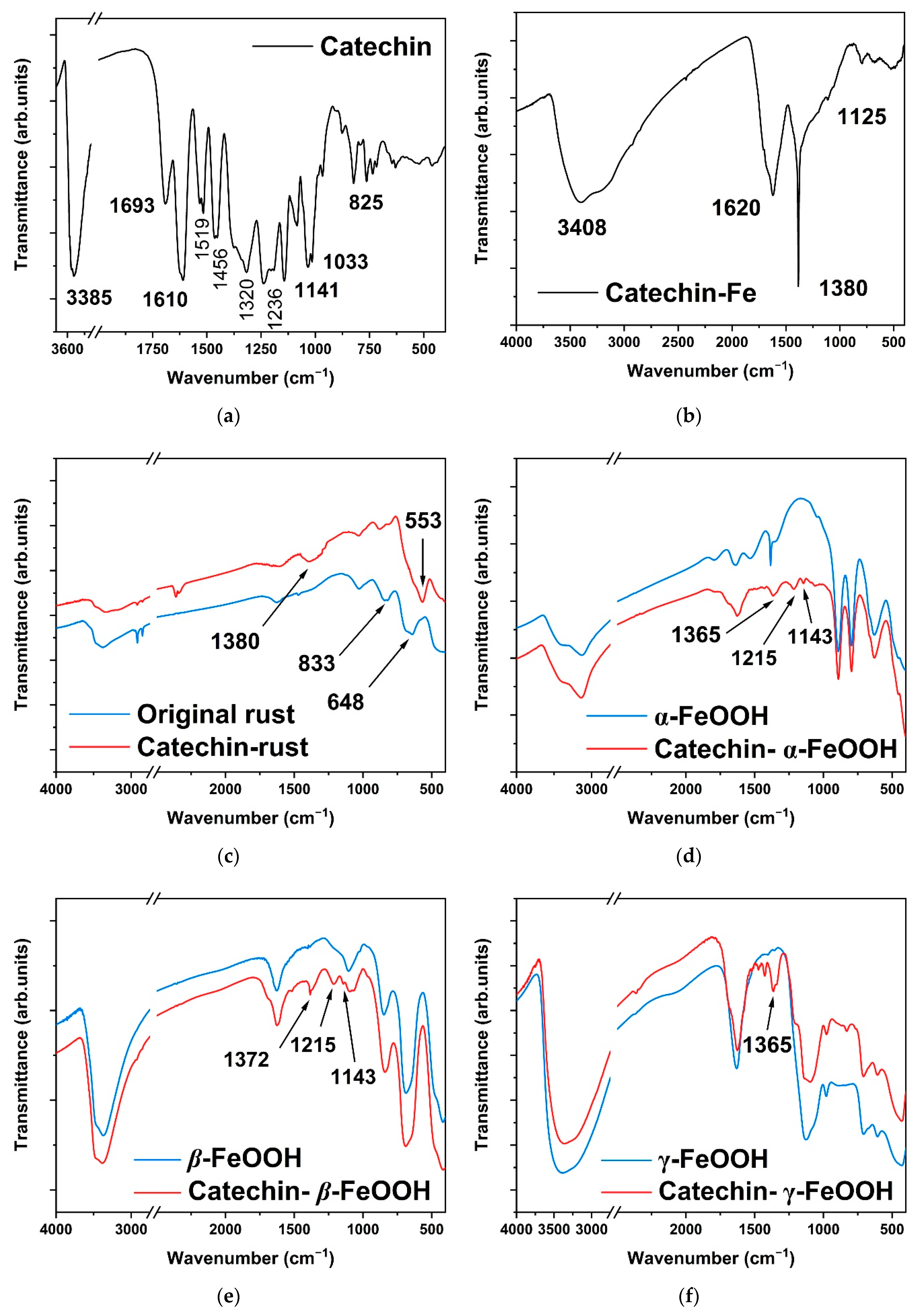
Figure 1. Archaeological iron artifacts coming from Nanhai I (Guangdong, China).
Metals 12 00714 g001
Figure 2. Metallographic diagrams of archaeological iron artifact from Nanhai I. Cross-section of the cast iron (A) and primary cementite and eutectic ledeburite (B).
Metals 12 00714 g002
Figure 3. Micro-Raman diagram of original rust layer.
Metals 12 00714 g003
Figure 4. XRD of original rust and three types of FeOOH before and after the catechin treatment. Original rust (a); catechin-modified rust (b); catechin-modified α-FeOOH (c); catechin-modified β-FeOOH (d); catechin-modified γ-FeOOH (e).
Metals 12 00714 g004
Figure 5. FTIR of original rust and three types of FeOOH before and after the catechin treatment. catechin (a); catechin-Fe (b); catechin-rust (c); catechin-α-FeOOH (d); catechin-β-FeOOH (e); catechin-γ-FeOOH (f).
Metals 12 00714 g005
Figure 6. SEM of original (a) and catechin-treated surface (b) of archaeological iron of Nanhai I.
Metals 12 00714 g006
Figure 7. Potentiodynamic polarization plots of archaeological iron in mixed solution.
Metals 12 00714 g007
Figure 8. Nyquist plots of original and catechin-treated archaeological iron in mixed solution.
Metals 12 00714 g008
Figure 9. Bode diagrams of original and catechin-treated archaeological iron in mixed solution. |Z| value (a) and phase shift (b) against frequency.
Metals 12 00714 g009
Figure 10. Equivalent circuits of EIS curves of archaeological iron in mixed solution.
Metals 12 00714 g010
Table 1. FTIR peaks and corresponding chemical bonds.
Chemical BondingIR Peaks (cm−1)
stretching vibration of the −OH bond3385
C=C bond of aromatic ring1456, 1519 and 1693
stretching vibration of the C−O−Fe bond
(in the pure catechin−Fe composite)
1380
stretching vibration of the C−O−Fe bond
(in the catechin−FeOOH composite)
1365–1372
bending vibration of the C−O bond
(connected to the −OH on phenolic substances)
1320
bending vibration of the C−O bond1236
stretching vibration of the phenolic C−O−C bond1141 and 1033
bending vibration of the =C−H bond
(on the out-of-plane benzene ring)
823
vibration of the Fe−O bond in β-FeOOH1627
bending vibration of the −OH bond in γ-FeOOH1026
bending vibration of the −OH bond in β-FeOOH833
vibration of iron oxides553
Table 2. Fitted parameters of polarization curves of archaeological iron in mixed solution.
SamplesEcorr (mV vs. SCE)Icorr (μA·cm−2)βa (mV·dec−1)βc (mV·dec−1)vcorr (mmPY)χ2
blank−656.25156.89257.87148.951.84350.2043
1.0 g/L−639.81135.14272.51135.291.57580.4098
3.0 g/L−623.5898.64289.47146.711.12290.4110
Table 3. Fitted parameters of EIS curves of archaeological iron in mixed solution.
SamplesRs
(Ω·cm2)
Rr
(Ω·cm2)
Rct
(Ω·cm2)
Rw
(Ω·cm2)
CPEr
(sn·Ω−1·cm−2 × 10⁶)
CPEdl
(sn·Ω−1·cm−2 × 10⁶)
nrndlχ2ηctηr
blank17.5428.1178.5281.921.67717.6370.590.340.013//
1.0 g/L23.4976.8785.59417.5222.0638.8670.530.480.0218.26%63.43%
3.0 g/L16.70105.12100.35518.4120.2126.0050.500.510.01921.75%73.26%
blank + corr.18.5221.7661.1478.961.45119.2710.600.310.014//
1.0 g/L+ corr.21.9070.4470.12391.6028.8549.5140.510.490.01512.81%69.11%
3.0 g/L+ corr.18.8497.3994.08410.4520.0236.6650.510.490.01835.01%77.66%
Publisher’s Note: MDPI stays neutral with regard to jurisdictional claims in published maps and institutional affiliations.

