Review

Recent Advances in the Development of Magnesium-Based Alloy Guided Bone Regeneration (GBR) Membrane

1
Key Laboratory for Biomechanics and Mechanobiology of Ministry of Education, Beijing Advanced Innovation Centre for Biomedical Engineering, School of Biological Science and Medical Engineering, Beihang University, Beijing 100083, China
2
Beijing Advanced Innovation Center for Materials Genome Engineering, Institute for Advanced Materials and Technology, University of Science and Technology Beijing, Beijing 100083, China
3
School of Materials Science and Engineering, Nanjing Institute of Technology, Nanjing 211167, China
*
Authors to whom correspondence should be addressed.
These authors contributed equally to this work.
Metals 2022, 12(12), 2074; https://doi.org/10.3390/met12122074
Submission received: 10 November 2022 / Revised: 25 November 2022 / Accepted: 29 November 2022 / Published: 2 December 2022
(This article belongs to the Special Issue Advanced Biomedical Materials)

Abstract

:
In dental implantology, the guided bone regeneration (GBR) membrane plays an active role in increasing alveolar bone volume. However, there are some drawbacks to the current commercial membranes, such as non-degradability for non-absorbable membranes and low mechanical strength for absorbable membranes. Recently, magnesium (Mg) alloys have been proposed as potential barrier membrane candidates. As a result, the purpose of this research is to assess the feasibility of Mg alloys as GBR membranes in terms of physicochemical properties and biological performance. Mg alloys were identified as potential membrane materials due to their adjustable degradation, adequate mechanical support, sound osteogenic property, good bacteriostatic activity, and favorable wound-healing ability. Nonetheless, rapid degradation and stress corrosion cracking (SCC)/corrosion fatigue (CF) are major concerns for the use of Mg-based membranes, which can be mitigated through alloying, heat treatment, thermomechanical deformation, and other methods. Finally, the prospects for the design and manufacture of Mg-based membranes in the future were put forth.

1. Introduction

Implant prosthesis is now an important way to repair dentition defects. In clinical surgeries, insufficient alveolar bone volume before dental implantology is frequently encountered, which can be caused by periodontitis, local alveolar process absorption, and other factors [1,2,3]. Distraction osteogenesis, autografts, guided bone regeneration (GBR), and other procedures have been used to solve the problem [4]. However, due to its immature development, traction osteogenesis may leave undesirable tissue scars [5]. Despite its extensive clinical history and obvious alveolar augmentation effect, autogenous bone graft damage and repair are urgent issues that must be addressed [6]. In contrast, the GBR technique uses the membrane as a barrier to prevent fibrous tissue infiltration while providing a suitable local environment for bone regeneration, making it one of the most effective operations for increasing alveolar bone volume during dental implantology [7].
In clinical practice, a combination of bone grafts and guided bone regeneration (GBR) techniques is typically used to treat periodontal defects, as illustrated in Figure 1. The missing bone in the defect area was primarily replaced with autogenous or allograft bone substitutes via bone grafts. The grafts may stimulate bone growth, with the new bone filling the defect and providing adequate mechanical support for the tooth [8,9]. Following that, the GBR technique was used, and a barrier membrane (Figure 1c) was placed between the gum and the bone defect area, with the goal of preventing the rapid proliferation of epithelial cells or fibroblasts while still allowing enough space for periodontal tissue regeneration [10,11,12]. The membrane can act as a carrier for growth factors such as BMP-2, IGF, and other bone-related factors [13,14]. Furthermore, the barrier membrane protects the integrity of the blood clot in the operation area, facilitates the transport of oxygen and other nutrients, promotes the establishment of microcirculation, and so on [15,16,17]. Furthermore, in addition to its barrier function, Omar et al. proposed that the membrane actively hosted and modulated the molecular activities of membrane-associated cells during the GBR process [14]. As a result, it is clear that the choice of GBR membrane has a significant impact on the therapeutic effect of GBR surgery.
Magnesium (Mg) and its alloys have recently been proposed as potential materials for oral and maxillofacial implants due to their sufficient mechanical strength, appropriate degradation rate, good biocompatibility, sound in vitro or in vivo osteogenesis performance, and so on [18,19,20,21,22,23,24,25,26,27,28,29,30]. However, there still exist some drawbacks for Mg-based membranes, such as rapid degradation rate, hydrogen evolution effect, stress corrosion cracking (SCC) susceptibility, premature mechanical integrity loss and undesirable pH increase, etc. [20]. Primarily, the rapid degradation rate of Mg-based alloys can result in accelerated hydrogen release, undesirable pH increase, and premature mechanical integrity loss. Beyond that, when Mg alloys are used as structural components, they will be subjected to the dual effects of stress and corrosion environment in the service process, causing stress corrosion cracking (SCC) and rapid loss of mechanical integrity. Until now, some methods, such as alloying, thermal deformation, heat treatment, surface modification, etc., have been adopted to enhance the SCC resistance of Mg-based alloys [31]. Many researchers are interested in the feasibility of using Mg-based alloys as GBR membranes. Despite significant progress in the design or fabrication of Mg-based membranes in recent years, there is still a gap between the bench and the bedside. As a result, this work will systematically demonstrate the feasibility of Mg-based alloys as GBR membranes from the standpoints of material science and biology. Besides that, the obstacles, improved measures, and prospects for future Mg-based membrane design and manufacturing will be discussed.

2. The Classification and Characteristics of Commercial GBR Membranes

With the advancement of clinical demands, the emergence of various types of GBR membranes has played various roles in clinical surgeries. The function of the membrane is determined by its physicochemical and biological properties. Similarly, the membrane selection is influenced by the characteristics and processing requirements of the materials, with each material presenting distinct advantages and disadvantages. As shown in Table 1, the GBR membrane can be classified into two types based on in vivo degradability: non-resorbable membranes and resorbable membranes [11,32,33,34,35,36,37,38,39,40,41,42,43,44,45,46,47].
Non-resorbable membranes are primarily made of polytetrafluoroethylene (PTFE) and titanium film. A large number of studies and experiments have shown that titanium films, as a representative of non-absorbable membranes, have good biocompatibility and excellent mechanical properties, which can help to restore the ideal anatomical structure and make adequate horizontal or vertical alveolar bone regeneration [48]. Furthermore, the titanium-reinforced PTFE membrane demonstrated good stiffness, adequate mechanical support, high stability, and superior anti-deformation ability [49]. However, some disadvantages have restricted their clinical applications. For example, the non-degradability of the membranes typically necessitates secondary surgery to remove the implant, which increases patients’ pain and financial burdens. The elastic modulus of titanium alloy differs from that of natural bone, resulting in the formation of a stress-shielding effect [18]. Furthermore, the material’s high stiffness causes soft tissue dehiscence and premature membrane exposure, leading to bacterial infection and implant failure [50,51,52].
Resorbable membranes, on the other hand, are primarily composed of synthetic or natural polymers [44]. Collagen from pigs, cows, or humans is one of the most commercially valuable natural polymers, and it is widely used in GBR due to its medical applicability. Collagen has high biocompatibility and bioabsorbability, allowing it to avoid secondary removal following bone regeneration [53,54]. At the same time, the absorbable film has selective permeability, which promotes early microcirculation [55]. In addition, absorbable membranes are appealing to both clinicians and patients because they reduce morbidity and the risk of tissue damage. Furthermore, some synthetic polymers, such as polylaticaeid (PLA), polyglycolicacid (PGA), poly-dl-lactic/co-glycolic acid (PLGA), and others, have been used in the fabrication of the membranes [11]. Synthetic polymers can be used as drug delivery vehicles and cause less inflammation in the body. Nonetheless, as shown in Table 1, the main factors limiting the applications of absorbable membranes are poor mechanical properties and resorption rate variability [11,44]. As a result, novel absorbable membranes with high mechanical strength, good malleability, and good biological effect are still desperately needed in clinical practice.

3. The Feasibility of Utilizing Mg-Based Alloys as GBR Membrane

The barrier membrane primarily performs the functions of increasing alveolar bone volume prior to surgery, repairing bone defects caused by immediate implantation, and treating pathological bone resorption caused by peri-implantitis after implantation during the GBR procedure [1]. As a result, the selection of membrane material is critical to the therapeutic effect of GBR surgery. Chen et al. [44] recently summarized that an ideal membrane should have the following characteristics: (1) benign biocompatibility, (2) adequate mechanical strength and integrity, (3) good occlusive function, (4) reasonable malleability, (5) sound osteogenic performance, (6) preferable antibacterial activity, and (7) promoted wound-healing ability. To address potential issues in commercial membranes, Mg and its alloys have recently been identified as potential candidates as GBR membranes, and thus the feasibility of the alloys as barrier membranes has been discussed from these perspectives.

3.1. Biocompatibility

Typically, the biocompatibility of the materials is the first factor to consider before applying them. Previous reports have confirmed the materials’ biocompatibility for Mg-based alloys [20,56]. Mg is an essential element for the human body, and it has been suggested that the recommended daily intake of Mg for an adult is approximately 240–420 mg/day, which is approximately 52.5 times higher than that of zinc (8–11 mg/day) and iron (8–18 mg/day) [26,28,57]. As shown in Figure 2, the distribution of Mg in the human body is approximately 55% in bone, 25% in muscle, 0.8% in extracellular fluid, and 0.3% in plasma [58]. The Mg2+ concentration in plasma has been reported to be about 0.5–1 mM, with the types of free Mg2+ (about 65%), inorganic or organic salts (15%), and protein (20%) [59]. Furthermore, with concentrations ranging from 5 to 20 mM, intracellular Mg is the second most abundant cation after Ca. Mg is primarily bound to adenosine triphosphate (ATP) in the cell [19].
In terms of physiological effects, Mg acts as an enzyme activator, a co-regulator of protein synthesis and muscle contraction, a DNA and RNA stabilizer, and so on [20,60]. Mg can act as a Ca impedance agent, preventing Ca accumulation in cells [61]. Mg is also involved in the bone metabolism process [62]. Mg deficiency may cause neuropathy, shivering, depression, delusion, restlessness, excitement, confusion, and other neuropsychiatric disorders, as well as blood circulation system disorders such as arrhythmia, increased-frequency pulse, and ventricular throbbing [63,64]. Excessive Mg, on the other hand, can cause nausea, vomiting, low muscle strength, and sluggish blood flow [61]. Fortunately, excess Mg can be excreted by kidneys and urine (as shown in Figure 2) [20,65].
Previous studies have also confirmed the cytocompatibility of Mg-based alloys [66,67,68]. Gu et al. [66], for example, recently successfully prepared a series of binary Mg-based alloys and investigated their cytocompatibility using fibroblasts (L-929 and NIH3T3) and osteoblasts (MC3T3-E1). The results indicate that the cell viability of some Mg-based alloys (Mg-1Zn, Mg-1Si, Mg-1Al, and so on) was greater than 70%, indicating that the alloys were non-toxic to cells according to the ISO 10993-5 standard. Furthermore, the body fluid is in a dynamic state, which can effectively dilute alloy extracts and improve cell viability. Previous research has also demonstrated that cells cultured with diluted Mg-based alloy extracts have higher viability [69,70,71]. As a result, the use of Mg-based alloy as a GBR membrane is medically safe.

3.2. Biodegradation of Mg-Based Alloys

One of the primary benefits of Mg-based alloys is their biodegradability, which allows them to perform their functions during the regeneration process before disappearing, either by being absorbed by tissue or excreted from the human body. This effectively avoids secondary surgery while also reducing patients’ pain. The barrier membrane is placed between the bone defect areas and the external soft tissues, resulting in biocorrosion at the materials/body fluid interface.
Mg is a highly reactive metal with a standard electrode potential of −2.372V (vs. SHE) [72]. Mg can corrode in physiological environments and is governed by the following reactions (Equations (1)–(3)), as shown in Figure 3. However, the physiological fluid contains a high concentration of Cl- (about 150 mM), causing corrosion of the surface oxide layer (Mg(OH)2) by Cl- and the formation of MgCl2 [20].
Anodic reaction:
Mg Mg 2 + + 2 e
Cathodic reaction:
2 H 2 O + 2 e H 2 + 2 OH
Overall reaction:
Mg + 2 H 2 O Mg OH 2 + H 2
The membrane acts as a barrier between bone defect areas and outer epithelial tissue. As a result, the corrosive media are mostly made up of bodily fluids. Until now, PBS, Hank’s, SBF, DMEM, DMEM+FBS, and other simulated body fluids have been used to stimulate the degradation behavior of Mg-based alloys [73,74]. The main components of the Mg alloy degradation products are Mg(OH)2, MgCl2, MgCO3, Mg3(PO4)2, Ca3(PO4)2, and other products [75,76]. Although these metal salts, such as MgCO3 and Mg3(PO4)2, have much lower water solubility and may be difficult to remove from the implantation site, a previous study found that they were biocompatible, particularly in bone tissues [20]. Furthermore, Jin et al. demonstrated that these degradation products are engulfed by macrophages via heterophagy and degraded in the phagolysosome [77]. As a result, it appears that the degradation products are biologically safe; however, a long-term study of the degradation particles’ degradability and biocompatibility is still required.
Actually, the use of barrier membranes has been linked to complications such as premature membrane exposure caused by insufficient tissue coverage for primary wound closure or wound dehiscence during healing [51,78]. Premature exposure of the barrier membrane will result in saliva–membrane contact. The pH of saliva is approximately 6.6–7.1 [79], indicating acidity and being lower than commonly used simulated body fluids (pH = 7.4). The acidic corrosive medium could potentially accelerate the degradation of Mg alloys. Zeng et al. [80] recently investigated the degradation behavior of pure Mg samples in artificial saliva, and the results show that a Ca-P layer was deposited on the sample surface, and pure Mg demonstrated good resistance in artificial saliva.
Another factor to consider with Mg alloys is their rapid degradation rate, which does not correspond well with the tissue healing process and may result in premature mechanical loss of implants. Erinc et al. recently proposed that the degradation rate of Mg alloys used as implants be less than 0.5 mm/yr [81]. Table 2 summarizes recent studies on the development of Mg-based GBR membranes [7,70,82,83,84,85,86,87,88,89,90,91,92,93,94,95,96]. The degradation rate of Mg-based membrane is found to be in the range of 0.02–1.76 mm/yr, indicating the great potential of Mg alloys as GBR membrane. Furthermore, alloying, grain refinement, heat treatment, surface modifications, and other methods can be adopted to improve the degradation resistance of Mg alloys [46,97]. As a result, the degradation rate of Mg alloys can be tuned to meet the specifications of an ideal membrane.

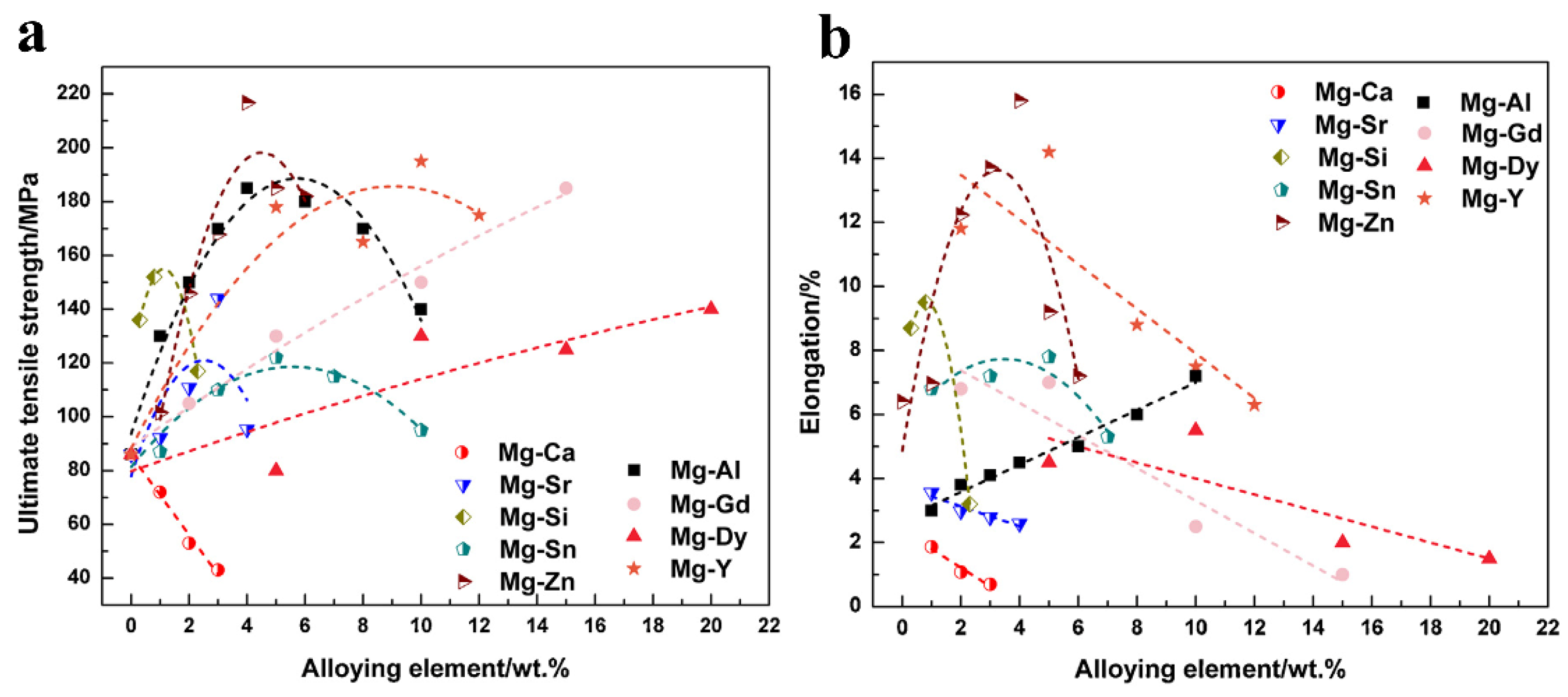
3.3. Mechanical Performance and Integrity

One of the main disadvantages of commercial absorbable membranes is their low mechanical strength, which cannot provide adequate mechanical support during the bone healing process. According to reports, the barrier membrane should perform its function for at least 3-6 months, retaining enough gaps for internal osteoblasts to proliferate while also enduring some external stresses (compression of overlying soft tissue) [11]. According to Table 1 and previous reports [35,44,98], the highest strength and elongation of commercial absorbable membranes are approximately 55 MPa and 20%, respectively.
As shown in Figure 4a, the strength of Mg alloys is much higher than that of commercial polymer membranes and sufficient for use as a GBR membrane, indicating adequate mechanical support during the regeneration period. On the other hand, elongation is an important factor to consider because it influences the membrane’s malleability. Figure 4b shows that the elongation of as-cast Mg alloys can reach around 16%. Conceivably, Mg alloys may be able to achieve greater elongation through severe plastic deformation, alloying, heat treatment, and other methods. Actually, as long as selected materials meet the basic requirements of a barrier membrane, their mechanical properties do not need to be particularly high.

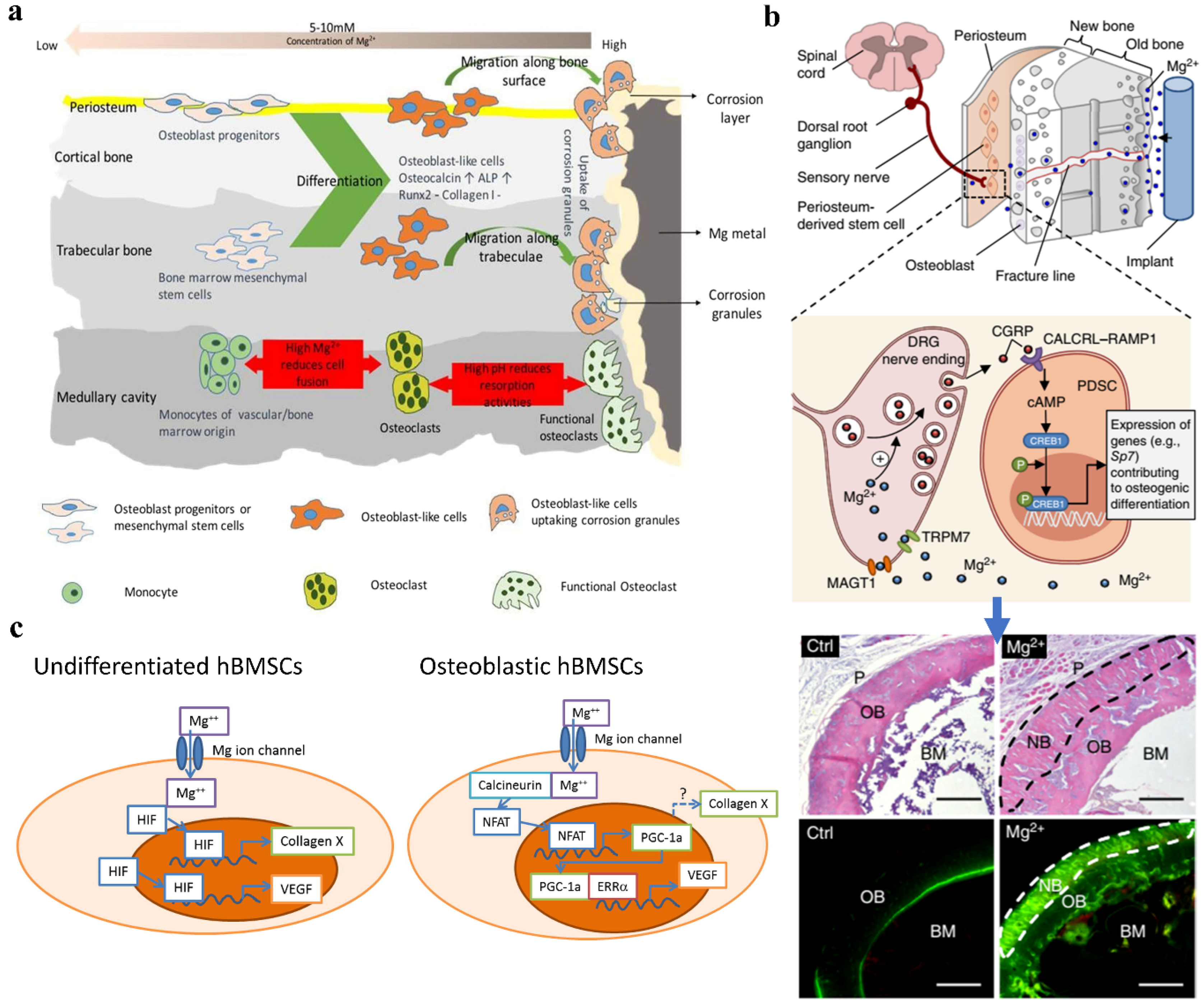
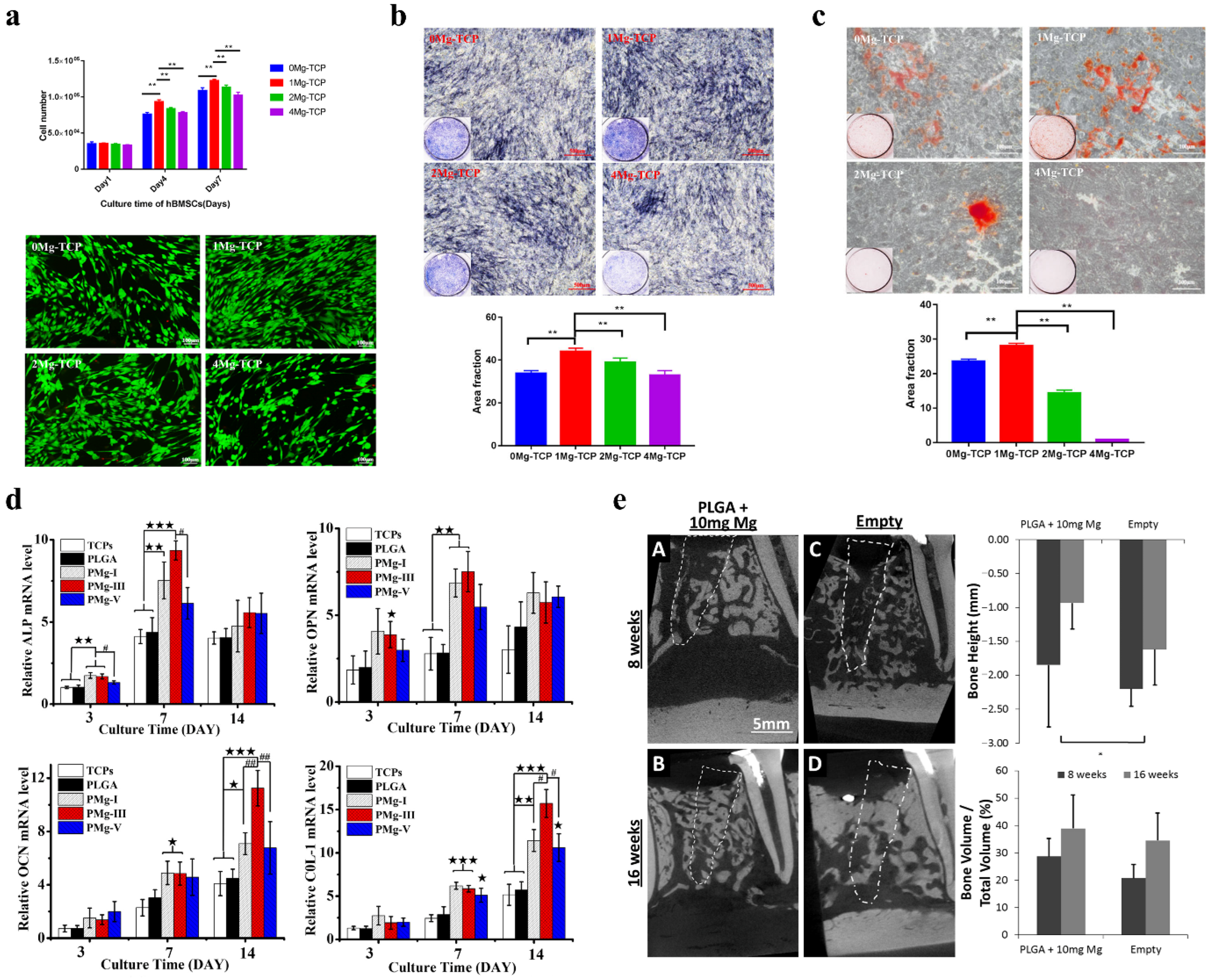
3.4. Osteopromotive Properties of Mg Alloys

One of the most important functions of the barrier membrane is its ability to increase alveolar bone volume prior to surgery, and the favorable osteogenic effect is beneficial to subsequent dental implantology. In clinical surgeries, a barrier membrane with a high osteogenesis-promoting ability is desired.
Bone and its surrounding microenvironment are well-known to be a complex system containing numerous stem cells, osteoblasts, osteoclasts, osteocytes, immune cells, and so on [99]. The cellular response to Mg-based implants is primarily determined by the material implantation sites. For example, when Mg-based screws were used to treat peri-tunnel bone loss following anterior cruciate ligament (ACL) reconstruction, promoted trabecular bone formation was observed [100,101]. This phenomenon can be attributed primarily to high Mg2+ concentrations and high pH values, which reduce fusion of pre-osteoclast cells and affect osteoclastogenesis [102], as shown in Figure 5a. The following are the primary reasons for Mg’s beneficial effects on osteogenic performance. First, Mg could stimulate the formation of neuronal calcitonin gene-related polypeptide-a (CGRP) in both the femoral peripheral cortex and the ipsilateral dorsal root ganglia (DRG) (Figure 5b), promoting osteogenic differentiation and improving bone-fracture healing [103]. Second, the addition of Mg would encourage osteogenic differentiation. Yoshizawa et al. [104] investigated the effect of different Mg2+ concentrations on the osteogenic activity of human bone marrow stromal cells (hBMSCs) and differentiating osteoblasts, and the results showed that the addition of 10 mM Mg2+ increased extracellular matrix (ECM) mineralization, which increases collagen type X protein production and vascular endothelial growth factor (VEGF) expression (Figure 5c). They also discovered that undifferentiated cells are stimulated by hypoxia-inducible factor 2a (HIF-2a), whereas osteogenic cells are stimulated by peroxisome proliferator-activated receptor gamma coactivator (PPAR-gamma) (PGC-1a). Furthermore, Hung et al. found that increasing the Mg2+ concentration increased the protein expression of active β-catenin, LEF1, and Dkk1, indicating activation of the canonical Wnt signaling pathway in hBMSCs [105]. Third, Mg2+-induced angiogenesis may promote bone formation. As shown in Figure 5c, the addition of Mg can increase VEGF expression, which is important in the development of type H capillaries, which are required for bone formation [106]. Previous researches have also discovered the phenomenon of synergistically enhanced osteogenesis and angiogenesis [99,107,108]. Furthermore, Mg supplementation can inhibit osteoclastogenesis, thereby enhancing angiogenesis and osteogenesis. Recently, Zhai et al. [109] discovered that Mg leach liquor (MLL) inhibits osteoclast formation, polarization, and bone resorption in vitro. Furthermore, in vivo results showed that MLL reduced wear particle-induced osteolysis by inhibiting nuclear factor-kB (NF-kB) activation, confirming Mg’s anti-osteoclastogenic effect. Finally, the presence of Mg may influence bone growth and regeneration by regulating macrophage polarization. Mg has been shown to promote M2 phased macrophage polarization and suppress M1 phased macrophages, indicating an anti-inflammatory effect [99,110]. This anti-inflammatory effect has been linked to Mg-regulated TRMP7 [111]. As a result, Mg plays a variety of roles in bone growth and regeneration. All of the cellular or molecular mechanisms mentioned above worked together to promote osteogenesis in various animal models.
Furthermore, recent reports in vitro and in vivo have confirmed the osteopromotive properties of Mg alloys. Gu et al. [112] recently investigated the osteogenesis and angiogenesis of xMg-doped β-TCP scaffolds with different Mg contents (x = 0, 1, 2, and 4 wt.%). As shown in Figure 6a, the supplementation of 1Mg-TCP scaffolds resulted in the highest proliferation rate and better cell morphology for hBMSCs than the other groups, owing to the proper Mg2+ concentration in 1Mg-TCP scaffold extracts (2.31 ± 0.12 mM). Wong et al. demonstrated that a low Mg2+ concentration of 50 ppm (approximately 2 mM) would significantly promote the proliferation and differentiation of pre-osteoblasts, upregulate osteogenic-related genes, and stimulate new bone formation in vivo [113], accounting for the higher cell proliferation and better in vitro osteogenic performance of the 1Mg-TCP group (Figure 6b,c). Yuan et al. recently created biodegradable microspheres (PMg) by embedding poly(lactide-co-glycolide) (PLGA) with varying MgO and MgCO3 contents (1:0, 3:1, 1:1, 1:3, 0:1) [114]. The results showed that PMg microspheres exhibited controlled release of Mg2+ by tuning the MgO/MgCO3 ratios, and PMg-III microspheres (MgO/MgCO3 in 1:1) were more effective than PLGA and control groups in promoting osteogenic differentiation of BMSCs (Figure 6d), which was attributed to a long-term moderate Mg2+ release behavior. Furthermore, many in vivo experiments have confirmed Mg’s osteopromotive properties. Brown et al. [115], for example, demonstrated that incorporating Mg (about 10 mg) into PLGA composite scaffolds increased bone height and bone volume/total volume (BV/TV) in a canine socket preservation model compared to the empty defects group, as shown in Figure 6e. As a result, all of the above findings suggest that a moderate amount of Mg introduced into the bone micro-environment plays a positive role in bone metabolism, which is beneficial to increasing alveolar bone volume and has great potential as the GBR membrane.

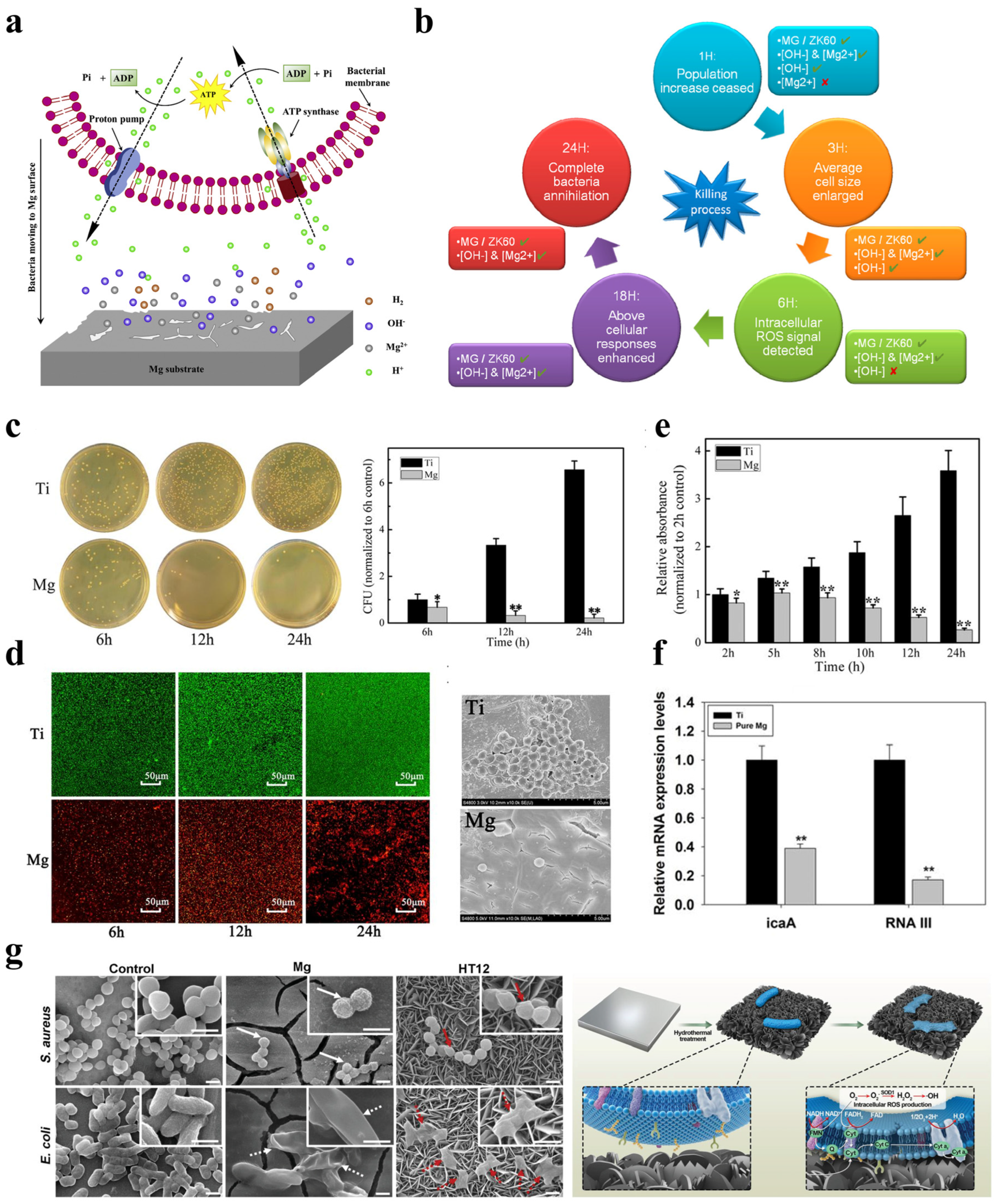
3.5. Bacteriostatic Activity

Infection after fracture fixation and insufficient osseointegration at the implant–bone interface remain major concerns for clinical orthopedic surgeries. Similarly, premature exposure of the GBR membrane and subsequent infection will impair bone defect regenerative outcomes [44]. Furthermore, the oral cavity is a non-sterile environment rich in bacteria, both aerobic and anaerobic [116]. As a result, good bacteriostatic activity is also required for certain barrier membrane materials.
According to Equations (1)–(3), the high antibacterial performance can be attributed to the high alkalinity and released Mg2+ during corrosion [117]. Most bacteria can survive in an appropriate pH range (pH = 6–8) [118], whereas Mg degradation typically causes the alkalinity of a solution to reach 9–10 [44,119], accounting for the high bacteriostatic activity of Mg alloys. Rahim et al. [120] recently reported that Mg granulate corrosion extracts could inhibit the growth of Pseudomonas aeruginosa (P. aeruginosa) and Staphylococcus aureus (S. aureus) strains. Furthermore, the results showed that increasing the pH value of the extracts could increase their bacteriostatic activity, whereas neutralized supernatants lost their antibacterial ability completely. Besides that, Li et al. [121] investigated the antibacterial properties of pure Mg (99.9%) against methicillin-resistant Staphylococcus aureus (MRSA), with Ti serving as the control group. The in vitro results showed that the Mg implants effectively reduced bacterial adhesion (Figure 7c,d) and prevented biofilm formation (Figure 7e), most likely due to increased local alkalinity caused by metal degradation [121]. Furthermore, as shown in Figure 7f, the real-time PCR results showed that increased alkalinity would significantly downregulate the biofilm-formation-related gene (icaA) and the cell-protein-related gene (RNAIII). As a result, it is possible that the antibacterial effect of Mg alloy is primarily due to the high alkalinity produced by its degradation. Lin et al. [117] recently proposed a possible antibacterial mechanism for Mg alloys, as shown in Figure 7a. It is assumed that a large number of H+ produced by bacteria can be neutralized by OH- produced by alloy degradation, causing damage to the proton electrochemical gradient in the bacterial intermembrane space. The electrochemical gradient of the proton drives the synthesis of adenosine triphosphate (ATP). As a result, damage to the transmembrane proton electrochemical gradient would impede ATP synthesis, eventually leading to bacterial death [122]. Besides this, the alkalinity near the sample surface may be higher [123], which can enhance the antibacterial effect and inhibit bacteria adhesion.
On the other hand, the Mg2+ released during the degradation process may play an active role in increasing the bacteriostatic activity of Mg alloys. High Mg2+ concentrations have been shown to increase bacteria’s sensitivity to antibacterial agents and adverse conditions, impairing bacterial adhesion and biofilm formation [124]. According to Matevosyan et al. [125], adding Mg2+ to lactic acid bacteria (LAB) isolates increased their antibacterial activity. In addition, Jess et al. [126] investigated the bactericidal effect of Mg2+ concentration on the survival ratio of Staphylococcus epidermidis (S. epidermidis) and Escherichia coli (E. coli) strains at constant pH (about 8.0). The results showed that higher Mg2+ concentrations resulted in stronger antibacterial activity due to the increased osmotic stress on the cell wall caused by Mg2+. However, some studies show that the bacteriostatic activity is not dependent on Mg2+. For example, Robinson et al. discovered that increasing Mg2+ concentration alone had no effect on antibacterial ability against E. coli, P. aeruginosa, and S. aureus [119]. Furthermore, Liu et al. reported that no antibacterial ability of Mg alloys was observed in a neutral solution [127]. It seems there exists a divergence in the antibacterial performance of Mg2+. In fact, intracellular Mg2+ concentrations in many cell types range from 15 to 30 mM [128]. As a result, when Mg ions are used as antibacterial agents, their concentration should be higher than that of bacterial cells.
Furthermore, the formation of surface hydroxide (Mg(OH)2, as shown in Equation (3)) also contributes to the antibacterial activity of Mg alloys. It has been demonstrated that positively charged Mg(OH)2 nanoparticles are easily absorbed into negatively charged bacteria and destroy the integrity of cell walls, resulting in bacterial death [129]. Similarly, Wang et al. [130] demonstrated that Mg(OH)2 nanoflakes could trap bacteria, resulting in bacterial death due to high surface tension force and the generation of intracellular reactive oxygen species (ROS) bursts, as shown in Figure 7g. In 2016, Feng et al. [131] systematically investigated the inherent antibacterial properties of Mg alloys and proposed a possible bacterial killing process in 24 h, as shown in Figure 7b. It was assumed that the high bacteriostatic activity of Mg alloys is primarily due to high alkalinity and released Mg2+ concentration, as well as the effect of ROS produced by Mg(OH)2.
Figure 7. The antibacterial behavior of Mg-based alloys. (a) A possible antibacterial mechanism of Mg-based alloys due to the generation of hydroxyl ions on surface. Reproduced with permission from [117]. Copyright 2020, Elsevier. (b) A diagram illustrating the bacterial killing process and cellular response of Mg-based alloys during 24 h. Reproduced with permission from [131]. Copyright 2016, American Chemical Society. (c) Representative images displaying viable bacteria adhered on samples after 24 h culture and normalized number of bacteria. (d) Fluorescent and SEM images illustrating the viable or dead bacteria adhered on the sample surface after 24 h culture. (e) the anti-biofilm ability of Mg and Ti tested by crystal violet staining method. (f) Expressions of the icaA and agr RNAIII genes derived from RT-PCR tests. * p < 0.05, ** p < 0.01. Reproduced with permission from [121]. Copyright 2014, American Society for Microbiology. (g) Morphology of S. aureus and E. coli cultivated on Ti-, Mg-, and HT12-treated samples for 3h and the antibacterial mechanism of surface Mg(OH)2. Scale bars: 1 µm. Reproduced with permission from [130]. Copyright 2014, Wiley-VCH GmbH.
Metals 12 02074 g007
Additionally, earlier studies have supported the in vivo antibacterial effectiveness of magnesium alloys [117,132]. As shown in Figure 8, Li et al. effectively created an MRSA-induced osteomyelitis model in rats, and the in vivo results reveal that, in contrast to Ti nails, almost no germs were seen on the Mg nail surfaces and bone tissue [121]. As a result, it can be said that Mg alloys have certain antibacterial properties and can be employed as a barrier membrane based on the results presented above.

3.6. Wound-Healing Ability

As a barrier membrane, the rigidity and jagged edges created during the bending process may also adversely stimulate the mucosal flaps, leading to a mucosal rupture and subsequent membrane exposure [48]. The prevention of wound dehiscence, early membrane exposure, and subsequent bacterial contamination are all advantages of favorable soft tissue closure. Therefore, the membrane’s capacity to heal wounds must therefore be taken into account as well.
Actually, adding the right amount of magnesium is beneficial. Mg ions can work as cofactors, aiding in the production of proteins and collagen during the healing process as well as the control and regulation of the integrin family’s function qualities, which in turn affects cell differentiation, wound healing, and hemostasis [133]. According to Lin et al. [134], the Mg2+ (about 13.2 mM) deteriorated from Mg-Zn-Zr alloys greatly increased fibroblast cell activity and aided in wound healing. Recently, Guo et al. [135] added Mg and polydopamine (PDA) to polyacrylamide (PAM) hydrogels, and the in vitro results suggestted that the existence of Mg2+ aided in the enhancement of wound healing and cell proliferation (Figure 9a). Additionally, as shown in Figure 9b, the in vivo data demonstrated that the PDA-PAM/Mg2+ group had the highest tissue mending ability in a subcutaneous bacterial infection scenario. Additionally, Mg-doped smectite was effectively created by Sasaki et al., and the Mg-smectite could aid in skin tissue regeneration by releasing Mg2+ and Si4+ ions [136]. Additionally, Mg was recently immobilized on a Ti substrate by Zhu et al. via the Mg-PIII method, and its capacity to heal wounds was examined [137]. The Mg-PIII-treated Ti appeared to have a greater capacity for wound healing, which can be mostly attributed to the regulating effect of Mg2+ on integrin expression, activation of the PI3k/AKT signaling pathway, and subsequent adhesion, proliferation, and migration of HGF cells (Figure 9c). Additionally, the high alkalinity brought on by the breakdown of magnesium would lessen the dangers of infection brought on by bacteria.

4. The Challenges Faced with Mg-Based GBR Membranes

At the moment, the primary limitations of Mg alloy applications are their rapid degradation rate and non-uniform degradation mode. The unexplained degradation behavior of Mg alloys can be attributed to two factors. First, there are second phases in the microstructure of Mg alloys, and these second phases will form galvanic cells with the matrix [138], accelerating the degradation rate. Second, the naturally generated surface oxide coating is not compact, and the Pilling–Bedworth ratio (P-B ratio) of pure magnesium is 0.81, which means that it cannot prevent additional substrate corrosion [139]. Moreover, the mechanical integrity and service life of implants made of magnesium are compromised by the localized breakdown mechanism of Mg alloys. For instance, as demonstrated in Figure 10, it was discovered that ZX50 alloys displayed a localized breakdown pattern in vivo and vanished after 12 weeks [45]. Furthermore, even after applying the micro-arc oxidation (MAO) treatment to ZX50 alloys, the pins still completely degraded after 12 weeks. These findings suggest that the substrate itself, in essence, controls the degrading behavior of magnesium alloys.
Furthermore, proper degradation behavior of Mg-based implants, including degradation rate and degradation mode, can provide sufficient mechanical support during the service stage and avoid premature failure of implants. Mechanical support is required as the barrier membrane to isolate the bone defect areas and epithelial tissue. The relationship between degradation behavior and mechanical integrity change of Mg-based implants during the bone healing process is depicted in Figure 11 [140]. It indicated that the degradation behavior of Mg-based implants should be optimized to correspond with the healing process of the fractured bone. The barrier membrane is expected to function for at least 3-6 months, leaving enough gaps for internal osteoblasts to proliferate while also enduring some external stresses [11]. As a result, the initial degradation rate of Mg should be tailored to provide adequate mechanical support for at least three months.
The effect of stress on the degradation profile of Mg alloys is also a source of concern. Stress can be caused by acute mechanical loadings during surgeries, residual stress after thermomechanical deformation, and other factors [31]. As a result of the synergistic presence of corrosive human body fluid and mechanical loading, stress corrosion cracking (SCC) and corrosion fatigue (CF) behavior can occur [141]. As a barrier membrane, it typically necessitates intraoperative bending or shaping treatment to adapt to various defects, resulting in stress concentration at the distortion sites. Furthermore, the membrane should prevent abrupt collapse caused by soft tissue pressure overlying the chewing function. As a result, the SCC or CF behavior of Mg alloys poses a significant challenge for their use as barrier membranes. Previous reports summarized the influence of many factors, such as loading, hydrogen embrittlement, microstructure, heat treatment, surface treatment, and so on, on SCC or CF behaviors of Mg alloys, and it indicated that degradation resistance and SCC/CF resistance can be tailored by high purification treatment, alloying, grain refinement, hot deformation, and other methods [31,141,142,143].

5. The Outlook for the Future

Figure 12 illustrates how Mg-based membranes have some advantages over currently available commercial membranes, including degradability, acceptable mechanical strength, good osteogenesis performance, bacteriostatic activity, and wound-healing capacity. However, before using Mg alloys in clinical settings, it is important to take into account their quick degradation rate and SCC/CF sensitivity. Mg-based alloy membrane development is still ongoing. Here are some potential directions for designing and producing Mg-based membranes in the future. First, by alloying, heat treatment, thermal formation, and other techniques, the microstructure of the alloys should be adjusted to increase the materials’ inherent resistance to degradation. Additionally, there is a difference between the biological response to Mg-based implants in vitro and in vivo. In order to endow the alloy with appropriate physiological function (antibacterial activity, angiogenesis, osteogenesis, and so on) and good biocompatibility (soft tissue healing properties of gingival tissue), functional surface coatings on Mg-based membranes are therefore necessary. Moreover, the SCC/CF sensitivity of Mg alloys is significantly influenced by the design, deformation, and surface modification of Mg-based membranes, hence consideration for their SCC/CF resistance is also necessary for these processes. Finally, the current in vivo animal model differs from the GBR membrane’s actual application scenario. It is therefore necessary to develop a better in vivo alveolar bone disease model for the barrier membrane.

Author Contributions

Conceptualization, K.C. and L.Z.; Methodology, K.C. and X.G.; Software, L.Z.; Validation, K.C. and X.G.; Formal Analysis, C.H., X.Y. and X.Z.; Investigation, K.C.; Resources, K.C. and X.G.; Data Curation, K.C.; Writing—Original Draft Preparation, K.C.; Writing—Review and Editing, K.C. and X.G.; Visualization, X.G. and P.L.; Supervision, X.G. and Y.F.; Project Administration, X.G.; Funding Acquisition, X.G. All authors have read and agreed to the published version of the manuscript.

Funding

This work was financially supported by the National Natural Science Foundation of China (52071008, 61871014, U20A20390).

Data Availability Statement

Not applicable.

Conflicts of Interest

The authors declare that they have no conflict of interest.

References

  1. Aprile, P.; Letourneur, D.; Simon-Yarza, T. Membranes for Guided Bone Regeneration: A Road from Bench to Bedside. Adv. Healthc. Mater. 2020, 9, 2000707. [Google Scholar] [CrossRef] [PubMed]
  2. Zakrzewski, W.; Dobrzynski, M.; Rybak, Z.; Szymonowicz, M.; Wiglusz, R.J. Selected Nanomaterials’ Application Enhanced with the Use of Stem Cells in Acceleration of Alveolar Bone Regeneration during Augmentation Process. Nanomaterials 2020, 10, 1216. [Google Scholar] [CrossRef] [PubMed]
  3. Pang, C.; Ding, Y.; Hu, K.; Zhou, H.; Qin, R.; Hou, R. Influence of preservation of the alveolar ridge on delayed implants after extraction of teeth with different defects in the buccal bone. Br. J. Oral Maxillofac. Surg. 2016, 54, 176–180. [Google Scholar] [CrossRef] [PubMed]
  4. Fok, M.R.; Pelekos, G.; Tonetti, M.S. Feasibility and needs for simultaneous or staged bone augmentation to place prosthetically guided dental implants after extraction or exfoliation of first molars due to severe periodontitis. J. Clin. Periodontol. 2020, 47, 1237–1247. [Google Scholar] [CrossRef]
  5. Watson, T.J. Distraction Osteogenesis. JAAOS J. Am. Acad. Orthop. Surg. 2006, 14, S168–S174. [Google Scholar] [CrossRef]
  6. Chen, F.-M.; Zhang, J.; Zhang, M.; An, Y.; Chen, F.; Wu, Z.-F. A review on endogenous regenerative technology in periodontal regenerative medicine. Biomaterials 2010, 31, 7892–7927. [Google Scholar] [CrossRef]
  7. Guo, Y.; Yu, Y.; Han, L.; Ma, S.; Zhao, J.; Chen, H.; Yang, Z.; Zhang, F.; Xia, Y.; Zhou, Y. Biocompatibility and osteogenic activity of guided bone regeneration membrane based on chitosan-coated magnesium alloy. Mater. Sci. Eng. C. 2019, 100, 226–235. [Google Scholar] [CrossRef]
  8. Saima, S.; Jan, S.; Shah, A.; Yousuf, A.; Batra, M. Bone grafts and bone substitutes in dentistry. J. Oral Res. Rev. 2016, 8, 36–38. [Google Scholar]
  9. Orsini, M.; Orsini, G.; Benlloch, D.; Aranda, J.J.; Sanz, M. Long-Term Clinical Results on the Use of Bone-Replacement Grafts in the Treatment of Intrabony Periodontal Defects. Comparison of the Use of Autogenous Bone Graft Plus Calcium Sulfate to Autogenous Bone Graft Covered with a Bioabsorbable Membrane. J. Periodontol. 2008, 79, 1630–1637. [Google Scholar] [CrossRef]
  10. Rodriguez, I.A.; Selders, G.S.; Fetz, A.E.; Gehrmann, C.J.; Stein, S.H.; Evensky, J.A.; Green, M.S.; Bowlin, G.L. Barrier membranes for dental applications: A review and sweet advancement in membrane developments. Mouth Teeth 2018, 2, 1–9. [Google Scholar]
  11. Bottino, M.C.; Thomas, V.; Schmidt, G.; Vohra, Y.K.; Chu, T.-M.G.; Kowolik, M.J.; Janowski, G.M. Recent advances in the development of GTR/GBR membranes for periodontal regeneration—A materials perspective. Dent. Mater. 2012, 28, 703–721. [Google Scholar] [CrossRef]
  12. Chiapasco, M.; Zaniboni, M. Clinical outcomes of GBR procedures to correct peri-implant dehiscences and fenestrations: A systematic review. Clin. Oral Implant. Res. 2009, 20, 113–123. [Google Scholar] [CrossRef]
  13. Venkatesan, J.; Anil, S.; Kim, S.-K.; Shim, M.S. Chitosan as a vehicle for growth factor delivery: Various preparations and their applications in bone tissue regeneration. Int. J. Biol. Macromol. 2017, 104, 1383–1397. [Google Scholar] [CrossRef]
  14. Omar, O.; Elgali, I.; Dahlin, C.; Thomsen, P. Barrier membranes: More than the barrier effect? J. Clin. Periodontol. 2019, 46, 103–123. [Google Scholar] [CrossRef] [Green Version]
  15. Leghissa, G.C.; Zaffe, D.; Assenza, B.; Botticelli, A.R. Guided bone regeneration using titanium grids: Report of 10 cases. Clin. Oral Implant. Res. 1999, 10, 62–68. [Google Scholar] [CrossRef]
  16. Mamalis, A.A.; Cochran, D.L. The therapeutic potential of oxygen tension manipulation via hypoxia inducible factors and mimicking agents in guided bone regeneration. A review. Arch. Oral Biol. 2011, 56, 1466–1475. [Google Scholar] [CrossRef]
  17. Milstein, D.M.J.; Mathura, K.R.; Lindeboom, J.A.H.; Ramsoekh, D.; Lindeboom, R.; Ince, C. The temporal course of mucoperiosteal flap revascularization at guided bone regeneration-treated implant sites: A pilot study. J. Clin. Periodontol. 2009, 36, 892–897. [Google Scholar] [CrossRef]
  18. Xia, D.; Yang, F.; Zheng, Y.; Liu, Y.; Zhou, Y. Research status of biodegradable metals designed for oral and maxillofacial applications: A review. Bioact. Mater. 2021, 6, 4186–4208. [Google Scholar] [CrossRef]
  19. Staiger, M.P.; Pietak, A.M.; Huadmai, J.; Dias, G. Magnesium and its alloys as orthopedic biomaterials: A review. Biomaterials 2006, 27, 1728–1734. [Google Scholar] [CrossRef]
  20. Zheng, Y.F.; Gu, X.N.; Witte, F. Biodegradable metals. Mater. Sci. Eng. R. 2014, 77, 1–34. [Google Scholar] [CrossRef]
  21. Geng, X.; Jiang, J.; Zhang, X. Corrosion Behavior of Mg-xGd-1Zn-0.4Zr Alloys with Different Gd Additions for Biomedical Application. Metals 2022, 12, 1763. [Google Scholar] [CrossRef]
  22. Zhang, X.; Dai, J.; Dong, Q.; Ba, Z.; Wu, Y. Corrosion behavior and mechanical degradation of as-extruded Mg–Gd–Zn–Zr alloys for orthopedic application. J. Biomed. Mater. Res. B. 2020, 108, 698–708. [Google Scholar] [CrossRef]
  23. Hong, L.; Wang, R.; Zhang, X. Effects of Nd on microstructure and mechanical properties of as-cast Mg-12Gd-2Zn-xNd-0.4Zr alloys with stacking faults. Int. J. Miner. Metall. Mater. 2022, 29, 1570–1577. [Google Scholar] [CrossRef]
  24. Nie, Y.; Dai, J.; Li, X.; Zhang, X. Recent developments on corrosion behaviors of Mg alloys with stacking fault or long period stacking ordered structures. J. Magnes. Alloy 2021, 9, 1123–1146. [Google Scholar] [CrossRef]
  25. Zhang, X.; Dai, J.; Zhang, R.; Ba, Z.; Birbilis, N. Corrosion behavior of Mg–3Gd–1Zn–0.4Zr alloy with and without stacking faults. J. Magnes. Alloy 2019, 7, 240–248. [Google Scholar] [CrossRef]
  26. Nie, Y.-J.; Dai, J.-W.; Zhang, X.-B. Effect of Ag Addition on Microstructure, Mechanical and Corrosion Properties of Mg–Nd–Zn–Zr Alloy for Orthopedic Application. Acta Metall. Sin. 2022; preprint. [Google Scholar] [CrossRef]
  27. Zhang, X.; Kairy, S.K.; Dai, J.; Birbilis, N. A Closer Look at the Role of Nanometer Scale Solute-Rich Stacking Faults in the Localized Corrosion of a Magnesium Alloy GZ31K. J. Electrochem. Soc. 2018, 165, C310–C316. [Google Scholar] [CrossRef]
  28. Zhang, J.; Zhang, X.; Liu, Q.; Yang, S.; Wang, Z. Effects of Load on Dry Sliding Wear Behavior of Mg–Gd–Zn–Zr Alloys. J. Mater. Sci. Technol. 2017, 33, 645–651. [Google Scholar] [CrossRef]
  29. Chen, K.; Lu, Y.; Tang, H.; Gao, Y.; Zhao, F.; Gu, X.; Fan, Y. Effect of strain on degradation behaviors of WE43, Fe and Zn wires. Acta Biomater. 2020, 113, 627–645. [Google Scholar] [CrossRef]
  30. Chen, K.; Xie, X.; Tang, H.; Sun, H.; Qin, L.; Zheng, Y.; Gu, X.; Fan, Y. In vitro and in vivo degradation behavior of Mg–2Sr–Ca and Mg–2Sr–Zn alloys. Bioact. Mater. 2020, 5, 275–285. [Google Scholar] [CrossRef]
  31. Winzer, N.; Atrens, A.; Song, G.; Ghali, E.; Dietzel, W.; Kainer, K.U.; Hort, N.; Blawert, C. A Critical Review of the Stress Corrosion Cracking (SCC) of Magnesium Alloys. Adv. Eng. Mater. 2005, 7, 659–693. [Google Scholar] [CrossRef]
  32. Kasaj, A.; Reichert, C.; Götz, H.; Röhrig, B.; Smeets, R.; Willershausen, B. In vitro evaluation of various bioabsorbable and nonresorbable barrier membranes for guided tissue regeneration. Head Face Med. 2008, 4, 22. [Google Scholar] [CrossRef] [PubMed]
  33. Milella, E.; Ramires, P.A.; Brescia, E.; la Sala, G.; di Paola, L.; Bruno, V. Physicochemical, mechanical, and biological properties of commercial membranes for GTR. J. Biomed. Mater. Res. 2001, 58, 427–435. [Google Scholar] [CrossRef] [PubMed]
  34. Donos, N.; Kostopoulos, L.; Karring, T. Alveolar ridge augmentation using a resorbable copolymer membrane and autogenous bone grafts. Clin. Oral Implant. Res. 2002, 13, 203–213. [Google Scholar] [CrossRef]
  35. Bowen, P.K.; Shearier, E.R.; Zhao, S.; Guillory, R.J.; Zhao, F.; Goldman, J.; Drelich, J.W. Biodegradable Metals for Cardiovascular Stents: From Clinical Concerns to Recent Zn-Alloys. Adv. Healthc. Mater. 2016, 5, 1121–1140. [Google Scholar] [CrossRef] [Green Version]
  36. Klinge, U.; Klosterhalfen, B.; Müller, M.; Schumpelick, V. Foreign body reaction to meshes used for the repair of abdominal wall hernias. Eur. J. Surg. 1999, 165, 665–673. [Google Scholar]
  37. Hou, L.-T.; Yan, J.-J.; Tsai, A.Y.M.; Lao, C.S.; Lin, S.J.; Liu, C.-M. Polymer-assisted regeneration therapy with Atrisorb® barriers in human periodontal intrabony defects. J. Clin. Periodontol. 2004, 31, 68–74. [Google Scholar] [CrossRef]
  38. Coonts, B.A.; Whitman, S.L.; O’Donnell, M.; Polson, A.M.; Bogle, G.; Garrett, S.; Swanbom, D.D.; Fulfs, J.C.; Rodgers, P.W.; Southard, G.L.; et al. Biodegradation and biocompatibility of a guided tissue regeneration barrier membrane formed from a liquid polymer material. J. Biomed. Mater. Res. 1998, 42, 303–311. [Google Scholar] [CrossRef]
  39. Bottino, M.C.; Jose, M.V.; Thomas, V.; Dean, D.R.; Janowski, G.M. Freeze-dried acellular dermal matrix graft: Effects of rehydration on physical, chemical, and mechanical properties. Dent. Mater. 2009, 25, 1109–1115. [Google Scholar] [CrossRef]
  40. Owens, K.W.; Yukna, R.A. Collagen Membrane Resorption in Dogs: A Comparative Study. Implant. Dent. 2001, 10, 49–58. [Google Scholar] [CrossRef]
  41. Coïc, M.; Placet, V.; Jacquet, E.; Meyer, C. Propriétés mécaniques des membranes de collagène. Rev. Stomatol. Chir. Maxillo-Faciale 2010, 111, 286–290. [Google Scholar] [CrossRef]
  42. Bunyaratavej, P.; Wang, H.-L. Collagen Membranes: A Review. J. Periodontol. 2001, 72, 215–229. [Google Scholar] [CrossRef] [PubMed] [Green Version]
  43. Yamada, M.; Kojima, N.; Att, W.; Minamikawa, H.; Sakurai, K.; Ogawa, T. Improvement in the osteoblastic cellular response to a commercial collagen membrane and demineralized freeze-dried bone by an amino acid derivative: An in vitro study. Clin. Oral Implant. Res. 2011, 22, 165–172. [Google Scholar] [CrossRef] [PubMed]
  44. Chen, K.; Zhao, L.; Sun, J.; Gu, X.; Huang, C.; Su, H.; Fan, Y. Utilizing biodegradable alloys as guided bone regeneration (GBR) membrane: Feasibility and challenges. Sci. China. Mater. 2022, 65, 2627–2646. [Google Scholar] [CrossRef]
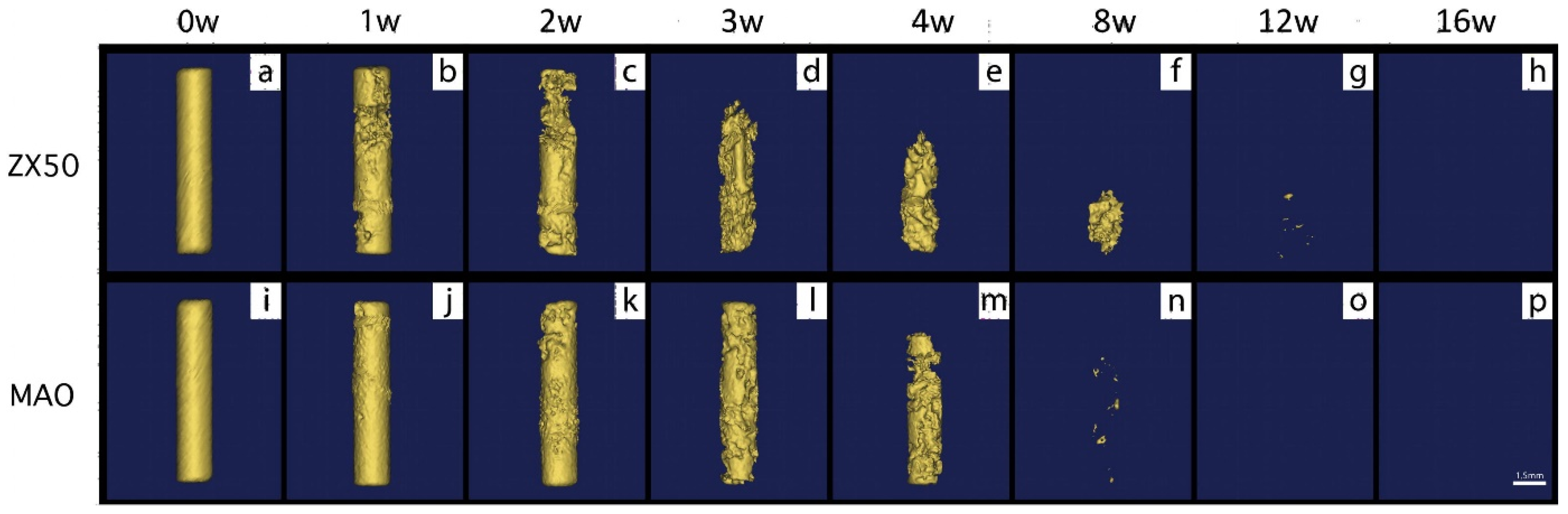
  45. Fischerauer, S.F.; Kraus, T.; Wu, X.; Tangl, S.; Sorantin, E.; Hänzi, A.C.; Löffler, J.F.; Uggowitzer, P.J.; Weinberg, A.M. In vivo degradation performance of micro-arc-oxidized magnesium implants: A micro-CT study in rats. Acta Biomater. 2013, 9, 5411–5420. [Google Scholar] [CrossRef] [PubMed]
  46. Chen, K.; Dai, J.; Zhang, X. Improvement of corrosion resistance of magnesium alloys for biomedical applications. Corros. Rev. 2015, 33, 101–117. [Google Scholar] [CrossRef]
  47. Haude, M.; Erbel, R.; Erne, P.; Verheye, S.; Degen, H.; Böse, D.; Vermeersch, P.; Wijnbergen, I.; Weissman, N.; Prati, F.; et al. Safety and performance of the drug-eluting absorbable metal scaffold (DREAMS) in patients with de-novo coronary lesions: 12 month results of the prospective, multicentre, first-in-man BIOSOLVE-I trial. Lancet 2013, 381, 836–844. [Google Scholar] [CrossRef]
  48. Xie, Y.; Li, S.; Zhang, T.; Wang, C.; Cai, X. Titanium mesh for bone augmentation in oral implantology: Current application and progress. Int. J. Oral Sci. 2020, 12, 37. [Google Scholar] [CrossRef]
  49. Elgali, I.; Omar, O.; Dahlin, C.; Thomsen, P. Guided bone regeneration: Materials and biological mechanisms revisited. Eur. J. Oral Sci. 2017, 125, 315–337. [Google Scholar] [CrossRef] [Green Version]
  50. Garcia, J.; Dodge, A.; Luepke, P.; Wang, H.-L.; Kapila, Y.; Lin, G.-H. Effect of membrane exposure on guided bone regeneration: A systematic review and meta-analysis. Clin. Oral Implant. Res. 2018, 29, 328–338. [Google Scholar] [CrossRef] [Green Version]
  51. Jung, R.E.; Fenner, N.; Hämmerle, C.H.F.; Zitzmann, N.U. Long-term outcome of implants placed with guided bone regeneration (GBR) using resorbable and non-resorbable membranes after 12–14 years. Clin. Oral Implant. Res. 2013, 24, 1065–1073. [Google Scholar] [CrossRef] [PubMed] [Green Version]
  52. Chen, K.; Zhou, G.; Li, Q.; Tang, H.; Wang, S.; Li, P.; Gu, X.; Fan, Y. In vitro degradation, biocompatibility and antibacterial properties of pure zinc: Assessing the potential of Zn as a guided bone regeneration membrane. J. Mater. Chem. B 2021, 9, 5114–5127. [Google Scholar] [CrossRef] [PubMed]
  53. Sheikh, Z.; Qureshi, J.; Alshahrani, A.M.; Nassar, H.; Ikeda, Y.; Glogauer, M.; Ganss, B. Collagen based barrier membranes for periodontal guided bone regeneration applications. Odontology 2017, 105, 1–12. [Google Scholar] [CrossRef] [PubMed]
  54. Behring, J.; Junker, R.; Walboomers, X.F.; Chessnut, B.; Jansen, J.A. Toward guided tissue and bone regeneration: Morphology, attachment, proliferation, and migration of cells cultured on collagen barrier membranes. A systematic review. Odontology 2008, 96, 1–11. [Google Scholar] [CrossRef] [PubMed]
  55. Joop, A.; Rahlf, B.; Gellrich, N.-C.; Kampmann, A.; von See, C.; Stoetzer, M. Examination of Local Periosteal Microcirculation After Application of Absorbable and Nonabsorbable Membranes. J. Oral Implantol. 2017, 43, 462–467. [Google Scholar] [CrossRef]
  56. Liu, Y.; Zheng, Y.; Chen, X.H.; Yang, J.A.; Pan, H.; Chen, D.; Wang, L.; Zhang, J.; Zhu, D.; Wu, S.; et al. Fundamental Theory of Biodegradable Metals—Definition, Criteria, and Design. Adv. Funct. Mater. 2019, 29, 1805402. [Google Scholar] [CrossRef]
  57. Chen, Y.; Xu, Z.; Smith, C.; Sankar, J. Recent advances on the development of magnesium alloys for biodegradable implants. Acta Biomater. 2014, 10, 4561–4573. [Google Scholar] [CrossRef]
  58. Yang, Y.; He, C.; Dianyu, E.; Yang, W.; Qi, F.; Xie, D.; Shen, L.; Peng, S.; Shuai, C. Mg bone implant: Features, developments and perspectives. Mater. Des. 2020, 185, 108259. [Google Scholar] [CrossRef]
  59. Knowles, A.F.; Leng, L. Purification of a low affinity Mg2+ (Ca2+)-ATPase from the plasma membranes of a human oat cell carcinoma. J. Biol. Chem. 1984, 259, 10919–10924. [Google Scholar] [CrossRef]
  60. Li, N.; Zheng, Y. Novel Magnesium Alloys Developed for Biomedical Application: A Review. J. Mater. Sci. Technol. 2013, 29, 489–502. [Google Scholar] [CrossRef]
  61. Guangyin, Y.; Jia, Z.; Wenjiang, D. Research Progress of Mg-Based Alloys as Degradable Biomedical Materials. Mater. China 2011, 30, 44–50. [Google Scholar]
  62. Gaffney-Stomberg, E. The Impact of Trace Minerals on Bone Metabolism. Biol. Trace Elem. Res. 2019, 188, 26–34. [Google Scholar] [CrossRef] [PubMed]
  63. Mauskop, A.; Varughese, J. Why all migraine patients should be treated with magnesium. J. Neural Transm. 2012, 119, 575–579. [Google Scholar] [CrossRef] [PubMed]
  64. Lonsdale, D. Thiamine and magnesium deficiencies: Keys to disease. Med. Hypotheses 2015, 84, 129–134. [Google Scholar] [CrossRef] [PubMed]
  65. Musso, C.G. Magnesium metabolism in health and disease. Int. Urol. Nephrol. 2009, 41, 357–362. [Google Scholar] [CrossRef] [PubMed]
  66. Gu, X.; Zheng, Y.; Cheng, Y.; Zhong, S.; Xi, T. In vitro corrosion and biocompatibility of binary magnesium alloys. Biomaterials 2009, 30, 484–498. [Google Scholar] [CrossRef]
  67. Ding, Y.; Wen, C.; Hodgson, P.; Li, Y. Effects of alloying elements on the corrosion behavior and biocompatibility of biodegradable magnesium alloys: A review. J. Mater. Chem. B 2014, 2, 1912–1933. [Google Scholar] [CrossRef]
  68. Walker, J.; Shadanbaz, S.; Woodfield, T.B.F.; Staiger, M.P.; Dias, G.J. The in vitro and in vivo evaluation of the biocompatibility of Mg alloys. Biomed. Mater. 2014, 9, 015006. [Google Scholar] [CrossRef]
  69. Tang, H.; Wang, F.; Li, D.; Gu, X.; Fan, Y. Mechanical properties, degradation behaviors and biocompatibility of micro-alloyed Mg-Sr-RE alloys for stent applications. Mater. Lett. 2020, 264, 127285. [Google Scholar] [CrossRef]
  70. Chen, K.; Zhao, Y.; Liu, C.; Li, Q.; Bai, Y.; Li, P.; Wang, C.; Gu, X.; Fan, Y. Novel Mg-Ca-La alloys for guided bone regeneration: Mechanical performance, stress corrosion behavior and biocompatibility. Mater. Today Commun. 2022, 32, 103949. [Google Scholar] [CrossRef]
  71. Gu, X.; Wang, F.; Xie, X.; Zheng, M.; Li, P.; Zheng, Y.; Qin, L.; Fan, Y. In vitro and in vivo studies on as-extruded Mg-5.25wt.%Zn-0.6wt.%Ca alloy as biodegradable metal. Sci. China. Mater. 2018, 61, 619–628. [Google Scholar] [CrossRef] [Green Version]
  72. Thomas, S.; Medhekar, N.V.; Frankel, G.S.; Birbilis, N. Corrosion mechanism and hydrogen evolution on Mg. Curr. Opin. Solid State Mater. Sci. 2015, 19, 85–94. [Google Scholar] [CrossRef]
  73. Mei, D.; Lamaka, S.V.; Lu, X.; Zheludkevich, M.L. Selecting medium for corrosion testing of bioabsorbable magnesium and other metals—A critical review. Corros. Sci. 2020, 171, 108722. [Google Scholar] [CrossRef]
  74. Chen, K.; Gu, X.; Sun, H.; Tang, H.; Yang, H.; Gong, X.; Fan, Y. Fluid-induced corrosion behavior of degradable zinc for stent application. J. Mater. Sci. Technol. 2021, 91, 134–147. [Google Scholar] [CrossRef]
  75. Kieke, M.; Feyerabend, F.; Lemaitre, J.; Behrens, P.; Willumeit-Römer, R. Degradation rates and products of pure magnesium exposed to different aqueous media under physiological conditions. BioNanoMaterials 2016, 17, 131–143. [Google Scholar] [CrossRef]
  76. Gonzalez, J.; Hou, R.Q.; Nidadavolu, E.P.S.; Willumeit-Romer, R.; Feyerabend, F. Magnesium degradation under physiological conditions—Best practice. Bioact. Mater. 2018, 3, 174–185. [Google Scholar] [CrossRef]
  77. Jin, L.; Chen, C.; Jia, G.; Li, Y.; Zhang, J.; Huang, H.; Kang, B.; Yuan, G.; Zeng, H.; Chen, T. The bioeffects of degradable products derived from a biodegradable Mg-based alloy in macrophages via heterophagy. Acta Biomater. 2020, 106, 428–438. [Google Scholar] [CrossRef]
  78. Moses, O.; Pitaru, S.; Artzi, Z.; Nemcovsky, C.E. Healing of dehiscence-type defects in implants placed together with different barrier membranes: A comparative clinical study. Clin. Oral Implant. Res. 2005, 16, 210–219. [Google Scholar] [CrossRef]
  79. Matzeu, G.; Naveh, G.R.S.; Agarwal, S.; Roshko, J.A.; Ostrovsky-Snider, N.A.; Napier, B.S.; Omenetto, F.G. Functionalized Mouth-Conformable Interfaces for pH Evaluation of the Oral Cavity. Adv. Sci. 2021, 8, 2003416. [Google Scholar] [CrossRef]
  80. Zeng, R.C.; Li, X.T.; Liu, L.J.; Li, S.Q.; Zhang, F. In vitro Degradation of Pure Mg for Esophageal Stent in Artificial Saliva. J. Mater. Sci. Technol. 2016, 32, 437–444. [Google Scholar] [CrossRef]
  81. Erinc, M.; Sillekens, W.H.; Mannens, R.G.T.M.; Werkhoven, R.J. Applicability of existing magnesium alloys as biomedical implant materials. In Proceedings of the Magnesium Technology 2009, San Francisco, CA, USA, 15–19 February 2009; pp. 209–214. [Google Scholar]
  82. Wu, S.; Jang, Y.-S.; Lee, M.-H. Enhancement of Bone Regeneration on Calcium-Phosphate-Coated Magnesium Mesh: Using the Rat Calvarial Model. Front. Bioeng. Biotechnol. 2021, 9, 652334. [Google Scholar] [CrossRef] [PubMed]
  83. Wu, S.; Jang, Y.-S.; Kim, Y.-K.; Kim, S.-Y.; Ko, S.-O.; Lee, M.-H. Surface Modification of Pure Magnesium Mesh for Guided Bone Regeneration: In Vivo Evaluation of Rat Calvarial Defect. Materials 2019, 12, 2684. [Google Scholar] [CrossRef] [PubMed] [Green Version]
  84. Jung, O.; Hesse, B.; Stojanovic, S.; Seim, C.; Weitkamp, T.; Batinic, M.; Goerke, O.; Kačarević, Ž.P.; Rider, P.; Najman, S.; et al. Biocompatibility Analyses of HF-Passivated Magnesium Screws for Guided Bone Regeneration (GBR). Int. J. Mol. Sci. 2021, 22, 12567. [Google Scholar] [CrossRef] [PubMed]
  85. Barbeck, M.; Kühnel, L.; Witte, F.; Pissarek, J.; Precht, C.; Xiong, X.; Krastev, R.; Wegner, N.; Walther, F.; Jung, O. Degradation, Bone Regeneration and Tissue Response of an Innovative Volume Stable Magnesium-Supported GBR/GTR Barrier Membrane. Int. J. Mol. Sci. 2020, 21, 3098. [Google Scholar] [CrossRef]
  86. Yan, Z.-Y.; Zhu, J.-H.; Liu, G.-Q.; Liu, Z.-C.; Guo, C.-B.; Cui, N.-H.; Han, J.-M. Feasibility and Efficacy of a Degradable Magnesium-Alloy GBR Membrane for Bone Augmentation in a Distal Bone-Defect Model in Beagle Dogs. Bioinorg. Chem. Appl. 2022, 2022, 4941635. [Google Scholar] [CrossRef]
  87. Byun, S.-H.; Lim, H.-K.; Kim, S.-M.; Lee, S.-M.; Kim, H.-E.; Lee, J.-H. The Bioresorption and Guided Bone Regeneration of Absorbable Hydroxyapatite-Coated Magnesium Mesh. J. Craniofacial Surg. 2017, 28, 518–523. [Google Scholar] [CrossRef]
  88. Peng, W.; Chen, J.-X.; Shan, X.-F.; Wang, Y.-C.; He, F.; Wang, X.-J.; Tan, L.-L.; Yang, K. Mg-based absorbable membrane for guided bone regeneration (GBR): A pilot study. Rare Met. 2019, 38, 577–587. [Google Scholar] [CrossRef]
  89. Zhang, S.; Sun, X.; Kang, C.; Yang, M.; Zhao, Y.; Wang, C. Study on repairing canine mandibular defect with porous Mg–Sr alloy combined with Mg–Sr alloy membrane. Regen. Biomater. 2020, 7, 331–336. [Google Scholar] [CrossRef] [Green Version]
  90. Guo, Y.; Liu, W.; Ma, S.; Wang, J.; Zou, J.; Liu, Z.; Zhao, J.; Zhou, Y. A preliminary study for novel use of two Mg alloys (WE43 and Mg3Gd). J. Mater. Sci. Mater. Med. 2016, 27, 82. [Google Scholar] [CrossRef]
  91. Chen, Y.; Ye, S.-H.; Sato, H.; Zhu, Y.; Shanov, V.; Tiasha, T.; D’Amore, A.; Luketich, S.; Wan, G.; Wagner, W.R. Hybrid scaffolds of Mg alloy mesh reinforced polymer/extracellular matrix composite for critical-sized calvarial defect reconstruction. J. Tissue Eng. Regen. Med. 2018, 12, 1374–1388. [Google Scholar] [CrossRef]
  92. Si, J.; Shen, H.; Miao, H.; Tian, Y.; Huang, H.; Shi, J.; Yuan, G.; Shen, G. In vitro and in vivo evaluations of Mg-Zn-Gd alloy membrane on guided bone regeneration for rabbit calvarial defect. J. Magnes. Alloy 2021, 9, 281–291. [Google Scholar] [CrossRef]
  93. Zhao, M.; Liu, G.; Li, Y.; Yu, X.; Yuan, S.; Nie, Z.; Wang, J.; Han, J.; Tan, C.; Guo, C. Degradation Behavior, Transport Mechanism and Osteogenic Activity of Mg–Zn–RE Alloy Membranes in Critical-Sized Rat Calvarial Defects. Coatings 2020, 10, 496. [Google Scholar] [CrossRef]
  94. Du, J.; Wang, G.; Song, D.; Jiang, J.; Jiang, H.; Gao, J. In-vitro degradation behavior and biocompatibility of superhydrophilic hydroxyapatite coating on Mg–2Zn–Mn–Ca–Ce alloy. J. Mater. Res. Technol. 2022, 17, 2742–2754. [Google Scholar] [CrossRef]
  95. Lin, D.-J.; Hung, F.-Y.; Lee, H.-P.; Yeh, M.-L. Development of a Novel Degradation-Controlled Magnesium-Based Regeneration Membrane for Future Guided Bone Regeneration (GBR) Therapy. Metals 2017, 7, 481. [Google Scholar] [CrossRef]
  96. Ge, W.; Chen, K.; Tang, H.; Arken, X.; Zhang, X.; Gu, X.; Zhu, C. Degradability and in vivo biocompatibility of micro-alloyed Mg-Ca-La alloys as orthopedic implants. Mater. Lett. 2022, 310, 131510. [Google Scholar] [CrossRef]
  97. Esmaily, M.; Svensson, J.E.; Fajardo, S.; Birbilis, N.; Frankel, G.S.; Virtanen, S.; Arrabal, R.; Thomas, S.; Johansson, L.G. Fundamentals and advances in magnesium alloy corrosion. Prog. Mater. Sci. 2017, 89, 92–193. [Google Scholar]
  98. Guo, H.; Xia, D.; Zheng, Y.; Zhu, Y.; Liu, Y.; Zhou, Y. A pure zinc membrane with degradability and osteogenesis promotion for guided bone regeneration: In vitro and in vivo studies. Acta Biomater. 2020, 106, 396–409. [Google Scholar] [CrossRef]
  99. Wang, J.-L.; Xu, J.-K.; Hopkins, C.; Chow, D.H.-K.; Qin, L. Biodegradable Magnesium-Based Implants in Orthopedics—A General Review and Perspectives. Adv. Sci. 2020, 7, 1902443. [Google Scholar] [CrossRef] [PubMed] [Green Version]
  100. Wang, J.; Xu, J.; Song, B.; Chow, D.H.; Yung, P.S.H.; Qin, L. Magnesium (Mg) based interference screws developed for promoting tendon graft incorporation in bone tunnel in rabbits. Acta Biomater. 2017, 63, 393–410. [Google Scholar] [CrossRef]
  101. Wang, J.; Wu, Y.; Li, H.; Liu, Y.; Bai, X.; Chau, W.; Zheng, Y.; Qin, L. Magnesium alloy based interference screw developed for ACL reconstruction attenuates peri-tunnel bone loss in rabbits. Biomaterials 2018, 157, 86–97. [Google Scholar] [CrossRef]
  102. Maradze, D.; Musson, D.; Zheng, Y.; Cornish, J.; Lewis, M.; Liu, Y. High Magnesium Corrosion Rate has an Effect on Osteoclast and Mesenchymal Stem Cell Role During Bone Remodelling. Sci. Rep. 2018, 8, 10003. [Google Scholar] [CrossRef] [Green Version]
  103. Zhang, Y.; Xu, J.; Ruan, Y.C.; Yu, M.K.; O’Laughlin, M.; Wise, H.; Chen, D.; Tian, L.; Shi, D.; Wang, J.; et al. Implant-derived magnesium induces local neuronal production of CGRP to improve bone-fracture healing in rats. Nat. Med. 2016, 22, 1160–1169. [Google Scholar] [CrossRef] [Green Version]
  104. Yoshizawa, S.; Brown, A.; Barchowsky, A.; Sfeir, C. Magnesium ion stimulation of bone marrow stromal cells enhances osteogenic activity, simulating the effect of magnesium alloy degradation. Acta Biomater. 2014, 10, 2834–2842. [Google Scholar] [CrossRef]
  105. Hung, C.-C.; Chaya, A.; Liu, K.; Verdelis, K.; Sfeir, C. The role of magnesium ions in bone regeneration involves the canonical Wnt signaling pathway. Acta Biomater. 2019, 98, 246–255. [Google Scholar] [CrossRef]
  106. Kusumbe, A.P.; Ramasamy, S.K.; Adams, R.H. Coupling of angiogenesis and osteogenesis by a specific vessel subtype in bone. Nature 2014, 507, 323–328. [Google Scholar] [CrossRef]
  107. Lai, Y.; Li, Y.; Cao, H.; Long, J.; Wang, X.; Li, L.; Li, C.; Jia, Q.; Teng, B.; Tang, T.; et al. Osteogenic magnesium incorporated into PLGA/TCP porous scaffold by 3D printing for repairing challenging bone defect. Biomaterials 2019, 197, 207–219. [Google Scholar] [CrossRef]
  108. Wang, M.; Yu, Y.; Dai, K.; Ma, Z.; Liu, Y.; Wang, J.; Liu, C. Improved osteogenesis and angiogenesis of magnesium-doped calcium phosphate cement via macrophage immunomodulation. Biomater. Sci. 2016, 4, 1574–1583. [Google Scholar] [CrossRef]
  109. Zhai, Z.; Qu, X.; Li, H.; Yang, K.; Wan, P.; Tan, L.; Ouyang, Z.; Liu, X.; Tian, B.; Xiao, F.; et al. The effect of metallic magnesium degradation products on osteoclast-induced osteolysis and attenuation of NF-κB and NFATc1 signaling. Biomaterials 2014, 35, 6299–6310. [Google Scholar]
  110. Chen, Z.; Mao, X.; Tan, L.; Friis, T.; Wu, C.; Crawford, R.; Xiao, Y. Osteoimmunomodulatory properties of magnesium scaffolds coated with β-tricalcium phosphate. Biomaterials 2014, 35, 8553–8565. [Google Scholar] [CrossRef]
  111. Ryazanova, L.V.; Rondon, L.J.; Zierler, S.; Hu, Z.; Galli, J.; Yamaguchi, T.P.; Mazur, A.; Fleig, A.; Ryazanov, A.G. TRPM7 is essential for Mg2+ homeostasis in mammals. Nat. Commun. 2010, 1, 109. [Google Scholar]
  112. Gu, Y.; Zhang, J.; Zhang, X.; Liang, G.; Xu, T.; Niu, W. Three-dimensional Printed Mg-Doped β-TCP Bone Tissue Engineering Scaffolds: Effects of Magnesium Ion Concentration on Osteogenesis and Angiogenesis In Vitro. Tissue Eng. Regen. Med. 2019, 16, 415–429. [Google Scholar] [CrossRef] [PubMed]
  113. Wong, H.M.; Wu, S.; Chu, P.K.; Cheng, S.H.; Luk, K.D.K.; Cheung, K.M.C.; Yeung, K.W.K. Low-modulus Mg/PCL hybrid bone substitute for osteoporotic fracture fixation. Biomaterials 2013, 34, 7016–7032. [Google Scholar] [CrossRef] [PubMed]
  114. Yuan, Z.; Wei, P.; Huang, Y.; Zhang, W.; Chen, F.; Zhang, X.; Mao, J.; Chen, D.; Cai, Q.; Yang, X. Injectable PLGA microspheres with tunable magnesium ion release for promoting bone regeneration. Acta Biomater. 2019, 85, 294–309. [Google Scholar] [CrossRef] [PubMed]
  115. Brown, A.; Zaky, S.; Ray, H.; Sfeir, C. Porous magnesium/PLGA composite scaffolds for enhanced bone regeneration following tooth extraction. Acta Biomater. 2015, 11, 543–553. [Google Scholar] [CrossRef]
  116. Doel, J.J.; Benjamin, N.; Hector, M.P.; Rogers, M.; Allaker, R.P. Evaluation of bacterial nitrate reduction in the human oral cavity. Eur. J. Oral Sci. 2005, 113, 14–19. [Google Scholar] [CrossRef] [PubMed]
  117. Lin, Z.; Sun, X.; Yang, H. The Role of Antibacterial Metallic Elements in Simultaneously Improving the Corrosion Resistance and Antibacterial Activity of Magnesium Alloys. Mater. Des. 2021, 198, 109350. [Google Scholar] [CrossRef]
  118. Padan, E.; Bibi, E.; Ito, M.; Krulwich, T.A. Alkaline pH homeostasis in bacteria: New insights. Biochim. Biophys. Acta Biomembr. 2005, 1717, 67–88. [Google Scholar] [CrossRef]
  119. Robinson, D.A.; Griffith, R.W.; Shechtman, D.; Evans, R.B.; Conzemius, M.G. In vitro antibacterial properties of magnesium metal against Escherichia coli, Pseudomonas aeruginosa and Staphylococcus aureus. Acta Biomater. 2010, 6, 1869–1877. [Google Scholar] [CrossRef]
  120. Rahim, M.I.; Eifler, R.; Rais, B.; Mueller, P.P. Alkalization is responsible for antibacterial effects of corroding magnesium. J. Biomed. Mater. Res. A 2015, 103, 3526–3532. [Google Scholar] [CrossRef]
  121. Li, Y.; Liu, G.; Zhai, Z.; Liu, L.; Li, H.; Yang, K.; Tan, L.; Wan, P.; Liu, X.; Ouyang, Z.; et al. Antibacterial properties of magnesium in vitro and in an in vivo model of implant-associated methicillin-resistant Staphylococcus aureus infection. Antimicrob. Agents Chemother. 2014, 58, 7586–7591. [Google Scholar] [CrossRef] [Green Version]
  122. Qin, H.; Zhao, Y.; Cheng, M.; Wang, Q.; Wang, Q.; Wang, J.; Jiang, Y.; An, Z.; Zhang, X. Anti-biofilm properties of magnesium metal via alkaline pH. RSC Adv. 2015, 5, 21434–21444. [Google Scholar] [CrossRef]
  123. Virtanen, S. Biodegradable Mg and Mg alloys: Corrosion and biocompatibility. Mater. Sci. Eng. B 2011, 176, 1600–1608. [Google Scholar] [CrossRef]
  124. Demishtein, K.; Reifen, R.; Shemesh, M. Antimicrobial Properties of Magnesium Open Opportunities to Develop Healthier Food. Nutrients 2019, 11, 2363. [Google Scholar] [CrossRef] [PubMed] [Green Version]
  125. Matevosyan, L.; Bazukyan, I.; Trchounian, A. Comparative analysis of the effect of Ca and Mg ions on antibacterial activity of lactic acid bacteria isolates and their associations depending on cultivation conditions. AMB Express 2019, 9, 32. [Google Scholar] [CrossRef] [PubMed] [Green Version]
  126. Rodríguez-Sánchez, J.; Pacha-Olivenza, M.Á.; González-Martín, M.L. Bactericidal effect of magnesium ions over planktonic and sessile Staphylococcus epidermidis and Escherichia coli. Mater. Chem. Phys. 2019, 221, 342–348. [Google Scholar] [CrossRef]
  127. Liu, C.; Fu, X.; Pan, H.; Wan, P.; Wang, L.; Tan, L.; Wang, K.; Zhao, Y.; Yang, K.; Chu, P.K. Biodegradable Mg-Cu alloys with enhanced osteogenesis, angiogenesis, and long-lasting antibacterial effects. Sci. Rep. 2016, 6, 27374. [Google Scholar] [CrossRef] [Green Version]
  128. Wolf, F.I.; Torsello, A.; Fasanella, S.; Cittadini, A. Cell physiology of magnesium. Mol. Asp. Med. 2003, 24, 11–26. [Google Scholar] [CrossRef]
  129. Pan, X.; Wang, Y.; Chen, Z.; Pan, D.; Cheng, Y.; Liu, Z.; Lin, Z.; Guan, X. Investigation of Antibacterial Activity and Related Mechanism of a Series of Nano-Mg(OH)2. ACS Appl. Mater. Inter. 2013, 5, 1137–1142. [Google Scholar] [CrossRef]
  130. Wang, G.; Jiang, W.; Mo, S.; Xie, L.; Liao, Q.; Hu, L.; Ruan, Q.; Tang, K.; Mehrjou, B.; Liu, M.; et al. Nonleaching Antibacterial Concept Demonstrated by In Situ Construction of 2D Nanoflakes on Magnesium. Adv. Sci. 2020, 7, 1902089. [Google Scholar] [CrossRef] [Green Version]
  131. Feng, H.; Wang, G.; Jin, W.; Zhang, X.; Huang, Y.; Gao, A.; Wu, H.; Wu, G.; Chu, P.K. Systematic Study of Inherent Antibacterial Properties of Magnesium-based Biomaterials. ACS Appl. Mater. Inter. 2016, 8, 9662–9673. [Google Scholar] [CrossRef]
  132. Gao, Z.; Song, M.; Liu, R.-L.; Shen, Y.; Ward, L.; Cole, I.; Chen, X.-B.; Liu, X. Improving in vitro and in vivo antibacterial functionality of Mg alloys through micro-alloying with Sr and Ga. Mater. Sci. Eng. C 2019, 104, 109926. [Google Scholar] [CrossRef] [PubMed]
  133. Li, X.; Gao, P.; Wan, P.; Pei, Y.; Shi, L.; Fan, B.; Shen, C.; Xiao, X.; Yang, K.; Guo, Z. Novel Bio-functional Magnesium Coating on Porous Ti6Al4V Orthopaedic Implants: In vitro and In vivo Study. Sci. Rep. 2017, 7, 40755. [Google Scholar] [CrossRef] [PubMed] [Green Version]
  134. Lin, D.-J.; Hung, F.-Y.; Yeh, M.-L.; Lui, T.-S. Microstructure-modified biodegradable magnesium alloy for promoting cytocompatibility and wound healing in vitro. J. Mater. Sci. Mater. Med. 2015, 26, 248. [Google Scholar] [CrossRef]
  135. Guo, Z.; Zhang, Z.; Zhang, N.; Gao, W.; Li, J.; Pu, Y.; He, B.; Xie, J. A Mg2+/polydopamine composite hydrogel for the acceleration of infected wound healing. Bioact. Mater. 2022, 15, 203–213. [Google Scholar] [CrossRef] [PubMed]
  136. Sasaki, Y.; Sathi, G.A.; Yamamoto, O. Wound healing effect of bioactive ion released from Mg-smectite. Mater. Sci. Eng. C 2017, 77, 52–57. [Google Scholar] [CrossRef]
  137. Zhu, Y.; Zhang, C.-n.; Gu, Y.-x.; Shi, J.-y.; Mo, J.-j.; Qian, S.-j.; Qiao, S.-c.; Lai, H.-c. The responses of human gingival fibroblasts to magnesium-doped titanium. J. Biomed. Mater. Res. A 2020, 108, 267–278. [Google Scholar] [CrossRef]
  138. Bahmani, A.; Arthanari, S.; Shin, K.S. Formulation of corrosion rate of magnesium alloys using microstructural parameters. J. Magnes. Alloy 2020, 8, 134–149. [Google Scholar] [CrossRef]
  139. Chen, T.; Yuan, Y.; Wu, J.; Liu, T.; Chen, X.; Tang, A.; Pan, F. Alloy Design Strategies of the Native Anti-corrosion Magnesium Alloy. In Magnesium Technology 2019; Joshi, V.V., Jordon, J.B., Orlov, D., Neelameggham, N.R., Eds.; Springer International Publishing: Cham, Switzerland, 2019; pp. 169–173. [Google Scholar]
  140. Zhao, D.; Witte, F.; Lu, F.; Wang, J.; Li, J.; Qin, L. Current status on clinical applications of magnesium-based orthopaedic implants: A review from clinical translational perspective. Biomaterials 2017, 112, 287–302. [Google Scholar] [CrossRef]
  141. Jafari, S.; Harandi, S.E.; Raman, R.K.S. A Review of Stress-Corrosion Cracking and Corrosion Fatigue of Magnesium Alloys for Biodegradable Implant Applications. J. Miner. 2015, 67, 1143–1153. [Google Scholar] [CrossRef]
  142. Wang, B.J.; Wang, S.D.; Xu, D.K.; Han, E.H. Recent progress in fatigue behavior of Mg alloys in air and aqueous media: A review. J. Mater. Sci. Technol. 2017, 33, 1075–1086. [Google Scholar] [CrossRef]
  143. Raman, R.K.S.; Jafari, S.; Harandi, S.E. Corrosion fatigue fracture of magnesium alloys in bioimplant applications: A review. Eng. Fract. Mech. 2015, 137, 97–108. [Google Scholar] [CrossRef]
Figure 1. A schematic diagram illustrating the treatment of periodontal defects using a combination of bone grafts and guided bone regeneration techniques. (a,b) The use of bone grafts to replace missing bone. The grafts may stimulate bone growth, and the new bone will fill the defect. (c,d) A barrier membrane was placed between the gum and the bone defect area to prevent rapid proliferation of epithelial cells or fibroblasts while allowing enough space for periodontal tissue regeneration. Reproduced with permission from [6]. Copyright 2010, Elsevier.
Metals 12 02074 g001
Figure 2. The diagram illustrates the dynamic absorption and excretion of Mg in the human body. Reproduced with permission from [58]. Copyright 2019, Elsevier.
Metals 12 02074 g002
Figure 3. Schematic diagrams displaying the biocorrosion at biodegradable metals/medium surface. Reproduced with permission from [20]. Copyright 2014, Elsevier.
Metals 12 02074 g003
Figure 4. Summary of (a) ultimate tensile strength and (b) elongation of as-cast binary Mg-X alloys as a function of alloying element content. Reproduced with permission from [20]. Copyright 2014, Elsevier.
Metals 12 02074 g004
Figure 5. (a) Schematic diagram illustrating the cellular response to the Mg and pH gradient after implantation of Mg-based alloy screw. Both bone marrow stem cells and periosteal stem cells differentiate into osteoblast-like cells and migrate to the implantation site to remove the corrosion granules. Reproduced with permission from [102]. Copyright 2018, Springer Nature. (b) Robust bone formation at the periosteal region demonstrating the differentiation of periosteum stem cells through the activation of Mg-induced calcitonin gene-related peptide (CGRP), as well as the new bone formation in the mid-shaft of rat femora intramedullary implanted with a Mg or SS (Ctrl) rod for 2 weeks. Reproduced with permission from [103]. Copyright 2016, Springer Nature. (c) Schematic of hypothesized intracellular signaling cascades by Mg ion stimulation of hBMSCs. Mg2+ will directly promote the expression of hypoxia-induced factor (HIF) in BMSCs, leading to enhanced chondrogenesis and osteogenesis. Reproduced with permission from [104]. Copyright 2014, Elsevier.
Metals 12 02074 g005
Figure 6. The osteopromitve performance of Mg both in vitro and in vivo. (a) The proliferation of hBMSCs after being co-cultured with Mg-doped β-TCP scaffold extracts for 7 days, as well as the fluorescence photographs of hBMSCs after 24 h culture. ** p < 0.01. (b) ALP staining and (c) ARS straining results of hBMSCs cultured in Mg-doped β-TCP scaffold extracts for 10 days and corresponding quantitative analysis data for positive area. Reproduced with permission from [112]. Copyright 2019, Springer Nature. (d) In vitro evaluation of the osteogenic differentiation of BMSCs cultured on PLGA or PMg size-III microspheres. TCPs were set as control group, and osteogenesis-related genes were examined by qPCR method. *, # (p < 0.05), **, ## (p < 0.01), *** (p < 0.001). Reproduced with permission from [114]. Copyright 2018, Elsevier. (e) The micro-CT analysis, bone height, and bone volume/total volume (BV/TV) results of the PLGA + 10 mg Mg scaffolds implanted into canine pre-molar tooth sockets for 8 and 16 weeks. * p < 0.05. Reproduced with permission from [115]. Copyright 2014, Elsevier.
Metals 12 02074 g006
Figure 8. The in vivo antibacterial activity of Mg compared with Ti. (a) SEM pictures illustrating the residual bacteria adhered on the Mg or Ti surface and surrounding tissues. (b) Representative images of viable bacteria on peri-implant bone tissues and implanted nails; (c) the cumulative numbers of bacterial colonies in the Mg group after normalization. ** p < 0.01. Reproduced with permission from [121]. Copyright 2014, American Society for Microbiology.
Metals 12 02074 g008
Figure 9. (a) The optical images displaying the effect of Mg on L929 cell migration and corresponding calculations on scratch area rate. (b) The in vivo wound-healing effects of the PAM, PAM-PDA, and PDA-PAM/Mg2+ hydrogels, including the micro photographs, calculated wound closure rate, histological section results (H&E and Masson), and derived collagen ratio of the wound tissue post-operation for 14 days. * p < 0.05, ** p < 0.01, ns indicates that the difference between groups is not significant. Reproduced with permission from [135]. Copyright 2021, Elsevier. (c) A diagram illustrating the possible action of Mg-modified surfaces on human gingival fibroblasts (HGFs). Reproduced with permission from [137]. Copyright 2019, Wiley Periodicals, Inc.
Metals 12 02074 g009
Figure 10. Micro-CT images of (ah) ZX50- and (ip) MAO-treated ZX50 pins after their implantation in the femoral mid-diaphyseal region of the rats for 16 weeks. Reproduced with permission from [45]. Copyright 2016, Elsevier.
Metals 12 02074 g010
Figure 11. A schematic diagram illustrating the relationship between degradation behavior and mechanical integrity change of Mg-based implants during the bone healing process. Reproduced with permission from [140]. Copyright 2016, Elsevier.
Metals 12 02074 g011
Figure 12. A schematic diagram showing the required characteristics of ideal Mg-based alloy GBR membranes.
Metals 12 02074 g012
Table 1. Comparison between the present commercial membranes and Mg-based membranes.
DegradabilityTechnicCommercial NameMaterialsMechanical StrengthDegradation rateRef.
Non-resorbableSynthetic
Metallic
Cytoplast® TXT-200
Cytoplast® Ti-250
Frios® BoneShields
High-density polytetrafluoroethylene (d-PTFE)
Titanium-reinforced PTFE
Titanium film
N/A
N/A
N/A
Non-degradable
Non-degradable
Non-degradable
[11,32]
ResorbableSyntheticResolut LT®
Vicryl®
Atrisorb®
Poly-dl-lactic/co-glycolic acid (PLGA)
Polyglactin 910
Poly-dl-lactide and solvent (N-methyl-2-pyrrolidone)
11.7–50 MPa
N/A
N/A
5–6 months
~9 months
6–12 months
[33,34,35,36,37,38]
Collagen-basedALLoDerm®
Bio-Gide®
BioMend Extend®
Cytoplast® RTM
Collagen Type-I derived from cadaveric human skin
Collagen derived from porcine skin (Types I and III)
Collagen Type-I derived from bovine tendon
9.4–21.5 MPa
7.75 MPa
3.5–22.5 MPa
N/A
~16 weeks
24 weeks
18 weeks
26–38 weeks
[39,40,41,42,43]
ResorbableMetallicN/AMagnesium and its alloys87–560 MPa3–12 months[44,45,46,47]
Table 2. Summary of the current research status of Mg-based GBR membranes. Data are derived from refs [7,70,82,83,84,85,86,87,88,89,90,91,92,93,94,95,96].
MaterialsUltimate Tensile Strength/MPaElongation/%In Vitro Degradation Rate/(mm/yr)CytocompatibilityPathological ModelImplantation Time/WeekImplanted Sample Size/mmTherapeutic EffectRef.
High pure Mg mesh
Ca-P-coated Mg mesh
More mouse osteoblastic cells (MC3T3-E1) could be found on the surface of Ca-P-coated Mg meshRat cranial bone defect model8Φ10 × 0.4Ca-P coating can endow Mg with higher surface energy and osteogenesis capability and lower degradation than pure Mg mesh[82]
Pure Mg mesh
PEO+HT-treated pure Mg mesh
Rat cranial bone defect model8Φ10 × 0.1The volume and mineral density around the PEO+HT-treated alloys are higher than that of untreated alloys[83]
HF-coated NovaMag® fixation screw HF-treated Mg screws showed better cytocompatibility with L-929 mouse fibroblasts and mouse osteoblast precursor cells (MC3T3) than untreated screwsRabbit femoral condyle defect model6Φ1.0 × 3.5HF-treated Mg screws implanted showed a reduction in gas formation, slower biodegradation, and better bony integration in comparison to the untreated Mg screws[84]
MgF2-coated AZ31 mesh MgF2-coated alloy showed higher cytocompatibility with L-929 mouse fibroblasts and mouse osteoblast precursor cells (MC3T3) than untreated alloyRabbit cranial bone defect model18 i. HF-Mg shows less corrosion and is degraded by phagocytosis
ii. The application of membranes did not result in higher bone regeneration
[85]
MgF2-coated Mg-2Zn-0.46Y-0.5Nd
Bio-Gide membrane
Distal bone-defect model in beagle dogs425 × 5 × 5i. Mg alloy did not increase the prevalence of infection, wound dehiscence, or subcutaneous emphysema compared with those using commercial Bio-Gide membrane
ii. Higher trabecular bone volume could be inspected in the Mg alloy group
[86]
HA-coated Mg mesh20011.8 Rat cranial bone defect model18Φ12 × 0.5i. The results show no quantitative difference in bone regeneration between the control and experimental groups
ii. Sufficient mechanical stability of HA-coated magnesium mesh for supporting overlying tissue and protecting bony defect during the resorption period
[87]
Ca-P-coated Mg 1.76 Rabbit cranial bone defect model128 × 8 × 0.05 Ca–P-coated Mg membrane exhibited better bioactivity than pure Mg and blank group[88]
Mg-1.5Sr Dog mandible buccal fenestration bone defects model1220 × 20 × 0.38After 12 weeks, the Mg-Sr alloy group showed a better osteogenesis property than that of the mineralized collagen group[89]
WE43 0.91i. Cell viability is influenced by the concentration of the extract
ii. Good cytocompatibility with human osteoblast-like MG63 cells
[90]
Mg3Gd 0.98i. Cell viability is influenced by the concentration of the extract
ii. Good cytocompatibility with human osteoblast-like MG63 cells
[90]
Chitosan-coated Mg3Gd Chitosan-coated alloys showed better biocompatibility with human osteoblast-like MG63 cells than uncoated alloysNew Zealand white rabbit calvarial critical-sized bone defect model1210 × 10 × 1Chitosan-coated alloys showed more newly formed trabecular and woven bone matrix[7]
Mg mesh (AZ31)-reinforced PLGA/DBM hybrid scaffold Rat calvarial bone defect model12Φ8 × 0.25The Mg-mesh-reinforced PLGA/DBM hybrid scaffold promoted in vitro osteogenic differentiation of BMSCs as well as stimulated bone regeneration in rat calvarial defects as compared with the other control scaffolds[91]
Mg-2.0Zn-1.0Gd
Ca-P-coated Mg-2.0Zn-1.0Gd
23522.50.26
0.13
i. Diluted alloy extracts showed good cytocompatibility with mouse osteoblastic cells (MC3T3-E1)
ii. Ca-P-coated alloys showed better cytocompatibility
New Zealand white rabbit calvarial critical-sized bone defect model1210 × 10 × 0.6Ca-P-coated alloy displayed the highest amount of new bone formation compared to all the other groups at 3 months after the surgery[92]
Mg-6.0Zn-2.7RE Rat calvarial bone defect model8Φ5 × 0.11The new bone thickness and volume in the alloy group were significantly higher than those of the blank group[93]
Mg–2Zn–Mn–Ca–Ce alloy
HA-coated Mg–2Zn–Mn–Ca–Ce alloy
0.63
0.02
HA-coated Mg–2Zn–Mn–Ca–Ce alloy further promoted cell adhesion and proliferation of murine monocyte-macrophage cell line (Raw 264.7), human bone mesenchymal stem cells (hBMSCs), and murine aortic endothelial cells (MAECs). [94]
Mg-5Zn-0.45Zr
HT-treated Mg-5Zn-0.45Zr
HT+ Surface fluoride-treated Mg-5Zn-0.45Zr
268
256
8.5
20.2
0.144
0.117
0.038
Rat calvarial bone defect model12Φ5 × 0.4HT-treated alloy presented the highest bone-regeneration capability[95]
Mg-0.2La
Mg-0.2Ca-0.2La
Mg-0.8Ca-0.2La
231.2
175.9
179.7
9.82
32.53
11.12
0.356
0.119
0.224
Mg-0.2Ca-0.2La alloys display a favorable human gingival fibroblast (HGF) cellular response with benign cell adhesion, subsequent proliferation, and cell migration, as well as a good MC3T3-E1 cytocompatibility in vitro, demonstrating the promising potential for GBR membrane applicationRat femoral condyle bone defect model12Φ2×4Mg-0.2Ca-0.2La promote new bone formation adjacent to the implant, suggesting good osteogenesis capability.[70,96]
Publisher’s Note: MDPI stays neutral with regard to jurisdictional claims in published maps and institutional affiliations.

