Article

Effect of Chloride and Iodide on the Corrosion Behavior of 13Cr Stainless Steel

1
School of New Energy and Materials, Southwest Petroleum University, Chengdu 610500, China
2
State key Laboratory of Oil and Gas Reservoir Geology and Exploitation, Southwest Petroleum University, Chengdu 610500, China
*
Author to whom correspondence should be addressed.
Metals 2022, 12(11), 1833; https://doi.org/10.3390/met12111833
Submission received: 4 September 2022 / Revised: 12 October 2022 / Accepted: 25 October 2022 / Published: 28 October 2022
(This article belongs to the Special Issue Corrosion Detection and Protection of Steel Pipelines)

Abstract

:
The corrosion behavior and mechanism of 13Cr stainless steel in the solution with 1 mol/L NaCl and 5 mmol/L KI were investigated by weight loss method, scanning electrochemical microscopy (SECM), the phase analysis (XRD) of inclusions, and surface analysis technique (SEM and EDS). Results showed that the corrosion rate was a linear relationship with the time and Cl concentration. The corrosion became serious with the increase in time and Cl concentration. The corrosion occurred in the unstable electroactive points that contained aluminum oxide and metallic phase inclusions. The generation and disappearance of the electroactive points simultaneously occurred with the corrosion. The active dissolved level on different areas of the surface of 13Cr stainless-steel sample was different. The oxidation current peak of the sample presented the strip shape. The corrosion dissolution was mainly caused by aluminum oxide inclusions (Al2O3) and FeAl phase.

1. Introduction

The exploitation of oil and gas field in China has been ongoing for decades. Based on the development of oil and gas exploration toward the deep wells and ultra-deep wells with high-temperature, high-pressure, and highly corrosive fluids, conventional tubing and casing cannot meet the requirements of exploration and development. Nowadays, common tubing and casing materials are eliminated, and high-corrosion-resistance materials are beginning to be applied in the oilfield. Components such as Cr, Ni, and Mo are added into 13Cr stainless steel to improve not only the uniform corrosion resistance of CO2 but also the resistance to stress corrosion cracking (SCC) and local corrosion. Some studies showed that common 13Cr stainless steel is widely used in the market, but limitations are showing up in resisting uniform corrosion and hydrogen sulfide stress corrosion cracking [1]. The valuable information from microscopic aspects on studying the local corrosion of materials can be obtained by scanning electrochemical microscopy technology as a new and in situ electrochemical method with high resolution [2,3]. Several research literatures about the initiation stage of pitting corrosion of stainless steel and the expansion of the metal coating under the condition of open circuit by SECM have been reported [4,5,6]. Lin et al. [7] studied the corrosion of 13Cr stainless steel in the simulation environment of the oil field by SECM, and the results show that the average corrosion rate firstly increases and then decreases with the Cl concentration increasing. The corrosion of 13Cr stainless steel is mainly ascribed to Cl. Ions will adsorb onto the surface of the passivation membrane when the polarization potential reaches the pitting potential with the Cl concentration increasing, causing the diffusion and local corrosion. With the increase in temperature, the hydrolysis of Fe2+ accelerates, the pH value of the solution decreases, and the autocatalytic effect strengthens [8]. This phenomenon increases the activity of the material and the tendency of pitting corrosion. The surface corrosion of metal involves general corrosion and local corrosion. General corrosion usually occurs and proceeds on the entire surface of the metal at a relatively low reaction rate (compared with local corrosion) [9]. However, local corrosion mainly occurs in some separate points or micro area of the metal surface or interior. It is difficult to be discovered and controlled because it does not evenly occur on the surface of the whole metal unlike in the case of general corrosion. Moreover, corrosion, such as pitting corrosion, galvanic corrosion, and crevice corrosion, is not easily discovered. Therefore, it will cause more serious perniciousness [10]. SECM technology can be used to study the local corrosion mechanism of the metal and can detect current or apply current between the microelectrode and the sample in the solution. The most obvious feature is in situ and actual time observation of the three-dimensional space to the research system. This is due to the feedback characteristic of the redox current from the scanning microprobe. It has a close relationship with the solution composition, the distance between the microprobe and substrate, and the feature of the working electrode surface. The scanning microprobe has unique sensitivity, and it can obtain the corrosion behavior characteristics of the metal from a microscale [3]. In recent years, the obvious progress on studying the corrosion of metal materials by SECM has been obtained. For example, the pitting of the stainless steel was caused by the inclusions, that is, the dynamic characteristics in the pitting process of stainless steel and the local decomposition process of the phase from the stainless steel [11,12,13,14]. In this paper, the corrosion behavior of 13Cr stainless steel immersing in the solution of 1mol/L NaCl and 5 mmol/L KI was investigated. The corrosion rate of stainless steel immersing in different concentrations of the Cl solution for different times was tested. The redox current of 13Cr stainless steel in 1 mol/L NaCl solution (5 mmol/L KI) was tested by SECM technology. The corrosion behavior and corrosion mechanism of 13Cr stainless steel were discussed in depth.

2. Experimental Procedures

2.1. Material

13Cr stainless steel used as casing was the test material obtained from the Tarim oilfield. It had the following chemical compositions: 0.20–0.35 Si, ≤0.03 C, 0.35–0.50 Mn, ≤0.015 P, ≤0.003 S, 13.00–14.00 Cr, 1.00–1.20 Mo, 4.00–5.00 Ni, ≤0.02 N, 0.02–0.05 V, 0.032 Al, and balanced Fe.

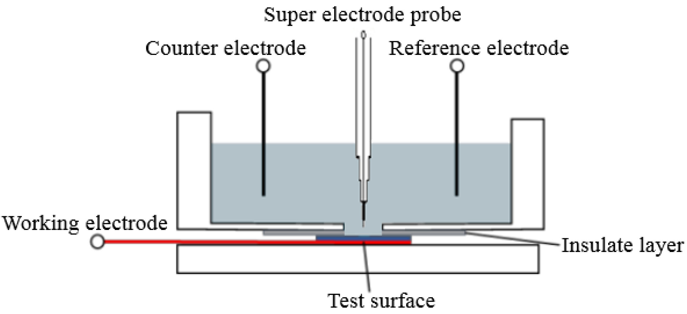
2.2. Methods and Techniques

Twelve cylindrical coupons of 5 mm × 5 mm× 20 mm were cut from the 13Cr stainless-steel casing and made into standard scanning electrochemical working electrodes by epoxy resin. The samples were grounded using an abrasive paper until #2000 grit and ultrasonically cleaned in acetone. They were exposed into the NaCl solution of 0.2 mol/L, 0.4 mol/L, 0.6 mol/L, and 1 mol/L. The nonmetallic inclusions were observed by a metallographic microscope and taken out for drying, weighing, and weight loss for different times (0 h, 48 h, 72 h, and 96 h). All experiments were performed with a research-grade digital microscope model Axio Scope A1, a scanning electron microscope (SEM) model EVO MA15 from Zeiss (Carl Zeiss, Oberkochen, Germany), a Philips X’Pert diffractometer Kα Cu = 1.54.56 A (Panaco, Almelo, Netherlands), and a scanning electrochemical microscopy (SECM) system model CHI 900C (CH Instruments Inc., Shanghai, China). Three electrode systems were adopted in the SECM system. A saturated calomel electrode (SCE) was used as the reference electrode. The platinum plate was used as the counter electrode. 13Cr stainless steel was used as the working electrode. Specimens were horizontally mounted facing upward. The accurate location was ensured by the three-dimensional positioning device of Pt superfine electrode probe 75 (r = 7.5 μm). A solution (10 mL) was poured into the electrolytic cell after the device installation according to Figure 1. The probe potential was 0.4 V, scan interval was 100 um × 100 um, and scanning rate was 30 μm/s during the SECM test process. The entire device should be stable in the measure process to ensure the test effect. The solution of 1 mol/L NaCl solution with 5 mmol/L KI was prepared from disposable distilled water. All tests were conducted at room temperature. The experimental reagent is chemically pure and bought from Chengdu Kelon chemical reagent company (Chengdu Kelong Chemical Reagent Factory, Chengdu, China). The experimental purpose is to simulate the corrosion behavior of metal materials in an ocean atmospheric environment. The surface microstructures and corrosion characteristics of 13Cr stainless-steel specimens after the SECM test were observed by a scanning electron microscope.

3. Results and Discussion

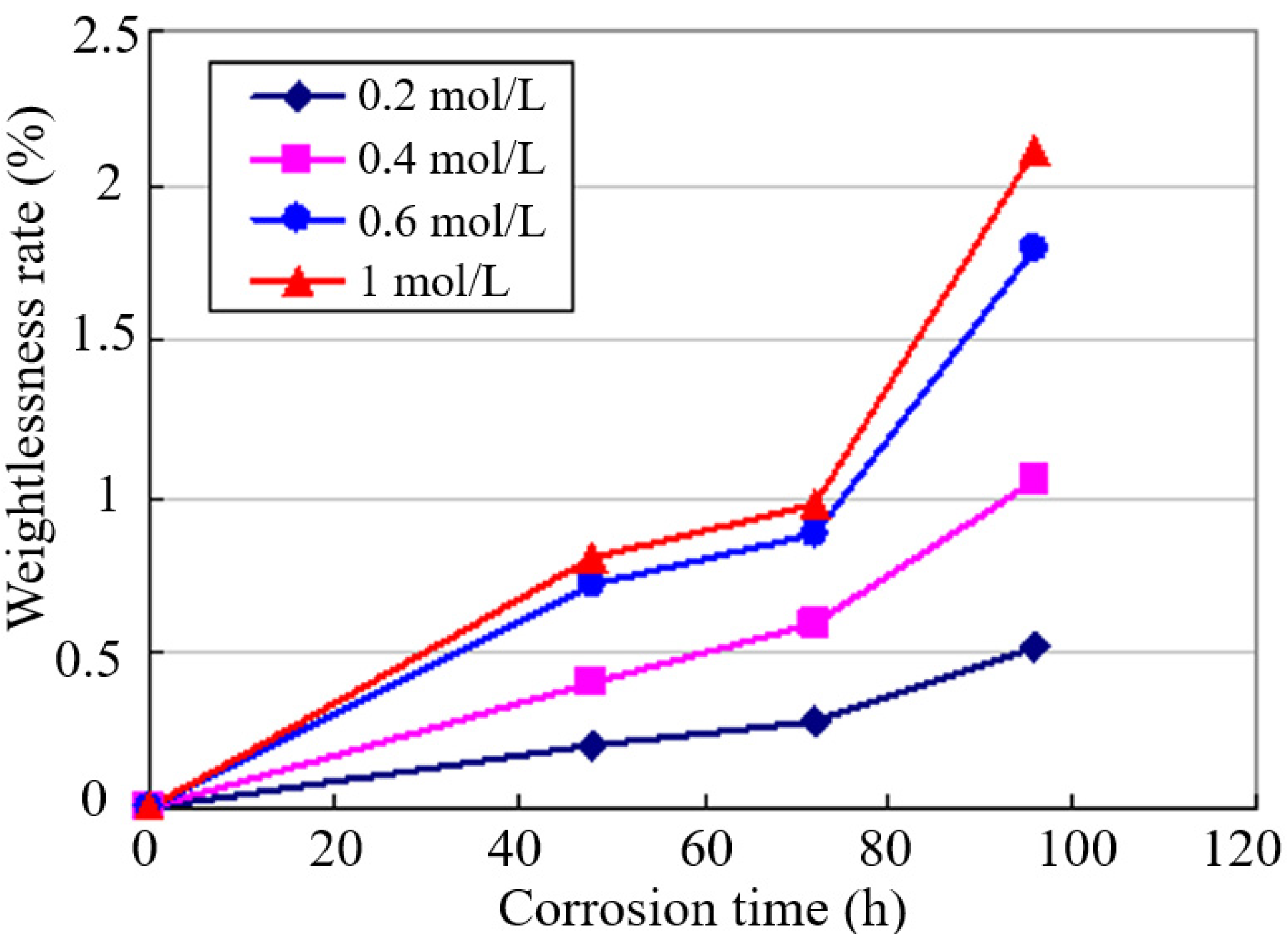
3.1. Weight Loss Test

The weight loss test result of 13Cr stainless-steel specimens under the solution with different Cl concentration is presented in Figure 2.
The corrosion weight loss rate of metal materials can be measured according to the quality variation of the samples before and after immersing in the solution with different Cl concentration. The calculation formula is as follows:
Δ m ( % ) = m 0 m 1 m 0 × 100 %
m 0 is the sample quality before it is corroded, mg; m 1 is the sample quality after it is corroded, mg.
The weight loss rate of the sample has a linear relation with the immersion time, as shown in Figure 2. The rate of weight loss sharply increases, and the corrosion rate accelerates with the rise of the immersion time. The corrosion gradually gets serious, but the corrosion rate starts to get slow due to the corrosion product film forming on the surface of the sample, which has the passivation effect. In addition, later, the protection of the corrosion passivation disappears and sharply accelerates the corrosion effect of Cl to the body of the substrate as time increases. The weight loss rapidly increases again, and corrosion becomes more serious. The corrosion products formed on the surface of the 13Cr stainless-steel sample immersing in the solution with different Cl concentration for different time. The Cl concentration in the NaCl solution is higher; the corrosion products formed under the same immersion time are much more. The corrosion rate increased with the increase in the Cl concentration, and the corrosion aggravated.

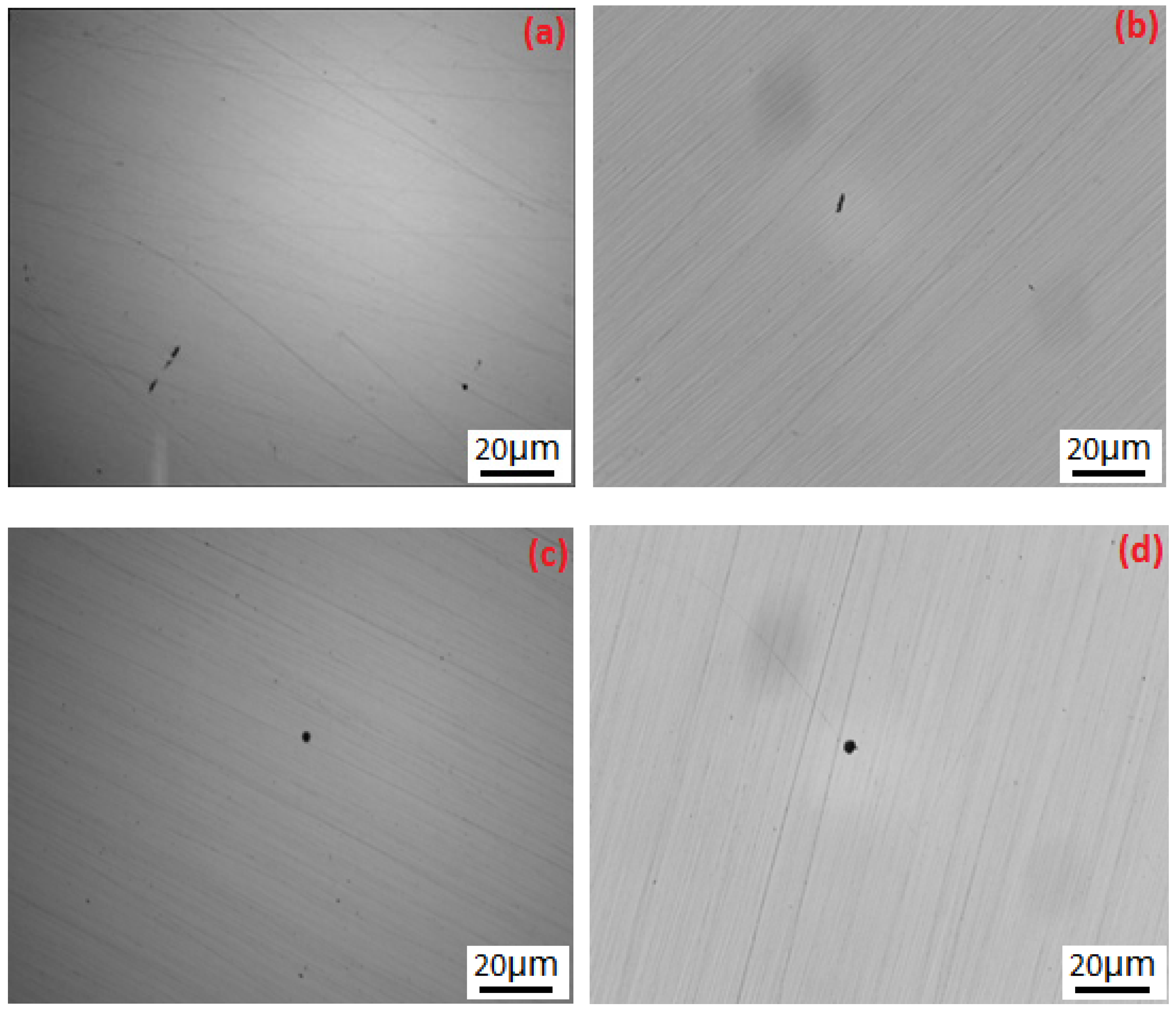
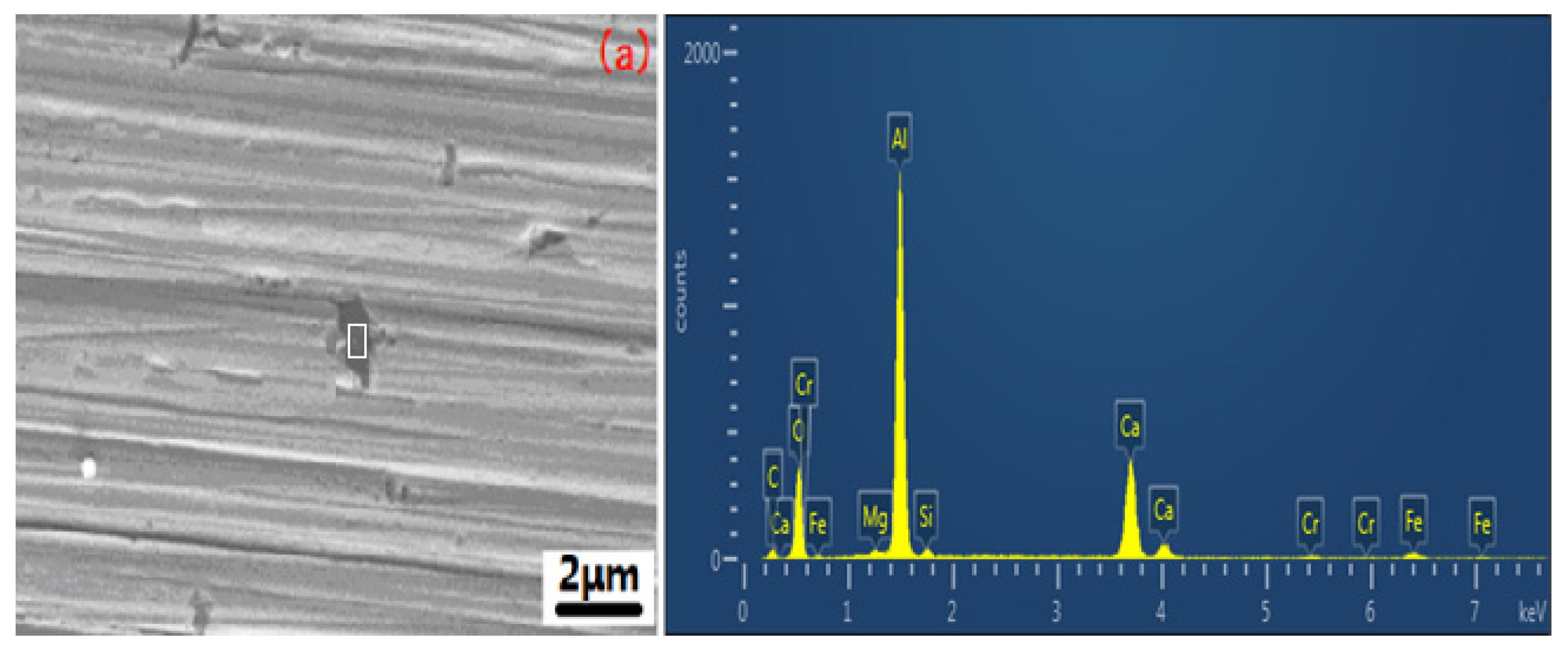
3.2. Surface SEM Morphology of the Inclusions

Inclusions of 13Cr stainless steel were observed and analyzed by a research-grade digital microscope model Axio Scope A1 after the samples were polished. The results show that the inclusion shapes were strip and ring. Strip shape inclusions are shown in Figure 3a,b. Ring shape inclusions are shown in Figure 3c,d. It can be speculated that the inclusions of ring and strip shapes are aluminum oxide inclusions and metallic phase inclusions according to the Chinese standard GB/T 10561-121 2005. The result of the observation is shown in Figure 3.

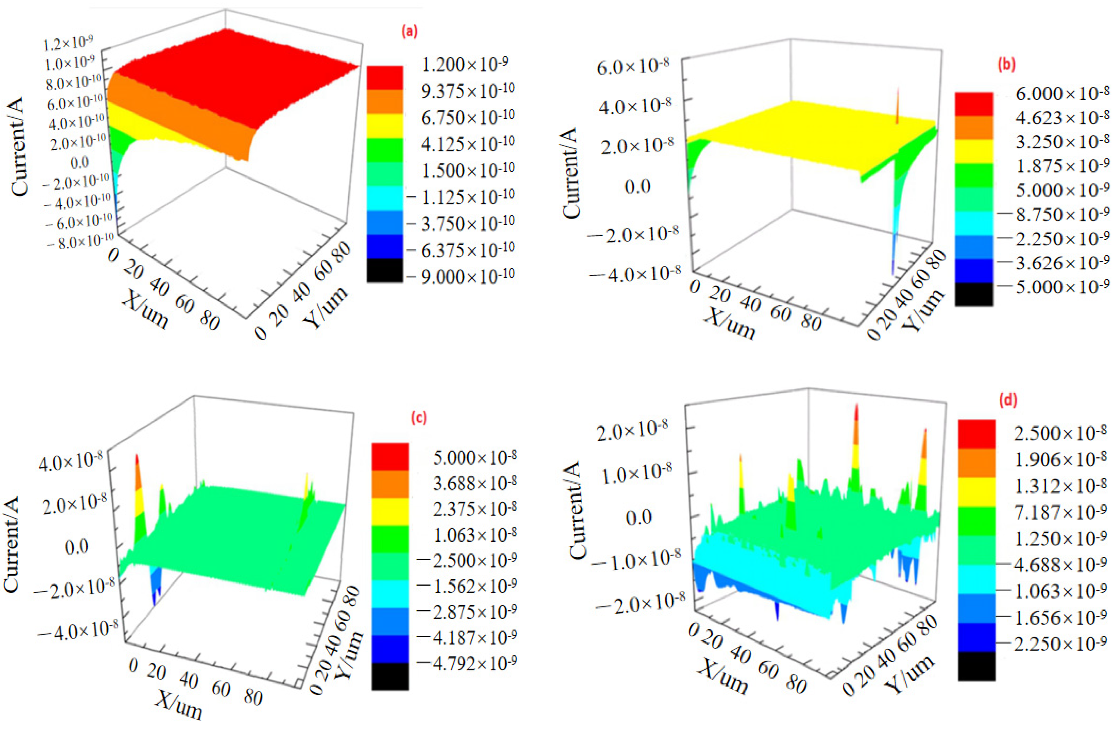
3.3. SECM Corrosion Behavior

Figure 4 shows the SECM morphologies of 13Cr stainless steel immersed in the solution of 1 mol/L NaCl and 5 mmol/L KI for different times. From Figure 4, it can be seen that the redox current has obvious differences in different regions of the samples and different corrosion times. Combined with the research results from Wang et al. [15], it can be obtained that stainless steel is continually corroded in different concentrations of the NaCl solution. The corrosion dissolution occurred in the local region of the surface. Meanwhile, the reduction reaction occurred. The possible electrode reaction is as follows:
F e F e 3 + + 3 e
I 3 + 2 e 3 I
Meanwhile, the reaction occurring on the probe is as follows:
3 I I 3 + 2 e
The local area of the 13Cr stainless-steel surface started to dissolve when Pt probe was scanning the substrate surface of 13Cr stainless steel. Then oxidizing reaction occurred after iron lost the electrons. I 3 was generated on the probe and F e 3 + dissolved due to the reaction from the substrate. The oxidation current on the probe increased. A convex peak from the surface-active points of the dissolved iron appeared just as what was shown in the plane-scanning figure. The quantity of oxidation current peaks was little, and the oxidation current value got large increasing at the beginning of corrosion (the time was 0 h). Therefore, active points were little. The quantity of oxidation current peaks increased, and the oxidation current value continued to increase after it was corroded for 48 h. It showed that active points increased and the tendency of corrosion dissolution strengthened. The corrosion gradually increased with the increase in time. The corrosion was relatively slow when the corrosion time was 72 h. However, the rate of corrosion dramatically increased when the time was more than 72 h. The quantity and value of corrosion current peaks on the surface of the sample constantly changed with the extension of corrosion time. It can be analyzed and shown that the surface active dissolution of 13Cr stainless steel was uneven in the corrosion process, and the corrosion dissolution was prior to the occurrence in the area with inclusions. It extended to other local areas from the view of the microperspective. The oxidation current peak was distributed in terms of strips and ribbons.
The reason for the occurrence of the metal corrosion reaction in the electrolyte was the local passivation membrane with protective effect on the damaged metal. At the moment, hydrogen atoms cause the reaction between the metal substrate and the interface of the passivation membrane as follows [15,16]:
H H o x + + e
2 H o x + + F e ( O H ) 2 F e 2 + + 2 H 2 O
H o x + + O 2 O H o x
H o x + + O H o x H 2 O
According to the literature, the oxidation of hydrogen decreases the pH value between the metal substrate and the interface of the passivation membrane. Therefore, the proportion of O H / O 2 was improved. The higher the O H / O 2 , the lower the pitting-resistance property of the passivation membrane. Moreover, the material was easy to be corroded by Cl. The clear figure can be obtained by SECM technology through detecting the current variation of Fe2+ in this area. This is because the concentration of Fe2+ in the pitting site was higher than those in the other sites when pitting occurred.

3.4. SEM Morphology

When the SECM test was over, the micromorphology of the samples was observed by a scanning electron microscope. The results showed that pitting corrosion firstly occurred in inclusions. The morphology of the inclusions includes strip (Figure 5a,b) and round inclusions (Figure 5c,d). The area containing strip inclusions on the surface of the sample displayed serious pitting corrosion. It should be known that the inclusions were oxide inclusions of the aluminum and FeAlx phase according to the analysis of the energy spectrum (EDS) test. Table 1 shows the EDS results of the inclusions from Figure 5a–d.

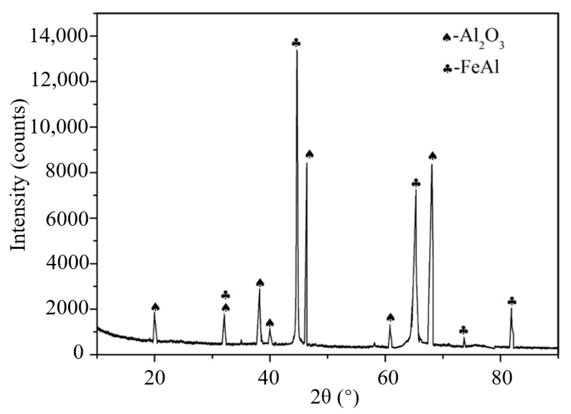
3.5. Phase Structure of Inclusions
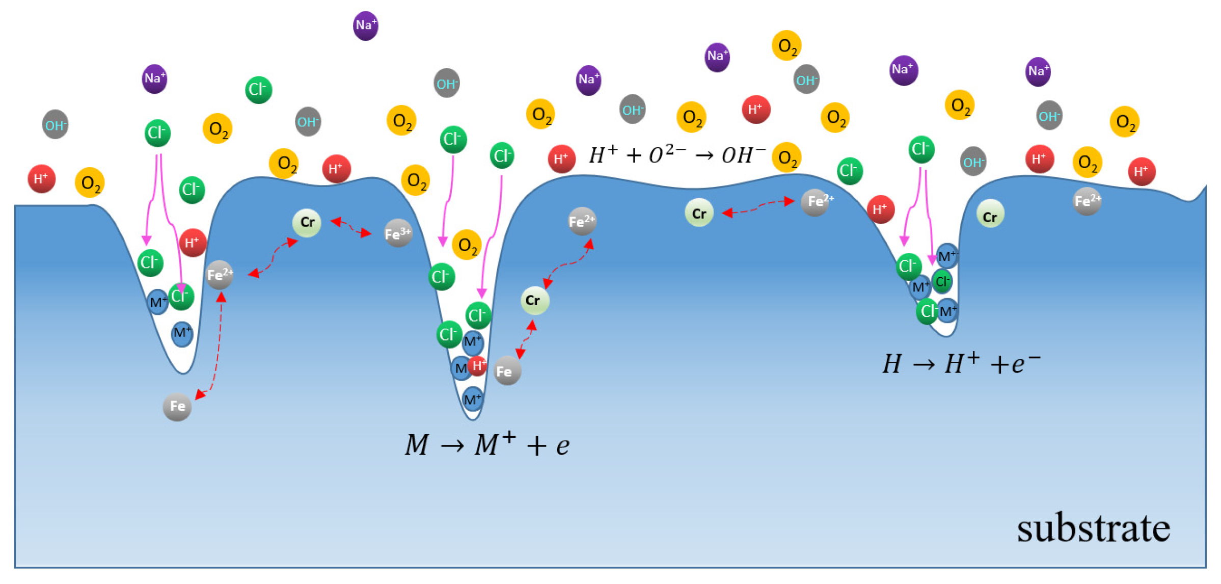
A low-angle XRD study of the metallic inclusions in the 13Cr stainless steel revealed the Al2O3 and FeAl phases as the main inclusions. It is clear that its peaks exhibited a higher intensity for Al2O3 and FeAl (Figure 6). The result is consistent with the results of the SECM and energy spectrum (EDS) test.

4. Discussion

The main reason why 13Cr stainless steel was showing local corrosion in the corrosion medium was the local dissolution of the materials. It had a relationship with the structure. The corrosion dissolution of the materials was associated with the precipitated phase, inclusions, and flaw. Figure 3 shows 13Cr stainless-steel materials containing ring and long strip aluminum oxide inclusions and inclusions of the iron and aluminum phase. Its main electrochemical corrosion mechanism was the microcell corrosion generation between the aluminum oxide inclusions and the iron substrate material because the corrosion potential is different among them. This accelerated the corrosion dissolution of the materials. On the basis of the literature analysis results [17,18,19], the corrosion dissolution occurred due to the intermetallic compounds firstly dissolving when the potential of intermetallic compounds such as those containing magnesium and aluminum was lower than that of the iron substrate material. When the potential of intermetallic compounds such as Cu and Pt was higher than that of the iron substrate, the iron substrate firstly dissolved. Then the corrosion pits formed when intermetallic compound particles fell off. This was ascribed to the local dissolution of iron and the different phase of the substrate causing the local corrosion of the material [20,21].
The aluminum oxide compound inclusions (Al2O3) and the FeAl phase compound are contained in 13Cr stainless steel as per the results of the scanning electron microscopy analysis and XRD test. The results show that the material contains the matrix phase of iron and intermetallic compounds of aluminum oxide inclusions, as shown in Figure 5 and Table 1. Because the corrosion potential of the Al2O3 phase was high in the 1 mol/L NaCl solution, it acted as a cathode in the process of corrosion microbattery between the solution and matrix steel. However, the iron matrix acted as an anode due to the low corrosion potential. This caused the anodic dissolution of iron around Al2O3 phase inclusions. Meanwhile, the corrosion active points formed. Aluminum ingots will form the local rich aluminum zone during the process of 13Cr stainless-steel distillation, and iron and aluminum improve with the increase in temperature. The increase in temperature was beneficial to the formation of iron-containing aluminum compounds, namely FeAl and Al2O3, in the low alumina high iron due to the different entropy. The FeAl phase material was similar to the β2 phase in the binary phase of iron aluminum and became the electroactive points [22]. Therefore, this area firstly displayed pitting corrosion because the self-corrosion current was large, and the self-corrosion potential was low. The study of passive film breakdown was important due to the autocatalytic nature of pitting corrosion. Once a corrosion pit generated on the surface of the metal, the attack would indefinitely continue, which is ascribed to the ample supply of aggressive ions (Figure 7). Once the corrosion pit formed on the metal surface, a localized site of high positive charge (the metal ion M+ being removed from the metal surface) would be built up in the pit. This excess positive charge attracted the aggressive ion Cl into the pit; thereby, fresh ions would be continually supplied to the metal surface. The corrosion pit continued to enlarge when a large number of aggressive ions gather in the pit [23].
It is shown in Figure 4 that electrochemical corrosion in the micro area is tested by a scanning electrochemical microscope. The adhesion between intermetallic compound and body material decreases because of the continual dissolution of iron around the intermetallic compound. When the corrosion arrives to the later period, the intermetallic compound falls off, and pitting occurs on the surface of the material (as shown in Figure 5). This is in agreement with the results of the Peguet study [24]. The conclusion of the corrosion dissolution mainly occurs in the Al2O3 phase inclusion and FeAl phase, and surrounding them can be easily obtained from the EDS analysis of inclusions in 13Cr stainless-steel combing with the analysis of the distribution of the REDOX peak current with strip from the scanning electrochemical. Figure 4a showed that the electroactive point did not appear because no current peaks occurred in the blank sample. The uniform forward and reverse electroactive point firstly appeared when samples soaked in the 1 mol/L NaCl solution for 48 h. Then a certain current peak occurred. After 72 h, 5 electroactive points appeared and pitting formed, as shown in Figure 4c. With the soaked time continually prolonging, the corrosion occurred in the original electroactive point, and new electroactive points appeared in the site with inclusions and flaws, as shown in Figure 4d. Electroactive points firstly generated on the surface of 13Cr stainless steel at the early stage of pitting. However, these electroactive points are unstable. They will disappear and generate pitting with the difference of the immersion time. The generation and disappearance of electroactive points occur simultaneously with the occurrence of pitting. The corrosion mechanism diagram in Figure 7 can also show that the Fe2+ concentration from the solution increases with the increase in the immersion time. This causes the probability of electroactive points and pitting occurrence to increase.

5. Conclusions

(1) The corrosion of 13Cr stainless steel got more serious with the Cl concentration increasing. The active points on the surface of the samples continually changed with the prolonging of the corrosion time. The sample surface was partially firstly dissolved, and then corroded and dissolved in a large area. The corrosion greatly varied in different areas.
(2) The pitting of 13Cr stainless steel firstly occurred in the electroactive points with metallic inclusions of the Al2O3 and FeAl phase. The generation and disappearance of electroactive points were simultaneous with the corrosion occurrence. The dissolution actively ascribed to the corrosion potential was different between the intermetallic compound such as Al2O3 and the iron matrix, and then corrosion microbattery formed. Corrosion dissolution mainly occurred in the inclusion and its surrounding areas.

Author Contributions

Conceptualization, W.L. and H.Y.; methodology, Y.L.; software, X.L.; validation, H.Y., X.L. and K.D.; formal analysis, W.L. and K.D.; resources, Y.L.; data curation, Z.Z.; writing—original draft preparation, W.L.; writing—review and editing, Y.L.; visualization, H.Y.; supervision, Z.Z.; project administration, W.L.; funding acquisition, W.L., Y.L., Z.Z. and K.D. All authors have read and agreed to the published version of the manuscript.

Funding

This research was funded by National Natural Science Foundation of China grant number 52074232, 52074234, 51904261, Sichuan Science and Technology Program grant number 2022NSFSC0028, Sichuan Youth Science and Technology Innovation Research Team Project Plan grant number 2020JDTD0016, the Scientific Research Starting Project of SWPU grant number 2021QHZ013, 2021 Provincial College Student Innovation and Entrepreneurship Training Program Project S202110615016, Open Fund (PLN2022-44) of State Key Laboratory of Oil and Gas Reservoir Geology and Exploitation (Southwest Petroleum University).

Data Availability Statement

Not applicable.

Conflicts of Interest

The authors declare no conflict of interest.

References

  1. Cai, W.; Zhao, G.; Zhao, D.; Wei, A. Corrosion resistance and semiconductor properties of passive films formed on super 13Cr stainless steel. J. Univ. Sci. Technol. Beijing 2011, 33, 1226–1230. [Google Scholar]
  2. Hampel, M.; Schenderlein, M.; Schary, C.; Dimper, M.; Ozcan, O. Efficient detection of localized corrosion processes on stainless steel by means of scanning electrochemical microscopy (SECM) using a multi-electrode approach. Electrochem. Commun. 2019, 101, 52–55. [Google Scholar] [CrossRef]
  3. Yule, L.C.; Bentley, C.L.; West, G.; Shollock, B.A.; Unwin, P.R. Scanning electrochemical cell microscopy: A versatile method for highly localised corrosion related measurements on metal surfaces. Electrochim. Acta 2019, 298, 80–88. [Google Scholar] [CrossRef]
  4. Zhou, H.R.; Ma, J.; Li, X.G.; Dong, C.F.; Lu, Q.K.; Feng, H. Corrosion study of pure aluminum in 0.6 mol/L NaCl solution by SECM. Hangkong Cailiao Xuebao 2009, 29, 8–12. [Google Scholar]
  5. Molina, J.; Fernández, J.; Inés, J.C.; Del Río, A.I.; Bonastre, J.; Cases, F. Electrochemical characterization of reduced graphene oxide-coated polyester fabrics. Electrochim. Acta 2013, 93, 44–52. [Google Scholar] [CrossRef]
  6. Yu, J.G.; Luo, J.L.; Norton, P.R. Investigation of hydrogen induced pitting active sites. Electrochim. Acta 2002, 47, 4019–4025. [Google Scholar] [CrossRef]
  7. Lin, C.J.; Xie, Z.X.; Tian, Z.W. Electrochemical scanning tunneling microscope studies of initial pitting corrosion 277 of stainless steel. Corros. Sci. Prot. Technol. 1997, 4, 3–8. [Google Scholar]
  8. Sun, P.; Zhang, Z.; Gao, Z.; Shao, Y. Probing fast facilitated ion transfer across an externally polarized liquid–liquid interface by scanning electrochemical microscopy. Angew. Chem. 2002, 114, 3595–3598. [Google Scholar] [CrossRef]
  9. Meng, F.; Wang, J.; Han, E.H.; Ke, W. Effects of scratching on corrosion and stress corrosion cracking of Alloy 690TT at 58 °C and 330 °C. Corros. Sci. 2009, 51, 2761–2769. [Google Scholar] [CrossRef]
  10. Reclaru, L.; Lerf, R.; Eschler, P.Y.; Blatter, A.; Meyer, J.M. Pitting, crevice and galvanic corrosion of REX stainless-steel/CoCr orthopedic implant material. Biomaterials 2002, 23, 3479–3485. [Google Scholar] [CrossRef]
  11. Olorundaisi, E.; Jamiru, T.; Adegbola, A.T. Mitigating the effect of corrosion and wear in the application of high strength low alloy steels (HSLA) in the petrochemical transportation industry—A review. Mater. Res. Express 2020, 6, 1265k9. [Google Scholar] [CrossRef]
  12. Amphlett, J.L.; Denuault, G. Scanning electrochemical microscopy (SECM): An investigation of the effects of tip geometry on amperometric tip response. J. Phys. Chem. B 1998, 102, 9946–9951. [Google Scholar] [CrossRef]
  13. Yuan, Y.; Li, L.; Wang, C.; Zhu, Y. Study of the effects of hydrogen on the pitting processes of X70 carbon steel with SECM. Electrochem. Commun. 2010, 12, 1804–1807. [Google Scholar] [CrossRef]
  14. Dong, C.F.; Luo, H.; Xiao, K.; Li, X.G.; Cheng, Y.F. In situ characterization of pitting corrosion of stainless steel by a scanning electrochemical microscopy. J. Mater. Eng. Perform. 2012, 21, 406–410. [Google Scholar] [CrossRef]
  15. Wang, F.; Shan, Q.; Zhang, F.; Lu, F.; Li, J.; Yu, T.; Qu, C. Pitting corrosion behavior of metal materials and research methods. IOP Conf. Ser. Earth Environ. Sci. 2021, 651, 032039. [Google Scholar] [CrossRef]
  16. Venugopal, A.; Raja, V.S. AC impedance study on the activation mechanism of aluminium by indium and zinc in 3.5% NaCl medium. Corros. Sci. 1997, 39, 2053–2065. [Google Scholar] [CrossRef]
  17. Aligizaki, K.K. Analytical Methods in Corrosion Science and Engineering. Corrosion 2008, 64, 86. [Google Scholar]
  18. Lister, T.E.; Pinhero, P.J. The effect of localized electric fields on the detection of dissolved sulfur species from Type 304 stainless steel using scanning electrochemical microscopy. Electrochim. Acta 2003, 48, 2371–2378. [Google Scholar] [CrossRef]
  19. Cruz, J.; Martınez, R.; Genesca, J.; Garcıa-Ochoa, E. Experimental and theoretical study of 1-(2-ethylamino)-2-methylimidazoline as an inhibitor of carbon steel corrosion in acid media. J. Electroanal. Chem. 2004, 566, 111–121. [Google Scholar] [CrossRef]
  20. Katemann, B.B.; Inchauspe, C.G.; Castro, P.A.; Schulte, A.; Calvo, E.J.; Schuhmann, W. Precursor sites for localised corrosion on lacquered tinplates visualised by means of alternating current scanning electrochemical microscopy. Electrochim. Acta 2003, 48, 1115–1121. [Google Scholar] [CrossRef]
  21. Lister, T.E.; Pinhero, P.J. Scanning electrochemical microscopy study of corrosion dynamics on type 304 stainless steel. Electrochem. Solid-State Lett. 2002, 5, B33. [Google Scholar] [CrossRef]
  22. Chen, G.; Liss, K.D.; Chen, C.; He, Y.; Qu, X.; Cao, P. Porous FeAl alloys via powder sintering: Phase transformation, microstructure and aqueous corrosion behavior. J. Mater. Sci. Technol. 2021, 86, 64–69. [Google Scholar] [CrossRef]
  23. Nagaoka, A.; Nose, K.; Nokami, K.; Kajimura, H. The role of micro pits in the initiation process of crevice corrosion of SUS304 stainless steel in an aqueous chloride solution. Mater. Trans. 2022, 63, 335–342. [Google Scholar] [CrossRef]
  24. Paik, C.H.; White, H.S.; Alkire, R.C. Scanning electrochemical microscopy detection of dissolved sulfur species from inclusions in stainless steel. J. Electrochem. Soc. 2000, 147, 4120. [Google Scholar] [CrossRef]
Figure 1. Schematic of test setup for SECM test.
Metals 12 01833 g001
Figure 2. Corrosion weight loss result.
Metals 12 01833 g002
Figure 3. Image of inclusions morphology: (a) location 1 of band inclusions; (b) location 2 of band inclusions; (c) location 1 of annular oxide inclusion; (d) location 2 of annular oxide inclusion.
Metals 12 01833 g003
Figure 4. SECM images of 13Cr stainless steel in 1 mol/L NaCl (5 mmol/L KI) solution for different times: (a) 0 h; (b) 48 h; (c) 72 h; (d) 96 h.
Metals 12 01833 g004
Figure 5. Images of inclusions morphology and EDS test results after SECM tests of 13Cr stainless steel immersing in different times: (a) 0 h; (b) 48 h; (c) 72 h; (d) 96 h.
Metals 12 01833 g005aMetals 12 01833 g005b
Figure 6. XRD pattern of metallic inclusions.
Metals 12 01833 g006
Figure 7. Local corrosion dissolution diagram of intermetallic compound.
Metals 12 01833 g007
Table 1. Spectrum analysis results of four inclusions (wt. %).
ElementCOAlCrFeNiSiCaMg
15.5934.2445.532.6911.951.0114.270.44
25.6123.5030.186.5232.510.9216.081.34
314.9435.0230.960.753.05
49.8036.7432.860.662.501.68
Publisher’s Note: MDPI stays neutral with regard to jurisdictional claims in published maps and institutional affiliations.

