Article

Surface Properties and Biocompatibility of Anodized Titanium with a Potential Pretreatment for Biomedical Applications

1
Biomedical Technology R & D Center, China Medical University Hospital, Taichung 404, Taiwan
2
Graduate Institute of Dental Science, College of Dentistry, China Medical University, Taichung 404, Taiwan
3
Implant Academy of Minimally Invasive Dentistry, Taipei 106, Taiwan
4
Division of Family and Operative Dentistry, Taipei Medical University Hospital, Taipei 110, Taiwan
5
Department of Oral Hygiene Care, Ching Kuo Institute of Management and Health, Keelung 203, Taiwan
6
Department of Oral and Maxillofacial Surgery, Faculty of Dentistry, Hasanuddin University, Makassar 90245, Indonesia
7
Department of Dentistry, Fu Jen Catholic University Hospital, Fu Jen Catholic University, New Taipei City 242, Taiwan
8
Department of Dentistry, Taipei Medical University-Shuang Ho Hospital, New Taipei City 235, Taiwan
9
Division of Clinical Cariology and Endodontology, Department of Oral Rehabilitation, School of Dentistry, Health Sciences University of Hokkaido, Hokkaido 061-0293, Japan
10
3D Global Biotech Inc. (Spin-off Company from Taipei Medical University), New Taipei City 221, Taiwan
11
Taiwan Society of Blood Biomaterials, New Taipei City 221, Taiwan
12
School of Dentistry, College of Oral Medicine, Taipei Medical University, Taipei 110, Taiwan
13
Graduate Institute of Biomedical Optomechatronics, College of Biomedical Engineering, Taipei Medical University, Taipei 110, Taiwan
14
Graduate Institute of Biomedical Materials and Tissue Engineering, College of Biomedical Engineering, Taipei Medical University, Taipei 110, Taiwan
*
Authors to whom correspondence should be addressed.
Co-First author: Yi-Jung Lu.
Metals 2021, 11(7), 1090; https://doi.org/10.3390/met11071090
Submission received: 10 June 2021 / Revised: 4 July 2021 / Accepted: 6 July 2021 / Published: 8 July 2021
(This article belongs to the Special Issue Advances in Biomedical Metallic Materials)

Abstract

:
The effects of anodized titanium (Ti) with a potential hydrogen fluoride (HF) acid pretreatment through cathodization on the formation of nano-porous Ti dioxide (TiO2) layer were characterized using field-emission scanning electron microscopy, grazing incidence X-ray diffractometer, and contact angle goniometer. The biocompatibility was determined by 3-[4,5-dimethylthiazol-2-yl]-2,5-diphenyltetrazolium bromide (MTT) test. Analytical results found that a well-aligned nano-porous structure was formed on the anodized Ti surface with HF pretreatment concentration above 0.5%. Microstructure of the nano-porous Ti dioxide surface generated by anodization with HF pretreatment was composed of anatase and rutile phases, while the anodized Ti sample with HF pretreatment concentration of 0.5% presented excellent hydrophilicity surface. An in-vitro biocompatibility also indicated that osteoblast cells grown on the surface of the anodized Ti sample with HF pretreatment increased with the increase of culture time. The filopodia of osteoblast cells not only adhered flat, but also tightly grabbed the nano-porous structure for promoting cell adhesion and proliferation. Therefore, the anodized Ti with HF pretreatment can form a functionalized surface with great biocompatibility for biomedical applications, particularly for dental implants.

1. Introduction

Demand for biomaterials in biomedical applications such as orthopedics and dental implants is increasing rapidly [1,2]. Titanium (Ti) is well-known as a biomaterial with excellent mechanical strength, resistance to body fluid effect, and biocompatibility [1,2,3,4,5]. However, Ti still needs to be modified through surface treatment to supply fast and durable osseointegration used in the human body [2]. The fundamental of osseointegration requires the ability of an implant to bond with the surrounding host bone [2,6]. Hence, the characteristics of the implant surface such as, topography, chemical composition, and surface energy will have an impact on cell interactions with the surrounding tissue of the implant [3,6].
Various surface modification methods were performed on Ti implants to obtain a more biocompatible surface [5,6]. Recent studies indicated that plasma electrolytic oxidation (PEO) is a promising anodization technique to modify metal surfaces [7,8]. These PEO modified surfaces provide suitable conditions for cell attachment, proliferation and antibacterial. Moreover, Yanovska et al. [9] reported that hydroxyapatite (HA) coatings can be formed on Ti alloy surface using a potential thermal deposition technique. HA coating makes surfaces more osteoconductive due to increasing collagen synthesis, while the latest research by Lan et al. [10] also proved that the anodized Ti with a hierarchical porous (micro and nano-porous) surface possessed great potential to enhance osteoblast cell adhesion ability. The anodized surface played an important role to promote osteoblast cells ingrowth into the nano-porous structure, which provided cell adhesion ability for enhancing osseointegration. Therefore, anodization is one of the potential modification methods of implants with several advantages, such as producing numerous surface morphologies, generating beneficial chemical species obtained from electrolytes used during the anodization process, adaptability to various implant shapes, and easy application in the biomedical fields [2,4,5].
In the present study, the anodization method is performed as a surface modification of Ti yet accompanied by pretreatment with different concentrations of hydrogen fluoride (HF) to determine which of these concentrations is effective in increasing the biocompatibility of Ti. The in vitro biocompatibility is evaluated by culturing with osteoblast cell because osteoblasts are known to play a crucial role in osseointegration [11]. In addition, the surface properties and microstructural characteristics were also investigated to understand the relationship between cellular adhesion behavior and anodized Ti surface in this study.

2. Materials and Methods

2.1. Materials Preparation

The biomedical grade-IV pure Ti sheet (obtained from Bio Tech One Inc., Kaohsiung, Taiwan) with a thickness of 1 mm served as the substrate in the present study. The sample was prepared as discs (10 mm in diameter) to perform the experiment analysis. The samples were mechanically ground and polished with 1500 grit paper, followed by 1 μm diamond abrasive, and finished with 0.04 μm colloidal silica abrasive. Prior to use, the samples were cleaned of various impurities and contaminants that remained from the machining process by being ultrasonically washed with acetone at 25 °C for 5 min and etched in a mixture solution of 2% ammonium fluoride (NH4F), 2% HF acid, and 10% nitric acid (HNO3) at 25 °C for 1 min. Afterwards, the samples were washed using distilled water in an ultrasonic bath at 25 °C for 10 min. Subsequently, the platinum bar with a diameter of 2.5 cm was placed in front of the samples at a distance of 2 cm to act as a cathode under constant current with current density of 1 A/dm2 for 10 min in different concentrations (0.01%, 0.05%, 0.1%, 0.5%, 1%, and 2%) of 1 M aqueous electrolyte HF solution, respectively. After cathodic pretreatment, the sample was anodized in sodium hydroxide (NaOH) solution. The anodization process was performed with magnetic stirring under a constant current of 15 A/dm2 for 10 min at 25 °C. Finally, the investigated samples were cleaned, rinsed with deionized water, and air-dried. The polished Ti disc without treatment acted as control for comparison.

2.2. Analysis of Surface Property

The sample was positioned on the copper holder and the surface was sputter-coated using platinum thin film with a thickness of 25 nm before loading into the vacuum chamber in order to provide an electrical conductivity. Subsequently, the surface morphology of the sample was determined by field-emission scanning electron microscope (FE-SEM; JEOL JSM6400, Tokyo, Japan) operating at 20 kV. The phase composition and crystallinity of the sample were analyzed by grazing incidence X-ray diffractometer (GIXRD; PHILIPS X’Pert Pro, Almelo, Netherlands) equipped with Cu Kα radiation source operated at 50 kV and 250 mA. The corresponding peaks of GIXRD spectrum were investigated according to the database from the Joint Committee on Powder Diffraction Standards.

2.3. Wettability Testing

The wettability of the sample was analyzed using static method by measuring the angle produced from the tangent line between the liquid drop and the surface of the sample (n = 5). Static method was carried out by dropping a 0.05 mL deionized water droplet onto the surface of the investigated sample (the distance of dropping was maintained constant at 10 mm), and subsequently, droplet profile inspected by goniometer (GBX Scientific LTD., Romans-sur-Isère, France).

2.4. Biocompatibility Evaluation

The osteoblast cell line (MG-63, ATCC-CRL1427) was obtained from the Bioresource Collection and Research Center, Hsinchu, Taiwan. The MG-63 cell line was maintained in Dulbecco’s Modified Eagle medium (DMEM; Gibco, Waltham, MA, USA) containing 10% fetal bovine serum (FBS), and antibiotic solution (1% penicillin-streptomycin). The cells were incubated at 37 °C with an atmosphere of 5% CO2 and 95% air. The cells were detached through trypsinization after the cell reach 90% confluency. The investigated samples (n = 5) were sterilized by washing in acetone for 15 min, soaking in ethanol for 15 min, air dried, and followed by exposure to ultraviolet lamp at room temperature for 24 h. The sterilized samples were placed in 24-well plates, inoculated with MG-63 cell at cell density of 1 × 10⁴ cells/well, and incubated at 37 °C for 8 h, 1 day, and 3 days, respectively. The 3-[4,5-dimethylthiazol-2-yl]-2,5-diphenyltetrazolium bromide (MTT; Sigma, Taipei, Taiwan) test was used to verified cellular growth on the samples. After 8 h, 1 day, and 3 days of MG-63 cell cultivation with investigated samples, 50 mL of MTT-dissolved in culture medium was added to the well plates and incubated for 4 h at 37 °C to form the formazan crystals. The medium containing MTT was removed, formazan crystals were dissolved in 150 mL dimethyl sulfoxide (DMSO, Sigma, Taipei, Taiwan), and quantified spectrophotometrically using an Epoch microplate reader (BioTek instruments Inc., Winooski, VT, USA) at the wavelength of 595 nm. Moreover, the morphology and colonization of MG-63 cells on sample surface after 48 h incubation were observed through the JSM-6500 FE-SEM operated at 25 kV.

2.5. Statistical Analysis

The experimental data were analyzed via SPSS statistic software (Version 19.0., SPSS Inc., Chicago, IL, USA). The difference between multiple groups were determined by one-way analysis of variance followed by Tukey’s HSD post hoc test. A p value of less than 0.05 was indicated as statistically significant.

3. Results

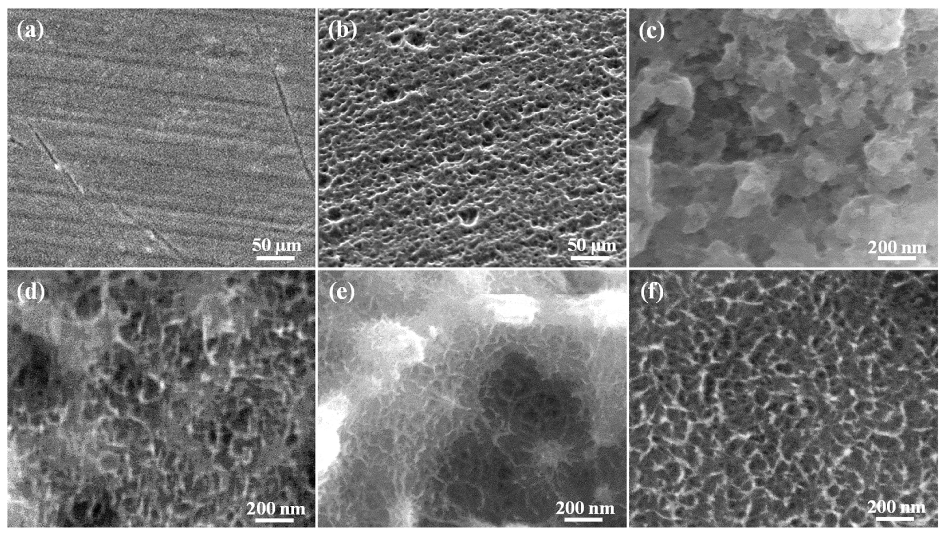
3.1. Morphology of the Investigated Samples

The surface morphology of the polished Ti, anodized Ti (A-Ti), and anodized Ti with cathodization pretreatment (AC-Ti) of various concentrations of HF are shown in Figure 1. Figure 1a presents surface morphology of the sample without any treatment with smooth machining polished Ti. Figure 1b depicts micro-porosity of the surface sample treated with anodization only. The nano-porous structure was generated on the surface of the sample (Figure 1c–f), which previously received HF treatment through cathodization before anodization. It was found that an irregular nano-porous structure was formed on the sample surface as the HF concentration below 0.1%. However, the formation of a well-aligned nano-porous structure can be seen on the surface of the AC-Ti with 0.5% HF sample. A similar feature could also be found in the AC-Ti with 1.0% and 2.0% HF samples.

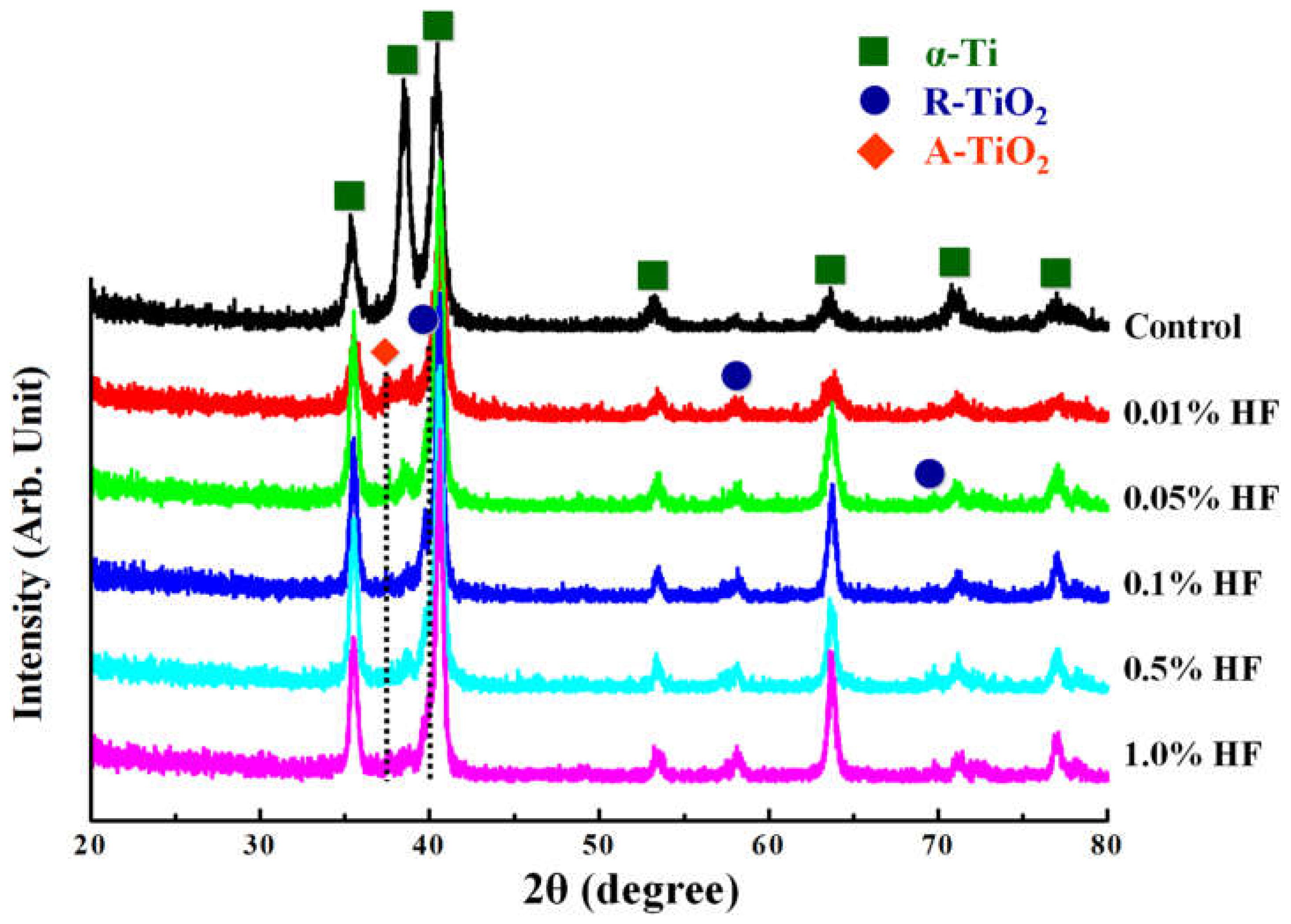
3.2. Microstructure of the Investigated Samples

The GIXRD pattern of the investigated sample as shown in Figure 2. Only the α-Ti peaks with a hexagonal crystalline structure were observed on the control sample. When Ti sample was subjected to anodization with HF pretreatment, the anatase (A-TiO2) and rutile (R-TiO2) crystalline structure peaks were detected on the surface of the treated samples.

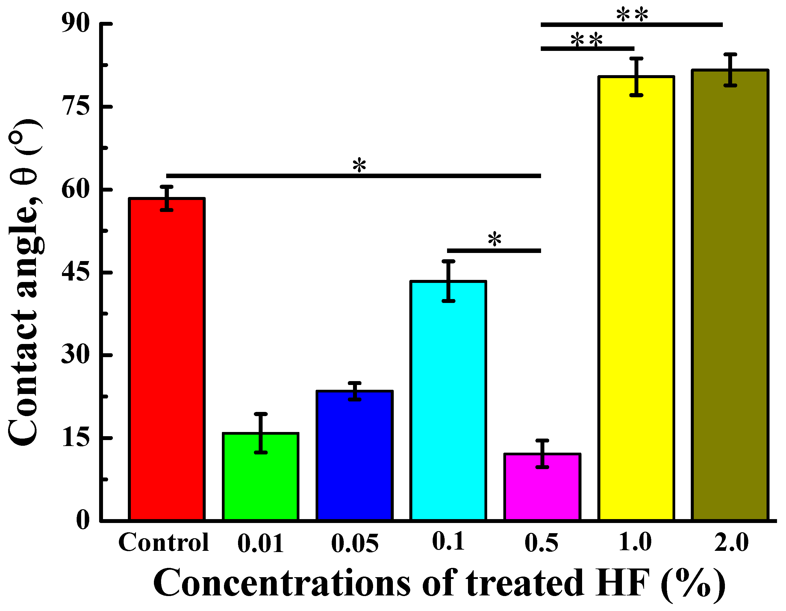
3.3. Wettability of the Investigated Samples

Figure 3 illustrates the average contact angle measurements of the investigated samples. Apparently, all measured contact angles are lower than 90°, which means that treated Ti surfaces are hydrophilic. However, the sample pretreated with 0.5% HF exhibited a smaller contact angle, indicating a higher hydrophilic surface than others sampled.

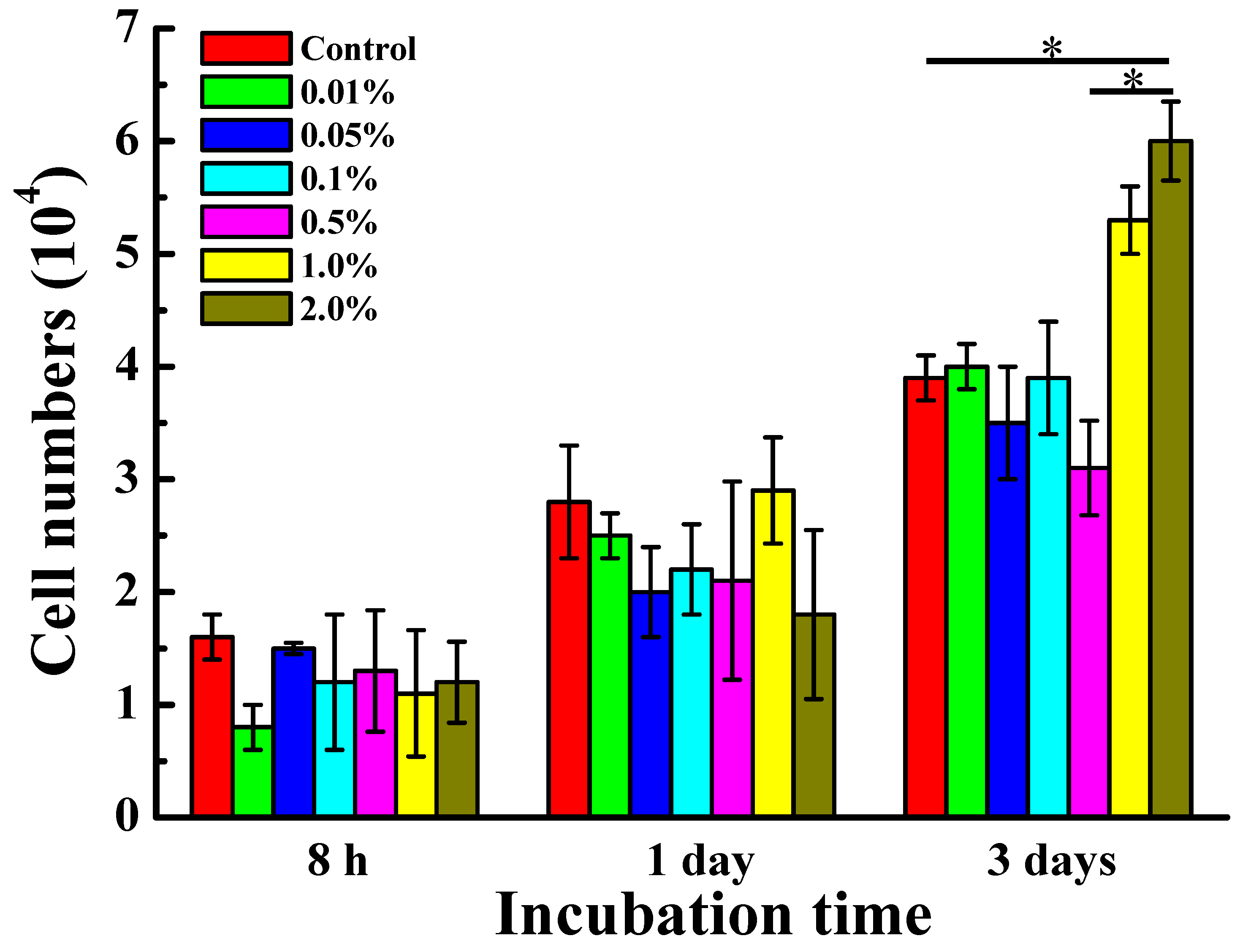
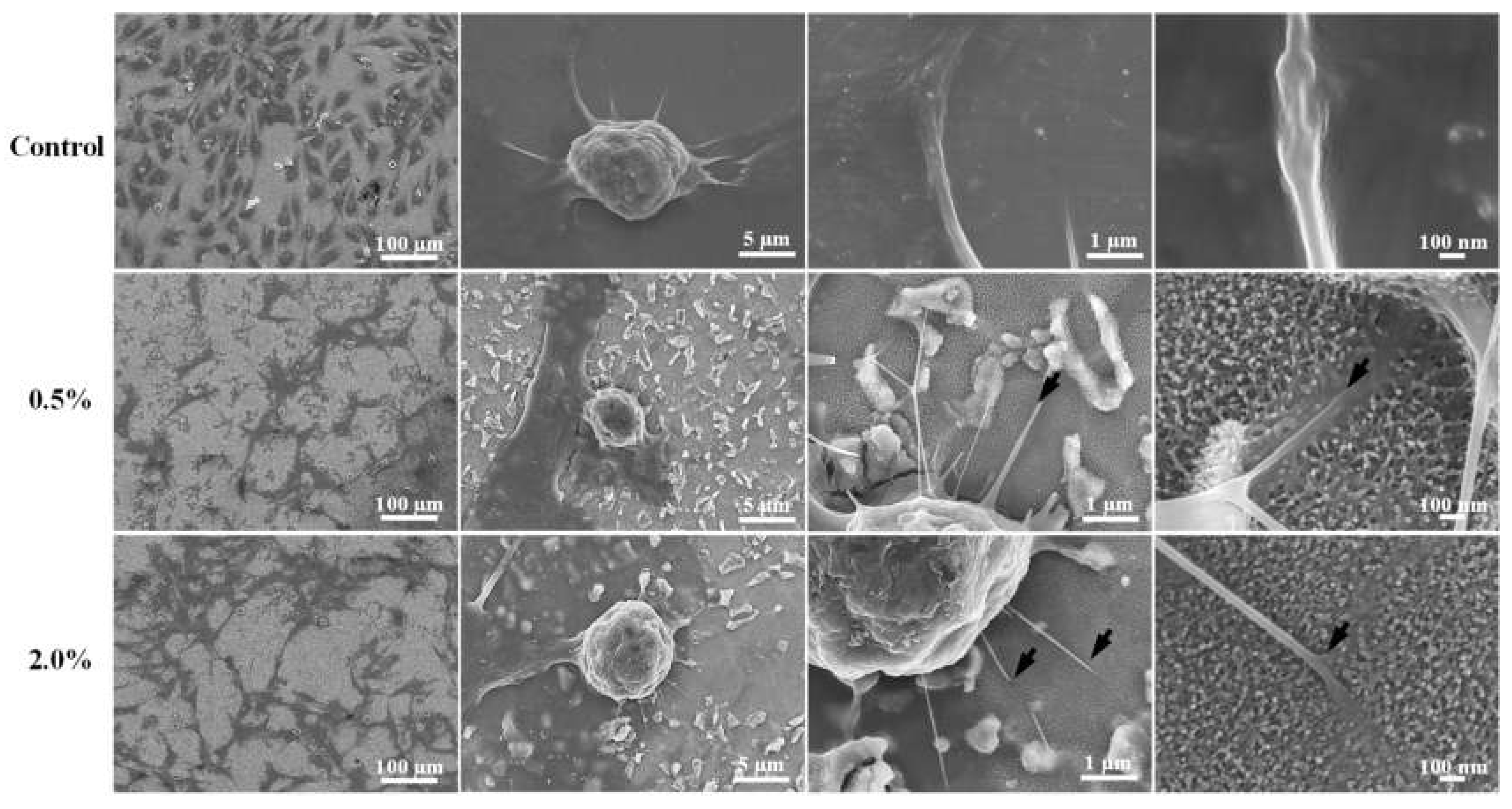
3.4. Cell Response and Adhesion Behavior of the Investigated Samples

The biocompatibility of the investigated samples, cultured with MG-63 cell, is displayed in Figure 4. According to the MG-63 cells growth result on the Ti surface treated with different conditions, it was found that the number of cells increased as the culture time increasing. No significant difference can be observed between the investigated samples at 8 h and day 1, while the AC-Ti with 2.0% HF sample exhibited a higher number of MG-63 cells grown on the surface as compared with other treated samples at day 3.
Figure 5 presents the FE-SEM images of MG-63 cells cultured for 48 h on the surface of Ti treated with different conditions. It is clearly seen that the cells did not have any changes in the cell morphology due to the control Ti surface without HF pretreatment. However, both samples pretreated with 0.5% and 2.0% HF showed more elongated filopodia than the Ti sample without HF pretreatment. In addition, the filopodia of cells not only adhered flat, but also tightly grabbed the nano-porous structure surface (as indicated by arrows). This cell response characteristic could also be observed in other HF pretreated samples.

4. Discussion

Surface modification of Ti implant greatly affected the success rate of implant placement. Since the nano-surface is known to have a necessary effect in promoting the living-cell response, the living cell can adhere, differentiate, grow, and regenerate the tissue on the Ti surface, and various surface modifications are made to the Ti surface to optimize its acceptability on the host and not cause multiple infections after implantation [12,13,14,15,16,17]. In the present study, the Ti surface was modified through chemical modification with anodization treatment preceded by cathodization using HF, which would produce nano-porous surfaces on the Ti. Without pretreatment, anodization will only produce microporous on the Ti surface, which is formed from immersion with NaOH at high temperatures for a long time [10,18,19,20]. Several studies have shown that anodization can produce a nano-surface [21,22]. Our previous study [10] also demonstrated that the Ti subjected to the HF pretreatment with different current densities showed the formation of a hierarchical porous (micro and nano-porous) surface. In the study with different concentrations of HF pretreatment, the results reveal that the Ti hydride sacrificial layer has melted away to produce nano-holes, and the Ti metal is anodized to achieve a TiO2 layer. The outcome of the oxide layer has anatase and rutile phases, which play a vital role in forming nano-porous TiO2 [23].
A surface has a good wettability if the surface is hydrophilic characterized by a contact angle that is smaller than 90° [24,25]. All investigated samples in this study showed a contact angle below 90°. However, it was found that the sample with 0.5% HF pretreatment showed superior hydrophilic performance with a contact angle of about 13°. This feature can be attributed that the surface with a well-aligned nano-porous structure can generate a larger surface free energy formation [26,27,28]. It is well known that the surface free energy related to the surface wettability. Accordingly, the rising effective surface area that will increase the hydrophilicity of the surface [1,26]. Good wettability will have an impact on the absorption of biological fluids, proteins, and cells on the Ti surface, as well as affecting cell morphology, adhesions, and proliferation [21,29,30].
Osteoblasts have been used to measure the biocompatibility of a material to be used as an implant since osteoblasts play a crucial role in bone regeneration. The MTT results indicated that the number of osteoblast cells adhering to the treated Ti surface increased with increasing culture time. In addition, the high concentration of HF proves better for osteoblast cell growth. This phenomenon occurs because HF has dose-dependent effects on osteoblast cell proliferation [31,32,33]. Moreover, Pham et al. [34] also found that low concentrations of HF can increase fibroblast cell proliferation, and high concentrations can reduce fibroblast cell proliferation and reduce the rate of bone mineralization. In this case, the Ti surface exposed to a range of 0.2–1.2% HF concentration would not have a cytotoxic effect on fibroblast cells. However, at these low concentrations, apart from increasing fibroblast proliferation, there was also a significant increase in interleukin 6, osteoprotegerin, and sclerostin, all of which play a role in bone formation and prevent bone resorption [34]. This event can be explained by HF affecting the stage of cell differentiation, which is more likely to affect osteoprogenitor cells or undifferentiated osteoblasts [35,36,37].
Surface properties of the material (hydrophilicity, composition, roughness, and morphology) will affect cell adhesion and spread, leading to faster osseointegration and implant stabilization [27]. Adhesion is the first step for cells to proliferate and differentiate, and failure of adhesion will cause cells not to survive [5]. In the present study, as seen in the FE-SEM micrographs of the cell morphology of MG-63, the Ti surface pretreated with different concentrations of HF exhibited elongated filopodia, meaning that the cell was in the adhesion process. The prolonged filopodia will strengthen the cell anchorage, while the prolonged filopodia will only elongate on a surface suitable for cell growth [5]. Our previous study has also demonstrated that the anodized Ti surface significantly increases osteoblast adhesion and proliferation [10]. Thus, the anodized Ti with HF pretreatment can form a nano-porous TiO2 surface to promote the bone cell ingrowth into the nano-porous structure for enhancing biocompatibility. Finally, further investigations must be performed to offer scientific information concerning surface properties and in vitro biocompatibility in the presence of anodized Ti.

5. Conclusions

The nano-porous TiO2 surface could be formed on the Ti substrate surface through the anodization with HF pretreatment method. All measured contact angles are less than 90°, which reveal that obtained nano-porous TiO2 surfaces are hydrophilic. The AC-Ti with 0.5% HF sample exhibited a well-aligned nano-porous topography and higher wettability. The cell response and adhesion behavior assessment also proved that the anodized Ti surface with nano-porous structure possessed great potential to increase cell adhesion ability. Thus, the research findings could provide useful scientific information in the surface modification of Ti.

Author Contributions

Writing -original draft, B.-H.H.; Investigation, B.-H.H. and Y.-J.L.; Data curation, Y.-C.C. and H.-Y.T.; Methodology, H.-Y.L. and W.-C.L.; Supervision, K.-L.O. and T.S.; Resources, C.-H.L.; Validation, M.R. and P.-J.H.; Writing—review & editing, T.-S.Y. and C.-M.L. All authors have read and agreed to the published version of the manuscript.

Funding

The authors would like to thank the 2021 Integrated Research Project of Innovative Translational Medicine, MOE Sprout Project, Ministry of Education, Taiwan for financially supporting this research under contract no. DP2-110-21121-01-O-06.

Institutional Review Board Statement

Not applicable.

Informed Consent Statement

Not applicable.

Data Availability Statement

Data is contained within the article.

Conflicts of Interest

The authors declare no conflict of interest.

References

  1. Kim, K.; Lee, B.A.; Piao, X.H.; Chung, H.J.; Kim, Y.J. Surface characteristics and bioactivity of an anodized titanium surface. J. Periodontal Implant Sci. 2013, 43, 198–205. [Google Scholar] [CrossRef]
  2. Kaczmarek, M.; Jurczyk, K.; Koper, J.K.; Paszel-Jaworska, A.; Romaniuk, A.; Lipińska, N.; Żurawski, J.; Urbaniak, P.; Jakubowicz, J.; Jurczyk, M.U. In vitro biocompatibility of anodized titanium with deposited silver nanodendrites. J. Mater. Sci. 2016, 51, 5259–5270. [Google Scholar] [CrossRef] [Green Version]
  3. Barjaktarević, D.R.; Cvijović-Alagić, I.L.; Dimić, I.D.; Đokić, V.R.; Rakin, M.P. Anodization of Ti-based materials for biomedical applications: A review. Metall. Mater. Eng. 2016, 22, 129–144. [Google Scholar] [CrossRef]
  4. Singh, A.; Singh, B.P.; Wani, M.R.; Kumar, D.; Singh, J.K.; Singh, V. Effect of anodization on corrosion behaviour and biocompatibility of Cp-titanium is simulated body fluid. Bull. Mater. Sci. 2013, 36, 8. [Google Scholar] [CrossRef] [Green Version]
  5. Chen, Z.X.; Takao, Y.; Wang, W.X.; Matsubara, T.; Ren, L.M. Surface characteristics and in vitro biocompatibility of titanium anodized in a phosphoric acid solution at different voltages. Biomed. Mater. 2009, 4, 065003. [Google Scholar] [CrossRef]
  6. Martinez, E.F.; Ishikawa, G.J.; de Lemos, A.B.; Barbosa Bezerra, F.J.; Sperandio, M.; Napimoga, M.H. Evaluation of a Titanium Surface Treated with Hydroxyapatite Nanocrystals on Osteoblastic Cell Behavior: An In Vitro Study. Int. J. Oral Maxillofac. Implant. 2018, 33, 597–602. [Google Scholar] [CrossRef] [Green Version]
  7. SergiyKyrylenko, S.; Warchoł, F.; Oleshko, O.; Husak, Y.; Kazek-Kęsik, A.; Korniienko, V.; Deineka, V.; Sowa, M.; Maciej, A.; Michalska, J.; et al. Effects of the sources of calcium and phosphorus on the structural and functional properties of ceramic coatings on titanium dental implants produced by plasma electrolytic oxidation. Mater. Sci. Eng. C 2020, 119, 111607. [Google Scholar]
  8. Myakinin, A.; Turlybekuly, A.; Pogrebnjak, A.; Mirek, A.; Bechelany, M.; Liubchak, I.; Oleshko, O.; Husak, Y.; Korniienko, V.; Leśniak-Ziółkowska, K.; et al. In vitro evaluation of electrochemically bioactivated Ti6Al4V 3D porous scaffolds. Mater. Sci. Eng. C 2021, 121, 111870. [Google Scholar] [CrossRef]
  9. Yanovska, A.; Husak, Y.; Mishchenko, O.; Gudakov, A.; Oleshko, O.; Yusupova, A.; Vielikov, M.; Radwan-Pragłowska, J.; Piątkowski, M.; Janus, Ł.; et al. Cell viability and collagen deposition on hydroxyapatite coatings formed on pretreated substrates. Mater. Chem. Phys. 2021, 258, 123978. [Google Scholar] [CrossRef]
  10. Lan, W.-C.; Wang, C.-H.; Huang, B.-H.; Cho, Y.-C.; Saito, T.; Huang, C.-C.; Huang, M.-S. Fabrication of a Promising Hierarchical Porous Surface on Titanium for Promoting Biocompatibility. Appl. Sci. 2020, 10, 1363. [Google Scholar] [CrossRef] [Green Version]
  11. Baxter, L.C.; Frauchiger, V.; Textor, M.; ap Gwynn, I.; Richards, R.G. Fibroblast and osteoblast adhesion and morphology on calcium phosphate surfaces. Eur. Cell. Mater. 2002, 4, 18. [Google Scholar] [CrossRef]
  12. Carrado, A.; Perrin-Schmitt, F.; Le, Q.V.; Giraudel, M.; Fischer, C.; Koenig, G.; Jacomine, L.; Behr, L.; Chalom, A.; Fiette, L.; et al. Nanoporous hydroxyapatite/sodium titanate bilayer on titanium implants for improved osteointegration. Dent. Mater. 2017, 33, 321–332. [Google Scholar] [CrossRef] [PubMed]
  13. Shayganpour, A.; Rebaudi, A.; Cortella, P.; Diaspro, A.; Salerno, M. Electrochemical coating of dental implants with anodic porous titania for enhanced osteointegration. Beilstein J. Nanotechnol. 2015, 6, 2183–2192. [Google Scholar] [CrossRef]
  14. Aboushelib, M.N.; Salem, N.A.; Taleb, A.L.; El Moniem, N.M. Influence of surface nano-roughness on osseointegration of zirconia implants in rabbit femur heads using selective infiltration etching technique. J. Oral Implantol. 2013, 39, 583–590. [Google Scholar] [CrossRef]
  15. El-Gammal, M.N.; El-Gammal, N.Y.; Fadhil, O.N.; Maria, O.M. Biological reactions to different dental implant surface treatments. Int. J. Contemp. Dent. Med. Rev. 2016, 2015, 9. [Google Scholar] [CrossRef]
  16. Tomsia, A.P.; Launey, M.E.; Lee, J.S.; Mankani, M.H.; Wegst, U.G.K.; Saiz, E. Nanotechnology approaches for better dental implant. Int. J. Oral Maxillofac. Implant. 2011, 26, 25. [Google Scholar]
  17. Zhang, W.; Cao, H.; Zhang, X.; Li, G.; Chang, Q.; Zhao, J.; Qiao, Y.; Ding, X.; Yang, G.; Liu, X.; et al. A strontium-incorporated nanoporous titanium implant surface for rapid osseointegration. Nanoscale 2016, 8, 5291–5301. [Google Scholar] [CrossRef]
  18. Ou, K.-L.; Lin, C.-T.; Chen, S.-L.; Huang, C.-F.; Cheng, H.-C.; Yeh, Y.-M.; Lin, K.-H. Effect of Multi-nano-titania Film on Proliferation and Differentiation of Mouse Fibroblast Cell on Titanium. J. Electrochem. Soc. 2008, 155. [Google Scholar] [CrossRef]
  19. Cheng, H.-C.; Lee, S.-Y.; Chen, C.-C.; Shyng, Y.-C.; Ou, K.-L. Influence of Hydrogen Charging on the Formation of Nanostructural Titania by Anodizing with Cathodic Pretreatment. J. Electrochem. Soc. 2007, 154. [Google Scholar] [CrossRef]
  20. Shih, Y.-H.; Lin, C.-T.; Liu, C.-M.; Chen, C.-C.; Chen, C.-S.; Ou, K.-L. Effect of nano-titanium hydride on formation of multi-nanoporous TiO 2 film on Ti. Appl. Surf. Sci. 2007, 253, 3678–3682. [Google Scholar] [CrossRef]
  21. Ross, A.P.; Webster, T.J. Anodizing color coded anodized Ti6Al4V medical devices for increasing bone cell functions. Int. J. Nanomedicine 2013, 8, 109–117. [Google Scholar] [CrossRef] [Green Version]
  22. Marenzi, G.; Spagnuolo, G.; Sammartino, J.C.; Gasparro, R.; Rebaudi, A.; Salerno, M. Micro-Scale Surface Patterning of Titanium Dental Implants by Anodization in the Presence of Modifying Salts. Materials 2019, 12, 1753. [Google Scholar] [CrossRef] [Green Version]
  23. Bezait, Z.; Tatar, D.; Ertugrul, M. Effect of oxygen addition on the formation of anatase TiO2 nano-coatings obtained by spray pyrolysis technique. J. Appl. Res. Technol. 2018, 16, 9. [Google Scholar]
  24. Chiang, H.-J.; Chou, H.-H.; Ou, K.-L.; Sugiatno, E.; Ruslin, M.; Waris, R.A.; Huang, C.-F.; Liu, C.-M.; Peng, P.-W. Evaluation of Surface Characteristics and Hemocompatibility on the Oxygen Plasma-Modified Biomedical Titanium. Metals 2018, 8, 513. [Google Scholar] [CrossRef] [Green Version]
  25. Kota, A.K.; Kwon, G.; Tuteja, A. The design and applications of superomniphobic surfaces. NPG Asia Mater. 2014, 6, e109. [Google Scholar] [CrossRef] [Green Version]
  26. Barbosa, T.P.; Naves, M.M.; Menezes, H.H.M.; Pinto, P.H.C.; De Mello, J.D.B.; Costa, H.L. Topography and surface energy of dental implants: A methodological approach. J. Braz. Soc. Mech. Sci. Eng. 2017, 39, 1895–1907. [Google Scholar] [CrossRef]
  27. Liu, X.M.; Wu, S.L.; Chu, P.K.; Chung, C.Y.; Chu, C.L.; Chan, Y.L.; Lam, K.O.; Yeung, K.W.; Lu, W.W.; Cheung, K.M.; et al. Nano-scale surface morphology, wettability and osteoblast adhesion on nitrogen plasma-implanted NiTi shape memory alloy. J. Nanosci. Nanotechnol. 2009, 9, 3449–3454. [Google Scholar] [CrossRef]
  28. Gong, D.; Grimes, C.A.; Varghese, O.K.; Hu, W.; Singh, R.S.; Chen, Z.; Dickey, E.C. Titanium oxide nanotube arrays prepared by anodic oxidation. J. Mater. Res. 2001, 16, 5. [Google Scholar] [CrossRef] [Green Version]
  29. Wu, F.; Xu, R.; Yu, X.; Yang, J.; Liu, Y.; Ouyang, J.; Zhang, C.; Deng, F. Enhanced Biocompatibility and Antibacterial Activity of Selective Laser Melting Titanium with Zinc-Doped Micro-Nano Topography. J. Nanomater. 2019, 2019, 5432040. [Google Scholar] [CrossRef]
  30. Syam, S.; Wu, C.-J.; Lan, W.-C.; Ou, K.-L.; Huang, B.-H.; Lin, Y.-Y.; Saito, T.; Tsai, H.-Y.; Chuo, Y.-C.; Yen, M.-L.; et al. The Potential of a Surface-Modified Titanium Implant with Tetrapeptide for Osseointegration Enhancement. Appl. Sci. 2021, 11, 2616. [Google Scholar] [CrossRef]
  31. Li, K.Q.; Jia, S.S.; Ma, M.; Shen, H.Z.; Xu, L.; Liu, G.P.; Huang, S.Y.; Zhang, D.S. Effects of fluoride on proliferation and mineralization in periodontal ligament cells in vitro. Braz. J. Med. Biol. Res. 2016, 49. [Google Scholar] [CrossRef] [Green Version]
  32. Wei, Y.; Wu, Y.; Zeng, B.; Zhang, H. Effects of sodium fluoride treatment in vitro on cell proliferation, BMP-2 and BMP-3 expression in human osteosarcoma MG-63 cells. Biol. Trace Elem. Res. 2014, 162, 18–25. [Google Scholar] [CrossRef]
  33. Lee, J.H.; Jung, J.Y.; Jeong, Y.J.; Park, J.H.; Yang, K.H.; Choi, N.K.; Kim, S.H.; Kim, W.J. Involvement of both mitochondrial- and death receptor-dependent apoptotic pathways regulated by Bcl-2 family in sodium fluoride-induced apoptosis of the human gingival fibroblasts. Toxicology 2008, 243, 340–347. [Google Scholar] [CrossRef] [PubMed]
  34. Pham, M.H.; Haugen, H.J.; Rinna, A.; Ellingsen, J.E.; Reseland, J.E. Hydrofluoric acid treatment of titanium surfaces the proliferation of human gingival fibroblasts. J. Tissue Eng. 2019, 10, 8. [Google Scholar] [CrossRef] [PubMed] [Green Version]
  35. Kumar, P.S.; Ks, S.K.; Grandhi, V.V.; Gupta, V. The Effects of Titanium Implant Surface Topography on Osseointegration: Literature Review. JMIR Biomed. Eng. 2019, 4. [Google Scholar] [CrossRef]
  36. Guo, J.; Padilla, R.J.; Ambrose, W.; De Kok, I.J.; Cooper, L.F. The effect of hydrofluoric acid treatment of TiO2 grit blasted titanium implants on adherent osteoblast gene expression in vitro and in vivo. Biomaterials 2007, 28, 5418–5425. [Google Scholar] [CrossRef]
  37. Hattar, S.; Berdal, A.; Asselin, A.; Loty, S.; Greenspan, D.C.; Sautier, J.M. Behaviour of moderately differentiated osteoblast-like cells cultured in contact with bioactive glasses. Eur. Cell. Mater. 2002, 4, 61–69. [Google Scholar] [CrossRef] [PubMed]
Figure 1. FE-SEM micrographs of the (a) polished Ti (control), (b) A-Ti, (c) AC-Ti with 0.01% HF, (d) AC-Ti with 0.05% HF, (e) AC-Ti with 0.1% HF, and (f) AC-Ti with 0.5% HF.
Metals 11 01090 g001
Figure 2. GIXRD pattern of control sample and samples that had been pretreated with various concentrations of HF.
Metals 11 01090 g002
Figure 3. Contact angle of control sample and samples that had been pretreated with various concentrations of HF (* p < 0.05 and ** p < 0.001).
Metals 11 01090 g003
Figure 4. Effect of HF at different concentrations on the proliferation of MG-63 cell of the investigated samples (* p < 0.05).
Metals 11 01090 g004
Figure 5. Cell morphologies of the investigated samples after culturing with MG-63 cells for 48 h. The HF pretreated samples with the most specific characteristic of the cells filopodia tightly grabbed the nano-porous structure are selected to present in this figure for comparison with the surface without any treatment.
Metals 11 01090 g005
Publisher’s Note: MDPI stays neutral with regard to jurisdictional claims in published maps and institutional affiliations.

