Review

What Are the Chances of Resilon to Dominate the Market Filling Materials for Endodontics?

by
Joanna Dobrzańska
1,2,
Lech B. Dobrzański
1,2,
Leszek A. Dobrzański
1,3,*,
Anna D. Dobrzańska-Danikiewicz
4 and
Klaudiusz Gołombek
5
1
Medical and Dental Engineering Centre for Research, Design and Production ASKLEPIOS, 12/1 King Jan III Sobieski St., 44-100 Gliwice, Poland
2
Medical and Dental Centre SOBIESKI, 12/1 King Jan III Sobieski St., 44-100 Gliwice, Poland
3
Department of Biomedical Engineering, Koszalin University of Technology, 2 Sniadeckich St., 75-453 Koszalin, Poland
4
Faculty of Mechanical Engineering, University of Zielona Góra, 4 Prof. Z. Szafran St., 65-516 Zielona Góra, Poland
5
Faculty of Mechanical Engineering, Silesian University of Technology, 18 A Konarski St., 44-100 Gliwice, Poland
*
Author to whom correspondence should be addressed.
Metals 2021, 11(11), 1744; https://doi.org/10.3390/met11111744
Submission received: 27 August 2021 / Revised: 23 September 2021 / Accepted: 27 October 2021 / Published: 30 October 2021
(This article belongs to the Special Issue Recent Biomedical Materials)

Abstract

:
This paper is a literature review with additional virtual analyses of the authors’ own experimental research results. Knowledge from various areas was synergistically combined, appropriately for concurrent engineering, presenting several possible methodological approaches used in research, optimizing the selection of engineering materials and the conditions of their application with particular application in endodontics. Particular attention was paid to the theoretical aspects of filling material strengths, weaknesses, opportunities, and threats SWOT analysis. Attention was paid to the original concepts of Sustainable Dentistry Development in conjunction with Dentistry 4.0, which includes endodontics as an important element. The dentists’ actions, among others, in conservative dentistry, along with endodontics, requires close cooperation with engineers and the enginering sciences. Methods of root canal preparation were described, together with selected tools, including those made of nitinol. Principles concerning the process of cleaning and shaping the pulp complex are presented. The importance of obturation methods, including the Thermo-Hydraulic-Condensation THC technique, and the selection of filling materials with the necessary sealants for the success of endodontic treatment are discussed. The experimental studies were carried out in vitro on human teeth removed for medical reasons, except for caries, for which two groups of 16 teeth were separated. After the root canal was prepared, it was filled with studs and pellets of a filling material based on polyester materials, which has gained the common trade name of resilon or, less frequently, RealSeal (SybronEndo) with an epiphany sealant. The teeth for the first group were obturated by cold lateral condensation. In the second case the obturation was performed using the Thermo-Hydraulic-Condensation technique using System B and Obtura III. The experimental leakage testing was done using a scanning electron microscope SEM and a light stereoscopic microscope LSM, as typical research tools used in materialography. The research results, in a confrontation with the data taken from the literature studies, do not indicate the domination of resilon in endodontics.

1. Introduction and Main Goals of the Paper

The paper is aimed at two separate professional groups. On the one hand, the recipients are dentists, and on the other, dental engineers. The success of clinical activities carried out for the benefit of patients requires the close cooperation of both these groups of specialists. Like any article from such a hybrid interdisciplinary subject area, on the one hand, it aims to equalize the level of detailed knowledge of both professional groups, and on the other hand, it may be dedicated for some readers, dentists or engineers, respectively. Not all the content presented in this paper is equally relevant to each of these professional groups. It may give the impression that some passages are less readable for one of these groups. Paradoxically, it is precisely these passages which, due to a learned and practised profession, that constitute the most valuable passages for the analysis of the problem by dentists or dental engineers, respectively.
The hybridity of this paper also lies in the differentiation of the form. However, it is considered wholly as a literature review, although it contains both an extensive review of the literature, the results of experimental research using the materialographic research methodology applied for this purpose, and the results of analyses made using knowledge engineering methods. These activities aim to use knowledge from various fields, which have been synergistically combined in a manner suitable for concurrent engineering, presenting several possible methodological approaches used in research, optimizing the selection of engineering materials and the conditions of their application. Nowadays, in many areas of life, the approach resulting from the current stage of the industrial revolution called Industry 4.0 [1,2,3,4,5,6,7,8,9,10,11,12,13,14,15,16,17,18,19,20,21,22,23,24,25,26,27,28,29,30,31,32,33] and the related idea of Dentistry 4.0 [34,35,36] have become popular. The essence of this approach is to pay special attention to the aspects of computerization, broadly understood in the elementary model represented by nine technologies [1,2,3,4], and in the aauthors’ own holistic model augmented to 12 technologies [34,35,36,37,38]. This state corresponds to the Japanese concept of the advanced information society “Society 5.0” [39,40,41,42,43,44]. Among the many descriptors of this developmental stage, one can mention the Internet of Things, the digital factory, and the concept of the “digital twin” [34,35,36,37,38]. These principles can be used as never before in the methodology of research planning, regardless of the subject matter. In general, the idea is to use the various available computational modelling and simulations as much as possible and finally to perform real experiments to as limited an extent possible. This saves both the time spent on research and the related financial outlays. The methods mentioned above were used in the comparative analysis, among other content procedural benchmarking and context matrices used in knowledge engineering and management sciences.
So far, the authors have widely used this approach and this methodology to solve the problems of surface engineering technology and other issues of materials engineering [5,9,37,45,46,47,48,49,50,51,52,53,54,55,56,57,58,59,60,61,62,63,64,65,66,67,68,69,70,71,72,73], independently of the work done by other authors [74,75,76,77,78,79,80,81,82,83,84,85,86,87]. It is possible to use several methods from a methodological point of view but also for the resolution of problems in endodontics, as was firstly the aim in the previous aauthors’ own studies [88,89]. This approach is continued in this paper and in fact, it concerns the application of these methods in the field of endodontics. It is therefore a paper about one of these methods used in endodontics. It may be a specific disappointment for dentists that the focus is not on their medical problems, but on the methodological aspect of assessing the method used and the materials and techniques used by them. They should be fully aware that without continuous progress in this area, no clinical measures would be feasible in dentistry.
It is the engineering and clinical aspects of endodontic treatment that constitute the main topic of this paper. This subject requires attention to seven factors and an analysis using the ceteris paribus method (Latin, in English: if everything else remains the same; other things being equal) (Figure 1). It might seem at first glance that the problem comes down only to the correct selection of the filling material available. Furthermore, identifying other important considerations required for each dentist’s decision on a treatment plan has been done. It is necessary to analyze the method of qualifying teeth for endodontic treatment, the method of developing the root canal, whilst taking into account the selection of endodontic tools and their sterilization. Tools made of the nitinol alloy are much better suited to preparing curved channels than those made of steel. Techniques of root canal obturation are an important factor, and the analyzes in this area, among others, both conventional cold consolidation and thermohydraulic condensation (THC) techniques are important. Each of these aspects have been analyzed in detail based on the available literature data, which allowed the authors to comprehensively illuminate all the premises that may be related to and/or affect the effective selection of the filling material, ensuring the best possible tightness of the root canal filling as a result of endodontic treatment.
Fulfilling the intention to meet the requirements contained in the title of this paper, it begins with a review of the literature on knowledge engineering methods, including contextual matrices analysis and the analysis of the background, which is the closer and further environment, and its characteristics, taking into account internal factors, both positive (strengths), as well as negative (weaknesses) as well as external positive (op-portunities) and negative (threats). Summing up all the converging elements of the hybrid interdisciplinary approach to solving the given problem, a quantitative SWOT analysis was performed for each factor mentioned above.
The research hypothesis of this work was adopted so that the optimization of the technology of preparing and filling root canals is made due to the effectiveness of these filling materials, measured by leakage and/or the minimum number of the smallest possible gaps on the border of the root canal wall and the filling material. Therefore, it becomes necessary to efficiently and reliably test the effectiveness of fillings. It turns out that the methods most commonly used by dentists for this purpose are of marginal importance or even turn out to be useless, as demonstrated in the work of the aauthors [89]. These marginal methods include quantitative methods, including a liquid transport model, a bleaching technique, a dye penetration test, a bacterial penetration test and their metabolites, a glucose penetration test, with little outlook and poor prognosis, and using dental light microscopes, commonly used in dentistry. The highest scores in the assessment of root canal tightness are achieved by the visualization methods used in high-resolution materialographic studies, which have been rarely used for this purpose. These methods use materialographic microscopes, especially scanning electron microscopy, and their application for assessement of the filling quality is valuable methodological contribution of this work. Dentists have difficult access to such research equipment. They find it difficult to master the basics of this research methodology and gain appropriate experience due to the fundamentally different nature of their daily work.
The performed virtual analysis [89] showed that among many possible materials used in endodontics, only some of them might be of practical importance. Each of the materials used have some disadvantages since only some reach strength indicators above the normal level. However, none of them achieve the level of normality in terms of the quality of the root canal filling. Detailed analysis shows that only four materials are found in the dendrological matrix quadrant defined as wide-stretching oak. Only two of them turn out to be worthy of attention and detailed experimental research. In the authors’ own work [88], the issues of filling material based on gutta-percha were considered due to the relatively high-quality index of root canal filling. The second filling material that also deserves attention is a material based on polymeric polyester materials, most often known under the trade name resilon. The virtual analysis shows that resilon achieves a rather high level of quality of the root canal filling, which can be considered satisfactory and an excellent result in terms of strength. It is an important premise for appropriate experimental research, which is also the purpose of this paper.
The main goal of the paper is, therefore, to focus on resilon as a filling material used in endodontics and to perform experimental tests with a precise assessment of the tightness of the filling, as well as quantitative SWOT analysis which was applied to endodontics firstly in the authors’ work [88] and the related strategic analysis of development trends, taking into account the results of the virtual analysis performed in [89]. The aim is also to confirm the feasibility and advisability of using the scanning electron microscope and the light stereoscopic microscope as typical tools used in materialographic research to assess the effectiveness of filling in endodontics. These research methods, as already mentioned, have so far been used relatively rarely for this purpose and are still of the experimental value [88].

2. Possibilities of Application in Endodontics of the Methods of Contextual Matrices’ Analysis to Optimize the Selection of Engineering Materials and the Conditions of Their Application

Knowledge from various areas was combined in a synergistic manner appropriate for concurrent engineering [52] and required the cooperation of specialists from different professions and their experience (Figure 2a). Both life and professional experience, which constitute expert knowledge, make it possible to transform it into explicit knowledge, personal knowledge, and therefore expert knowledge is hidden and difficult to measure [52]. In this way, it is possible to describe and publish numerous elements of previously unknown knowledge, which, moreover, can be quantified in many cases (Figure 2b). While one expert’s opinion may present a view or trend, the use of the opinions of many specialists ensures the objectification of assessments. The use of qualitative, semi-quantitative, and quantitative methods is dedicated to this task. In the aauthors’ own work [89], the method of benchmarking was adopted, comparing various possible solutions, and most importantly, comparing them with the situation of a leader in a given area, to determine the optimal solution to one’s own problem. The result of such activities is developing a contextual matrix in the system of axes of the generalized potential and the generalized attractiveness of the action or technology. Such a matrix is one of the methods characterizing the product portfolio offered by the company and was first developed by the Boston Consulting Group—BCG [90] (Figure 2c). This matrix quickly gained widespread popularity, enabling intuitive inference through references to simple associations. The star is associated with the dominant market entity. At the same time, the cash cow is a symbol of market success, when the dog or dog’s tail are symbols of a failed venture, and the question mark does not cancel the venture but also does not provide any positive prognosis.
Using contextual matrices, it is also possible to perform quantitative analyzes. In these cases, a universal ten-point unipolar positive scale of relative states is used for quantification. Ten is the maximum possible rating, one is the lowest, and zero is not used (Figure 3).
Contextual matrices of this type are now often used in management sciences, as well asauthorin other areas of knowledge, thanks to the efforts of the authors of this paper. In their works, the aauthors have widely used contextual matrices in surface engineering [52,63], gaining extensive experience in applying this approach to solving optimization problems in the field of materials engineering. In the aauthors’ work [89], this approach was used to solve endodontic problems virtually. Figure 4a presents the attractiveness matrix of materials for filling root canals used in endodontics.
According to the earlier works of the aauthors [89], each quadrant of this matrix, defined as dendrological, was assigned an appropriate symbolizing tree, the main features of objects that are located there as a result of the analysis. The potential represented here by strength and attractiveness represented by the quality of the endodontic filling on each axis of the coordinate system, selected according to the actual needs in each specific case, here concern the characterization of individual endodontic methods and materials, are generalized.
The quarter adequate to wide oak O, due to the high potential and attractiveness with the ranges (5.5; 10), guarantees future success and expansion in the market.
In the quadrant, soaring cypress C, the potential is limited in the range (1; 5.5), while the attractiveness is high in the range (5.5; 10). Success in such a case is possible, although it may turn out to be an apparent advantage in the face of significantly limited real application possibilities.
Limited attractiveness in the range (1; 5.5), and significant potential in the range (5.5; 10) are characteristic of the quadrant rooted dwarf mountain pine P, which makes future success highly probable, however, on the condition of improving the practical applicability of the additional activity that requires expenditure.
The quarter quivering aspen A is characterized by the weakest possibilities with limited both the potential in the range (1; 5.5) and the attractiveness in the range (1; 5.5). In this case, any success is impossible or at most unlikely, leading to abandonment engaging in the practical application of such a solution.
Among the many possible materials used in endodontics, the virtual analysis performed by the aauthors [89] showed that only some of them but practically only two have practical importance. In the aauthors’own work [88], the issues of filling material based on gutta-percha were considered, and the material named resilon based on polymeric polyester materials is also worth special attention. It is an indication not to deal with other filling materials, but only to analyse the two above-mentioned.
A more detailed analysis shows that all technologies and materials, including those used in endodontics, are subject to the general principles of technology development. The attractiveness and popularity of the application of various technologies significantly depend on the phases of the technology life cycle (Figure 4a). This figure also shows a ten-point scale, compatible with the universal scale of relative states, introduced for the sake of consistency. This scale is used to objectively assess the life phase of a given technology or group of technologies. On this scale, one is declining technology, and 10 is embryonic technology. The stages of developing new technology are accompanied by expenditure on materials, construction of new devices and remuneration of personnel carrying out scientific and research works, which gradually increase, reaching the maximum at the stage of designing and testing prototype installations. If the solutions developed in this way meet the manufacturer’s expectations, a series of activities related to applying a given technology occurs. The technology is being implemented successively, generating the first profits, partially compensating for the incurred financial outlays for research and implementation works, until the break-even point is reached when the profits are balanced with the outlays. The new technology then goes into the growth phase, gaining more and more importance among all implemented processes. Considering the specificity of endodontics, such considerations should be made on the scale of the industry, not a single company. Such technologies, which are in the growth phase, are analyzed here. Although the technology begins to generate significant profits, the costs incurred for its improvement, the progressive modernization of the machine park usually associated with automation and robotization, adjusting the product to the needs and expectations of the client, and promotion still consume large amounts. It applies to the scale of manufacturers of materials used in endodontics, but also to the development of technology on the scale of a single dental clinic, where, admittedly, financial outlays are not incurred to develop new technological variants, but to gain experience in the use of new materials and technologies. Over time, these proportions change as technologies entering the maturity phase generate more and more profits, and expenditures decrease, which is a long-awaited moment by the manufacturer, defined on the BCG matrix as the star and/or cash cow related to prosperity. After this period, the profits from the use of technology decrease, which usually mobilizes the managers to repair, modernize and improve activities, usually accompanied by intensive promotional activities. After a temporary improvement, these activities are typically found to be insufficiently effective and the technology then in the baseline phase 3 degrades, quickly leading to the technology becoming obsolete and declining. Unfortunately, this agonizing phase is sometimes alarmingly prolonged in the absence of sufficiently developed new technologies that enter the maturity or at least growth phase. Contextual analyzes show that this is also the case in endodontics.
In paper [89], the author’s own authorcontextual matrices for endodontics are presented, in addition to filling materials selection, also including root canal development technique selection, techniques of obturation selection and assessing of the selection of the methods of the tightness of root canal fillings (Figure 5).
The success of endodontic treatment is ensured by the correct selection of the technique of root canal preparation (Figure 5a). The performed analysis allowed for eliminating laser and ultrasonic techniques and indicated the best results provided by mechanical methods, although they are not free from weak points. The use of stainless-steel tools provides a better quality indicator than tools made of a nickel-titanium alloy of the nitinol type. It always requires the dentist’s experience and preferences, although nitinol tools are better suited to prepare curvilinear canals. The results of these analyzes indicate the need to develop a new, breakthrough method in this area. From among the methods of filling root canals (Figure 5b), based on the analysis results, the methods of filling the canal with paste were eliminated from further considerations. Despite the very good strength of the filling material, the cold side condensation technique does not provide the required effectiveness. Heat lateral condensation offers significantly better results, and the Thermo-Hydraulic-Condensation THC technique is the most attractive technique of root canal obturation. Among the methods of assessing the tightness of root canal fillings (Figure 5c), quantitative methods, including liquid transport model, brightening technique, dye penetration test, bacterial penetration test and their metabolites, glucose penetration test, are marginal, with little prospect and poor prognosis, as well as a method that uses an optical microscope, usually used in dentistry. Among the methods of assessing the tightness of root canal fillings, the highest values are obtained by visualization methods using materialographic microscopes, especially the scanning electron microscope. The preparations were prepared as longitudinal fractures of the teeth incised on one side with a thin diamond disc and fractured at the liquid nitrogen temperature. The fractures were vacuum dusted with gold to ensure electrical conductivity. The stereoscopic light microscope was used to observe teeth after decalcification and to observe longitudinal fractures. Among other things, gaps between the dentin and the filling material, and between the dentin and the sealant were disclosed. The gaps between the filling material and the root canal wall were also measured, professionally performed using a scanning electron microscope.

3. Approach for the Strategic Development Analysis of the Filling Materials

As part of knowledge engineering, the analysis requires each time the background, which is the closer and further environment, and its characteristics, taking into account internal factors, both positive (strengths) and negative (weaknesses), as well as external positive (opportunities) and negative (threats) (Figure 6a). A set of sub-criteria to make a quantitative analysis for each of the factors mentioned, is established, assigning an appropriate value on the scale of relative states and appropriate weights summing up to one for each of the factors. The products of the numerical evaluation value multiplied by appropriate weights are summed up to obtain a weighted average characterizing each of the four SWOT factors. The result of the multi-criteria analysis in four assessments numerically expresses strengths and weaknesses and opportunities and threats. The resultant vector of these impacts within the Strengths-Weaknesses-Opportunities-Threats SWOT analysis is the basis for an integrated strategic analysis and selection of the most appropriate strategy for the future. This method was used in this paper to evaluate the applicability of resilon in endodontics. It is possible to compare the obtained results of this analysis with the results of previous works in the authors’ paper on the filling material based on gutta-percha (Figure 6) [89].
The quantitative multi-criteria SWOT analysis indicates the choice of an aggressive MAXI-MAXI strategy for the development of filling material based on gutta-percha (Figure 6c) [88], because the assessment of strengths (8.30) is greater than the weaknesses (5.70). In comparison, the chances carried by the environment (7.45) outweigh the threats (6.95) (Figure 6b). It requires diversification and searching for new groups of recipients and strong expansion, among others, by promoting positive application examples, with substantive analysis of the actions of competitors promoting other solutions. Hence, it is necessary to provide a factual and reliable analysis of the importance of resilon as the main competitor of gutta-percha, which is the subject of this paper.
The most important strengths of the material based on gutta-percha as a filling material in endodontics [88] include the lack of resorption over time (S3), the universality, availability, and thus the popularity of this filling material (S4), as well as the ease of removing this material from the root canal in if it is necessary to endodontic treatment of the same tooth (S2). Among the main weaknesses of gutta-percha, attention was drawn to its relatively lower mechanical strength after application in the root canal (W3), to the strict dependence of the effectiveness of filling the root canal with this material on the possible techniques of developing and obturating the root canals (W5) and the inevitability of using sealants on the root canal based on synthetic resins, including this material (W4). Continuous improvement of the technology of production and application of filling material based on gutta-percha is one of the greatest opportunities (O5), as is the continual modernization of the chemical compositions of gutta-percha-based filling materials and the associated reserves of material properties and its potential, improving both bactericidal activity and reducing toxicity (O4), as well as the dissemination of the THC method for obturation to ensure the required sealing (O2). The threats include the indications in the literature concerning difficulties in proper bonding of the filling material on the gutta-percha matrix with dentin (T4) and the activity of the industry lobby for the dissemination of materials competitive to gutta-percha (T5). The intensive development of competing materials, mainly resilon based on polymeric polyester materials (T1), on the one hand is the greatest threat from the environment. On the other hand, it is the basis for the implementation of this paper in order to objectify opinions about endodontic filler materials appearing in the media space.

4. The Global Threat of Oral Disease and the General Concept of Sustainable Dentistry Development

There is no doubt that the development and spread of caries are greatly weakened by proper dental care. Still, its elimination as a global contagious disease is impossible only through prevention. The International Caries Detection and Assessment System (ICDAS) [91,92,93,94] and International Caries Classification and Management System (ICCMS) [94,95,96,97] facilitate the prevention and long-term treatment of caries based on the Precious Caries Management. There are three grades of caries severity [98,99] as part of the so-called caries continuum [99]. The 5D caries management cycle rule (CMCR) indicates that actions should be taken before the first cavitation symptoms appear to detect the earliest possible stages of the disease and assess the risk of its development along with the selection of adequate countermeasures [100,101,102,103]. The CDP caries development pyramid illustrates different stages of advancement in a single patient (Figure 7).
The issue discussed in this paper is extremely important from the point of view of the Sustainable Development Goals designated by the United Nations, as the third one concerns good and long health and well-being [104]. It turns out, however, that out of almost 8 billion people inhabiting the globe, according to various estimates, 3–5 billion are affected by caries [37]. The countries with the most common caries in the 5–14 age group include Kyrgyzstan, Uzbekistan, Romania, Bulgaria, Poland, and Ecuador [105]. France, Spain, Iceland, Greece, and Georgia complete this group across the age range [37]. Many behavioural, social, and psychological factors influence the development of this disease. Still, its essence lies in the interaction between the tooth surface and bacterial biofilm in the presence of saliva and genes and dietary carbohydrates, including sugars and starch [106,107,108]. Systemic errors, such as socio-economic degradation due to nutritional deficiencies and the lack of proper dental care, and hygiene negligence or even negligence in this regard, are among the most important risk factors for caries. It is also determined by individual negligence consisting of excessive sugar consumption, lack of fluoride due to negligence in daily tooth brushing and/or the use of improper toothpaste or its non-application, and disturbances in the flow of saliva [37]. Periodontal diseases often bring further health complications [37].
Oral diseases in themselves constitute a serious health perturbation, but they can also be a direct cause of systemic complications. Many diseases have their origins in primary caries lesions in the oral cavity, such as endocarditis, ischemic heart disease, bacterial pneumonia, nephritis, rheumatoid arthritis, cerebral abscess, osteoporosis, premature delivery, reduction in infant birth weight, and even direct patient death [66,109,110,111,112,113]. Due to the necessary tooth extractions, partial or even complete toothlessness is also intensified, and the average age of people with dominant edentulousness is systematically decreasing. Obviously, it causes a loss of aesthetic values and gastric diseases or ailments. Edentulousness caused by caries is also responsible for numerous systemic complications and life-shortening [114,115,116,117,118,119,120,121,122,123,124,125,126,127,128]. The list of diseases of such genesis is considerable. It includes, among others, cardiovascular diseases and hypertension and stroke, diseases of the stomach, duodenum, kidneys and pancreas, rheumatoid arthritis, neoplastic lesions of the oesophagus and upper gastrointestinal tract and other neoplasms, diabetes, and complications during pregnancy. The range of neurological diseases, mood disorders, obstructive sleep apnea, cervical spine pain, migraine and headaches, hippocampal lesions, memory impairment and general dementia, are considerable [129,130,131,132,133]. These numerous diseases affect many people and have a strong impact on the employment situation in the labor markets, the organization of health care and social security systems in many countries. The mentioned aspects fully justify the necessity of caries treatment. This process is multifaceted and requires, at the same time, care for global dental prevention, the development of advanced interventionist dentistry 4.0, and the improvement of the dental safety system for patients, medical staff, and dentists, as part of the concept of sustainable dentistry development presented in [37]. Obviously, every possible effort should be made to prevent the development of this dangerous disease effectively. However, it cannot be an alternative to interventional dentistry, as suggested in [134,135]. The operation of dentists, not only in the field of implantology and prosthetics, but also in the field of conservative dentistry and endodontics, requires close cooperation with the engineers and engineering sciences, including in the field of Dentistry 4.0 [34,35] as activities within Industry 4.0 [1,2,3,4,5,6,7,8,9,10,11,12,13,14,15,16,17,18,19,20,21,22,23,24,25,26,27,28,29,30,31,32,33]. Dental prosthetic restorations manufacturing centers as intelligent factories are an example of the implementation of this idea [34,35].

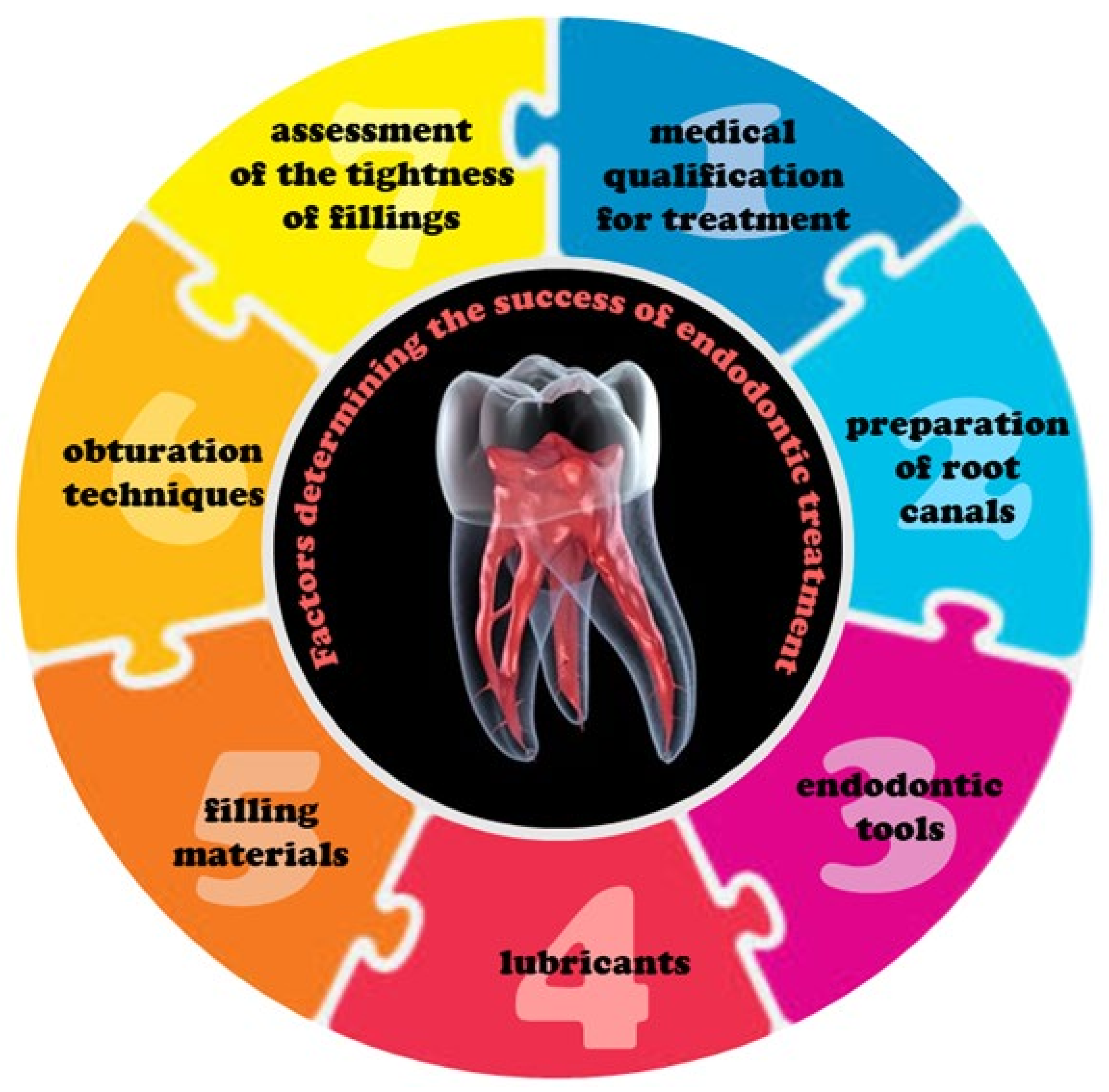
5. Goals, Expectations and Tooth Qualification for Root Canals Preparation

For thousands of years, mankind has had to deal with disease and toothache, which dates almost from the beginning of the mastery of the cultivation of cereals in the course of progress for civilization. The only radical solution at that time was the removal of diseased teeth. The situation did not change until the 19th century. Although already at the beginning of the 17th century, anesthesia with clove oil, and devitalization with arsenic trioxide or cauterization with a heated element were made, the breakthrough was not until the action of E. Maynard in 1836 [88]. He was the first in history to use a tool to remove the pulp from the inside of a tooth and since then endodontic treatment has been developed. From this time an alternative to the extraction of teeth affected by caries and the inevitable implant and prosthetic treatment at that time is an endodontic treatment to keep the tooth in the oral cavity in a functional state when it is impossible to keep it alive. Under ethical and pragmatic reasons, dentists prefer now to undertake such treatment rather than tooth extraction [136,137]. When attacked by caries bacteria, dental pulp causes pain, especially in the maxillary and mandibular incisors of the first molars, which are most affected [138]. It applies to cases when caries is advanced above the second stage according to the ICDAS. It is too late for any preventive measures, so endodontic treatment is undertaken [139,140,141] to eliminate or at least reduce apical periodontitis [142]. It prevents the spread of caries bacteria with possible toxins [143,144,145], including saliva [146], towards the root apex. Factors determining the systematically growing importance of endodontics include the global growth of the elderly population [147], the need to treat numerous groups of people with low incomes and inadequate prevention of oral diseases [148], a general systematic increase in awareness of oral hygiene and the need for treating oral diseases in higher-income societies [149], as well as increasing government support in many countries in this area, an increasing number of dentists and dental clinics in many countries and increasing demand for dental services, including due to motivations resulting not so much from health as from aesthetics [149]. However, in many countries, especially low-income countries, the tendency is the opposite [127]. The global endodontics market is forecast to grow from 1.7 billion USD in 2020 to 2.1 billion USD by 2026, with a compound annual growth rate CAGR of 4.1% [148]. The global dental consumables market, which to some extent relates to endodontics, will receive 55.6 billion USD in 2023 with a CAGR of 5.2% in 2017 to 2023 [149].
As part of endodontic treatment, the tooth pulp complex should be removed, which is a continuum ranging from occlusal at the pulp horns to the apical foramina [150], after prior preparation of the root canal and shaping it into an appropriate shape, which, after disinfection, will ensure the required encapsulation of the root canal by filling it with material replacing living tissues [151]. Qualification for endodontic treatment requires identifying all root canal orifices in the pulp chamber, using the right anatomy of the pulp chamber floor, after obtaining full access and visualization of the pulp chamber floor and its all-around contact with the walls [152]. Initial analysis and diagnostic evaluation of the teeth require removing the pulp chamber roof, identifying the pulp chamber opening and the bottom of the root canal opening, and root canal instrumentation.
Figure 8 presents the principles of the process of cleaning and shaping the pulp complex and the associated identification of the coronal part of the system, the pulp chamber, and the root pulp. The position of the pulp chamber at the centre of the tooth at the level of the cementoenamel junction CEJ is determined by the law of centrality [152], requiring a wide application of available medical imaging methods using a cone-beam computer tomography CBCT to trace the path of the entire root canal all the way to the apex. The law of concentricity [152] is alternatively used to locate CEJ, thus establishing the tooth’s angulation and a faciolingual direction. Measurements of the distance between the furcation and the cusp tip as well as the cusp tip to pulpal floor distance CPFD made on the radiograph and the CBCT method and the appropriate direction of the drill allow to prevent the chamber perforation in the furcation [153], regardless of whether the hole is triangular, trapezoidal, or even irregular [152]. Complete removal of the pulp chamber roof [154] makes it possible to search for canal openings in a given tooth, which is necessary to determine to prevent iatrogenic damage. Law of the colour chamber [152] indicates that access is full when the shade difference between the darker pulp chamber floor occurs throughout the entire range around it at the junction with the lighter walls. The law of symmetry indicates that the orifices of the canals are equidistant from a line drawn in a mesial-distal direction through the pulp-chamber floor, except for maxillary molars. Based on the laws of orifices location, it is possible to determine the position of the canal orifices by drawing a mesial-distal line through the centre of the pulp chamber floor. The law of floor anatomy indicates the number of channel openings.

6. The Importance of the Endodontic Tools and Lubricants Selection

In the root canal preparation phase, the correct selection of endodontic tools is extremely important. The issue has been discussed in detail in the authors’ own works [88,89] and is presented in Figure 4a. The best effects of removing the root canal content, its disinfection, shaping it the required shape, and the best encapsulation with filling material is ensured by the removal of dentin by laser or ultrasound techniques [155,156,157,158,159] and very often by classical methods of mechanical cutting of dentin using hand or rotary tools. It is required to use the most suitable rinsing fluids and lubricants. The lavage fluid most commonly used in endodontic treatment is sodium hypochlorite at a concentration of 0.5 to 5.25% [160,161,162,163], which has strong bactericidal properties and dissolves organic debris. Irrigation reduces the microhardness of the root canal dentine [162], less with sodium hypochlorite and more severe with sodium edetate or Ethylenediaminetetraacetic acid disodium salt dihydrate [164]. Each tool inserted into a root canal requires lubricant to lubricate the tool [165] and prevent jamming and breaking. Lubricants contain glycerin and either sodium edetate or urea peroxide, always accompanied by sodium hypochlorite solution [166,167].
As previously indicated, the important factors determining the success of endodontic treatment include the appropriate selection of tools for the preparation of the root canal. This selection is made each time by the dentists, based on their professional experience. It is not their task to analyze the details of the selection of the chemical composition, as well as the processes of production and processing of materials. However, on the other hand, the complete ignorance of the fact that such problems exist at all will most certainly lead to a huge number of errors in the purchase of these tools and, consequently, to the many, in most cases, unjustified iatrogenic errors. For obvious reasons, all details regarding the selection of the type and technology of materials for endodontic instruments must be decided by material engineers and instrument manufacturers. In turn, dentists need to be sure that these tools are actually the best they can be, and dentists need to be provided with a full body of true, comprehensive and reliable information to prevent them from making mistakes in this regard. The fact is that the commonly used tools made of steel, in many situations, especially in the development of curved root canals, show significantly worse functional properties than those made of the nitinol alloy.
According to dentists’ experience [168], when preparing root canals, it is preferable to use the crown-down technique [160,169]. Hand and rotary tools made of corrosion-resistant steel are often used. However, the risk of breaking them inside the root canal is significant [169], as they do not show the expected elastic properties. Tools for manual or rotary preparation [169,170,171,172] of a nickel-titanium alloy [173,174,175] known as nitinol (abbreviation made from words Nickel Titanium—Naval Ordnance Laboratory means the chemical alloy composition and place where the alloy was obtained) [176,177,178]. Nitinol endodontic instruments can provide a natural course and maintain a stable working length of the root canal while minimizing the removal of dentin from the root canal [179]. Due to the shape memory effect and superelasticity, nitinol is characterized by high fatigue strength [168,173,174,175,180,181,182,183,184,185,186,187,188,189,190,191,192,193,194,195,196,197,198,199,200,201,202] and mechanical resistance to fractures [174]. These features are issues very far from the interests of dentists and far beyond their influence. However, incorrectly ordered tools can greatly limit the possibilities of their application in clinical settings. The significance of the chemical composition of nitinol alloys was analyzed [203,204], and the requirements in this regard were given in the standard [205]. In [206] it was stated that relatively small changes in the mass concentration of nickel within the range of approximately 48–51% significantly determine the performance properties of the alloy nitinol, and therefore also including endodontic tools.
The manufacturing method, especially vacuum smelting [173,176,194,197,199,207,208,209,210,211,212,213,214,215,216,217,218,219,220,221,222,223,224,225,226,227,228,229,230,231,232,233,234,235,236,237,238,239,240,241,242,243,244,245,246,247,248,249,250,251,252,253,254,255,256,257,258,259,260,261,262,263,264,265,266], reduction of the share of non-metallic Ti4Ni2Ox [210,253,267,268,269,270,271,272,273,274,275,276,277,278,279,280,281], heat treatment conditions [282,283] and technological processes affecting the grain refinement of the austenitic matrix [284,285], have a significant impact on the properties of the nitinol alloy, processes of the dispersive precipitation of Ni4Ti3 in austenite B2 [286,287,288] and its martensitic transformation into phases with the B19′ or R structure, respectively [288,289]. Coherent or semi-coherent boundaries of the Ti3Ni4 precipitates with the B2 matrix [290,291,292,293] determine the course of the direct B2 → B19′ transformation [294,295,296] when the lack or reduction of these stresses causes the martensitic transformation according to the scheme B2 → R [283,297] during cooling [294,295,296,298,299]. Significant stress field generated by the precipitation processes of Ti3Ni4 provides a two-variant course of direct or two-stage transformation [291,292]. According to the scheme B2 → R [300] described in many works [207,300,301,302,303,304], martensitic transformation usually results from the previous ageing associated with separating Ti3Ni4 phase particles in the B2 matrix. Coarse-grained precipitates of Ti3Ni4, their growth and coagulation, and the lack of coherence, on the other hand, favour the direct transformation B2 → B19′ [305,306,307]. The course of the B2 → R transformation, in turn, provides important application possibilities, including increased fatigue strength [209,308,309], considerable stability with cyclic changes in temperature and loads [209,310,311,312,313], quick reaction to temperature changes [302,308,311,314,315,316] and a narrow hysteresis loop [302,317]. For the desired activation of B2 → R transformation, annealing after plastic deformation [318,319,320], ageing of alloys [28,283,298,303,321,322,323,324], as well as cyclic temperature changes [310,312,325,326] and stress [327,328] are appropriate. Increasing supercooling and cooling rate during annealing [298] is favourable for direct B2 → B19′ transformation and two-stage B2 → R → B19′ transformation [297,298,299,319,329,330,331,332,333], which occurs only in polycrystals [298]. The differentiation of nickel concentration at the boundaries and inside the grains of Ni-Ti alloys as a result of the Ti3Ni4 phase separation and in a privileged way at the B2 phase boundaries causes a parallel course in the zone adjacent to the grain boundaries of the two-stage transformation B2 → R → B19′ [298] and direct transformation B2 → B19′ inside the grains with a relatively higher concentration of nickel [298]. The normal course of the single-stage B2 → B19′ transformation corresponds to the conditions of homogeneous precipitation in the entire volume of B2 austenite grains, Ti3Ni4 particles during aging, which takes place with even a slight excess of nickel concentration above equilibrium in the alloy and with a longer ageing time, favouring the growth and coagulation of precipitates and lack of differentiation of nickel concentration in different zones of B2 austenite grains [298].
Like all other nitinol endodontic instruments [334,335,336,337,338,339,340,341,342,343,344,345,346,347,348,349,350,351,352,353,354,355,356], they require sterilization to meet sanitary and epidemiological requirements [334,335,336,337], as described in more detail in the authors’ own work [207]. Sterilization clearly showed a significant decrease in cutting ability and efficiency for nitinol instruments [334,338,339,342], unlike conventional stainless-steel instruments [341,342]. As a measure of the influence of sterilization on the properties of endodontic instruments, the cutting efficiency was taken as the ratio of the cut weight of the PMMA plate with the simulated curved root canal, with the accuracy of 3 × 10⁻⁵ g, related to the unit of energy consumed during cutting in Mg/J [340,343]. It is a significant decrease in this effectiveness when testing nitinol rotary tools under the influence of repeated 14 or 7 sterilization cycles compared to non-sterilized tools [334]. Then, there is a significant increase in the share of titanium oxide on the tool surface [334]. Treatment by chemical disinfection with sodium hypochlorite NaOCl did not significantly affect the cutting performance of endodontic instruments [340].

7. The Modern Obturation Techniques in Endodontics

In addition to the method of root canal preparation described above, the success of endodontic treatment is significantly influenced by other factors, including the method of obturation and the filling materials with the necessary sealants. In the authors’ own work [88] the generalization of, among others, in the field of obturation techniques, illustrated in the form of an umbrella (Figure 9a). Obturation techniques have evolved over the years and are essential elements in the success of endodontic treatment. Modern obturation techniques usually require specialized instrumentation. In the authors’ own work [89], a virtual analysis was also made in this respect (Figure 5b). Among the root canal filling methods, only some deserve attention, and practically only two of them can refer to resilon, i.e., the cold side condensation technique and the Thermo-Hydraulic-Condensation THC technique. Both of these techniques were developed for gutta-percha filling material, were connected with the development of this filling material [88] and found that they can be easily adapted to resilon. In the cold lateral condensation method, the main cone is initially fitted with an apical size corresponding to the size of the root canal preparation tool. Then, the canal is supplemented with smaller studs using a manual spreader [357]. The advantage of this technique is that the material is pushed past the apex less than the other, especially thermoplastic obturation techniques [358]. During condensation, however, the root canal’s working length may be exceeded by the expander, in the case of incorrectly selected tools [359].
Beginning in 1976, practical attempts were made to apply plasticized gutta-percha to the root canal [360], which turned out to be attractive due to the greater chance of three-dimensional obturation of the root canal. Not all of these methods were used in the case of resilon because it was possible to reach for the most advanced Elements Obturation Unit immediately (SybronEndo) system, combining two devices, i.e., System B and Obtura, equipped with a cauter and allowing the application of the material with a micromotor tip. The root canal filling technique using this system is derived from Buchanan [361] and popularized as the Continuous Wave of Condensation Obturation Technique or CWT for short [362]. The scheme of the method is presented in Figure 9b–k, and it is described in detail in the authors’ own work [88], where the importance of sealants is also emphasized.
The THC method is applied in a similar way to resilon. When filling the canal, small portions of liquid filling material should be introduced, done in stages, and condensed each time, as in the case of gutta-percha [363]. The use of System B and Obtura as part of the Thermo-Hydraulic Condensation technique THC [364,365] is one of the best methods of filling root canals with both gutta-percha and resilon materials, allowing the use of sealant to be absolutely minimized [366], which may be additionally supported by hot water supplies pluggers into the vibration tip of the DownPak. The results of the own virtual analysis [89] and the conducted literature review justify the experimental studies in this paper to compare the variant of root canal obturation with the Thermo-Hydraulic-Condensation THC technique with the use of System B and Obtura with the standard cold lateral condensation method. The use of the THC technique to resilon as filling material is, therefore, one of this paper’s main goals.

8. The Filling Materials Selection and Monoblock Idea Reality

Numerous requirements are placed on the filling materials, including biocompatibility, bactericide or at least prevention of temperature increase, sterility, good adaptability to the geometric features of the root canal, removable for endodontic treatment renewal [367,368], no tooth discoloration [368,369,370], the ability to strengthen the strength of the prepared tooth root [371]. The material based on gutta-percha is recognized and most commonly used for filling root canals [45,372,373,374,375,376]. Therefore, it is not surprising that any possible new solutions are compared and even confronted with this filling material. This material used in endodontics most often contains 18–22% of the pure form of β gutta-percha, 59–75% of zinc oxide, 1.1–31.2% of barium and strontium sulfate, as well as other polymers and wax in the proportion of 1–4.1% [37]. The general importance of this material among others used for this purpose was analyzed in [89], while in [88] extensive experimental studies of this material were performed, along with a SWOT analysis. Sealing materials are used, including custom ones [371,377,378,379]. Initially, the material was used on a matrix of plasticized gutta-percha using a heat conveyor, heated over the burner until it was red, and repeatedly inserted into the canal next to the gutta-percha cone, then withdrawn [136,137,138]. Then, the Touch and Heat electric heat conveyor was implemented, activated when the tip was inserted into the root canal, eliminating the need for a burner, but allowing heating to a temperature not higher than 350 °C [380,381,382]. The Thermo-Hydraulic-Condensation technique [365,383,384,385,386,387] is most often used, which was confirmed by the study results [88]. However, there have been alarming signs that in some cases, it is not properly bonded to dentin [388,389,390,391,392,393,394,395,396,397], which could warrant looking for other solutions, although dentist obstructive errors may only cause this.
An alternative to gutta-percha is a synthetic thermoplastic filler based on polyester materials [398,399,400,401,402,403,404]. This material is supplied as studs and pellets. This system includes [398,405,406] a self-etching primer containing a sulfonic acid-terminated functional monomer, HEMA hydroxyelylmethacrylate, water, and a polymerization initiator, a dual-curing resin sealant containing approximately 70% calcium hydroxide, bismuth oxychloride, barium glass and silica, and a synthetic core thermoplastic filling material based on polyester materials, containing approx. 65% of fillers and also bismuth oxychloride, bioactive glass, and barium sulfate. This material gained the common trade name of resilon after implementing Resilon Research, LLC, Madison, Connecticut, the USA, in 2004 [399]. Epiphany sealant implemented by Sybron Dental Specialties, Orange, California, USA, is used. Analogous RealSeal materials from Pentron Clinical Technologies, Wallingford, Connecticut, USA, or AH-26/AH-plus from Dentsply Maillefer, Ballaigues, Switzerland, and Endorez from Ultradent Products, South Jordan, Utah, USA are also used. The advantages of this material include the possibility of dissolving in some solvents, such as chloroform, or thermal softening, which is of particular importance when endodontic retreatment is required. It is possible to apply obturation techniques to this material, including thermoplastic techniques developed for gutta-percha [167]. Reports of the advantage of this material over gutta-percha [399,407,408] and the implementation of new sealants [409] were promising. It has been argued that resilon with dentin forms a monoblock [410,411,412], which would indicate that it is the best filling material available, were it not for the fact that this monoblock concept was not valid [413].
The problem of the monoblock is discussed in more detail in [414]. This term means the formation of a solid, bonded, continuous dentin material from one root canal wall to another, usually mechanically forming a homogeneous unit with the root dentine [415], and was introduced to orthodontics in 1902 by Pierre Robin [416], and to endodontics by Franklin R. Tay [417,418]. Despite this, the author believes that due to the lack of the required tightness and insufficient ability of the filling materials used to strengthen the tooth roots, the credibility of this concept is still controversial and requires further research [418]. It indicates two prerequisites to be met: a strong connection of the monoblock components with the reinforcing material is needed. The modulus of elasticity of the monoblock and this material should have similar values. He classified monoblocks according to the number of interfaces between the bulk material core and bonding substrate and characterized three types of monoblocks (Figure 10).
A primary monoblock has a single interface between the material and the root canal wall. An example of such a material was Hydron, which is a 2-hydroxyethyl methacrylate (HEMA) containing root filling material produced in the 1970s by Hydron Technologies (Pompano Beach, FL, USA) [419]. As a modern version of the primary monoblock, the mineral trioxide aggregate MTA can be considered, strengthening the immature tooth roots, and consists mainly of Portland cement and bismuth oxide, which provides X-ray impermeability [420,421]. Examples include Bio-gutta and Polyethylene fibre pose core systems. The secondary monoblock has two interfaces, between the core material and cement and the other between cement and dentin. It is the case with gutta-percha fillers due to the presence of a sealant [422], although the modulus of elasticity is lower than that of dentin [423,424,425]. Conventional and resin-modified glass ionomer cement is also used as root canal sealants, adhering to dentin, but not to gutta-percha, and therefore does not provide a monoblock [426,427]. Thus, adhesives were introduced as substitutes for gutta-percha to meet the monoblock requirements [428]. Since resilon is applied with a methacrylate-based sealant to the self-etching root dentine with a primer, the two interfaces are therefore considered a secondary monoblock. The differentiation of the assessment of the effectiveness of the filling with resilon and its tightness is presented later. Still, it is undeniable that there is a strong shrinkage during polymerisation, which is the cause of the leak [418], contradicting the monoblock concept. A high C-factor makes it difficult to seal a resin-filled root canal due to its polymerization stress. The ability of resilon to strengthen the tooth roots is comparable to that of gutta-percha, which gives it no advantage. The contrary makes it inferior to it [371,408]. Tertiary monoblocks have a third interface between the abutment material and bonding substrate. The resin polymerisation causes gaps formed between the fibre post and the resin coating which is the tertiary interface but could dislodge the fibre post [429]. EndoRez is a resin-coated gutta-percha in which a self-etching adhesive Clearfil Liner Bond 2V is used, giving a good apical seal and high tensile bond to the dentin [430,431]. The polymerization of adhesive and thermoplastic properties of gutta-percha jointed with the adhesive is too thermoplastic. The gutta-percha could be stiffer as tertiary monoblock using ActiV GP where the gutta-percha cone surfaces are glazed with fillers of glass ionomer [430,432]. However, there are doubts as to whether this is a better solution than using conventional gutta-percha.
The general conclusions of the analysis made in [414] are consistent with the assessment in [413] where it was found that the monoblock concept is not true. Although this concept has established a stable position in the literature, as shown above, it poses a clinical challenge that seems impossible to achieve. It is possible to imagine a continuum of filling materials that could asymptotically approach the ideal state, but it is difficult or impossible to achieve in practice. Two almost opposite conditions of tightness of joints and compliance of the modulus of elasticity seem to be incompatible in reality by any of the previously known materials, even if they are layered gutta-percha composites with appropriate polymer sealants because it is the polymerization shrinkage that is an obstacle to the pursuit of the ideal. Resilon is not equal to these solutions in any case, so it is further away from the asymptote, although it makes a better monoblock than the MTA. The polymerization of the resin causes contraction stresses and, as a result, creates gaps in the walls of the root canal. It could become possible if the resin were non-shrink, which seems impossible, and therefore it becomes correct to say that the monoblock concept is not true as stated in work [413].
Due to polymerization shrinkage, resilon often showed leakage at the material boundary with the root canal walls [45,373]. There are numerous publications in the literature in which different properties of resilon and gutta-percha are compared. In the works [399,409,416,433,434,435,436,437,438,439,440,441,442,443,444,445,446,447,448,449,450,451,452,453,454,455,456,457,458,459,460,461,462,463,464,465], the advantage of resilon as a filling material in endodontics was clearly indicated in the direct comparison of various properties of this material and gutta-percha. The data is obviously not confirmed in the authors’ works [88,89,207] (Figure 4a), although this material obtains the highest possible strength properties. Many studies [371,377,408,437,438,442,466,467,468,469,470,471,472,473,474,475,476,477,478,479,480,481,482,483,484,485,486,487,488,489,490,491,492,493,494,495,496] also confirmed the thesis contradicting the advantage of resilon over gutta-percha in endodontic applications and indicated that gutta-percha is a better filling material. The authors of this paper confirmed in their work [88] the very good suitability of the gutta-percha-based material for use as a filling material in endodontics. In many studies [412,439,440,441,464,483,490,491,497,498,499,500,501,502,503,504,505,506,507,508,509,510,511,512,513,514,515,516,517,518,519,520,521,522,523,524,525] it was found that it does not matter which of these materials will be used, which is also not confirmed by the results of previous authors’ own works [89], taking into account the data contained in Figure 4a. In several papers [512,526,527] it was also stated that the technique of cold obturation [512,527] or hot obturation [528] is irrelevant for the comparison of the obtained properties and applicability of the two compared materials [528], which seems completely unlikely, considering the results of the work [89] (Figure 4b). It may, however, indicate serious methodological errors committed by the authors of these works [512,527,528]. Conflicting information and research results are reported in the literature also regarding the possibility of removing the filling if it is necessary to repeat endodontic procedures, as there may not be any differences between resilon and gutta-percha [464,490,491,492,493,523,524,525], gutta-percha may be advantageous [490,491,492,493,494,495,496] or on the contrary, resilon gains the upper hand [460,461,462,463,464,465].
Regardless of the fact that the works cited above come from different periods, they analyze other aspects of applicability and different functional and technological properties of both materials and use different assumptions and methodological possibilities, no doubt adopting various simplifications and making multiple mistakes methodical and interpretative, the issue is still highly debatable. The opinions presented, often presented as conclusions, are both ambiguous and questionable. The problem is of fundamental practical importance, and at the same time carries a huge moral responsibility, because everyday thousands of dentists around the world apply filling materials to hundreds of thousands of patients as part of endodontic treatment to remove the effects of teeth degradation caused by caries, so they should have complete and objective knowledge on this subject, and not rely on assumptions and information not fully verified. It indicates the purposefulness of dispelling the growing doubts. Therefore, the aim of this paper was to clarify the importance of resilon in endodontics, using the most appropriate imaging methods taken from materials engineering, using scanning electron microscopes and light stereoscopic microscopes, ensuring precise identification and measurement of the leaks between the dentin and the filling material.

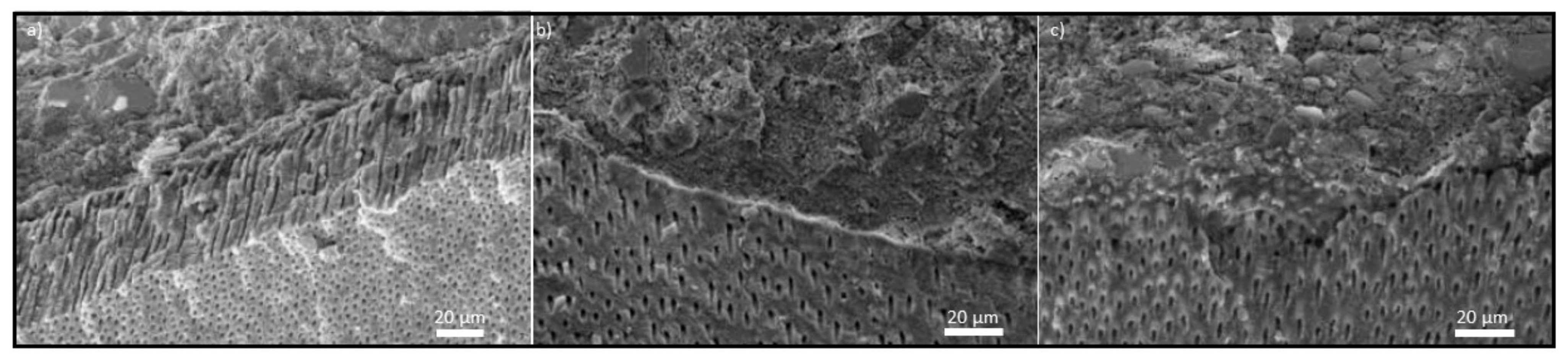
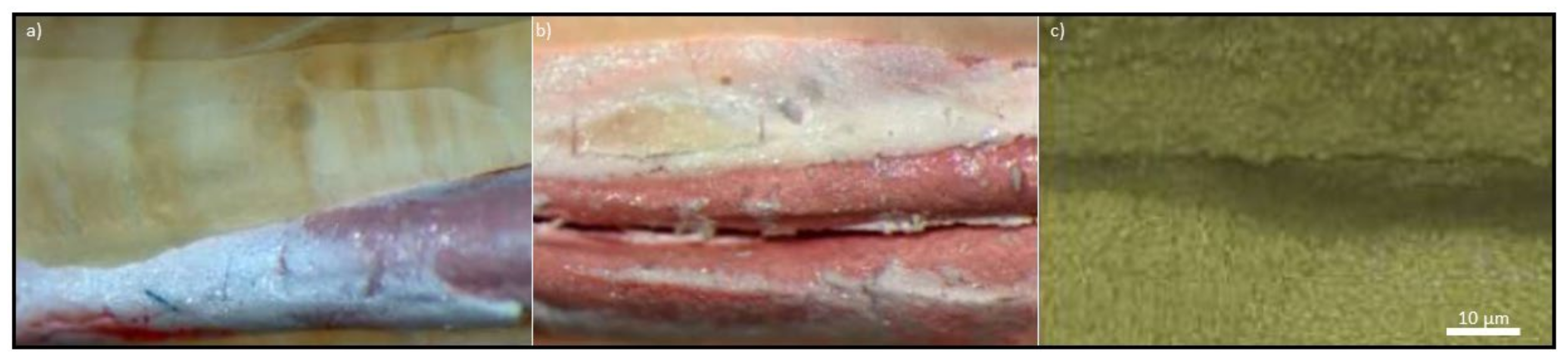
9. The Scope and Methodology of Experimental Work

In this paper, experimental studies were performed to determine the effect of obturation conditions on the tightness of the connection of the root canal filled with resilon using two obturation techniques. The results of virtual analysis contained in the authors’ own work [89] were used. Experimental studies were carried out in vitro on human teeth removed for orthodontic, prosthetic, and periodontal indications after obtaining the consent of the Bioethics Committee. The set includes maxillary and mandibular canines, as well as single-canal incisors in the maxilla and premolars in the maxilla and mandible. Each tooth was cleaned, rinsed in 0.9% NaCl solution, to cut off the crowns of the teeth at the level of the neck of each tooth using a diamond separator placed on the prosthetic handpiece. Then, the root canals were prepared using hand and rotary tools, including those made of nitinol and a lubricant containing glycerin, sodium edetate, and urea peroxide RC-Prep (Primer). Each insertion of the next instrument into the root canal is preceded by irrigation with 2.25% sodium hypochlorite solution and 0.9% saline solution. After careful selection and the rejection of teeth that did not meet the expectations or incorrectly prepared, 32 teeth were left for the study, from which two groups of 16 teeth were separated.
In both cases, after proper preparation of the root canal as described below for each group separately, the canal was filled with studs and Real Seal pellets (SybronEndo). The canals were thoroughly rinsed with 0.9% saline solution to neutralize the residual sodium hypochlorite and then dried with paper points. The main Real Seal pin (SybronEndo) was then selected with a taper of 4 and 2%, respectively, corresponding to the last Master Apical File MAF tool. When introduced to full working length, the cone would be wedged in the periapical area. The self-etching lubricant RC-Prep (Primer) included in the RealSeal kit (SybronEndo) was applied to the root canal walls using a paper filter. A thin layer of RealSeal (SybronEndo) was placed on the canal walls using a paper filter, and a previously selected pin was inserted into the canal.
Before filling the root canal with Real Seal studs and pellets (SybronEndo), they were prepared using manual ProTaper tools (Dentsply/Maillefer) in the case of the first group of teeth. In the first stage, the pulp chamber’s part was widened using the S1 tool, and then the SX tool with the highest 19% taper was introduced, and the canal length was measured with the Kerr file with ISO 10 number which 1 mm was subtracted. In the second stage, the canal was prepared with the S1 and S2 tools to the full working length, and the F1, F2, F3, and F4 tools were introduced to the full working length, widening, and smoothing the root canal walls with each canal recapitulation with the Keer tool. In this case, for obturation, using 4 and 2% tapers, respectively, a 4% tapered root was fitted to each canal, corresponding to the last MAF tool to prepare the canal apical. After applying the self-etching conditioner and placing a thin layer of sealant on the canal walls, a previously selected main stud was inserted into the canal. A plunger of 20 ISO size with a taper of 2% was chosen successively, and a length 1 mm shorter than the length over which the main stud was inserted was determined using a silicone stopper on the plunger. After a manual plunger prepared the site, the canal was refilled with RealSeal studs (SybronEndo) with a taper of 2% to the size of the plunger used. After completion of filling the canal, the mouth of the canal was irradiated with the light of a polymerization lamp for 40 s in order to seal the canal from the outlet side immediately.
In the case of the second group of teeth, before filling each root canal with Real Seal studs and pellets (SybronEndo), it was prepared with K3 rotary tools (SybroEndo) in two stages. In the first stage, the K3 tools with the symbols 10/25 and 08/25 with a taper of 10 and 8%, respectively, were used to develop the mouth of the canal. By inserting a 10 ISO Keer file and subtracting 1 mm from the full length, the working length of the root canal was measured as the average distance between the anatomical apex and the physiological narrowing of the root canal. For the preparation of the root canal to its full working length, the following tools were used successively in sizes 0.4/20; 0.4/25; 0.4/30; 0.4/35; 0.4/40, with each insertion of a new tool requiring recapitulation of the canal with the Keer tool. After complete root canal preparation, obturation was performed using the Thermo-Hydraulic-Condensation THC technique [403,404,405,406,407,408] using System B and Obtura III by introducing System B plugger heated to 150 °C through the RealSeal (SybronEndo) pin in the root canal up to the length marked with a stopper. After 3 s, the heating is turned off, and the pressure of the tool towards the root tip is maintained for 10 s. The plunger is then reheated to 300 °C with immediate removal of the plunger and excess non-condensed filling material. The apical portion of RealSeal (SybronEndo) is condensed with pulsating motions for the next 10 s using a cold Buchanan plugger (SybronEndo) selected in advance. The apical root canal filled as described is supplemented with RealSeal pellets (SybronEndo) fed into the canal with Obtura III (SybronEndo) at 160 °C.
In order to eliminate the methodological defects cited in many works, preventing objective assessment of the tightness of the filling and the absence of leaks, this paper uses for this purpose as basic materialographic tests using a scanning electron microscope and a light stereoscopic microscope, which methods were selected virtually in the authors’ own work. [89]. On the Stereo Discovery V12 stereo microscope with the AxioCam HRC digital camera by Zeiss, preliminary tests were performed, and the results were recorded using digital photography at 50× magnification. While digitally recording the results of basic tests, they were performed using a SUPRA 35 high-resolution scanning microscope by Zeiss with a WDS, EDS spectrometer and an EBSD TRIDENT XM4 camera from EDAX in the range of 2000–5000× magnification, and the test results were digitally archived. When assessing the structure after the performed obturation operations, attention was paid to the following elements:
  • connection of the filling material with the dentine of the root canal,
  • connection of the main stud with complementary material,
  • filling of the lateral tubules,
  • filling the root delta,
  • connection of the material with dentin tubules,
  • volume fraction of the sealant,
  • leakage on the border of the root canal wall and the filling material.
The mouths of the root canals of the teeth were selected for materialographic observations after obturation were secured with Ketac Molar glass ionomer cement (3M ESPE) and were stored for seven consecutive days at room temperature in a humid environment until the sealant was completely bound. Each tooth was wrapped in gauze soaked in physiological saline and sealed in polymer containers. The samples were cut longitudinally, along the root, to a depth of 1 mm using a diamond disc placed on a prosthetic handpiece. A longitudinal fracture was made after cooling in liquid nitrogen. After the breakthroughs were made, they were sputtered in a BAL—TEC SCD050 sputter by Oerlikon Balzers with a thin layer of gold as a conductive material. Alternatively, teeth filled with Real Seal (SybronEndo) were decalcified by immersion for 14 days in an aqueous solution of a mixture of 7% formic acid, 3% hydrochloric acid and 8% sodium citrate. After rinsing with running water, the samples were immersed in 99% acetic acid for 12 h and then rinsed again with distilled water. After that, the samples were held sequentially for 30 min each in an ethanol solution of successively increasing concentrations of 25, 50, 70, 90, 95 and 100%. The samples were stored in methyl salicylate before microscopic observations.
Using the capabilities of the scanning electron microscope, the measurements of the width of the slits were made. The results were statistically processed by calculating the mean and the confidence interval.
A SWOT point analysis was made (strengths, weaknesses, opportunities, threats), described, among others, by the authors’ own methodical work [89]. Five key factors relating to each factor in this analysis were defined, creating a list of factors with the most significant importance and the greatest impact on the development of resilon as a filling material, divided into four groups, assigning them weights reflecting their importance. Each of these factors was assessed with the use of the universal scale of relative states (Figure 2), assigning them appropriate weight. The sum of the products of the weight and the unit grade within each of the factors allows it to be quantified, which allows for the definition of a management strategy to the subject of analysis.

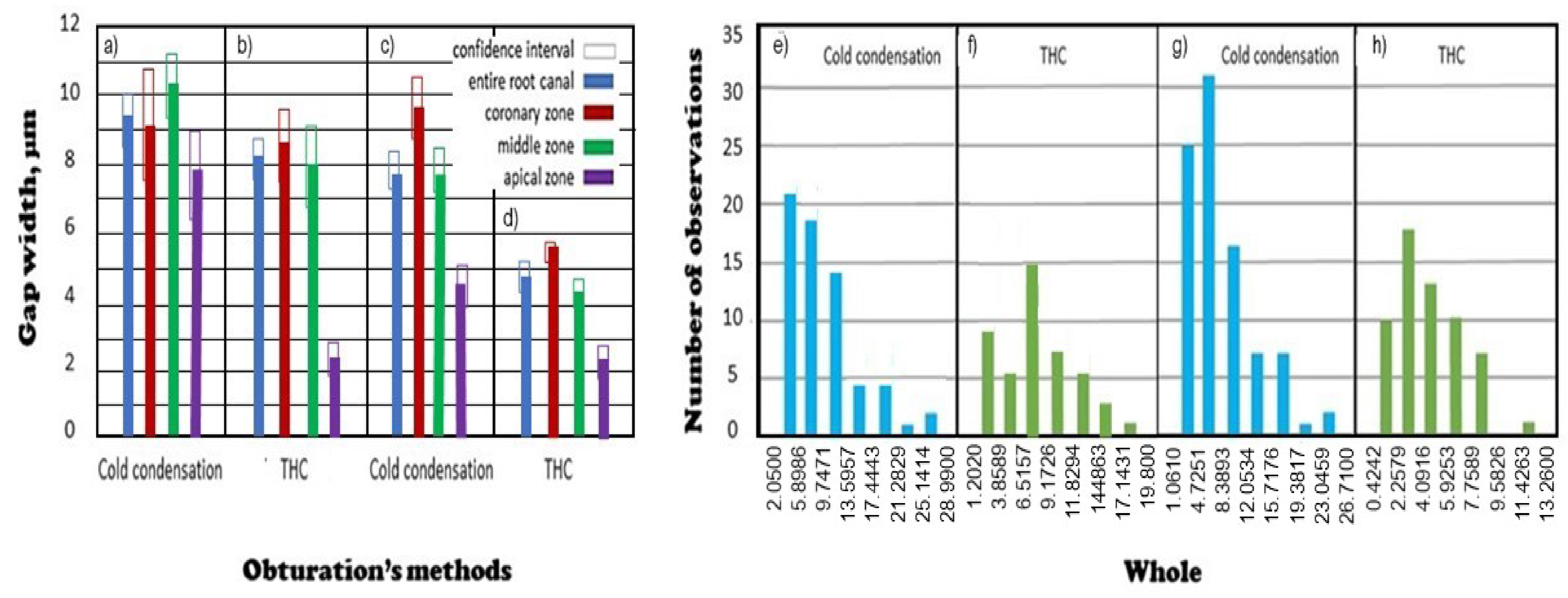
10. Materialographic Test Results of Teeth Filled with Resilon by Cold Lateral Condensation

As a result of the performed experimental studies, the selection of resilon as a filling material was assessed, combined with the choice of cold or thermoplastic obturation, the THC method, using an advanced methodology for assessing the effectiveness of root canal filling. Both the disinfection of the internal space and the hermetic three-dimensional hermetization are the criteria for the correctness of endodontic tooth treatment. The condition expected by the dentist undertaking endodontic treatment and the patient is the tightness of the connection of the filling material with the dentin of the root canal, i.e., a state in which the components of the filling adhere exactly to each other. The quality of the connection of the filling material with the dentine of the root canal depends on the tightness of the filling and the tightness of the root canal filling, and in practice on the low surface density of the leaks and their small size. In order to determine the influence of the chosen obturation method, the structure of the processed teeth was analyzed by the cold lateral coding method and then by applying the THC technique of thermoplastic condensation. Seven criteria were specified against which the research material was assessed in each of the studied cases.
The method of cold obturation of the root canal by the lateral condensation method with the use of resilon pins (RealSeal) and sealant (RealSeal) (Figure 11) ensures a gap-free connection of the material with the dentin of the root canal. The main stud or additional studs surrounded by a sealant may adhere to the root canal wall. The main cone is connected to the root canal dentin by a relatively thick (Figure 11b) or thin intermediate layer of sealant (Figure 11c). The close connection of the root dentine with the filling material occurs in the apical section (Figure 11). The filling material adheres closely to the border of the root canal wall (Figure 12), which confirms the correct connection of dentin and replacement material.
The quality of endodontic treatment of the tooth depends on the quality of the root canal filling and the quality of the connection of the main point with the complementary material, as one of the important factors. Both the technology of root canal preparation and obturation and the type of sealing materials used have an impact on this. The connection of the main stud made of resilon with a 4% conicity with additional studs with a 2% conicity was analyzed (Figure 13). The purpose of connecting the studs with each other is to create a uniform structure that fills the canal space of any shape (Figure 12a). The studs are chemically connected with each other by means of a sealant and mechanically due to condensation, i.e., lateral pressure of the plunger to the studs placed in the root canal. The correct connection of the central stud with additional studs was found, which guarantees the formation of a uniform filling encapsulating the root canal of any shape (Figure 13a). More often, however, we can find incorrect adhesion of resilon studs, which led to leakage on the edge of the stud (Figure 13b) or on the border of the filling material and root canal dentin (Figure 13c).
Tight three-dimensional obturation of the root canal and filling the main canal space also includes filling the spaces constituting a branch of the central canal, including lateral canals, and in the periapical area, also a complex system of canals with an irregular structure called the root delta. With this structure, it is impossible to fit a suitable stud since such a space can only be filled by the plasticized material forced into it under the appropriate pressure generated by the condensation of the filling material. The material filling the root delta can be resilon, centripetal sealant, and centrifugal resilon, or a sealant alone.
In the case of cold lateral condensation, a gap-free connection of the filling material with the dentine of the root canal occurs (Figure 14). The close adherence of resilon to the mouth of the dentinal tubules proves that their mouth is blocked (Figure 14c). Right next to the material-dentin boundary, obtured oval openings are visible, constituting a cross-section through the dentinal tubule. In the case of cold obturation methods, it is most likely to fill the lateral tubules with sealant, due to its lower density compared to the stud. However, the intense condensation of the studs also allows for the effect of filling the lateral tubules with resilon. The filling of the lateral tubules in the cold lateral condensation method proves that the resilon studs are pressed very well against the root canal wall and that a non-plasticized stud is forced into the main canal branch. However, no movement of the sealing material along the depth of the dentinal tubules was found. The results of these own studies only partially correspond to the observations of other works, regardless of the choice of the filling material [363,528], because in the cold side condensation technique at least 60% of the side channels are filled only with sealant.
Tight obturation of the root canal requires the use of a sealant in addition to the filling material (Figure 15). The root canal sealant is used to interconnect the studs and connect the studs to the root canal wall. In the cold obturation technique, the proportion of sealant tightly covering the filling material is relatively large.
The even distribution of the sealant on the resilon studs ensures a gap-free connection of the material with the root canal wall (Figure 14a,b), while the lack of the sealant causes the main cone to not stick to the root canal dentin (Figure 14a). Insufficient volume of sealant between the main cone and the auxiliary cones also results in a lack of bonding of the material (Figure 14b).
As a result of the examination in the scanning electron microscope, the presence of a thick layer of sealant tightly connecting the dentine of the root canal with the filling material was confirmed (Figure 16a,b). The presence of a gapless connection of the resilon stud, evenly covered with sealant, with the root canal wall was confirmed (Figure 16c).
The connection between the root canal wall and the filling material does not occur in all cases and is not always tight or gap-free. However, it is the tightness of the root canal filling that determines the quality of endodontic treatment. A leak is a stratification of two boundaries that are to adhere closely to each other. Despite the presence of three layers on the border of the tested samples, better adhesion of two layers was found compared to the third one. The delamination of the thick layer of sealant, adhering to the root canal wall on one side, separates the filling material from the dentine of the root canal (Figure 17).
Leaks between the root dentine and the central stud occur in places where it is not covered with a thick layer of sealant (Figure 18), on the border of the filling material and root canal dentin, where there is no tight connection between the sealant and resilon. There is no leakage (Figure 18a,b). Leaks at the root dentine and filling material border occur when the main stud is covered with both a thin (Figure 18c) and a thicker layer of sealant (Figure 18a).
Measurements on the border of the sealant and the dentine of the root canal indicate the formation of gaps with a width of 9.191, 9.368 and 111.64 µm, respectively (Figure 19a–c). Leaks on the root dentine and filling material border also occur when the resilon studs are covered with a relatively thinner layer of sealant (Figure 18a), where the gap width is 3.182 µm.
Leaks appear not only on the border of the root canal wall and the filling material, but also between the elements of the filling material. In the cold side condensation method, the boundary of the filling material is determined by two types of leakage. It is possible to unseal at the border of the studs and at the border of the studs and the sealant. The gaps formed on the border of the main cone and complementary cones were identified, in places where the share of the sealant is insufficient for the joining of two structural components (Figure 17c). Although in the test material filled with resilon studs, the sealant shows a greater tendency to bond with the filling material than with the root dentine, unsealing was found on the border of the sealant and resilon covered with sealant (Figure 18b,c).
Overall, 65 fractures were identified, of which 32 were recorded in the middle segment. The comparative results of the statistical analysis are presented in Figure 20, separately analyzing all three sections of the root canals: near-crown, middle and apical. Along the entire length of the root canal, the average value of the fissure width in the case of resilon obturation by cold lateral condensation is 9.44 ± 5.76 µm. The obtained values differ for the individual sections: near-crown, middle and apical sections of the completed longitudinal fracture. The average value of the fissure width along the entire length of the near-crown area is 9.24 ± 6.08 µm, along the whole length of the middle section it is 10.30 ± 5.87 µm, and along the entire length of the apical section, it is the smallest and amounts to 7.85 ± 4.84 µm.
The results of these calculations are presented in Figure 20, comparing them with the results obtained for root canals filled with resilon by the method of thermoplastic condensation using the Thermo-Hydraulic-Condensation technique and the results obtained in [88] in relation to the material based on gutta-percha.
The Results of Materialographic Tests of Teeth Filled with Resilon by the Thermoplastic Condensation Method Using the Thermo-Hydraulic-Condensation Technique
The method of thermoplastic condensation of resilon using the Thermo-Hydraulic-Condensation THC technique, which uses System B and Obtura III devices, is also characterized by the presence of a tight connection of the material with the root canal wall in each of its sections (Figure 21).
The chance of minimizing polymerization shrinkage increases, because the application of the material to the root canal is divided into three or four portions, between which there is vertical condensation of the material during its cooling. It ensures that the root canal sealant adheres to the root dentine (Figure 22). After plasticizing, cutting and condensing, the fitted central stud is connected with the extragranular plasticized resilon. For example, in the case of joining two differently applied portions of resilon, correct vertical condensation with a cold plunger takes place, as a result of which the materials are pressed against each other and polymerization shrinkage is reduced, there are no leaks on the border of the filling material and the root canal wall (Figure 21c).
Tight, three-dimensional obturation of the root canal consists in filling the main canal space and the spaces constituting a branch of the central canal (Figure 23). The thermoplastic THC method with resilon causes tight, three-dimensional obturation of the root canal. It fills the main canal space, including filling the spaces constituting a branch of the central canal, including lateral canals (Figure 23a), and in the periapical area, also a complex system of canals with an irregular structure called the root delta (Figure 23b). In this case, the resilon can be filling centrally the root delta, and the sealant centrifugally from the main canal (Figure 23b).
Tight obturation of the root canal, apart from filling material, requires a root canal sealant to connect the studs and the studs with the root canal wall. Undoubtedly, the sealant used in this case has a very limited ability to penetrate deep into the dentinal tubules due to its structure. However, this penetration occurs due to thermoplastic obturation. It was indicated in the literature [480] that in this respect, the material based on gutta-percha shows slightly better properties than resilon, e.g., after 16 months of follow-up [438]. However, this penetration may be considered relatively shallow. The study results [529] seem to confirm it, although this information relates mainly to the apical section and to a lesser extent to the remaining sections of the root canal. As a result of obturation with the method of THC, in addition to filling the main canal space, the filling material also penetrates the dentinal tubules, which are the smallest anatomical branches in the root dentine, which proves the close combination of both components. In this case, the filling material adheres closely to the root canal wall, with the mouths of the dentinal tubules closed (Figure 24). Each time, the border between two layers of the root dentine and the sealant and the sealant tightly coated with resilon is shown, which can be treated as an example of a dentine monoblock, sealant and the main filling material (Figure 24), despite the justified criticism of this concept presented earlier in this paper.
The THC thermoplastic method is characterized by a relatively small volume share of a sealant covering a resilon by the very thin layer in the root canal, which is revealed in the form of a shiny coating covering the material (Figure 24a,b). A relatively thin layer of sealant was shown, bonding the filling material to the root canal wall (Figure 25b). There is a characteristic combination of three layers consisting of root canal dentin, sealant, and filling material. The results of these own studies correspond with the observations of other works, regardless of the choice of the filling material [363,528], because in the THC thermoplastic technique, even 83–90.62% of the filling is made with the filling material and the sealant. On the other hand, Figure 24c shows a relatively small thickness of the sealant layer covering the resilon. It can be ascertained thanks to several surfaces of the filling with an uncovered sealant.
Although there should be three layers at the boundary of the tested samples, better adhesion of the two layers was found compared to the third. Resilon covered with a thin layer of sealant also shows gaps, and the sealant sticks tightly to the resilon more often than to the dentin (Figure 26 and Figure 27).
The measurements of the width of these gaps are 7.070; 5.833 and 3.535 µm, respectively (Figure 27). Leaks appear not only on the border of the root canal wall and resilon, but also between the filling material itself. Although there is a connection of the cut-off cone with the complementary material, there is no leakage at the point of their connection, however, only leaks were found on the border between the filling material and the sealant. The sealant adheres better to resilon than to the root canal wall, and gaps are formed between the root dentine sealant and the resilon sealant (Figure 27a), and the gap width is 5.833 µm (Figure 25b).
The total number of fractures found was 45, of which only 4 were in the apical segment. The results of measurements of the width of all these gaps were statistically evaluated (Figure 20). The average value of the fissure width is 7.86 ± 4.07 µm along the entire length of the root canal subjected to resilon obturation using the THC method. Along the entire length of the mear-crown section, the average value of the fissure width is 8.70 ± 3.74 µm, the middle section is 7.96 ± 4.17 µm, and the apical section is 2.77 ± 0.84 µm. While there are no significant differences between most of the measurements concerning the applied obturation methods, the width of the gaps in the apical-stitched section after obturation with the thermoplastic method of THC is definitely smaller than after obturation by lateral to cold condensation. This entitles the general conclusion to be drawn that this same THC method is more favourable than the two. Comparing these results with the analogous method using a material based on gutta-percha [88] shows that the average value of the fissure width along the entire length of the root canal is 4.76 ± 2.55 µm. These values differ for individual sections: the near-crown section with an average fissure width of 5.74 ± 1.97 µm, the middle (4.88 ± 2.75 µm) and the apical (2.33 ± 1.41 µm) longitudinal fracture (Figure 20). The analysis of the significance level of differences between the width of the gaps in all analyzed sections for the entire root (Figure 20) shows that in all sections along the entire length, the differences between the THC method do not have significant values for resilon and the material based on gutta-percha, despite significantly higher mean values for resilon. Therefore, the research requires increasing the test frequency to demonstrate that the material based on gutta-percha provides better tightness of the THC filling.

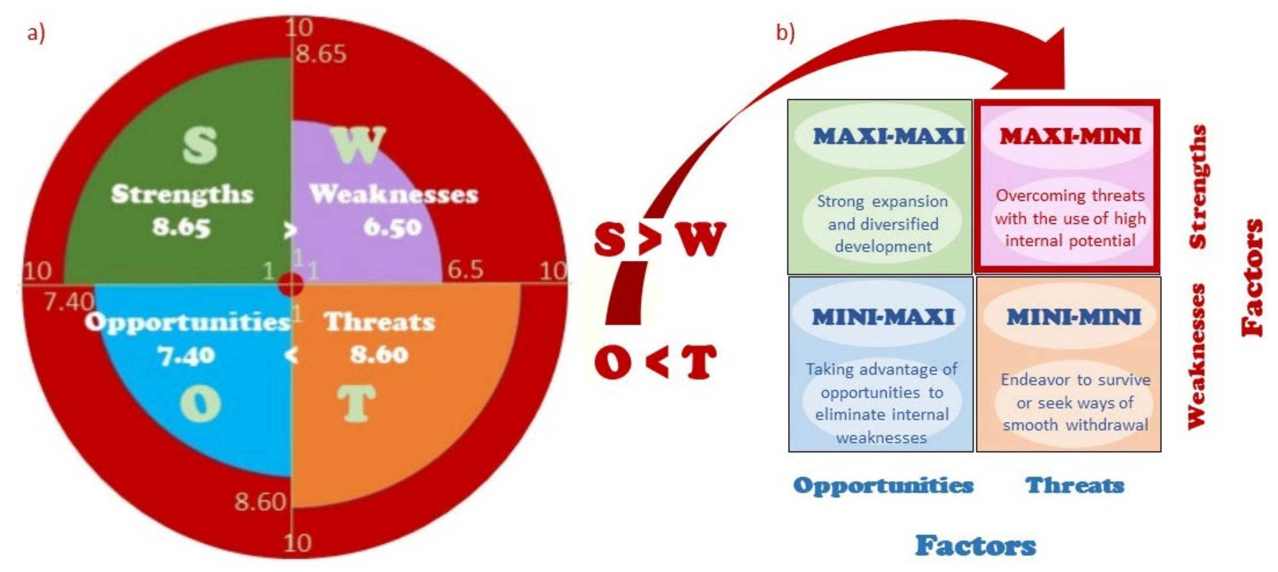
11. Strengths, Weaknesses, Opportunities, and Threats SWOT Analysis of the Resilon Application in Endodontics

In order to compare the strengths and weaknesses of the material based on polymeric polyester materials, known in circulation as resilon, as well as to determine the opportunities and threats that flow to it from the environment, a SWOT point analysis was performed. The analysis began with selecting five key internal factors with the greatest positive and negative impact (Figure 28) and external factors that have a decisive impact, positive and negative, on the future situation of resilon on the endodontic materials market. Factors were weighted and rated using the ten-point universal scale of relative states. According to this scale, the maximum value is 10 and the minimum value is 1 (Figure 3).
The most important strengths of the material based on polymeric polyester materials include excellent strength properties (S1), which the results of numerous scientific experiments have confirmed. The strength of resilon is also quite high (7 points) quality of the root canal filling (S2). However microscopic examinations indicate that it is possible to create leaks both on the edge of the studs and on the border of the filling material and the dentine of the root canal, which lowers the criterion assessment in this case. The advantage of resilon is also the complete lack of resorption over time (S3), assessed at 10 points. The ease of removal of resilon from the root canal in the case of revision (S4) was assessed quite high (7 points), by dissolving it in a solvent, such as chloroform or thermal softening, which is crucial if it is necessary to endodontic re-treatment. In the time of the COVID-19 pandemic, the possibility of sterilization (S5) became of particular importance, hence this feature was included among the most important strengths of the material based on polymeric polyester materials, awarding 10 points in this criterion.
The weaknesses of resilon (Figure 28), which were given the greatest importance, include its niche (W3), which should be understood as the lack of widespread availability and popularity in specialized endodontic clinics, which is dictated on the one hand by the manufacturer’s strategy for products distribution, and on the other hand by the moderate knowledge on this material by endodontists who, therefore, use it to a limited extent. An element that contributes to this is also the price of resilon (W4), which is higher than the price of standard gutta-percha. Another diagnosed weakness, very important from the technological point of view, is the polymerization shrinkage (W5), which causes leakage during the polymerization process at the border of the filling material and the root canal wall. Another identified disadvantage of the analyzed material is toxicity (W1), which was assessed as low (3 points) compared to materials based on zinc oxide with eugenol, for example. The weakness is also the lack of bactericidal activity (W2). At the same time, there are obstructive fillings, such as materials based on zinc oxide with eugenol, synthetic resins, or calcium hydroxide, which have a beneficial bactericidal effect, facilitating and accelerating the treatment process.
Among the external positive factors, the greatest chance was considered to break the oligopoly of producers (O1). At present, there are only a few entities that offer resilon on the market. Such a situation is not beneficial for the product recipients, as it is the producers who dictate both prices and terms of sale. De-oligopolization would certainly contribute to a significant development of the material based on polymeric polyester materials, by balancing the balance between the producer and the recipient. Another factor contributing to the development of resilon would be disseminating the THC (O2) thermoplastic condensation method. This technique provides better fill efficiency than the cold side condensation method, especially in minimizing the width of gaps in the apical section.
The effect of scale and the accompanying cost reduction of this obturation technique would reduce the overall cost of endodontic treatment with resilon. The quality of fillings should, in turn, be influenced by the improvement of the structure of the sealant (O3) in terms of the ability to penetrate deep into the dentinal tubules, as well as the improvement of the method of applying resilon (O5) to the root canal in terms of minimizing polymerization shrinkage, which may cause leakage at the boundary of the filling material with the walls of the root canal, which, however, seems unlikely at present. This fact is reflected in the assessment. Opportunities brought by the environment are also promotional activities (O4), consisting in disseminating among denetists and patients’ information about the benefits of resilon, using both traditional methods, such as fairs, conferences, seminars, scientific publications, and modern ones—using electronic media.
The most important factors that will hinder the development of resilon in the market of endodontic filling materials in the future include the dominant market position of gutta-percha (T1). This material, which is popular, available, and common, both in terms of specialist dentists’ attachment to it and numerous distribution channels, is still considered the gold standard [88]. The chance that this situation will change soon is slim.
The development of a material based on resilon would also not be favoured by the strengthening of the oligopoly of producers (T4), tantamount to further weakening the position of recipients, which is not conducive to building a positive image, increasing popularity and wide dissemination of the product. Over time, too drastic protection by oligopolists of the product will inevitably lead to unfair actions of the competition (T5), manifested by the appearance of counterfeits of the original product on the market, which will not only take part of the market from the oligopolists, but also lower the standard of the basic product in the eyes of the recipients, because the counterfeit always it is “more or less the same”, only inferior in quality. In the context of the intensive development of material engineering, including nanotechnology, the invention of completely new material for obturation of root canals (T2) is not excluded. Therefore, this factor was considered a significant threat. The development of resilon will also not be favored by the development and cost reduction of cold obturation (T3), because this method does not provide adequate tightness as a result of the incorrect connection of the central stud with additional studs, or failure to fill the side canals and root delta.
The next step of the SWOT analysis consisted in performing a multi-criteria analysis, which resulted in the award of four scores expressing strengths numerically (8.65) and weaknesses (6.80) of a material based on polymeric polyester materials, as well as opportunities (7.40) and threats (8.60) that flow from the environment (Figure 29). The strengths of resilon significantly exceed the identified weaknesses, while the threats are greater than the opportunities. Therefore, an adequate strategy for the development of resilon is a conservative strategy known as MAXI-MINI. This strategy assumes the use of the high potential of the material, characterized by excellent mechanical strength and fairly high quality of fillings, as well as the possibility of sterilization, no resorption over time and the possibility of removing from the root canal in the event of a need for revision and endodontic re-treatment. At the same time, it is necessary to overcome the threats posed by the environment concerning the market position, competitors’ activities, and the development of correlated methods and techniques while taking advantage of the emerging opportunities, of which many may also happen. High values of both threats and opportunities indicate that the projected material development is subject to risk, and thus a sudden return is possible, bringing both unexpected failures and unexpected successes.

12. Summary

Oral diseases are an extremely serious global problem. Estimates show that 3–5 billion people suffer from tooth caries [37,134,135]. Tooth caries and toothlessness, which is the natural consequence of caries, as well as periodontal diseases that often accompany them, are the cause of various systemic complications, and therefore have a significant impact on health insurance systems around the world whilst also putting pressure on health care and medical treatment systems in all countries of the world. Caries absolutely requires treatment on a global scale, and health threats in this area should be removed in a manner analogous to a pandemic of any other infectious disease. There is controversy about the importance and scope of oral disease prevention. Still, the notion that interventionist dentistry should be abandoned, promoted by the prestigious Lancet [134,135], clearly does not stand up to criticism, even in countries with the best-organized healthcare systems and the highest possible spending for health protection per capita, for which data is given in detail in other studies by the author [37,38]. An acceptable alternative to this hypothetical and utopian proposal is the original author’s concept of the Dentistry Sustainable Development (DSD) [37] and the related idea of Dentistry 4.0 [34,35], which is part of the current stage of the industrial revolution Industry 4.0 [1,2,3,4,5,6,7,8,9,10,11]. The adopted assumptions include Global Dental Prevention (GDP), Advanced Interventionist Dentistry 4.0 and Dentistry Safety System (DSS) complementary models [37].
An alternative to the extraction of teeth affected by caries and the inevitable toothlessness and the need for implantological and prosthetic treatment is endodontic treatment to keep the tooth in the oral cavity only functional when it is impossible to keep it fully alive. For ethical reasons and guided by pragmatism and the well-understood health interest of patients, dentists prefer such therapy instead of tooth extraction, which until relatively recently had no other alternative. This way, it is possible to preserve endodontically treated teeth for many years in the mouth of many patients. Such measures must be taken when caries are advanced beyond the second stage [139,140,141], when it is too late for any preventive measures. Due to many factors, including the ageing population and the increase in the number of patients significantly exceeding the limits of the working-age, systematic increases in awareness of oral hygiene and the importance of health protection in general, universal declarations by both the United Nations [91] and the European Union, and governments of many countries regarding increasing care for health (including oral health), the importance of endodontics is steadily growing. It is predicted that the global endodontics market will soon exceed the value of 2 billion USD with a compound annual growth rate CAGR not lower than 4% [148].
The principles of the process of cleaning and shaping the pulp complex have been widely popularized to such an extent that a huge number of endodontologists around the world can perform appropriate clinical procedures in a repeatable and successful manner. The method of developing the root canal with the selection of hand and rotary tools commonly available is of fundamental importance, and it is essential that they also include tools made of Ni-Ti alloys of the shape memory nitinol type and super-elastic [207], which have been shown to the number and probability of iatrogenic errors. These tools largely ensure a sufficiently large bend during the preparation of curved root canals without the risk of breaking and remaining at the bottom of the prepared canal. Contemporary methods of Thermo-Hydraulic-Condensation THC technique using System B and Obtura III [362,363] provide the greatest possible tightness of endodontic fillings. In terms of the methods of development and obturation of root canals, it seems that a sufficiently high level has been achieved, which is confirmed by the results of research and analyzes quoted and performed in this paper.
The most controversial issue seems to be selecting the filling material and the necessary sealants. On the one hand, as confirmed by the results of detailed research presented in the previous authors’ work [88] and supported by a virtual analysis [89], the most commonly and successfully used material is based on gutta-percha, considered the gold standard of endodontics. On the other hand, the undeniable fact is that the highest strength properties are represented by synthetic material based on polyester materials, most commonly known as resilon. In much of the literature, the advantages of the material based on gutta-percha is indicated, which is based on dentistss trust in the effectiveness of the treatment and the best possible tightness. On the other hand, many publications indicate that it is resilon that provides the best possible strength properties, as if ignoring the obvious drawbacks of this material, which undoubtedly include significant polymerization shrinkage resulting in a large number of leaks. Adaptation to resilon of the THC technique developed for materials based on gutta-percha, as well as the possibility of dividing the obturation process with this method into several stages cannot improve the situation. Instead, the adverse effect is mitigated. However neither this filling material nor the sealants used with it can provide evenat least partially filling of the smallest root tubules, e.g., in the region of the root delta. They can only be sealed at the interface with the central canal.
Therefore, there remains a problem, not only theoretical, but above all, of practical importance for every endodontist: which of these two important filling materials should be applied, considering the minimization of risk and the ethical responsibility for the health entrusted to the dentist by the patient. Of course, a philosophical answer could be that it is the third one, which synergistically combines the positive features of both materials mentioned above while eliminating the risks. There is no such material so far and the world undoubtedly faces a challenge to develop it. The signaled problems should also be accompanied by one more: the fact that there is a lot of residual information, signaling, groundlessly generalized, untrue, and even simply untrue information on this subject, also in specialist literature. One of the reasons is the lack of use of adequate and appropriate methods of assessing the effectiveness of endodontic treatment with the proper procedure for evaluation and measurement in many of these literature reports, together with a statistical assessment of the tightness and size of fissures and determining in what circumstances they may occur. The appropriate method turns out to be the typical methods of materialographic examinations on longitudinal fractures of endodontically treated teeth made in liquid nitrogen, using a light stereoscopic microscope, especially a scanning electron microscope, which, unfortunately, are methodically not used at all, or at most incidentally or very rarely.
As part of this paper, a series of independent experimental studies were carried out. The effectiveness of filling the root canal with resilon-type material was assessed using the methods mentioned above of materialographic research. Experimental research included determining to what extent the filling material based on polyester resilon materials is useful in endodontics and whether it is competitive with other materials used so far, especially gutta-percha-based material. The research results carried out in a confrontation with the data are taken from the literature studies [88] and do not indicate the real competitiveness of resilon compared with material based on gutta-percha. The legitimacy and feasibility of the monoblock concept was also analyzed, which, despite its theoretical attractiveness, is not confirmed in practice by the results of experimental research, and, furthermore, is not true, as it was written in a few of the publications [413,414]. The theoretical analyzes of synergistically combined knowledge from various areas were presented in a manner appropriate for concurrent engineering, showing several possible methodological approaches used in research, optimizing the selection of engineering materials and the conditions of their application, with particular application in endodontics [89]. Such methods include, among others, methods used in knowledge engineering and management science, such as procedural benchmarking and context matrices used in comparative analysis. Particular attention was paid to the theoretical aspects of the quantitative analysis of strengths, weaknesses, opportunities, and threats SWOT [88].
The multi-criteria SWOT analysis of the studied series of resilon filled teeth showed that the strengths of the resilon (8.65) had a significant advantage over the weak ones (6.80). At the same time, the chances (7.40) are much smaller than the threats (8.60) from the environment. It only indicates the possibility of a conservative development strategy for resilon, referred to as MAXI-MINI, allowing the use of the relatively large potential of this filling material, but with the need to overcome significant external threats posed by the environment. The development of this material carries a risk that could result in either unexpected success or spectacular failure.
It should be noted that it is not possible to simply and directly compare the results obtained in this way with the results of other studies concerning other materials, e.g., those given in the authors’ own work [88]. The criteria used in the quantitative SWOT analysis of different filler materials are different, as they have to be different. For this reason, the corresponding absolute values for each factor of different filler materials cannot be directly and indiscriminately compared. The only platform for comparison can be the matrix of possible development strategies for each material. It requires separate work to address this issue. However, the conducted analysis of experimental data resulting from the SWOT analysis does not indicate the real competitiveness of resilon to other filling materials. Hence, the resilon based material cannot be considered the dominant filler material in terms of functional features.

13. Conclusions

Based on extensive literature studies, the analysis of the results of experimental research with the use of an infrequently used methodological materialographical approach, as well as the results of analyzes using knowledge engineering methods, mainly contex-tual matrices, and especially SWOT analysis, the following basic conclusions can be formulated:
  • Treating the worldwide epidemic effects of oral cavity diseases, especially tooth de-cay, requires radical actions following the third UN Sustainable Development Goal.
  • The general goal of endodontics, as a part of interventional dentistry within Sus-tainable Dentistry Development, is to save teeth affected by advanced caries and pre-serve the functional features of the stomatognathic system in this way, or at least to be natural pillars of prosthetic restorations, despite the loss of the natural biological properties of these teeth.
  • The effectiveness of endodontic treatment and its long-term positive impact to save human health are determined by numerous factors regarding the selection of the site and method of endodontic intervention, the method of preparation and obturation of the root canal together with the selection of appropriate devices, as well as the correct and adequate selection of filling materials.
  • Resilon is one of the possible filling materials popularized in endodontic treatment, characterized by the advantage of strengths over weaknesses, but with a simultaneous advantage of threats over opportunities, which indicates the possibility of using the MAXI-MINI development strategy for this material, which in the case of unfavourable development events may fail in further development of this material, although if appropriate paths are found, it may also bring the success that has not yet been achieved.
  • The fact that it is not possible to apply the aggressive development strategy of MAXI-MAXI to resilon indicates that this filling material cannot be considered dominant in terms of clinical suitability among endodontic materials.

Author Contributions

Conceptualization, literature review, presentation design, resources, data curation, software, formal analysis, practical verification, writing—original draft preparation, visualization, writing—review and editing, supervision, project administration—J.D., L.B.D., K.G., L.A.D. and A.D.D.-D.; funding acquisition—J.D., L.B.D. and L.A.D. All authors have read and agreed to the published version of the manuscript.

Funding

This research was not directly financed by external funding.

Conflicts of Interest

The authors declare no conflict of interest.

Notice

The paper is prepared due to the implementation Project POIR.01.01-00-0485/16-00 on “IMSKA-MAT Innovative dental and maxillofacial implants manufactured using the innovative additive technology supported by computer-aided materials design ADD-MAT” realized by the Medical and Dental Engineering Center for Research, Design, and Production ASKLEPIOS in Gliwice, Poland. The project was implemented in 2017–2021 and is co-financed by the Operational Program Intelligent Development of the European Union.

References

  1. Hermann, M.; Pentek, T.; Otto, B. Design Principles for Industrie 4.0 Scenarios: A Literature Review; Technische Universität Dortmund: Dortmund, Germany, 2015. [Google Scholar]
  2. Kagermann, H.; Wahlster, W.; Helbig, J. Recommendations for Implementing the Strategic Initiative INDUSTRIE 4.0: Final Report of the Industrie 4.0 Working Group; Federal Ministry of Education and Research: Bonn, Germany, 2013.
  3. Rüßmann, M.; Lorenz, M.; Gerbert, P.; Waldner, M.; Justus, J.; Engel, P.; Harnisch, M. Industry 4.0: The Future of Productivity and Growth in Manufacturing Industries; Boston Consulting Group: Boston, MA, USA, 2015. [Google Scholar]
  4. Kagermann, H. Chancen von Industrie 4.0 Nutzen. In Industrie 4.0 in Produktion, Automatisierung und Logistik; Springer: Wiesbaden, Germany, 2014; pp. 603–614. [Google Scholar]
  5. Dobrzański, L.A.; Dobrzański, L.B.; Dobrzańska-Danikiewicz, A.D. Overview of conventional technologies using the powders of metals, their alloys and ceramics in Industry 4.0 stage. JAMME 2020, 98, 56–85. [Google Scholar] [CrossRef]
  6. Dobrzański, L.A. Role of materials design in maintenance engineering in the context of industry 4.0 idea. JAMME 2019, 96, 12–49. [Google Scholar] [CrossRef]
  7. Dobrzański, L.A.; Dobrzańska-Danikiewicz, A.D. Applications of Laser Processing of Materials in Surface Engineering in the Industry 4.0 Stage of the Industrial Revolution. Mater. Perform. Charact. 2019, 8, 1091–1129. [Google Scholar] [CrossRef]
  8. Dobrzański, L.A.; Dobrzańska-Danikiewicz, A.D. Why Are Carbon-Based Materials Important in Civilization Progress and Especially in the Industry 4.0 Stage of the Industrial Revolution. Mater. Perform. Charact. 2019, 8, 337–370. [Google Scholar] [CrossRef]
  9. Dobrzański, L.A.; Dobrzański, L.B.; Dobrzańska-Danikiewicz, A.D.; Kraszewska, M. Manufacturing powders of metals, their alloys and ceramics and the importance of conventional and additive technologies for products manufacturing in Industry 4.0 stage. AMSE 2020, 102, 13–41. [Google Scholar] [CrossRef]
  10. Lee, J.; Kao, H.-A.; Yang, S. Service Innovation and Smart Analytics for Industry 4.0 and Big Data Environment. Proc. CIRP 2014, 16, 3–8. [Google Scholar] [CrossRef] [Green Version]
  11. Jose, R.; Ramakrishna, S. Materials 4.0: Materials Big Data Enabled Materials Discovery. Appl. Mater. Today 2018, 10, 127–132. [Google Scholar] [CrossRef]
  12. Buer, S.-V.; Strandhagen, J.O.; Chan, F.T.S. The Link between Industry 4.0 and Lean Manufacturing: Mapping Current Research and Establishing a Research Agenda. Int. J. Product. Res. 2018, 56, 2924–2940. [Google Scholar] [CrossRef] [Green Version]
  13. Brettel, M.; Friederichsen, N.; Keller, M.; Rosenberg, M. How Virtualization, Decentralization, and Network-Building Change the Manufacturing Landscape: An Industry 4.0 Perspective. Int. J. Mech. Aerospac. Ind. Mechatron. Manuf. Eng. 2014, 8, 37–44. [Google Scholar]
  14. Tay, S.I.; Lee, T.C.; Hamid, N.A.A.; Ahmad, A.N.A. An Overview of Industry 4.0: Definition, Components, and Government Initiatives. J. Adv. Res. Dyn. Control. Syst. 2018, 10, 1379–1387. [Google Scholar]
  15. Xu, X. Machine Tool 4.0 for the New Era of Manufacturing. Int. J. Adv. Manuf. Techn. 2017, 92, 1893–1900. [Google Scholar] [CrossRef]
  16. Sipsas, K.; Alexopoulos, K.; Xanthakis, V.; Chryssolouris, G. Collaborative Maintenance in Flow-Line Manufacturing Environments: An Industry 4.0 Approach. Proc. CIRP 2016, 55, 236–241. [Google Scholar] [CrossRef] [Green Version]
  17. Posada, J.; Toro, C.; Barandiaran, I.; Oyarzun, D.; Stricker, D.; de Amicis, R.; Pinto, E.B.; Eisert, P.; Döllner, J.; Vallarino, I. Visual Computing as a Key Enabling Technology for Industrie 4.0 and Industrial Internet. IEEE Comp. Graph Appl. 2015, 35, 26–40. [Google Scholar] [CrossRef]
  18. Hozdic, E. Smart Factory for Industry 4.0: A Review. Int. J. Modern Manuf. Tech. 2015, 7, 28–35. [Google Scholar]
  19. Zhong, R.Y.; Xu, X.; Klotz, E.; Newman, S.T. Intelligent Manufacturing in the Context of Industry 4.0: A Review. Engineering 2017, 3, 616–630. [Google Scholar] [CrossRef]
  20. Łobaziewicz, M. Zarządzanie Inteligentnym Przedsiębiorstwem W Dobie Przemysłu 4.0; Towarzystwo Naukowe Organizacji i Kierownictwa: Toruń, Poland, 2019. [Google Scholar]
  21. Bahrin, M.A.K.; Othman, M.F.; Azli, N.H.N.; Talib, M.F. Industry 4.0: A Review on Industrial Automation and Robotic. J. Teknol. 2016, 78, 137–143. [Google Scholar] [CrossRef] [Green Version]
  22. Vaidya, S.; Ambad, P.; Bhosle, S. Industry 4.0–A Glimpse. Proc. Manuf. 2018, 20, 233–238. [Google Scholar] [CrossRef]
  23. Lee, J.; Bagheri, B.; Kao, H.-A. A Cyber-Physical Systems Architecture for Industry 4.0-Based Manufacturing Systems. Manuf. Lett. 2015, 3, 18–23. [Google Scholar] [CrossRef]
  24. Stock, T.; Seliger, G. Opportunities of Sustainable Manufacturing in Industry 4.0. Proc. CIRP 2016, 40, 536–541. [Google Scholar] [CrossRef] [Green Version]
  25. Schumacher, A.; Erol, S.; Sihn, W. A Maturity Model for Assessing Industry 4.0 Readiness and Maturity of Manufacturing Enterprises. Proc. CIRP 2016, 52, 161–166. [Google Scholar] [CrossRef]
  26. Kumar, K.; Zindani, D.; Davim, J.P. Industry 4.0: Developments towards the Fourth Industrial Revolution; Springer Nature: Singapore, 2019. [Google Scholar]
  27. Pfeiffer, S. Robots, Industry 4.0 and Humans, or Why Assembly Work Is More than Routine Work. Societies 2016, 6, 16. [Google Scholar] [CrossRef]
  28. Wang, S.; Wan, J.; Zhang, D.; Li, D.; Zhang, C. Towards Smart Factory for Industry 4.0: A Self-Organized Multi-Agent System with Big Data Based Feedback and Coordination. Comp. Netw. 2016, 101, 158–168. [Google Scholar] [CrossRef] [Green Version]
  29. Ardito, L.; Petruzzelli, A.M.; Panniello, U.; Garavelli, A.C. Towards Industry 4.0: Mapping Digital Technologies for Supply Chain Management-Marketing Integration. Bus. Proc. Manag. J. 2019, 25, 323–346. [Google Scholar] [CrossRef]
  30. Mosterman, P.J.; Zander, J. Industry 4.0 as a Cyber-Physical System Study. Softw. Syst. Model. 2016, 15, 17–29. [Google Scholar] [CrossRef]
  31. Almada-Lobo, F. The Industry 4.0 Revolution and the Future of Manufacturing Execution Systems (MES). J. Innov. Manag. 2015, 3, 16–21. [Google Scholar] [CrossRef]
  32. Lu, Y. Industry 4.0: A Survey on Technologies, Applications and Open Research Issues. J. Ind. Infor. Integrat. 2017, 6, 1–10. [Google Scholar] [CrossRef]
  33. Qin, J.; Liu, Y.; Grosvenor, R. A Categorical Framework of Manufacturing for Industry 4.0 and Beyond. Proc. CIRP 2016, 52, 173–178. [Google Scholar] [CrossRef] [Green Version]
  34. Dobrzański, L.A.; Dobrzański, L.B. Approach to the Design and Manufacturing of Prosthetic Dental Restorations According to the Rules of Industry 4.0. Mater. Perform. Charact. 2020, 9, 394–476. [Google Scholar] [CrossRef]
  35. Dobrzański, L.A.; Dobrzański, L.B. Dentistry 4.0 Concept in the Design and Manufacturing of Prosthetic Dental Restorations. Processes 2020, 8, 525. [Google Scholar] [CrossRef]
  36. Dobrzański, L.A.; Dobrzański, L.B.; Achtelik-Franczak, A.; Dobrzańska, J. Application Solid Laser-Sintered or Machined Ti6Al4V Alloy in Manufacturing of Dental Implants and Dental Prosthetic Restorations According to Dentistry 4.0 Concept. Processes 2020, 8, 664. [Google Scholar] [CrossRef]
  37. Dobrzański, L.A.; Dobrzański, L.B.; Dobrzańska-Danikiewicz, A.D.; Dobrzańska, J. The Concept of Sustainable Development of Modern Dentistry. Processes 2020, 8, 1605. [Google Scholar] [CrossRef]
  38. Dobrzański, L.A.; Dobrzańska-Danikiewicz, A.D.; Dobrzański, L.B. Effect of Biomedical Materials in the Implementation of a Long and Healthy Life Policy. Processes 2021, 9, 865. [Google Scholar] [CrossRef]
  39. Japan Business Federation. Toward Realization of the New Economy and Society (Outline). Keidanren. 2016. Available online: https://www.keidanren.or.jp/en/policy/2016/029_outline.pdf (accessed on 15 May 2021).
  40. Fukuyama, M. Society 5.0: Aiming for a New Human-Centered Society. Jpn. Spotlight 2018, July/August 47–50. Available online: https://www.jef.or.jp/journal/pdf/220th_Special_Article_02.pdf (accessed on 15 May 2021).
  41. Harayama, Y. Society 5.0: Aiming for a New Human-Centered Society. Hitachi Rev. 2017, 66, 8–13. Available online: https://www.hitachi.com/rev/archive/2017/r2017_06/pdf/p08-13_TRENDS.pdf (accessed on 15 May 2021).
  42. Japan Business Federation. Society 5.0: Co-Creating the Future (Excerpt). Keidanren. 2018. Available online: https://www.keidanren.or.jp/en/policy/2018/095_outline.pdf (accessed on 15 May 2021).
  43. Government of Japan Cabinet Office. “Society 5.0”, Cabinet Office. 2019. Available online: https://www8.cao.go.jp/cstp/english/society5_0/index.html (accessed on 15 May 2021).
  44. Japan Business Federation. “Japan’s Initiatives–Society 5.0”, Keidanren. Available online: http://www.keidanren.or.jp/en/policy/2017/010_overview.pdf (accessed on 15 May 2021).
  45. Dobrzańska, J. Analiza Szczelności Wypełnień Kanałów Korzeniowych. Ph.D. Thesis, Śląski Uniwersytet Medyczny w Katowicach, Zabrze, Poland, 2011. [Google Scholar]
  46. Dobrzański, L.A.; Dobrzański, L.B.; Dobrzańska-Danikiewicz, A.D. Manufacturing technologies thick-layer coatings on various substrates and manufacturing gradient materials using powders of metals, their alloys and ceramics. JAMME 2020, 99, 14–41. [Google Scholar] [CrossRef]
  47. Dobrzańska-Danikiewicz, A.D.; Dobrzański, L.A.; Szindler, M.; Achtelik-Franczak, A.; Dobrzański, L.B. Obróbka powierzchni materiałów mikroporowatych wytworzonych metodą selektywnego spiekania laserowego w celu uefektywnienia proliferacji żywych komórek. In Metalowe Materiały Mikroporowate I Lite Do Zastosowań Medycznych I Stomatologicznych; Open Access Library VII(1), Dobrzański, L.A., Dobrzańska-Danikiewicz, A.D., Eds.; International OCSCO World Press: Gliwice, Poland, 2017; pp. 289–375. [Google Scholar]
  48. Dobrzańska-Danikiewicz, A. The methodological fundaments of development state analysis of surface engineering technologies. JAMME 2010, 40, 203–210. [Google Scholar]
  49. Dobrzańska-Danikiewicz, A.D.; Hajduczek, E.; Polok-Rubiniec, M.; Przybył, M.; Adamaszek, K. Evaluation of selected steel thermochemical treatment technologies using foresight methods. JAMME 2011, 46, 115–146. [Google Scholar]
  50. Dobrzańska-Danikiewicz, A.D. The development perspectives of Physical Vapour Deposition technologies. JAMME 2012, 54, 103–109. [Google Scholar]
  51. Dobrzańska-Danikiewicz, A.D. Metodologia Komputerowo Zintegrowanego Prognozowania Rozwoju Inżynierii Powierzchni Materiałów; Open Access Library 1(7), Dobrzański, L.A., Eds.; International OCSCO World Press: Gliwice, Poland, 2012; pp. 1–289. [Google Scholar]
  52. Dobrzańska-Danikiewicz, A.D. Księga Technologii Krytycznych Kształtowania Struktury i Własności Powierzchni Materiałów Inżynierskich; Open Access Library 8(26), Dobrzański, L.A., Eds.; International OCSCO World Press: Gliwice, Poland, 2013; pp. 1–823. [Google Scholar]
  53. Dobrzańska-Danikiewicz, A.D. (Ed.) Materials Surface Engineering Development Trends; Open Access Library 6; International OCSCO World Press: Gliwice, Poland, 2011; pp. 1–594. [Google Scholar]
  54. Dobrzańska-Danikiewicz, A.D.; Dobrzański, L.A.; Sękala, A. Results of Technology Foresight in the Surface Engineering Area. AMM 2014, 657, 916–920. [Google Scholar] [CrossRef]
  55. Dobrzańska-Danikiewicz, A.D.; Dobrzański, L.A.; Mazurkiewicz, J.; Tomiczek, B.; Reimann, Ł. E-transfer of materials surface engineering e-foresight results. AMSE 2011, 52, 87–100. [Google Scholar]
  56. Dobrzańska-Danikiewicz, A.D.; Tański, T.; Malara, S.; Domagała-Dubiel, J. Technology Foresight Results Concerning Laser Surface Treatment of Casting Magnesium Alloys. In New Features on Magnesium Alloys; Monteiro, W.A., Ed.; IntechOpen: Rijeka, Croatia, 2012; pp. 1–30. [Google Scholar] [CrossRef] [Green Version]
  57. Dobrzańska-Danikiewicz, A.D. Foresight of Material Surface Engineering as a Tool Building a Knowledge-Based Economy. MSF 2012, 706–709, 2511–2516. [Google Scholar] [CrossRef]
  58. Dobrzański, L.A. (Ed.) 1st Workshop on Foresight of Surface Properties Formation Leading Technologies of Engineering Materials and Biomaterials; International OCSCO World Press: Gliwice, Poland, 2009; pp. 1–272. [Google Scholar]
  59. Dobrzański, L.A. (Ed.) 2nd Workshop on Foresight of Surface Properties Formation Leading Technologies of Engineering Materials and Biomaterials; International OCSCO World Press: Gliwice, Poland, 2009; pp. 1–324. [Google Scholar]
  60. Dobrzański, L.A.; Dobrzańska-Danikiewicz, A.D. (Eds.) 3rd Workshop on Foresight of Surface Properties Formation Leading Technologies of Engineering Materials and Biomaterials; Raport z realizacji zadania 2. "Analiza Istniejącej Sytuacji w Zakresie Rozwoju Technologii Oraz Uwarunkowań Społeczno-Gospodarczych w Odniesieniu Do Przedmioty Foresightu pt. FORSURF-Foresight Wiodących Technologii Kształtowania Własności Powierzchni Materiałów Inżynierskich i Biomedycznych"; International OCSCO World Press: Gliwice, Poland, 2010; pp. 1–184. [Google Scholar]
  61. Dobrzański, L.A.; Achtelik-Franczak, A. Struktura i własności tytanowych szkieletowych materiałów mikroporowatych wytworzonych metodą selektywnego spiekania laserowego do zastosowań w implantologii oraz medycynie regeneracyjnej. In Metalowe Materiały Mikroporowate i Lite Do Zastosowań Medycznych i Stomatologicznych; Open Access Library VII(1); Dobrzański, L.A., Dobrzańska-Danikiewicz, A.D., Eds.; International OCSCO World Press: Gliwice, Poland, 2017; pp. 186–244. [Google Scholar]
  62. Dobrzański, L.A. Effect of heat and surface treatment on the structure and properties of the Mg-Al-Zn-Mn casting alloys. In Magnesium and Its Alloys; Dobrzański, L.A., Totten, G.E., Bamberger, M., Eds.; CRC Press: Boca Raton, FL, USA, 2019; pp. 91–202. [Google Scholar]
  63. Dobrzański, L.A.; Dobrzańska-Danikiewicz, A.D. Perspektywy i trendy rozwojowe inżynierii powierzchni materiałów. In Inżynieria Powierzchni Materiałów: Kompendium Wiedzy i Podręcznik Akademicki; Open Access Library VIII(1), Dobrzański, L.A., Eds.; International OCSCO World Press: Gliwice, Poland, 2018; pp. 89–157. [Google Scholar]
  64. Dobrzański, L.A.; Nieradka-Buczek, B. Transparent conductive nanocomposite layers with polymer matrix and silver nanowires reinforcement. AMSE 2018, 93, 59–84. [Google Scholar] [CrossRef]
  65. Dobrzański, L.A.; Dobrzańska-Danikiewicz, A.D.; Czuba, Z.P.; Dobrzański, L.B.; Achtelik-Franczak, A.; Malara, P.; Szindler, M.; Kroll, L. Metallic skeletons as reinforcement of new composite materials applied in orthopaedics and dentistry. AMSE 2018, 92, 53–85. [Google Scholar] [CrossRef]
  66. Dobrzański, L.A.; Dobrzański, L.B.; Dobrzańska-Danikiewicz, A.D.; Dobrzańska, J.; Rudziarczyk, K.; Achtelik-Franczak, A. Non-Antagonistic Contradictoriness of the Progress of Advanced Digitized Production with SARS-CoV-2 Virus Transmission in the Area of Dental Engineering. Processes 2020, 8, 1097. [Google Scholar] [CrossRef]
  67. Dobrzański, L.A.; Dobrzańska-Danikiewicz, A.; Achtelik-Franczak, A. The structure and properties of aluminium alloys matrix composite materials with reinforcement made of titanium skeletons. AMSE 2016, 80, 16–30. [Google Scholar] [CrossRef] [Green Version]
  68. Dobrzański, L.A.; Prokopiuk vel Prokopowicz, M. The influence of reduced graphene oxide on the structure of the electrodes and the properties of dye-sensitized solar cells. AMSE 2016, 77, 12–30. [Google Scholar] [CrossRef]
  69. Dobrzański, L.A.; Hudecki, A.; Chladek, G.; Król, W.; Mertas, A. Biodegradable and antimicrobial polycaprolactone nanofibers with and without silver precipitates. AMSE 2015, 76, 5–26. [Google Scholar]
  70. Dobrzański, L.A.; Hudecki, A. Structure, geometrical characteristics and properties of biodegradable micro- and polycaprolactone nanofibers. AMSE 2014, 70, 5–13. [Google Scholar]
  71. Dobrzański, L.A.; Pawlyta, M.; Hudecki, A. Conceptual study on a new generation of the high-innovative advanced porous and composite nanostructural functional materials with nanofibers. JAMME 2011, 49, 550–565. [Google Scholar]
  72. Dobrzańska-Danikiewicz, A.D.; Żmudzki, J. Development trends of mucous-borne dentures in the aspect of elastomers applications. AMSE 2012, 55, 5–13. [Google Scholar]
  73. Dobrzański, L.B. Struktura i Własności Materiałów Inżynierskich Na Uzupełnienia Protetyczne Układu Stomatognatycznego Wytwarzane Metodami Przyrostowymi i Ubytkowymi. Ph.D. Thesis, Akademia Górniczo-Hutnicza im. Stanisława Staszica w Krakowie, Kraków, Poland, 2017. [Google Scholar]
  74. Rudziarczyk, K. Modelowanie Stanu Naprężeń w Wybranych Rodzajach Implantów Stomatologicznych Oraz w Otaczającej Je Tkance Kostnej. Master’s Thesis, Politechnika Śląska, Gliwice, Poland, 2020. [Google Scholar]
  75. Berliński, M. Modelowanie Zależności Między Warunkami Wytapiania w Wielkim Piecu i Składem Chemicznym Surówki z Wykorzystaniem Sieci Neuronowych. Ph.D. Thesis, Politechnika Śląska, Gliwice, Poland, 2018. [Google Scholar]
  76. Nieradka-Buczek, B. Zastosowanie Nanodrutów Srebra Jako Wzmocnienia Transparentnych Warstw Nanokompozytowych. Ph.D. Thesis, Politechnika Śląska, Gliwice, Poland, 2018. [Google Scholar]
  77. Kędzierski, K. Charakterystyka Ekologicznych Ośrodków Polimerowych Do Chłodzenia Nawęglonych Stali Niskowęglowych. Ph.D. Thesis, Politechnika Śląska, Gliwice, Poland, 2017. [Google Scholar]
  78. Szindler, M. Polimerowe Warstwy Półprzewodnikowe Zol-Żel Do Zastosowań Fotowoltaicznych. Ph.D. Thesis, Politechnika Śląska, Gliwice, Poland, 2017. [Google Scholar]
  79. Achtelik-Franczak, A. Inżynierskie Materiały Kompozytowe o Wzmocnieniu z Mikroporowatego Tytanu Selektywnie Spiekanego Laserowo. Ph.D. Thesis, Politechnika Śląska, Gliwice, Poland, 2016. [Google Scholar]
  80. Macek, M. Struktura i Własności Aluminiowych Materiałów Nanokompozytowych Wzmacnianych Nanorurkami Węglowymi. Ph.D. Thesis, Politechnika Śląska, Gliwice, Poland, 2016. [Google Scholar]
  81. Prokopiuk vel Prokopowicz, M. Wpływ Zredukowanego Tlenku Grafenu Na Strukturę Elektrod i Własności Barwnikowych Ogniw Fotowoltaicznych. Ph.D. Thesis, Politechnika Śląska, Gliwice, Poland, 2016. [Google Scholar]
  82. Czaja, I. Wpływ Nanodrutów i Nanoproszków Miedzi Na Strukturę i Własności Nanokompozytowych Materiałów Polimerowych. Ph.D. Thesis, Politechnika Śląska, Gliwice, Poland, 2015. [Google Scholar]
  83. Hudecki, A. Nanowłókna Kompozytowe o Bioaktywnym Rdzeniu i Antybakteryjnej Powłoce Na Rusztowania Tkankowe. Ph.D. Thesis, Politechnika Śląska, Gliwice, Poland, 2015. [Google Scholar]
  84. Mucha, A. Wpływ Nanorurek Węglowych Na Własności Barwnikowych Ogniw Fotowoltaicznych. Ph.D. Thesis, Politechnika Śląska, Gliwice, Poland, 2015. [Google Scholar]
  85. Górniak, M. Wpływ Organofilizacji Powierzchni Nanorurek Haloizytowych i Nanopłytek Montmorylonitowych Na Własności Kompozytów Polimerowych. Ph.D. Thesis, Politechnika Śląska, Gliwice, Poland, 2014. [Google Scholar]
  86. Szindler, M. Struktura i Własności Nanostrukturalnych Warstw Antyrefleksyjnych Otrzymanych Metodą ALD Oraz Zol-Żel Na Krzemowych Ogniwach Fotowoltaicznych. Ph.D. Thesis, Politechnika Śląska, Gliwice, Poland, 2014. [Google Scholar]
  87. Reimann, Ł. System Komputerowego Wspomagania Doboru Materiałów Na Wieloczłonowe Częściowe Stałe Protezy Stomatologiczne. Ph.D. Thesis, Politechnika Śląska, Gliwice, Poland, 2013. [Google Scholar]
  88. Dobrzańska, J.; Dobrzański, L.B.; Dobrzański, L.A.; Gołombek, K.; Dobrzańska-Danikiewicz, A.D. Is Gutta-Percha Still the “Gold Standard” among Filling Materials in Endodontic Treatment? Processes 2021, 9, 1467. [Google Scholar] [CrossRef]
  89. Dobrzańska, J.; Dobrzański, L.B.; Gołombek, K.; Dobrzański, L.A.; Dobrzańska-Danikiewicz, A.D. Virtual Approach to the Comparative Analysis of Biomaterials Used in Endodontic Treatment. Processes 2021, 9, 926. [Google Scholar] [CrossRef]
  90. Henderson, B. The Product Portfolio. Available online: https://www.bcg.com/publications/1970/strategy-the-product-portfolio (accessed on 8 April 2021).
  91. ICDAS Website. Available online: https://www.icdas.org/ (accessed on 6 April 2021).
  92. Featherstone, J.D. The continuum of dental caries—evidence for a dynamic disease process. J. Dent. Res. 2004, 83, C39–C42. [Google Scholar] [CrossRef]
  93. Ismail, A.I.; Sohn, W.; Tellez, M.; Amaya, A.; Sen, A.; Hasson, H.; Pitts, N.B. The International Caries Detection and Assessment System (ICDAS): An integrated system for measuring dental caries. Community Dent. Oral Epidemiol. 2007, 35, 170–178. [Google Scholar] [CrossRef] [Green Version]
  94. Ismail, A.; Tellez, M.; Pitts, N.B.; Ekstrand, K.R.; Ricketts, D.; Longbottom, C.; Eggertsson, H.; Deery, C.; Fisher, J.; Young, D.A.; et al. Caries management pathways preserve dental tissues and promote oral health. Community Dent. Oral Epidemiol. 2013, 41, e12–e40. [Google Scholar] [CrossRef] [Green Version]
  95. Pitts, N.B.; Ekstrand, K.R. International Caries Detection and Assessment System (ICDAS) and its International Caries Classification and Management System (ICCMS)—Methods for staging of the caries process and enabling dentists to manage caries. Community Dent. Oral Epidemiol. 2013, 41, e41–e52. [Google Scholar] [CrossRef]
  96. Ormond, C.; Douglas, G.; Pitts, N. The use of the International Caries Detection and Assessment System (ICDAS) in a National Health Service general dental practice as part of an oral health assessment. Prim. Dent. Care 2010, 17, 153–159. [Google Scholar] [CrossRef]
  97. Ismail, A.; Pitts, N.B.; Tellez, M. The international caries classification and management system (ICCMSTM) an example of a caries management pathway. BMC Oral Health 2015, 15, S9. [Google Scholar] [CrossRef] [Green Version]
  98. Marthaler, T.M. A standardized system of recording dental conditions. Helv. Odontol. Acta 1966, 10, 1–18. [Google Scholar]
  99. Dirks, O.B.; van Amerongen, J.; Winkler, K.C. A reproducible method for caries evaluation. J. Dent. Res. 1951, 30, 346–359. [Google Scholar] [CrossRef]
  100. Clarkson, B.H.; Exterkate, R.A.M. Noninvasive dentistry: A dream or reality? Caries Res. 2015, 49, 11–17. [Google Scholar] [CrossRef]
  101. Pitts, N.B.; Zero, D.T.; White paper on dental caries prevention and management. FDI World Dental Federation. Available online: http://www.fdiworlddental.org/sites/default/files/media/documents/2016-fdi_cpp-white_paper.pdf (accessed on 6 April 2021).
  102. Wierichs, R.J.; Meyer-Lueckel, H. Systematic review on noninvasive treatment of root caries lesions. J. Dent. Res. 2015, 94, 261–271. [Google Scholar] [CrossRef] [PubMed]
  103. Marsh, P.D.; Head, D.A.; Devine, D.A. Prospects of oral disease control in the future—an opinion. J. Oral Microbiol. 2014, 6, 261–276. [Google Scholar] [CrossRef] [PubMed]
  104. The 17 Goals. Available online: https://sdgs.un.org/goals (accessed on 1 March 2021).
  105. GBD Compare. Viz Hub. Available online: https://vizhub.healthdata.org/gbd-compare/ (accessed on 6 April 2021).
  106. Fejerskov, O. Concepts of dental caries and their consequences for understanding the disease. Community Dent. Oral Epidemiol. 1997, 25, 5–12. [Google Scholar] [CrossRef] [PubMed]
  107. Reisine, S.; Litt, M. Social and psychological theories and their use for dental practice. Int. Dent. J. 1993, 43, 279–287. [Google Scholar]
  108. Selwitz, R.H.; Ismail, A.I.; Pitts, N.B. Dental caries. Lancet 2007, 369, 51–59. [Google Scholar] [CrossRef]
  109. Li, X.; Tornstad, L.; Olsen, I. Brain abscesses caused by oral infection. Dent. Traumatol. 1999, 15, 95–101. [Google Scholar] [CrossRef]
  110. Scannapieco, F.A.; Bush, R.B.; Paju, S. Associations between periodontal disease and risk for nosocomial bacterial pneumonia and chronic obstructive pulmonary disease. A systemic review. Ann. Periodontol. 2003, 8, 54–69. [Google Scholar] [CrossRef] [Green Version]
  111. Aleksander, M.; Krishnan, B.; Shenoy, N. Diabetes mellitus and odontogenic infections-an exaggerated risk? Oral Maxillofac. Surg. 2008, 12, 129–130. [Google Scholar] [CrossRef]
  112. Scannapieco, F.A. Role of oral bacteria in respiratory infection. J. Periodontol. 1999, 70, 793–802. [Google Scholar] [CrossRef]
  113. Mueller, A.A.; Saldami, B.; Stübinger, S.; Walter, C.; Flückiger, U.; Merlo, A.; Schwenzer-Zimmerer, K.; Zeilhofer, H.F.; Zimmerer, S. Oral bacterial cultures in nontraumatic brain abscesses: Results of a first line study. Oral Surg. Oral Med. Oral Pathol. Oral Radiol. Endod. 2009, 107, 469–476. [Google Scholar] [CrossRef]
  114. Buset, S.L.; Walter, C.; Friedmann, A.; Weiger, R.; Borgnakke, W.S.; Zitzmann, N.U. Are periodontal diseases really silent? A systematic review of their effect on quality of life. J. Clin. Periodontol. 2016, 43, 333–344. [Google Scholar] [CrossRef]
  115. Sierpinska, T.; Golebiewska, M.; Dlugosz, J.W.; Kemona, A.; Laszewicz, W. Connection between masticatory efficiency and pathomorphologic changes in gastric mucosa. Quintessence Int. 2007, 38, 31–37. [Google Scholar]
  116. Al-Nawas, B.; Maeurer, M. Severe versus local odontogenic bacterial infections: Comparison of microbial isolates. Eur. Surg. Res. 2008, 40, 220–224. [Google Scholar] [CrossRef]
  117. Pallasch, T.J.; Wahl, M.J. Focal infection: New age or ancient history? Endodon. Top. 2003, 4, 32–45. [Google Scholar] [CrossRef] [Green Version]
  118. De Pablo, P.; Dietrich, T.; McAlindon, T.E. Association of periodontal disease and tooth loss with rheumatoid arthritis in the US population. J. Rheumatol. 2008, 35, 70–76. [Google Scholar]
  119. Felton, D.A. Edentualism and comorbid factors. J. Prosthodont. 2009, 18, 88–96. [Google Scholar] [CrossRef]
  120. Volzke, H.; Schwahn, C.; Hummel, A.; Wolff, B.; Kleine, V.; Robinson, D.M.; Dahm, J.B.; Felix, S.B.; John, U.; Kocher, T. Tooth loss is independently associated with the risk of acquired aortic valve sclerosis. Am. Heart J. 2005, 150, 1198–1203. [Google Scholar] [CrossRef]
  121. Bagchi, S.; Tripathi, A.; Tripathi, S.; Kar, S.; Tiwari, S.C.; Singh, J. Obstructive sleep apnea and neurocognitive dysfunction in edentulous patients. J. Prosthodont. 2019, 28, e837–e842. [Google Scholar] [CrossRef]
  122. Nagpal, R.; Yamashiro, Y.; Izumi, Y. The two-way association of periodontal infection with systemic disorders: An overview. Mediat. Inflamm. 2015, 2015, 793898. [Google Scholar] [CrossRef] [Green Version]
  123. Abnet, C.C.; Qiao, Y.L.; Dawsey, S.M.; Dong, Z.W.; Taylor, P.R.; Mark, S.D. Tooth loss is associated with increased risk of total death and death from upper gastrointestinal cancer, heart disease, and stroke in a Chinese population-based cohort. Int. J. Epidemiol. 2005, 34, 467–474. [Google Scholar] [CrossRef]
  124. Burzyńska, B.; Mierzwińska-Nastalska, E. Rehabilitacja protetyczna pacjentów bezzębnych. Nowa Stomatol. 2011, 4, 167–199. [Google Scholar]
  125. Bui, F.Q.; Almeida-da-Silva, C.L.C.; Huynh, B.; Trinh, A.; Liu, J.; Woodward, J.; Asadi, H.; Ojcius, D.M. Association between periodontal pathogens and systemic disease. Biomed. J. 2019, 42, 27–35. [Google Scholar] [CrossRef]
  126. Holmlund, A.; Holm, G.; Lind, L. Number of teeth as a predictor of cardiovascular mortality in a cohort of 7674 subjects followed for 12 years. J. Periodontol. 2010, 81, 870–876. [Google Scholar] [CrossRef]
  127. Takata, Y.; Ansai, T.; Matsumura, K.; Awano, S.; Hamasaki, T.; Sonoki, K.; Kusaba, A.; Akifusa, S.; Takehara, T. Relationship between tooth loss and electrocardiographic abnormalities in octogenarians. J. Dent. Res. 2001, 80, 1648–1652. [Google Scholar] [CrossRef]
  128. Felton, D.A. Complete edentulism and comorbid diseases: An update. J. Prosthodont. 2016, 25, 5–20. [Google Scholar] [CrossRef]
  129. Chen, H.; Iinuma, M.; Onozuka, M.; Kubo, K.-Y. Chewing maintains hippocampus-dependent cognitive. Int. J. Med. Sci. 2015, 12, 502–509. [Google Scholar] [CrossRef] [Green Version]
  130. Stein, P.S.; Desrosiers, M.; Donegan, S.J.; Yepes, J.F.; Kryscio, R.J. Tooth loss, dementia and neuropathology in the Nun study. J. Am. Dent. Assoc. 2007, 138, 1314–1322. [Google Scholar] [CrossRef]
  131. Henke, K. A model for memory systems based on processing modes rather than consciousness. Nat. Rev. Neurosci. 2010, 11, 523–532. [Google Scholar] [CrossRef]
  132. Lexomboon, D.; Trulsson, M.; Wårdh, I.; Parker, W.G. Chewing ability and tooth loss: Association with cognitive impairment in an elderly population study. J. Am. Geriatr. Soc. 2012, 60, 1951–1956. [Google Scholar] [CrossRef]
  133. Hirano, Y.; Obata, T.; Takahashi, H.; Tachibana, A.; Kuroiwa, D.; Takahashi, T.; Ikehira, H.; Onozuka, M. Effects of chewing on cognitive processing speed. Brain Cognit. 2013, 81, 376–381. [Google Scholar] [CrossRef] [PubMed]
  134. Watt, R.G.; Daly, B.; Allison, P.; Macpherson, L.M.D.; Venturelli, R.; Listl, S.; Weyant, R.J.; Mathur, M.R.; Guarnizo-Herreño, C.C.; Celeste, R.K.; et al. Ending the neglect of global oral health: Time for radical action. Lancet 2019, 394, 261–272. [Google Scholar] [CrossRef]
  135. Peres, M.A.; Macpherson, L.M.D.; Weyant, R.J.; Daly, B.; Venturelli, R.; Mathur, M.R.; Listl, S.; Celeste, R.K.; Guarnizo-Herreño, C.C.; Kearns, C.; et al. Oral diseases: A global public health challenge. Lancet 2019, 394, 249–260. [Google Scholar] [CrossRef]
  136. Alwadani, M.; Mashyakhy, M.H.; Jali, A.; Hakami, A.O.; Areshi, A.; Daghriri, A.A.; Shaabi, F.I.; Al Moaleem, M.M. Dentists and Dental Intern’s Preferences of Root Canal Treatment with Restoration Versus Extraction then Implant-Supported Crown Treatment Plan. Open Dent. J. 2019, 13, 93–100. [Google Scholar] [CrossRef]
  137. Estrela, C.; Holland, R.; Estrela, C.R.; Alencar, A.H.; Sousa-Neto, M.D.; Pécora, J.D. Characterization of successful root canal treatment. Braz. Dent. J. 2014, 25, 3–11. [Google Scholar] [CrossRef]
  138. De Oliveira, B.P.; Câmara, A.C.; Aguiar, C.M. Prevalence of endodontic diseases: An epidemiological evaluation in a Brazilian subpopulation. Braz. J. Oral Sci. 2016, 15, 119–123. [Google Scholar] [CrossRef] [Green Version]
  139. Peroz, I.; Blankenstein, F.; Lange, K.-P.; Naumann, M. Restoring endodontically treated teeth with post and cores–A review. Quintessence Int. 2005, 36, 737–746. [Google Scholar]
  140. Shutzky-Goldberg, I.; Shutzky, H.; Gorfil, C.; Smidt, A. Restoration of endodontically treated teeth review and treatment recommendations. Int. J. Dent. 2009, 2009, 150251. [Google Scholar] [CrossRef] [Green Version]
  141. Schwartz, R.S.; Robbins, J.W. Post placement and restoration of endodontically treated teeth: A literature review. J. Endodon. 2004, 30, 289–301. [Google Scholar] [CrossRef]
  142. Nair, P.N.R. On the cause of persistent apical periodontitis: A review. Int. Endod. J. 2006, 34, 249–281. [Google Scholar] [CrossRef]
  143. Siqueira, J.F.; Rocas, I.N.; Lopes, H.P.; de Uzeda, M. Coronal leakage of two root canal sealers containing calcium hydroxide after exposure to human saliva. J. Endod. 1999, 25, 14–16. [Google Scholar] [CrossRef]
  144. Hirsch, J.M.; Ahlstrom, U.; Henrikson, P.A.; Peterson, L.E. Periapical surgery. Int. J. Oral Surg. 1979, 8, 173–185. [Google Scholar] [CrossRef]
  145. Sundqvist, G.; Figdor, D.; Persson, S.; Sjörgren, U. Microbiological analysis of teeth with failed endodontic treatment and the outcome of conservative re-treatment. Oral Surg. Oral Med. Oral Pathol. Oral Radiol. Endod. 1998, 85, 86–93. [Google Scholar] [CrossRef]
  146. Carrotte, P. Endodontics: Part 1. The modern concept of root canal treatment. Br. Dent. J. 2004, 197, 181–183. [Google Scholar] [CrossRef] [Green Version]
  147. Dental Endodontics Market (Product-Instruments (Endodontic Scalers & Lasers, Motors, Apex Locators, and Machine Assisted Obturation Systems) and Consumables (Obturation, Shaping and Cleaning, and Access Cavity Preparation); End User: Dental Hospitals, Dental Clinics, and Dental Academic & Research Institutes)-Global Industry Analysis, Size, Share, Growth, Trends, and Forecast 2017–2025. Available online: https://www.transparencymarketresearch.com/dental-endodontics-market.html (accessed on 6 April 2021).
  148. Endodontic Devices Market Size, Share & Trends Analysis Report By Type (Instruments, Consumables), By End Use (Hospitals, Clinics, Dental Academic & Research Institutes), And Segment Forecasts, 2019–2026. Available online: https://www.grandviewresearch.com/industry-analysis/endodontic-devices-market (accessed on 6 April 2021).
  149. Dental Consumables Market by Product [Dental Implants (Root Form Dental Implants and Plate Form Dental Implants), Dental Prosthetics (Crowns, Bridges, Dentures, Abutments, Veneers, and Inlays & Onlays), Endodontics (Endodontic Files, Obturators, and Permanent Endodontic Sealers), Orthodontics (Brackets, Archwires, Anchorage Appliances, and Ligatures), Periodontics (Dental Sutures and Dental Hemostats), Retail Dental Care Essentials (Specialized Dental Pastes, Dental Brushes, Dental Wash Solutions, Whitening Agents, and Dental Floss), and Other Dental Consumables (Dental Splints, Dental Sealants, Dental Burs, Dental Impression Materials, Dental Disposables, Bonding Agents, Patient Bibs, and Aspirator Tubes & Saliva Ejectors)]-Global Opportunity Analysis and Industry Forecast, 2017–2023. Available online: https://www.alliedmarketresearch.com/dental-consumables-market (accessed on 6 April 2021).
  150. Cohen, S.; Hargreaves, K. Pathways of the Pulp, 9th ed.; Mosby: St. Louis, MO, USA, 2006. [Google Scholar]
  151. Drabarczyk-Nasińska, M.; Kacprzak, M. Nowoczesne leczenie endodontyczne–materiały i metody wypełniania kanału korzeniowego. Borgis-Nowa Stomatol. 2001, 3, 11–14. [Google Scholar]
  152. Krasner, P.; Rankow, H.J.; Abrams, E.S. Endodontics. Colleagues for Excellence. Access Opening and Canal Location; American Association of Endodontists: Chicago, IL, USA, 2010. [Google Scholar]
  153. Moreinis, S.A. Avoiding perforation during endodontic access. J. Am. Dent. Assoc. 1979, 98, 707–712. [Google Scholar] [CrossRef]
  154. Weller, R.N.; Hartwell, G.R. The impact of improved access and searching techniques on detection of the mesiolingual canal in maxillary molars. J. Endod. 1989, 15, 82–83. [Google Scholar] [CrossRef]
  155. Stabholz, A.; Sahar-Helft, S.; Moshonov, J. Lasers in endodontics. Dent. Clin. N. Am. 2004, 48, 809–832. [Google Scholar] [CrossRef]
  156. Kimura, Y.; Wilder-Smith, P.; Matsumoto, K. Lasers in endodontics: A review. Int. Endod. J. 2000, 33, 173–185. [Google Scholar] [CrossRef]
  157. Van der Sluis, L.W.M.; Versluis, M.; Wu, M.K.; Wasserlink, P.R. Passive ultrasonic irrigation of the root canal: A review of the literature. Int. Endod. J. 2007, 40, 415–426. [Google Scholar] [CrossRef]
  158. Roy, R.A.; Ahmad, M.; Crum, L.A. Physical mechanisms governing the hydrodynamic response of an oscillating ultrasonic file. Int. Endod. J. 1994, 27, 197–207. [Google Scholar] [CrossRef]
  159. Huque, J.; Kota, K.; Yamaga, M.; Iwaku, M.; Hoshino, E. Bacterial eradication from root dentine by ultrasonic irrigation with sodium hypochloride. Int. Endod. J. 1998, 31, 242–250. [Google Scholar] [CrossRef] [PubMed]
  160. Hülsmann, M.; Hahn, W. Complications during root canal irrigation-literature review and case reports. Int. Endod. J. 2000, 33, 186–193. [Google Scholar] [CrossRef] [PubMed]
  161. Siqueira, J.F.; Rôcas, I.N.; Favieri, A.; Lima, K.C. Chemomechanical reduction of the bacterial population in the root canal instrumentation and irrigation with 1%, 2.5% and 5.25% sodium hypochlorite. J. Endod. 2000, 26, 331–334. [Google Scholar] [CrossRef] [PubMed]
  162. Saleh, A.A.; Ettaman, W.M. Effect of endodontic irrigation solutions on microhardness of root canal dentine. J. Dent. 1999, 27, 43–46. [Google Scholar] [CrossRef]
  163. Walsh, L.J.; George, R. Activation of Alkaline Irrigation Fluids in Endodontics. Mater. 2017, 10, 1214. [Google Scholar] [CrossRef] [Green Version]
  164. Hart, J.R. Ethylenediaminetetraacetic Acid and Related Chelating Agents. In Ullmann’s Encyclopedia of Industrial Chemistry; Ullmann, F., Ed.; Wiley-VCH: Weinheim, Germany, 2000. [Google Scholar] [CrossRef]
  165. Wujec, P.; Pawlicka, H. Standardowe środki płuczące polecane w leczeniu endodontycznym – przegląd piśmiennictwa. Dent. Med. Probl. 2008, 45, 466–472. [Google Scholar]
  166. Heling, I.; Irani, E.; Karni, S.; Steinberg, D. In vitro antimicrobial effect of RC-Prep within dentinal tubules. J. Endod. 1999, 25, 782–785. [Google Scholar] [CrossRef]
  167. Morris, M.D.; Lee, K.W.; Bouillaguet, S.; Pashley, D.H. Effects of sodium hypochlorite and RC-Prep on bond strenghts of resin cement to endodontic surfaces. J. Endod. 2001, 27, 753–757. [Google Scholar] [CrossRef]
  168. Hryniewicz, T.; Rokosz, K. On the wear inspection and endurance recovery of Nitinol endodontic files. PAK 2009, 55, 247–250. [Google Scholar]
  169. Hülsmann, M.; Paters, O.A.; Dummer, P.M.H. Mechanical preparation of root canals: Shaping goals, techniques and means. Endod. Topics 2005, 10, 30–76. [Google Scholar] [CrossRef]
  170. Zuolo, M.L.; Walton, R.E. Instrument deterioration with usage: Nickel-titanium versus stainless steel. Quint. Int. 1997, 28, 397–402. [Google Scholar]
  171. Tanalp, J.; Kaplan, F.; Sert, S.; Kayahan, B.; Bayirl, G. Quantitative evaluation of the amount of apically extruded debris using 3 different rotary instrumentation systems. Oral Surg. Oral Med. Oral Pathol. Oral Radiol. Endod. 2006, 101, 250–257. [Google Scholar] [CrossRef]
  172. Pawlicka, H.; Ebert, J.; Prociów, A. Systematyka rotacyjnych narzędzi niklowo-tytanowych. Czas. Stomatol. 2005, 58, 709–713. [Google Scholar]
  173. Walia, H.M.; Brantley, W.A.; Gerstein, H. An initial investigation of the bending and torsional properties of Nitinol root canal files. J. Endod. 1988, 14, 346–351. [Google Scholar] [CrossRef]
  174. Anderson, M.E.; Price, J.H.W.; Parashos, P. Fracture resistance of electropolished rotary Nickel-Titanium endodontic instruments. J. Endod. 2007, 33, 1212–1216. [Google Scholar] [CrossRef]
  175. Duerig, T.; Pelton, A.; Stöckel, D. An overview of nitinol medical applications. Mater. Sci. Eng. A 1999, 273–275, 149–160. [Google Scholar] [CrossRef]
  176. Buehler, W.J.; Gilfrich, J.W.; Wiley, R.C. Effects of Low-Temperature Phase Changes on the Mechanical Properties of Alloys Near Composition TiNi. J. App. Phys. 1963, 34, 1475–1477. [Google Scholar] [CrossRef]
  177. Wang, F.E.; Buehler, W.J.; Pickart, S.J. Crystal Structure and a Unique Martensitic Transition of TiNi. J. App. Phys. 1965, 36, 3232–3239. [Google Scholar] [CrossRef]
  178. Metallurgy: The Alloy That Remembers. Available online: http://content.time.com/time/subscriber/article/0,33009,838687,00.html (accessed on 6 May 2021).
  179. Bergmans, L.; van Cleynenbreugel, J.; Wevers, M.; Lambrechts, P. Mechanical root canal preparation with NiTi rotary instruments: Rationale, performance and safety. Status report for the American Journal of Dentistry. Am. J. Dent. 2001, 14, 324–333. [Google Scholar]
  180. Hryniewicz, T.; Rokosz, K.; Rokicki, R. Magnetoelectropolishing process improves characteristics of finished metal surfaces: Intensity of externally applied magnetic field, plus oxygen control, manipulates rate of dissolution in electropolishing. Met. Finish. 2006, 104, 26–31, 33. [Google Scholar] [CrossRef]
  181. Hryniewicz, T. Wstęp Do Obróbki Powierzchniowej Biomateriałów Metalowych; Wyd. Politechniki Koszalińskiej: Koszalin, Poland, 2007; pp. 1–155. [Google Scholar]
  182. Rokicki, R.; Hryniewicz, T. Nitinol Surface Finishing by Magnetoelectropolishing. Trans. Inst. Met. Finish. 2008, 86, 280–285. [Google Scholar] [CrossRef]
  183. Hryniewicz, T.; Rokicki, R.; Rokosz, K. Modifying Metallic Implants with Magnetoelectropolishing. Med. Dev. Diagn. Ind. 2008, 30, 102–111. [Google Scholar]
  184. Mtwo. The efficient NiTi sytem. Available online: https://www.vdw-dental.com/en/products/detail/mtwo/ (accessed on 7 May 2021).
  185. Shi, P.; Cheng, F.T.; Man, H.C. Improvement in corrosion resistance of NiTi by anodization in acetic acid. Mater. Lett. 2007, 61, 2385–2388. [Google Scholar] [CrossRef]
  186. Ibris, N.; Rosca, J.C.M. EIS study of Ti and its alloys in biological media. J. Electroanal. Chem. 2002, 526, 53–62. [Google Scholar] [CrossRef]
  187. Tan, L.; Dodd, R.A.; Crone, W.C. Corrosion and wear-corrosion behaviour of NiTi modified by plasma source implantation. Biomater. 2003, 24, 3931–3939. [Google Scholar] [CrossRef]
  188. Cheng, F.T.; Shi, P.; Man, H.C. A preliminary study of TiO2 deposition on NiTi by a hydrothermal method. Surf. Coat. Technol. 2004, 187, 26–32. [Google Scholar] [CrossRef]
  189. Figueira, N.; Silva, T.M.; Carmezim, M.J.; Fernandes, J.C.S. Corrosion behaviour of NiTi alloy. Electrochim. Acta 2009, 54, 921–926. [Google Scholar] [CrossRef]
  190. Shabalovskaya, S.A. Surface, corrosion and biocompatibility aspects of Nitinol as an implant material. Biomed. Mater. Eng. 2002, 12, 69–109. [Google Scholar]
  191. Venugopalan, R.; Trépanier, C. Assessing the corrosion behaviour of Nitinol for minimally-invasive device design. Min. Invas. Therap. Allied Technol. 2000, 9, 67–74. [Google Scholar] [CrossRef]
  192. Rondelli, G.; Brunella, M.F.; De Nardo, L.; Cigada, A. Corrosion Behaviour of Nitinol Vascular Stents. Adv. Sci. Technol. 2006, 49, 252–257. [Google Scholar] [CrossRef]
  193. McInnes, P.M.; Wendt, S.L., Jr.; Retief, D.H.; Weinberg, R. Effect of dentin surface roughness on shear bond strength. Dent. Mater. 1990, 6, 204–207. [Google Scholar] [CrossRef]
  194. Thompson, S.A. An overview of nickel–titanium alloys used in dentistry. Int. Endod. J. 2000, 33, 297–310. [Google Scholar] [CrossRef] [PubMed] [Green Version]
  195. Himel, V.T.; Ahmed, K.M.; Wood, D.M.; Alhadainy, H.A. An evaluation of nitinol and stainless steel files used by dental students during a laboratory proficiency exam. Oral Surg. Oral Med. Oral Pathol. Oral Radiol. Endod. 1995, 79, 232–237. [Google Scholar] [CrossRef]
  196. Pelton, A.R.; Stöckel, D.; Duerig, T.W. Medical Uses of Nitinol. MSF 2000, 327–328, 63–70. [Google Scholar] [CrossRef]
  197. Civjan, S.; Huget, E.F.; DeSimon, L.B. Potential applications of certain nickel-titanium (nitinol) alloys. J. Dent. Res. 1975, 54, 89–96. [Google Scholar] [CrossRef]
  198. Karacay, S.; Akin, E.; Olmez, H.; Gurton, A.U.; Sagdic, D. Forsus Nitinol Flat Spring and Jasper Jumper corrections of Class II division 1 malocclusions. Angle Orthod. 2006, 76, 666–672. [Google Scholar] [CrossRef]
  199. Lee, D.H.; Park, B.; Saxena, A.; Serene, T.P. Enhanced surface hardness by boron implantation in Nitinol alloy. J. Endod. 1996, 22, 543–546. [Google Scholar] [CrossRef]
  200. Wadood, A. Brief Overview on Nitinol as Biomaterial. Adv. Mater. Sci. Eng. 2016, 2016, 4173138. [Google Scholar] [CrossRef] [Green Version]
  201. Cutright, D.E.; Bhaskar, S.N.; Perez, B.; Johnson, R.M.; Cowan, G.S., Jr. Tissue reaction to nitinol wire alloy. Oral Surg. Oral Med. Oral Pathol. 1973, 35, 578–584. [Google Scholar] [CrossRef]
  202. Jensen, O.T.; Jansen, C.E.; Seo, Y.; Yellich, G. Guided Nitinol-Retained (Smileloc) Single-Tooth Dental Restorations. Oral Maxillofac. Surg. Clin. North Am. 2019, 31, 437–446. [Google Scholar] [CrossRef]
  203. GT Rotary Files 20/.06 Yellow 25mm. Available online: https://www.dentsplysirona.com/en-ca/products/endodontics/glide-path-shaping.html/Endodontics/Glide-Path-%26-Shaping/Rotary-%26-Reciprocating-Files/Shaping/GT-Rotary-Files/p/TUL-GTR0602025/c/1000671.html (accessed on 7 May 2021).
  204. ASTM 2063 Shape Memory Ni Ti Alloy Nitinol 55 Nitinol 60 Wire. Available online: https://www.nitinolcn.com/showroom/astm-2063-shape-memory-ni-ti-alloy-nitinol-55-nitinol-60-wire.html (accessed on 7 May 2021).
  205. ASTM F2063-18: Standard Specification for Wrought Nickel-Titanium Shape Memory Alloys for Medical Devices and Surgical Implants; ASTM International: West Conshohocken, PA, USA, 2018.
  206. Bojarski, Z.; Morawiec, H. Metale z Pamięcią Kształtu. PWN: Warszawa, Poland, 1989. [Google Scholar]
  207. Dobrzański, L.A.; Dobrzański, L.B.; Dobrzańska, J.; Dobrzański, J.; Jung, T. Application of nitinol type alloys in teeth endodontic treatment. Processes 2021. (prepared for printing). [Google Scholar]
  208. Yoneyama, T.; Doi, H.; Hamanaka, H.; Okamoto, Y.; Mogi, M.; Miura, F. Super-elasticity and thermal behavior of Ni-Ti alloy orthodontic arch wires. Dent. Mater. J. 1992, 11, 1–10. [Google Scholar] [CrossRef]
  209. Launey, M.; Robertson, S.W.; Vien, L.; Senthilnathan, K.; Chintapalli, P.; Pelton, A.R. Influence of microstructural purity on the bending fatigue behavior of VAR-melted superelastic Nitinol. J. Mech. Behav. Biomed. Mater. 2014, 34, 181–186. [Google Scholar] [CrossRef]
  210. Lin, Z.; Pike, K.; Schlun, M.; Zipse, A.; Draper, J. Nitinol Fatigue Life for Variable Strain Amplitude Fatigue. J. Mater. Eng. Perform. 2012, 21, 2628–2632. [Google Scholar] [CrossRef]
  211. Thompson, S.A.; Dummer, P.M. Shaping ability of Quantec Series 2000 rotary nickel-titanium instruments in simulated root canals: Part 2. Int. Endod. J. 1998, 31, 268–274. [Google Scholar] [CrossRef]
  212. Walia, H.; Costas, J.; Brantley, W.; Gerstein, H. Torsional ductility and cutting efficiency of the Nitinol file. J. Endod. 1989, 15, 174. [Google Scholar]
  213. Mayhew, M.J.; Kusy, R.P. Effects of sterilization on the mechanical properties and the surface topography of nickel-titanium arch wires. Am. J. Orthod. Dentofacial Orthop. 1988, 93, 232–236. [Google Scholar] [CrossRef]
  214. Mercier, O.; Torok, E. Mechanical properties of the coldworked martensitic NiTi type alloys. J. Physique 1982, 43, C4–C267. [Google Scholar] [CrossRef]
  215. Miura, F.; Mogi, M.; Ohura, Y.; Hamanaka, H. The super-elastic property of the Japanese NiTi alloy wire for use in orthodontics. Am. J. Orthod. Dentofacial Orthop. 1986, 90, 1–10. [Google Scholar] [CrossRef]
  216. Miura, F.; Mogi, M.; Okamoto, Y. New application of superelastic NiTi rectangular wire. J. Clin. Orthod. 1990, 24, 544–548. [Google Scholar]
  217. Saburi, T.; Tatsumi, T.; Nenno, S. Effects of heat treatment on mechanical behaviour of Ti-Ni alloys. J. Physique 1982, 43, C4–C261. [Google Scholar] [CrossRef]
  218. Sachdeva, R.; Fukuyo, S.; Suzuki, K.; Oshida, Y.; Miyazaki, S. Shape memory NiTi alloys–applications in dentistry. Mater. Sci. Forum 1990, 56–58, 693–698. [Google Scholar] [CrossRef]
  219. Kapila, S.; Haugen, J.W.; Watanabe, L.G. Load-deflection characteristics of nickel-titanium alloy wires after clinical recycling and dry heat sterilization. Am. J. Orthod. Dentofacial Orthop. 1992, 102, 120–126. [Google Scholar] [CrossRef]
  220. Kimura, H.; Sohmura, T. Pure Ti thermal spray coating on Ti-Ni shape memory alloys and Ti. J. Osaka Univ. Dent. School 1987, 6, 672–678. [Google Scholar]
  221. Thompson, S.A.; Dummer, P.M. Shaping ability of Lightspeed rotary nickel-titanium instruments in simulated root canals. Part 1. J. Endod. 1997, 23, 698–702. [Google Scholar] [CrossRef]
  222. Thompson, S.A.; Dummer, P.M. Shaping ability of Lightspeed rotary nickel-titanium instruments in simulated root canals. Part 2. J. Endod. 1997, 23, 742–747. [Google Scholar] [CrossRef]
  223. Thompson, S.A.; Dummer, P.M. Shaping ability of ProFile.04 Taper Series 29 rotary nickel-titanium instruments in simulated root canals. Part 1. Int. Endod. J. 1997, 30, 1–7. [Google Scholar] [CrossRef]
  224. Thompson, S.A.; Dummer, P.M. Shaping ability of ProFile.04 Taper Series 29 rotary nickel-titanium instruments in simulated root canals. Part 2. Int. Endod. J. 1997, 30, 8–15. [Google Scholar] [CrossRef]
  225. Thompson, S.A.; Dummer, P.M. Shaping ability of NT Engine and McXim rotary nickel-titanium instruments in simulated root canals. Part 1. Int. Endod. J. 1997, 30, 262–269. [Google Scholar] [CrossRef]
  226. Thompson, S.A.; Dummer, P.M. Shaping ability of NT Engine and McXim rotary nickel-titanium instruments in simulated root canals. Part 2. Int. Endod. J. 1997, 30, 270–278. [Google Scholar] [CrossRef]
  227. Thompson, S.A.; Dummer, P.M. Shaping ability of Mity Roto 360 degrees and Naviflex rotary nickel-titanium instruments in simulated root canals. Part 1. J. Endod. 1998, 24, 128–134. [Google Scholar] [CrossRef]
  228. Thompson, S.A.; Dummer, P.M. Shaping ability of Mity Roto 360 degrees and Naviflex rotary nickel-titanium instruments in simulated root canals. Part 2. J. Endod. 1998, 24, 135–142. [Google Scholar] [CrossRef]
  229. Hasegawa, K. The studies of Ti-Ni shape memory alloy for dental use–the influence of shape memory alloy post on the stress of post hole. J. Dent. Mater. 1991, 10, 509–517. [Google Scholar]
  230. Marsicovetere, E.S.; Clement, D.J.; del Rio, C.E. Morphometric video analysis of the engine-driven nickel-titanium Lightspeed instrument system. J. Endod. 1996, 22, 231–235. [Google Scholar] [CrossRef]
  231. Esposito, P.T.; Cunningham, C.J. A comparison of canal preparation with nickel-titanium and stainless steel instruments. J. Endod. 1995, 21, 173–176. [Google Scholar] [CrossRef]
  232. Evans, T.J.; Durning, P. Aligning archwires, the shape of things to come?—A fourth and fifth phase of force delivery. Br. J. Orthod. 1996, 23, 269–275. [Google Scholar] [CrossRef]
  233. Gould, J.V. Machinability of Nickel–Titanium Alloys; Report No, 573–4062–1; Report No, AD-419009; Metcut Research Associates: Cincinnati, OH, USA; Office of Technical Services, U.S. Dept of Commerce: Springfield, MO, USA, 1963.
  234. Glossen, C.R.; Haller, R.H.; Dove, S.B.; del Rio, C.E. A comparison of root canal preparations using Ni-Ti hand, Ni-Ti engine-driven, and K-Flex endodontic instruments. J. Endod. 1995, 21, 146–151. [Google Scholar] [CrossRef]
  235. Hamanaka, H.; Doi, H.; Kohno, O.; Miura, I. Dental castings of NiTi alloys. Part 2. New casting techniques for NiTi alloys. J. Dent. Mater. 1985, 4, 573–579. [Google Scholar]
  236. Hasegawa, K. Ti-Ni shape memory alloy for dental use. Trial production of prefabricated straight-slit type posts by electric discharge machining. J. Dent. Mater. 1989, 8, 388–409. [Google Scholar] [CrossRef]
  237. Wang, F.E.; Pickart, S.J.; Alperin, H.A. Mechanism of the TiNi martensitic transformation and the crystal structures of TiNi-II and TiNi-III phases. J. Appl. Phys. 1972, 43, 97–112. [Google Scholar] [CrossRef]
  238. Sarkar, N.K.; Redmond, W.; Schwaninger, B.; Goldberg, A.J. The chloride corrosion behaviour of four orthodontic wires. J. Oral Rehabil. 1983, 10, 121–128. [Google Scholar] [CrossRef]
  239. Thompson, S.A.; Dummer, P.M. Shaping ability of Quantec Series 2000 rotary nickel-titanium instruments in simulated root canals: Part 1. Int. Endod. J. 1998, 31, 259–267. [Google Scholar] [CrossRef]
  240. Andreasen, G.F.; Hilleman, T.B. An evaluation of 55 cobalt substituted Nitinol wire for use in orthodontics. J. Am. Dent. Assoc. 1971, 82, 1373–1375. [Google Scholar] [CrossRef]
  241. Andreasen, G.F.; Morrow, R.E. Laboratory and clinical analyses of nitinol wire. Am. J. Orthod. 1978, 73, 142–151. [Google Scholar] [CrossRef]
  242. Andreasen, G.; Wass, K.; Chan, K.C. A review of superelastic and thermodynamic nitinol wire. Quintessence Int. 1985, 16, 623–626. [Google Scholar]
  243. Bryant, S.T.; Thompson, S.A.; al-Omari, M.A.; Dummer, P.M. Shaping ability of Profile rotary nickel-titanium instruments with ISO sized tips in simulated root canals: Part 1. Int. Endod. J. 1998, 31, 275–281. [Google Scholar] [CrossRef]
  244. Bryant, S.T.; Thompson, S.A.; al-Omari, M.A.; Dummer, P.M. Shaping ability of ProFile rotary nickel-titanium instruments with ISO sized tips in simulated root canals: Part 2. Int. Endod. J. 1998, 31, 282–289. [Google Scholar] [CrossRef]
  245. Edie, J.; Andreasen, G. Surface corrosion of Nitinol and stainless wires. J. Dent. Res. 1980, 59, 528. [Google Scholar]
  246. Kimura, H.; Sohmura, T. Improvement in corrosion resistance of Ti-Ni shape memory alloy by oxide film coating. J. Dent. Mater. 1988, 7, 106–110. [Google Scholar]
  247. Kuo, P.; Yang, P.; Zhang, Y.; Yang, H.; Yu, Y.; Dai, K.; Hong, W.Q.; Ke, M.Z.; Cai, T.D.; Tao, J.C. The use of nickel–titanium alloy in orthopaedic surgery in China. Orthopaedics 1989, 12, 111–116. [Google Scholar] [CrossRef]
  248. Kusy, R.P.; Stush, A.M. Geometric and material parameters of a nickel-titanium and a beta titanium orthodontic arch wire alloy. Dent. Mater. 1987, 3, 207–217. [Google Scholar] [CrossRef]
  249. Kusy, R.P. A review of contemporary archwires: Their properties and characteristics. Angle Orthod. 1997, 67, 197–207. [Google Scholar] [CrossRef]
  250. Lee, J.H.; Park, J.B.; Andreasen, G.F.; Lakes, R.S. Thermomechanical study of Ni-Ti alloys. J. Biomed. Mater. Res. 1988, 22, 573–588. [Google Scholar] [CrossRef]
  251. Stoeckel, D.; Yu, W. Superelastic Ni-Ti wire. Wire J. Int. 1991, 24, 45–50. [Google Scholar]
  252. Edie, J.W.; Andreasen, G.F.; Zaytoun, M.P. Surface corrosion of nitinol and stainless steel under clinical conditions. Angle Orthod. 1981, 51, 319–324. [Google Scholar] [CrossRef]
  253. Lin, Z.; Pike, K.; Zipse, A.; Schlun, M. Nitinol Fatigue Investigation on Stent-Finish Specimens Using Tension-Tension Method. J. Mater. Eng. Perform. 2011, 20, 591–596. [Google Scholar] [CrossRef]
  254. Schäfer, E. Root canal instruments for manual use: A review. Dent. Traumatol. 1997, 13, 51–64. [Google Scholar] [CrossRef]
  255. Schäfer, E.; Tepel, J.; Hoppe, W. Properties of endodontic hand instruments used in rotary motion. Part 2. Instrumentation of curved canals. J. Endod. 1995, 21, 493–497. [Google Scholar] [CrossRef]
  256. Schettler, D.; Baumgart, F.; Bensmann, G.; Haasters, J. Method of alveolar bracing in mandibular fractures using a new form of fixation made from memory alloy (preliminary report). J. Maxillofac. Surg. 1979, 7, 51–54. [Google Scholar] [CrossRef]
  257. Serene, T.P.; Adams, J.D.; Saxena, A. Nickel–Titanium Instruments: Applications in Endodontics; Ishiyaku Euro America, Inc.: St Louis MO, USA, 1995. [Google Scholar]
  258. Buehler, W.J.; Wang, F.E. A summary of recent research on the Nitinol alloys and their potential application in ocean engineering. Ocean Eng. 1968, 1, 105–120. [Google Scholar] [CrossRef]
  259. Buehler, W.J.; Cross, W.B. 55-Nitinol unique wire alloy with a memory. Wire J. 1969, 2, 41–49. [Google Scholar]
  260. Burstone, C.J.; Goldberg, A.J. Beta titanium: A new orthodontic alloy. Am. J. Orthod. 1980, 77, 121–132. [Google Scholar] [CrossRef]
  261. Sarkar, N.K.; Schwaninger, B. The in vivo corrosion of Nitinol wire. J Dent. Res. 1980, 59, 528. [Google Scholar]
  262. Burstone, C.J. Variable-modulus orthodontics. Am. J. Orthod. 1981, 80, 1–16. [Google Scholar] [CrossRef]
  263. Clinard, K.; von Fraunhofer, J.A.; Kuftinec, M.M. The corrosion susceptibility of modern orthodontic spring wires. J. Dent. Res. 1981, 60, 628. [Google Scholar]
  264. Drake, S.R.; Wayne, D.M.; Powers, J.M.; Asgar, K. Mechanical properties of orthodontic wires in tension, bending, and torsion. Am. J. Orthod. 1982, 82, 206–210. [Google Scholar] [CrossRef]
  265. Duerig, W. Applications of shape memory. Mater. Sci. Forum 1990, 56–58, 679–692. [Google Scholar] [CrossRef]
  266. Smith, G.A.; von Fraunhofer, J.A.; Casey, G.R. The effect of clinical use and sterilization on selected orthodontic arch wires. Am. J. Orthod. Dentofacial Orthop. 1992, 102, 153–159. [Google Scholar] [CrossRef]
  267. Kai, W.-Y.; Chang, K.-C.; Wu, H.-F.; Chen, S.-W.; Yeh, A.-C. Formation mechanism of Ni2Ti4Ox in NITI shape memory alloy. Materialia 2019, 5, 100194. [Google Scholar] [CrossRef]
  268. Schaffer, J.E.; Plumley, D.L. Fatigue Performance of Nitinol Round Wire with Varying Cold Work Reductions. J. Mater. Eng. Perform. 2009, 18, 563–568. [Google Scholar] [CrossRef] [Green Version]
  269. Pelton, A.R.; Schroeder, V.; Mitchell, M.R.; Gong, X.Y.; Barney, M.; Robertson, S.W. Fatigue and durability of Nitinol stents. J. Mech. Behav. Biomed. Mater. 2008, 1, 153–164. [Google Scholar] [CrossRef]
  270. Elahinia, M.H.; Hashemi, M.; Tabesh, M.; Bhaduri, S.B. Manufacturing and processing of NiTi implants: A review. Prog. Mater. Sci. 2012, 57, 911–946. [Google Scholar] [CrossRef]
  271. Haberland, C.; Elahinia, M.; Walker, J.M.; Meier, H.; Frenzel, J. On the development of high quality NiTi shape memory and pseudoelastic parts by additive manufacturing. Smart Mater. Struct. 2014, 23, 104002. [Google Scholar] [CrossRef]
  272. Schetky, L.M.; Wu, M. Issues in the further development of Nitinol properties and processing for medical device applications. In Medical Device Materials, In Proceedings of the Materials & Processes for Medical Devices Conference, Anaheim, CA, USA, 8–10 September 2003; ASM International: Novelty, OH, USA, 2004; p. 271. [Google Scholar]
  273. Morgan, N.; Wick, A.; DiCello, J.; Graham, R. Carbon and oxygen levels in nitinol alloys and the implications for medical device manufacture and durability. In Proceedings of the International Conference on Shape Memory and Superelastic Technologies, Pacific Grove, CA, USA, 7–11 May 2006; ASM International: Novelty, OH, USA, 2006; pp. 821–828. [Google Scholar]
  274. Shugo, Y.; Hanada, S.; Honma, T. Effect of oxygen content on the martensite transformation and determination of defect structure in TiNi alloys. Bull. Res. Inst. Miner. Dress. Metall. 1985, 41, 23–34. [Google Scholar]
  275. Frenzel, J.; George, E.P.; Dlouhy, A.; Somsen, C.; Wagner, M.F.X.; Eggeler, G. Influence of Ni on martensitic phase transformations in NiTi shape memory alloys. Acta Mater. 2010, 58, 3444–3458. [Google Scholar] [CrossRef]
  276. Toro, A.; Zhou, F.; Wu, M.H.; Van Geertruyden, W.; Misiolek, W.Z. Characterization of non-metallic inclusions in superelastic NiTi tubes. J. Mater. Eng. Perform. 2009, 18, 448–458. [Google Scholar] [CrossRef]
  277. Rahim, M.; Frenzel, J.; Frotscher, M.; Pfetzing-Micklich, J.; Steegmüller, R.; Wohlschlögel, M.; Mughrabi, H.; Eggeler, G. Impurity levels and fatigue lives of pseudoelastic NiTi shape memory alloys. Acta Mater. 2013, 61, 3667–3686. [Google Scholar] [CrossRef]
  278. Robertson, S.W.; Pelton, A.R.; Ritchie, R.O. Mechanical fatigue and fracture of Nitinol. Int. Mater. Rev. 2012, 57, 1–37. [Google Scholar] [CrossRef]
  279. Mentz, J.; Frenzel, J.; Wagner, M.F.X.; Neuking, K.; Eggeler, G.; Buchkremer, H.P.; Stover, D. Powder metallurgical processing of NiTi shape memory alloys with elevated transformation temperatures. Mater. Sci. Eng. A 2008, 491, 270–278. [Google Scholar] [CrossRef]
  280. Frenzel, J.; Zhang, Z.; Neuking, K.; Eggeler, G. High quality vacuum induction melting of small quantities of NiTi shape memory alloys in graphite crucibles. J. Alloys Compd. 2004, 385, 214–223. [Google Scholar] [CrossRef]
  281. Steegmuller, R.; Ulmer, J.; Quellmalz, M.; Wohlschlogel, M.; Schussler, A. Analysis of new nitinol ingot qualities. J. Mater. Eng. Perform. 2014, 23, 2450–2456. [Google Scholar] [CrossRef]
  282. Wang, Z.; Everaerts, J.; Salvati, E.; Korsunsky, A.M. Evolution of thermal and mechanical properties of Nitinol wire as a function of ageing treatment conditions. J. Alloys Compd. 2020, 819, 153024. [Google Scholar] [CrossRef]
  283. Chad Hornbuckle, B.; Yu, X.X.; Noebe, R.D.; Martens, R.; Weaver, M.L.; Thompson, G.B. Hardening behavior and phase decomposition in very Ni-rich Nitinol alloys. Mater. Sci. Eng. A 2015, 639, 336–344. [Google Scholar] [CrossRef]
  284. Wang, X.; Kustov, S.; Verlinden, B.; Van Humbeeck, J. Fundamental development on utilizing the R-phase transformation in NiTi shape memory alloys. Shap. Mem. Superelasticity 2015, 1, 231–239. [Google Scholar] [CrossRef] [Green Version]
  285. Shamimi, A.; Amin-Ahmadi, B.; Stebner, A.; Duerig, T. The effect of low temperature aging and the evolution of R-phase in Ni-rich NiTi. Shap. Mem. Superelasticity 2018, 4, 417–427. [Google Scholar] [CrossRef]
  286. Velmurugan, C.; Senthilkumar, V.; Dinesh, S.; Arulkirubakaran, D. Review on phase transformation behavior of NiTi shape memory alloys. Mater. Today Proc. 2018, 5, 14597–14606. [Google Scholar] [CrossRef]
  287. Kim, J.I.; Miyazaki, S. Effect of nano-scaled precipitates on shape memory behavior of Ti-50.9at.% Ni alloy. Acta Mater. 2005, 53, 4545–4554. [Google Scholar] [CrossRef]
  288. Liang, X.; Xiao, F.; Chen, H.; Li, Z.; Li, Z.; Jin, X.; Fukuda, T. Internal friction of the R-phase in single crystalline Ti-50.8Ni (at.%) alloy containing controlled precipitate of Ti3Ni4. Scr. Mater. 2019, 166, 44–47. [Google Scholar] [CrossRef]
  289. Pourbabak, S.; Orekhov, A.; Samaee, V.; Verlinden, B.; Van Humbeeck, J.; Schryvers, D. In-Situ TEM stress induced martensitic transformation in Ni50.8Ti49.2 microwires. Shap. Mem. Superelasticity 2019, 5, 154–162. [Google Scholar] [CrossRef]
  290. Hirsch, P.; Howie, A.; Nicholson, R.B.; Pashley, D.W.; Whelan, M.J. Electron Microscopy of Thin Crystals, 2nd ed.; R. E. Krieger Publishing Company: Malabar, FL, USA, 1977; pp. 169, 317. [Google Scholar]
  291. Honma, T. The effect of aging, on the spontaneous shape change and the all-round shape memory effect in Ni-rich TiNi alloy. In Proceedings of the International Conference on Martensitic Transformations ICOMAT-86, Nara, Japan, 26–30 August 1986; pp. 709–716. [Google Scholar]
  292. Kainuma, R.; Matsumoto, M.; Honma, T. The mechanism of the all-round shape memory effect in a Ni-rich TiNi alloy. In Proceedings of the International Conference on Martensitic Transformations ICOMAT-86, Nara, Japan, 26–30 August 1986; pp. 717–722. [Google Scholar]
  293. Koskimaki, D.; Marcinkowski, M.J.; Sastri, A.S. Solid State Diffusional Transformations in the Near Equiatomic Ni-Ti Alloys. Trans. Metall. Soc. AIME 1969, 245, 1883–1890. [Google Scholar]
  294. Chen, Q.; Wu, X.F.; Ko, T. The effects of Ti3Ni4 precipitates on the R-phase transformation. Scr. Metall. Mater. 1993, 29, 49–53. [Google Scholar] [CrossRef]
  295. Xie, C.Y.; Zhao, L.C.; Lei, T.C. Effect of precipitates on the electrical resistivity-temperature curves in an aged Ti-51.8 at % Ni shape memory alloy. Scr. Metall. 1989, 23, 2131–2136. [Google Scholar] [CrossRef]
  296. Xie, C.Y.; Zhao, L.C.; Lei, T.C. Effect of Ti3Ni4 precipitates on the phase transitions in an aged Ti-51.8at% Ni shape memory alloy. Scr. Metall. Mater. 1990, 24, 1753–1758. [Google Scholar] [CrossRef]
  297. Khalil-Allafi, J.; Ren, X.; Eggeler, G. The mechanism of multistage martensitic transformations in aged Ni-rich NiTi shape memory alloys. Acta Mater. 2002, 50, 793–803. [Google Scholar] [CrossRef]
  298. Fan, G.; Chen, W.; Yang, S.; Zhu, J.; Ren, X.; Otsuka, K. Origin of abnormal multi-stage martensitic transformation behavior in aged Ni-rich Ti–Ni shape memory alloys. Acta Mater. 2004, 52, 4351–4362. [Google Scholar] [CrossRef]
  299. Van Humbeeck, J. Non-medical applications of shape memory alloys. Mater. Sci. Eng. A 1999, 273–275, 134–148. [Google Scholar] [CrossRef]
  300. Ren, X.; Miura, N.; Taniwaki, K.; Otsuka, K.; Suzuki, T.; Tanaka, K.; Chumlyakov, Y.I.; Asai, M. Understanding the martensitic transformations in TiNi-based alloys by elastic constants measurement. Mater. Sci. Eng. A 1999, 273–275, 190–194. [Google Scholar] [CrossRef]
  301. Otsuka, K.; Ren, X. Martensitic transformations in nonferrous shape memory alloys. Mater. Sci. Eng. A 1999, 273–275, 89–105. [Google Scholar] [CrossRef]
  302. Otsuka, K.; Ren, X. Physical Metallurgy of Ti-Ni-based Shape Memory Alloys. Prog. Mater. Sci. 2005, 50, 511–678. [Google Scholar] [CrossRef]
  303. Wang, X.; Li, K.; Schryvers, D.; Verlinden, B.; Van Humbeeck, J. R-phase transition and related mechanical properties controlled by low-temperature aging treatment in a Ti–50.8at.% Ni thin wire. Scr. Mater. 2014, 72–73, 21–24. [Google Scholar] [CrossRef]
  304. Dautovich, D.P.; Purdy, G.R. Phase Transformations in TiNi. Canad. Metall. Quart. 1965, 4, 129–143. [Google Scholar] [CrossRef]
  305. Tadaki, T.; Nakata, Y.; Shimizu, K.; Otsuka, K. Crystal Structure, Composition and Morphology of a Precipitate in an Aged Ti-51 at%Ni Shape Memory Alloy. Trans. JIM 1986, 27, 731–740. [Google Scholar] [CrossRef] [Green Version]
  306. Frick, C.P.; Ortega, A.M.; Tyber, J.; Gall, K.; Maier, H.J. Multiscale structure and properties of cast and deformation processed polycrystalline NiTi shape-memory alloys. Metall. Mater. Trans. A 2004, 35, 2013–2025. [Google Scholar] [CrossRef]
  307. Tirry, W.; Schryvers, D. Quantitative determination of strain fields around Ni4Ti3 precipitates in NiTi. Acta Mater. 2005, 53, 1041–1049. [Google Scholar] [CrossRef]
  308. Šittner, P.; Landa, M.; Lukáš, P.; Novák, V. R-phase transformation phenomena in thermomechanically loaded NiTi polycrystals. Mech. Mater. 2006, 38, 475–492. [Google Scholar] [CrossRef]
  309. Ha, J.H.; Kim, S.K.; Cohenca, N.; Kim, H.C. Effect of R-phase heat treatment on torsional resistance and cyclic fatigue fracture. J. Endod. 2013, 39, 389–393. [Google Scholar] [CrossRef]
  310. Pelton, A.R.; Huang, G.H.; Moine, P.; Sinclair, R. Effects of thermal cycling on microstructure and properties in Nitinol. Mater. Sci. Eng. A 2012, 532, 130–138. [Google Scholar] [CrossRef]
  311. Tobushi, H.; Yamada, S.; Hachisuka, T.; Ikai, A.; Tanaka, K. Thermomechanical properties due to martensitic and R-phase transformations of TiNi shape memory alloy subjected to cyclic loadings. Smart Mater. Struct. 1996, 5, 788. [Google Scholar] [CrossRef]
  312. Uchil, J.; Kumara, K.G.; Mahesh, K.K. Effect of thermal cycling on R-phase stability in a NiTi shape memory alloy. Mater. Sci. Eng. A 2002, 332, 25–28. [Google Scholar] [CrossRef]
  313. Miyazaki, S.; Igo, Y.; Otsuka, K. Effect of thermal cycling on the transformation temperatures of Ti-Ni alloys. Acta Metall. 1986, 34, 2045–2051. [Google Scholar] [CrossRef]
  314. Miyazaki, S.; Otsuka, K. Deformation and transition behavior associated with the R-phase in Ti-Ni alloys. Metall. Mater. Trans. A 1986, 17, 53–63. [Google Scholar] [CrossRef]
  315. Miyazaki, S.; Kimura, S.; Otsuka, K. Shape-memory effect and pseudoelasticity associated with the R-phase transition in Ti-50·5 at.% Ni single crystals. Philos. Mag. A 1988, 57, 467–478. [Google Scholar] [CrossRef]
  316. Tobushi, H.; Kimura, K.; Sawada, T.; Hattori, T.; Lin, P.H. Recovery Stress Associated with R-Phase Transformation in TiNi Shape Memory Alloy: Properties under Constant Residual Strain. JSME Int. J. A 1994, 37, 138–142. [Google Scholar] [CrossRef] [Green Version]
  317. Miyazaki, S.; Otsuka, K. Mechanical behaviour associated with the premartensitic rhombohedral-phase transition in a Ti50Ni47Fe3 alloy. Philos. Mag. A 1985, 50, 393–408. [Google Scholar] [CrossRef]
  318. Tadayyon, G.; Mazinani, M.; Guo, Y.; Zebarjad, S.M.; Tofail, S.A.M.; Biggs, M.J. The effect of annealing on the mechanical properties and microstructural evolution of Ti-rich NiTi shape memory alloy. Mater. Sci. Eng. A 2016, 662, 564–577. [Google Scholar] [CrossRef]
  319. Huang, X.; Liu, Y. Effect of annealing on the transformation behavior and superelasticity of NiTi shape memory alloy. Scr. Mater. 2001, 45, 153–160. [Google Scholar] [CrossRef]
  320. Gall, K.; Tyber, J.; Wilkesanders, G.; Robertson, S.W.; Ritchie, R.O.; Maier, H.J. Effect of microstructure on the fatigue of hot-rolled and cold-drawn NiTi shape memory alloys. Mater. Sci. Eng. A 2008, 486, 389–403. [Google Scholar] [CrossRef]
  321. Khalil-Allafi, J.; Eggeler, G.; Dlouhy, A.; Schmahl, W.W.; Somsen, C.H. On the influence of heterogeneous precipitation on martensitic transformations in a Ni-rich NiTi shape memory alloy. Mater. Sci. Eng. A 2004, 378, 148–151. [Google Scholar] [CrossRef]
  322. Nishida, M.; Wayman, C.M. Electron microscopy studies of the “Premartensitic” transformations in an aged Ti-51 at%Ni shape memory alloy. Metallography 1988, 21, 255–273. [Google Scholar] [CrossRef]
  323. Favier, D.; Liu, Y.; McCormick, P.G. Three stage transformation behaviour in aged NiTi. Scr. Metall. Mater. 1993, 28, 669–672. [Google Scholar] [CrossRef]
  324. Liu, X.; Wang, Y.; Yang, D.; Qi, M. The effect of ageing treatment on shape-setting and superelasticity of a nitinol stent. Mater. Charact. 2008, 59, 402–406. [Google Scholar] [CrossRef]
  325. Matsumoto, H. Appearance of an intermediate phase with thermal cycling on the transformation of NiTi. J Mater Sci Lett 1991, 10, 408–410. [Google Scholar] [CrossRef]
  326. Matsumoto, H. Transformation behaviour with thermal cycling in NiTi alloys. J. Alloys Compd. 2003, 350, 213–217. [Google Scholar] [CrossRef]
  327. Dilibal, S.; Hamilton, R.F.; Lanba, A. The effect of employed loading mode on the mechanical cyclic stabilization of NiTi shape memory alloys. Intermetallics 2017, 89, 1–9. [Google Scholar] [CrossRef]
  328. Piekielny, M.; Jarmołowicz, M.; Dobrzyński, M. Odporność na cykliczne zmęczenie wybranych endodontycznych narzędzi maszynowych w świetle piśmiennictwa. Inż. Fiz. Med. 2020, 9, 143–144. [Google Scholar]
  329. Khalil-Allafi, J.; Eggeler, G.; Schmahl, W.W. Quantitative phase analysis in microstructures which display multiple step martensitic transformations in Ni-rich NiTi shape memory alloys. Mater. Sci. Eng. A 2006, 438–440, 593–596. [Google Scholar] [CrossRef]
  330. Zou, W.H.; Han, X.D.; Wang, R.; Zhang, Z.; Zhang, W.-Z.; Lai, J.K.L. TEM and HREM study of the interphase interface structure of Ti3Ni4 precipitates and parent phase in an aged TiNi shape memory alloy. Mater. Sci. Eng. A 1996, 219, 142–147. [Google Scholar] [CrossRef]
  331. Cao, S.; Nishida, M.; Schryvers, D. Quantitative three-dimensional analysis of Ni4Ti3 precipitate morphology and distribution in polycrystalline Ni-Ti. Acta Mater. 2011, 59, 1780–1789. [Google Scholar] [CrossRef]
  332. Xue, D.; Zhou, Y.; Ren, X. The effect of ageing on the B2-R transformation behaviors in Ti-51at%Ni alloy. Intermetal. 2011, 19, 1752–1758. [Google Scholar] [CrossRef]
  333. Jordan, L.; Chandrasekaran, M.; Masse, M.M.; Bouquet, G. Study of the phase transformation in Ni-Ti based shape memory alloys. J. Phys. IV France 1995, 05, C2–C489. [Google Scholar] [CrossRef] [Green Version]
  334. Rapisarda, E.; Bonaccorso, A.; Tripi, T.R.; Condorelli, G.G. Effect of sterilization on the cutting efficiency of rotary nickel-titanium endodontic files. Oral Surg. Oral Med. Oral Pathol. Oral Radiol. Endod. 1999, 88, 343–347. [Google Scholar] [CrossRef]
  335. Plotino, G.; Costanzo, A.; Grande, N.M.; Petrovic, R.; Testarelli, L.; Gambarini, G. Experimental Evaluation on the Influence of Autoclave Sterilization on the Cyclic Fatigue of New Nickel-Titanium Rotary Instruments. J. Endod. 2012, 38, 222–225. [Google Scholar] [CrossRef]
  336. La Chapelle, C.F.; Veersema, S.; Brölmann, H.A.M.; Jansen, F.W. Effectiveness and feasibility of hysteroscopic sterilization techniques: A systematic review and meta-analysis. Fertil. Steril. 2015, 103, 1516–1525.e3. [Google Scholar] [CrossRef]
  337. Dioguardi, M.; Laneve, E.; Di Cosola, M.; Cazzolla, A.P.; Sovereto, D.; Aiuto, R.; Laino, L.; Leanza, T.; Alovisi, M.; Troiano, G.; et al. The Effects of Sterilization Procedures on the Cutting Efficiency of Endodontic Instruments: A Systematic Review and Network Meta-Analysis. Materials 2021, 14, 1559. [Google Scholar] [CrossRef]
  338. Schafer, E. Effect of sterilization on the cutting efficiency of PVD-coated nickel-titanium endodontic instruments. Int. Endod. J. 2002, 35, 867–872. [Google Scholar] [CrossRef]
  339. Seago, S.T.; Bergeron, B.E.; Kirkpatrick, T.C.; Roberts, M.D.; Roberts, H.W.; Himel, V.T.; Sabey, K.A. Effect of Repeated Simulated Clinical Use and Sterilization on the Cutting Efficiency and Flexibility of Hyflex CM Nickel-Titanium Rotary Files. J. Endod. 2015, 41, 725–728. [Google Scholar] [CrossRef]
  340. Haïkel, Y.; Serfaty, R.; Wilson, P.; Speisser, J.M.; Allemann, C. Cutting efficiency of nickel-titanium endodontic instruments and the effect of sodium hypochlorite treatment. J. Endod. 1998, 24, 736–739. [Google Scholar] [CrossRef]
  341. Neal, R.G.; Craig, R.G.; Powers, J.M. Effect of sterilization and irrigants on the cutting ability of stainless steel files. J. Endod. 1983, 9, 93–96. [Google Scholar] [CrossRef]
  342. Morrison, S.W.; Newton, C.W.; Brown, C.E. The effects of steam sterilization and usage on cutting efficiency of endodontic instruments. J. Endod. 1989, 15, 427–431. [Google Scholar] [CrossRef]
  343. Haïkel, Y.; Serfaty, R.; Lwin, T.T.; Allemann, C. Measurement of the cutting efficiency of endodontic instruments: A new concept. J. Endod. 1996, 22, 651–656. [Google Scholar] [CrossRef]
  344. Webber, J.; Moser, J.B.; Heuer, M.A. A method to determine the cutting efficiency of root canal instruments in linear motion. J. Endod. 1980, 6, 829–834. [Google Scholar] [CrossRef]
  345. Villalobos, R.L.; Moser, J.B.; Heuer, M.A. A method to determine the cutting efficiency of root canal instruments in rotary motion. J. Endod. 1980, 6, 667–671. [Google Scholar] [CrossRef]
  346. Molven, O. A comparison of the dentin-removing ability of five root canal instruments. Scand. J. Dent. Res. 1970, 78, 500–511. [Google Scholar] [CrossRef]
  347. Oliet, S.; Sorin, S.M. Cutting efficiency of endodontic reamers. Oral Surg. Oral Med. Oral Pathol. 1973, 36, 243–252. [Google Scholar] [CrossRef]
  348. Fromme, H.G.; Riedel, H. Treatment of Dental Root Canals and the Marginal Contact between Filling Material and Tooth, studied by Scanning Electronic Microscopy. Int. Endod. J. 1972, 6, 17–20. [Google Scholar] [CrossRef]
  349. Stefanescu, T.; Popovici, R.A.; Antoniac, I.V.; Galuscan, A.; Tirca, T. Ni-Ti Rotary Instrument Fracture Analysis after Clinical Use. Structure Changes in Used Instruments. Environ. Eng. Manag. J. 2016, 15, 981–988. [Google Scholar] [CrossRef]
  350. Matei, A.; Pencea, I.; Stanciu, S.; Hristu, R.; Antoniac, I.; Coman, E.C.; Sfat, C.; Stanciu, G. Structural characterization and adhesion appraisal of TiN and TiCN coatings deposited by CAE-PVD technique on a new carbide composite cutting tool. J. Adhes. Sci. Technol. 2015, 29, 2576–2589. [Google Scholar] [CrossRef]
  351. Inan, U.; Keskin, C. Torsional Resistance of ProGlider, Hyflex EDM, and One G Glide Path Instruments. J. Endod. 2019, 45, 1253–1257. [Google Scholar] [CrossRef]
  352. Spagnuolo, G.; Ametrano, G.; d’Antò, V.; Rengo, C.; Simeone, M.; Riccitiello, F.; Amato, M. Effect of autoclaving on the surfaces of TiN-coated and conventional nickel-titanium rotary instruments. Int. Endod. J. 2012, 45, 1148–1155. [Google Scholar] [CrossRef]
  353. Razavian, H.; Iranmanesh, P.; Mojtahedi, H.; Nazeri, R. Effect of Autoclave Cycles on Surface Characteristics of S-File Evaluated by Scanning Electron Microscopy. Iran. Endod. J. 2015, 11, 29–32. [Google Scholar] [CrossRef]
  354. Nair, A.S.; Tilakchand, M.; Naik, B.D. The effect of multiple autoclave cycles on the surface of rotary nickel-titanium endodontic files: An in vitro atomic force microscopy investigation. J. Conserv. Dent. 2015, 18, 218–222. [Google Scholar] [PubMed] [Green Version]
  355. Fayyad, D.M.; Elgendy, A.A.E. Cutting Efficiency of Twisted versus Machined Nickel-Titanium Endodontic Files. J. Endod. 2011, 37, 1143–1146. [Google Scholar] [CrossRef] [PubMed]
  356. Hilt, B.R.; Cunningham, C.J.; Shen, C.; Richards, N. Torsional Properties of Stainless-Steel and Nickel-Titanium Files After Multiple Autoclave Sterilizations. J. Endod. 2000, 26, 76–80. [Google Scholar] [CrossRef] [PubMed]
  357. Shahi, S.; Zand, V.; Oskoee, S.S.; Abdolrahimi, M.; Rahnema, A.H. An in vitro study of the effect of spreader penetration depth and apical microleakage. J. Oral Sci. 2007, 49, 283–286. [Google Scholar] [CrossRef] [Green Version]
  358. Silva, D.; Endal, U.; Reynaud, A.; Portenier, I.; Ǿstravik, D.; Haapasalo, M. A comparative study of lateral condensation, heat-softened gutta-percha and modified master cone heat-softened backfilling technique. Int. Endod. J. 2002, 35, 1005–1111. [Google Scholar] [CrossRef]
  359. Nielsen, B.A.; Baumgartner, J.C. Spreader penetration during lateral compaction of resilon and gutta-percha. J. Endod. 2006, 32, 52–54. [Google Scholar] [CrossRef]
  360. Johnson, W.B. A new gutta-percha technique. J. Endod. 1978, 4, 184–188. [Google Scholar] [CrossRef]
  361. Buchanan, S.L. The continuous wave of obturation technique: ”Centered” condensation of warm gutta-percha in 12 seconds. Dent. Today 1996, 15, 60–67. [Google Scholar]
  362. Bugea, C. Continuous Wave of Condensation. Available online: http://endodontics.styleitaliano.org/continuous-wave-of-condensation/ (accessed on 17 May 2021).
  363. Barbosa, F.O.G.; Gusman, H.; Pimenta de Araujo, M.C. A comparative study on the frequency location and direction of accessory canals filled with the hydraulic vertical condensation and continuous wave of condensation techniques. J. Endod. 2009, 35, 397–400. [Google Scholar] [CrossRef]
  364. Nahmias, Y.; Mab, T.; Dovgan, J.S. The Thermo-Hydraulic Condensation Technique. Available online: https://www.endoexperience.com/documents/YTHTTechnique2001.pdf (accessed on 17 May 2021).
  365. Barattolo, R.; Santarcangelo, F. Otturazione del sistema dei canali radicolari con guttaperca termoplasticizzata: Principi, materiali e tecniche. G. Italia. Endod. 2011, 25, 112–124. [Google Scholar] [CrossRef] [Green Version]
  366. DuLac, K.A.; Nielsen, C.J.; Tomazic, T.J.; Ferrillo, P.J.; Hatton, J.F. Comparison of the obturation of lateral canals by six techniques. J. Endod. 1999, 25, 376–380. [Google Scholar] [CrossRef]
  367. Karamifar, K.; Tondari, A.; Saghiri, M.A. Endodontic Periapical Lesion: An Overview on the Etiology, Diagnosis and Current Treatment Modalities. Eur. Endod. J. 2020, 2, 54–67. [Google Scholar] [CrossRef]
  368. Grossman, L. Endodontic Practice, 11th ed; Lea & Fibiger: Philadelphia, PA, USA, 1988; p. 242. [Google Scholar]
  369. Muliyar, S.; Shameem, K.A.; Thankachan, R.P.; Francis, P.G.; Jayapalan, C.S.; Hafiz, K.A. Microleakage in endodontics. J. Int. Oral Health. 2014, 6, 99–104. [Google Scholar]
  370. Krastl, G.; Allgayer, N.; Lenherr, P.; Filippi, A.; Taneja, P.; Weiger, R. Tooth discoloration induced by endodontic materials: A literature review. Dent. Traumatol. 2013, 29, 2–7. [Google Scholar] [CrossRef]
  371. Teixeira, F.B.; Teixeira, E.C.; Thompson, J.Y.; Trope, M. Fracture resistance of roots endodontically treated with a new resin filling material. J. Am. Dent. Assoc. 2004, 135, 646–652. [Google Scholar] [CrossRef]
  372. Ferreira, C.M.; Silva, J.B.A., Jr.; Monteiro de Paula, R.C.; Andrade Feitosa, J.P.; Negreiros Cortez, D.G.; Zaia, A.A.; de Souza-Filho, F.J. Brazilian gutta-percha points. Part I: Chemical composition and X-ray diffraction analysis. Braz. Oral Res. 2005, 19, 193–197. [Google Scholar] [CrossRef] [Green Version]
  373. Dobrzańska, J.; Gołombek, K.; Dobrzański, L.B. Polymer materials used in endodontic treatment–in vitro testing. AMSE 2012, 58, 110–115. [Google Scholar]
  374. Schilder, H.; Goodman, A.; Winthrop, A. The termomechanical properties of gutta-percha. Determination of phase transition temperatures for gutta-percha. Oral Surg. Oral Med. Oral Pathol. 1974, 38, 109–114. [Google Scholar] [CrossRef]
  375. Ferreira, C.M.; Gurgel-Filho, E.D.; Silva, J.B.A., Jr.; Monteiro de Paula, R.C.; Pessoa Andrade Feitosa, J.; Figueiredo de Almeida Gomes, B.P.; de Souza-Filho, F.J. Brazilian gutta-percha points. Part II: Thermal properties. Braz. Oral Res. 2007, 21, 29–34. [Google Scholar] [CrossRef] [Green Version]
  376. Combe, E.C.; Cohen, B.D.; Cumming, K. Alpha- and beta-forms of gutta-percha in products for root canal filling. Int. Endodon. J. 2001, 34, 447–451. [Google Scholar] [CrossRef]
  377. Li, G.H.; Niu, L.N.; Selem, L.C.; Eid, A.A.; Bergeron, B.E.; Chen, J.H.; Pashley, D.H.; Tay, F.R. Quality of obturation achieved by an endodontic core-carrier system with crosslinked gutta-percha carrier in single-rooted canals. J. Dent. 2014, 42, 1124–1134. [Google Scholar] [CrossRef] [Green Version]
  378. Donnermeyer, D.; Bürklein, S.; Dammaschke, T.; Schäfer, E. Endodontic sealers based on calcium silicates: A systematic review. Odontol. 2019, 107, 421–436. [Google Scholar] [CrossRef]
  379. Gandolfi, M.G.; Siboni, F.; Prati, C. Properties of a novel polysiloxane-guttapercha calcium silicate-bioglass-containing root canal sealer. Dent. Mater. 2016, 32, e113–e126. [Google Scholar] [CrossRef]
  380. Schilder, H. Filling root canals in three dimensions. Dent. Clin. North Am. 1967, 11, 723–744. [Google Scholar] [CrossRef] [PubMed]
  381. Buchanan, L.S. The continuous wave of condensation technique: A convergence of conceptual and procedural advances in obturation. Dent. Today 1994, 13, 80, 82, 84, 85. [Google Scholar]
  382. Hand, R.E.; Huget, E.F.; Tsakinis, P.J. Effects of a warm gutta-percha technique on the lateral periodontium. Oral Surg. Oral Med. Oral Pathol. 1976, 42, 395–401. [Google Scholar] [CrossRef]
  383. Nahmias, Y.; Mab, T.; Dovgan, J.S. The Thermo Hydraulic Condensation Technique. Oral Health 2001, 91, 11–15. [Google Scholar]
  384. Nahmias, Y.; Serota, K.S.; Watson, W.R., Jr. Predictable Endodontic Success: Part II—Microstructural Replication. Available online: http://www.ecoweek.ca/issues/PrinterFriendly.asp?aid=1000156065&RType=&PC=&issue=12012003 (accessed on 6 April 2021).
  385. Nahmias, Y.; Mab, T.; Dovgan, J.S. La tecnica di condensazione termoidraulica. L’infor. Endod. 2002, 5, 28–33. [Google Scholar]
  386. Nahmias, Y.; Bery, P. Due radici palatine nei primi molari superiori. L’infor. Endod. 2007, 10, 48–51. [Google Scholar]
  387. Carvalho-Sousa, B.; Almeida-Gomes, F.; Carvalho, P.R.; Maníglia-Ferreira, C.; Gurgel-Filho, E.D.; Albuquerque, D.S. Filling lateral canals: Evaluation of different filling techniques. Eur. J. Dent. 2010, 4, 251–256. [Google Scholar] [CrossRef]
  388. Siqueira, J.F.; Roças, I.N.; Favieri, A.; Abad, E.C.; Castro, A.J.; Gahyva, S.M. Bacterial leakage in coronally unsealed root canals obturated with 3 different techniques. Oral Surg. Oral Med. Oral Pathol. Oral Radiol. Endod. 2000, 90, 647–650. [Google Scholar] [CrossRef] [PubMed]
  389. Limkangwalmongkol, S.; Burtscher, P.; Abbot, P.; Sandler, A.; Bishop, B. A comparative study of the apical leakage of four root canals sealers and laterally condensed gutta percha. J. Endod. 1991, 17, 495–499. [Google Scholar] [CrossRef]
  390. Swanson, K.; Madison, S. An evaluation of coronal microleakage in endodontically treated teeth. Part 1. Time periods. J. Endod. 1987, 13, 56–59. [Google Scholar] [CrossRef]
  391. Friedman, S.; Torneck, C.; Komorowsji, R.; Ouzounian, Z.; Syrtash, P.; Kaufman, A. In vivo model for assessing the functional efficacy of endodontic materials and techniques. J. Endod. 1997, 23, 557–561. [Google Scholar] [CrossRef]
  392. Torabinejad, M.; Ung, B.; Kettering, J. In vitro bacterial penetration of coronally unsealed endodontically treated teeth. J. Endod. 1990, 16, 566–569. [Google Scholar] [CrossRef]
  393. Shipper, G.; Trope, M. In vitro microbial leakage of endodontically treated teeth using new and standard obturation techniques. J. Endod. 2004, 30, 154–158. [Google Scholar] [CrossRef]
  394. Magura, M.E.; Kafrawy, A.H.; Brown, C.E.; Newton, C.W. Human saliva coronal microleakeage in obturated root canals: An in vitro study. J. Endod. 1991, 17, 324–331. [Google Scholar] [CrossRef]
  395. Oliver, C.; Abbott, P. An in vitro study of apical and coronal micro leakage of laterally condensed gutta-percha with Ketac-Endo and AH-26. Aust. Dent. J. 1998, 43, 262–268. [Google Scholar] [CrossRef]
  396. Madison, S.; Wilcox, L. An evaluation of coronal microleakge in endodontically treated teeth. Part III. In vivo study. J. Endod. 1988, 14, 455–458. [Google Scholar] [CrossRef]
  397. Khayat, A.; Lee, S.J.; Torabinejad, M. Human saliva penetration of coronally unsealed obturated root canals. J. Endod. 1993, 19, 458–461. [Google Scholar] [CrossRef]
  398. Gatwood, R.S. Endodontic materials. Dent. Clin. North Am. 2007, 15, 695–712. [Google Scholar] [CrossRef]
  399. Shipper, G.; Ørstavik, D.; Teixeira, F.B.; Trope, M. An evaluation of microbial leakage in roots filled with a thermoplastic synthetic polymer-based root canal filling material (Resilon). J. Endod. 2004, 30, 342–347. [Google Scholar] [CrossRef]
  400. Resilon™ Obturation Material—The New Standard of Care? Available online: https://www.endoexperience.com/filecabinet/Clinical%20Endodontics/Obturation/Resilon/Resilon.Fact.Sheet.pdf (accessed on 1 April 2021).
  401. Lipski, M.; Woźniak, K.; Buczkowska-Radlińska, J.; Łagocka, R.; Bochińska, J.; Nowicka, A. Resilon i Epiphany nowy materiał do wypełniania kanałów korzeniowych zębów: Badania wstępne w SEM. Mag. Stomat. 2005, 9, 108–112. [Google Scholar]
  402. Lotfi, M.; Ghasemi, N.; Rahimi, S.; Vosoughhosseini, S.; Saghiri, M.A.; Shahidi, A. Resilon: A comprehensive literature review. J. Dent. Res. Dent. Clin. Dent. Prospects 2013, 7, 119–130. [Google Scholar] [CrossRef]
  403. Pawińska, M.; Kierklo, A.; Marczuk-Kolada, G. New technology in endodontics—The Resilon-Epiphany system for obturation of root canals. Adv. Med. Sci. 2006, 51, 154–157. [Google Scholar]
  404. Pocket Dentistry. Gutta-percha Substitute: Resilon. Available online: https://pocketdentistry.com/33-gutta-percha-substitute-resilon/ (accessed on 1 April 2021).
  405. Mohammadi, Z.; Jafarzadeh, H.; Shalavi, S.; Bhandi, S.; Kinoshita, J. Resilon: Review of a New Material for Obturation of the Canal. J. Contemp. Dent. Pract. 2015, 16, 407–414. [Google Scholar] [CrossRef]
  406. Barnett, F.; Trope, M. Resilon™: A novel material to replace gutta-percha. Contemp. Endod. 2004, 1, 16–19. [Google Scholar]
  407. Hammad, M.; Qualtrough, A.; Silikas, N. Effect of a new obturating material on vertical root fracture resistance of endodontically treated teeth. J. Endod. 2007, 33, 732–736. [Google Scholar] [CrossRef]
  408. Teixeira, F.B.; Teixeira, E.C.N.; Thompson, J.Y.; Leinfelder, K.F.; Trope, M. Dentinal bonding reaches the root canal system. J. Esthet. Restor. Dent. 2004, 16, 348–354. [Google Scholar] [CrossRef]
  409. Shipper, G.; Teixeira, F.B.; Arnold, R.R.; Trope, M. Periapical inflammation after coronal microbial inoculation of dog roots filled with gutta-percha or resilon. J. Endod. 2005, 31, 91–96. [Google Scholar] [CrossRef]
  410. Shrestha, D.; Wei, X.; Wu, W.-C.; Ling, J.-Q. Resilon: A methacrylate resin-based obturation system. J. Dent. Sci. 2010, 5, 47–52. [Google Scholar] [CrossRef] [Green Version]
  411. Resende, L.M.; Rached-Junior, F.J.; Versiani, M.A.; Souza-Gabriel, A.E.; Miranda, C.E.; Silva-Sousa, Y.T.; Sousa Neto, M.D. A comparative study of physicochemical properties of AH Plus, Epiphany, and Epiphany SE root canal sealers. Int. Endod. J. 2009, 42, 785–793. [Google Scholar] [CrossRef]
  412. Rocha, A.W.; de Andrade, C.D.; Leitune, V.C.; Collares, F.M.; Samuel, S.M.; Grecca, F.S.; de Figueiredo, J.A.; dos Santos, R.B. Influence of endodontic irrigants on resin sealer bond strength to radicular dentin. Bull. Tokyo Dent. Coll. 2012, 53, 1–7. [Google Scholar] [CrossRef] [Green Version]
  413. Strange, K.A.; Tawil, P.Z.; Phillips, C.; Walia, H.D.; Fouad, A.F. Long-term Outcomes of Endodontic Treatment Performed with Resilon/Epiphany. J. Endod. 2019, 45, 507–512. [Google Scholar] [CrossRef]
  414. Nair, S.S.; Patil, A.H.; Jain, A.K.; Mali, S.D.; Yadav, P.A.; Agrawal, S.S. Fundamental concept of monoblock in endodontics. Int. J. Adv. Res. 2021, 9, 539–546. [Google Scholar] [CrossRef]
  415. Andreasen, J.O.; Farik, B.; Munksgaard, E.C. Long-term calcium hydroxide as a root canal dressing may increase risk of root fracture. Dent. Traumatol. 2002, 18, 134–137. [Google Scholar] [CrossRef]
  416. Aptekar, A.; Ginnan, K. Comparative analysis of microleakage and seal for 2 obturation materials: Resilon/ Epiphany and gutta-percha. J. Can. Dent. Assoc. 2006, 72, 245. [Google Scholar]
  417. Benkel, B.H.; Rising, D.W.; Goldman, L.B.; Rosen, H.; Goldman, M.; Kronman, J.H. Use of a hydrophilic plastic as a root canal filling material. J. Endod. 1976, 2, 196–202. [Google Scholar] [CrossRef]
  418. Tay, F.R.; Pashley, D. Monoblocks in Root Canals: A Hypothetical or a Tangible Goal. J. Endod. 2007, 33, 391–398. [Google Scholar] [CrossRef] [Green Version]
  419. Gillespie, W.T.; Loushine, R.J.; Weller, R.N.; Mazzoni, A.; Doyle, M.D.; Waller, J.L.; Pashley, D.H.; Tay, F.R. Improving the performance of EndoREZ root canal sealer with a dual-cured two-step self-etch adhesive. II. Apical and coronal seal. J. Endod. 2006, 32, 771–775. [Google Scholar] [CrossRef]
  420. Koch, K.; Min, P.S.; Stewart, G.G. Comparison of apical leakage between Ketac Endo sealer and Grossman sealer. Oral Surg. Oral Med. Oral Pathol. 1994, 78, 784–787. [Google Scholar] [CrossRef] [PubMed]
  421. Koch, K.; Brave, D. A new endodontic obturation technique. Dent. Today 2006, 25, 102, 104–107. [Google Scholar] [PubMed]
  422. Lee, K.W.; Williams, M.C.; Camps, J.J.; Pashley, D.H. Adhesion of endodontic sealers to dentin and gutta-percha. J. Endod. 2002, 28, 684–688. [Google Scholar] [CrossRef] [PubMed]
  423. Li, L.L.; Wang, Z.Y.; Bai, Z.C.; Mao, Y.; Gao, B.; Xin, H.T.; Zhou, B.; Zhang, Y.; Liu, B. Three-dimensional finite element analysis of weakened roots restored with different cements in combination with titanium alloy posts. Chin. Med. J. 2006, 119, 305–311. [Google Scholar] [CrossRef] [PubMed]
  424. Feilzer, A.J.; de Gee, A.J.; Davidson, C.L. Setting stress in composite resin in relation to configuration of the restoration. J. Dent. Res. 1987, 66, 1636–1639. [Google Scholar] [CrossRef]
  425. Nakashima, M.; Akamine, A. The application of tissue engineering to regeneration of pulp and dentin in endodontics. J. Endod. 2005, 31, 711–718. [Google Scholar] [CrossRef] [Green Version]
  426. Lertchirakarn, V.; Timyam, A.; Messer, H.H. Effects of root canal sealers on vertical root fracture resistance of endodontically treated teeth. J. Endod. 2002, 28, 217–219. [Google Scholar] [CrossRef]
  427. Lertchirakarn, V.; Palamara, J.E.; Messer, H.H. Finite element analysis and strain-gauge studies of vertical root fracture. J. Endod. 2003, 29, 529–534. [Google Scholar] [CrossRef]
  428. Lang, H.; Korkmaz, Y.; Schneider, K.; Raab, W.H. Impact of endodontic treatments on the rigidity of the root. J. Dent. Res. 2006, 85, 364–368. [Google Scholar] [CrossRef]
  429. Teixeira, F.B. Ideal obturation using synthetic root-filling systems: Coronal sealing and fracture resistance. Pract. Proced. Aesthet. Dent. 2006, 18, S7–S11. [Google Scholar]
  430. Williams, C.; Loushine, R.J.; Weller, R.N.; Pashley, D.H.; Tay, F.R. A comparison of cohesive strength and stiffness of Resilon and gutta-percha. J. Endod. 2006, 32, 553–555. [Google Scholar] [CrossRef]
  431. Watanabe, T.; Miyazaki, M.; Inage, H.; Kurokawa, H. Determination of elastic modulus of the components at dentin-resin interface using the ultrasonic device. Dent. Mater. J. 2004, 23, 361–367. [Google Scholar] [CrossRef] [Green Version]
  432. Wu, M.K.; van der Sluis, L.W.; Wesselink, P.R. Comparison of mandibular premolars and canines with respect to their resistance to vertical root fracture. J. Dent. 2004, 32, 265–268. [Google Scholar] [CrossRef]
  433. Pawińska, M.; Kierklo, A.; Tokajuk, G.; Sidun, J. New endodontic obturation systems and their interfacial bond strength with intraradicular dentine-ex vivo studies. Adv. Med. Sci. 2011, 56, 327–333. [Google Scholar] [CrossRef]
  434. Kqiku, L.; Miletic, I.; Gruber, H.J.; Anic, I.; Städtler, P. Dichtigkeit von Wurzelkanalfüllungen mit GuttaFlow und Resilon im Vergleich zur lateralen Kondensation. Wien Med. Wochenschr. 2010, 160, 230–234. [Google Scholar] [CrossRef]
  435. Shokouhinejad, N.; Sabeti, M.A.; Hasheminasab, M.; Shafiei, F.; Shamshiri, A.R. Push-out bond strength of Resilon/Epiphany self-etch to intraradicular dentin after retreatment: A preliminary study. J. Endod. 2010, 36, 493–496. [Google Scholar] [CrossRef]
  436. De-Deus, G.; Namen, F.; Galan, J., Jr.; Zehnder, M. Soft chelating irrigation protocol optimizes bonding quality of Resilon/Epiphany root fillings. J. Endod. 2008, 34, 703–705. [Google Scholar] [CrossRef]
  437. Shin, S.J.; Jee, S.W.; Song, J.S.; Jung, I.Y.; Cha, J.H.; Kim, E. Comparison of regrowth of Enterococcus faecalis in dentinal tubules after sealing with gutta-percha or Resilon. J. Endod. 2008, 34, 445–448. [Google Scholar] [CrossRef]
  438. Paqué, F.; Sirtes, G. Apical sealing ability of Resilon/Epiphany versus gutta-percha/AH Plus: Immediate and 16 month leakage. Int. Endod. J. 2007, 40, 722–729. [Google Scholar] [CrossRef]
  439. Silveira, F.F.; Soares, J.A.; Nunes, E.; Mordente, V.L. Negative influence of continuous wave technique on apical sealing of the root canal system with Resilon. J. Oral Sci. 2007, 49, 121–128. [Google Scholar] [CrossRef] [Green Version]
  440. Kocak, M.M.; Er, O.; Saglam, B.C.; Yaman, S. Apical leakage of epiphany root canal sealer combined with different master cones. Eur. J. Dent. 2008, 2, 91–95. [Google Scholar] [CrossRef] [Green Version]
  441. Bodrumlu, E.; Tunga, U. The apical sealing ability of a new root canal filling material. Am. J. Dent. 2007, 20, 295–298. [Google Scholar]
  442. Bodrumlu, E.; Tunga, U. Coronal sealing ability of a new root canal filling material. J. Can. Dent. Assoc. 2007, 73, 623. [Google Scholar]
  443. Bodrumlu, E.; Tunga, U. Apical leakage of Resilon obturation material. J. Contemp. Dent. Pract. 2006, 7, 45–52. [Google Scholar] [CrossRef]
  444. Verissimo, D.M.; do Vale, M.S.; Monteiro, A.J. Comparison of apical leakage between canals filled with gutta-percha/AH-plus and Resilon/Epiphany System, when submitted to two filling techniques. J. Endod. 2007, 33, 291–294. [Google Scholar] [CrossRef]
  445. Moura-Netto, C.; Pinto, T.; Davidowicz, H.; de Moura, A.A.M. Apical leakage of three resin-based endodontic sealers after 810-nm-diode laser irradiation. Photomed. Laser Surg. 2009, 27, 891–894. [Google Scholar] [CrossRef]
  446. Wedding, J.R.; Brown, C.E.; Legan, J.J.; Moore, B.K.; Vail, M.M. An in vitro comparison of micro leakage between Resilon and gutta-percha with a fluid filtration model. J. Endod. 2007, 33, 1447–1449. [Google Scholar] [CrossRef]
  447. Ishimura, H.; Yoshioka, T.; Suda, H. Sealing ability of new adhesive root canal filling materials measured by new dye penetration method. Dent. Mater. J. 2007, 26, 290–295. [Google Scholar] [CrossRef] [Green Version]
  448. Nawal, R.R.; Parande, M.; Sehgal, R.; Rao, N.R.; Naik, A. A comparative evaluation of 3 root canal filling systems. Oral Surg. Oral Med. Oral Pathol. Oral Radiol. Endod. 2011, 111, 387–393. [Google Scholar] [CrossRef]
  449. Punia, S.K.; Nadig, P.; Punia, V. An in vitro assessment of apical microleakage in root canals obturated with guttaflow, Resilon, thermafil and lateral condensation: A stereomicroscopic study. J. Conserv. Dent. 2011, 14, 173–177. [Google Scholar] [CrossRef] [Green Version]
  450. Tunga, U.; Bodrumlu, E. Assessment of the sealing ability of a new root canal obturation material. J. Endod. 2006, 32, 876–878. [Google Scholar] [CrossRef] [PubMed]
  451. Shashidhar, C.; Shivanna, V.; Shivamurthy, G.; Shashidhar, J. The comparison of microbial leakage in roots filled with resilon and gutta-percha: An in vitro study. J. Conserv. Dent. 2011, 14, 21–27. [Google Scholar] [CrossRef] [PubMed]
  452. Sagsen, B.; Er, O.; Kahraman, Y.; Orucoglu, H. Evaluation of micro leakage of roots filled with different techniques with a computerized fluid filtration technique. J. Endod. 2006, 32, 1168–1170. [Google Scholar] [CrossRef] [PubMed]
  453. Kqiku, L.; Städtler, P.; Gruber, H.J.; Baraba, A.; Anic, I.; Miletic, I. Active versus passive microleakage of Resilon/Epiphany and gutta-percha/AH Plus. Aust. Endod. J. 2011, 37, 141–146. [Google Scholar] [CrossRef] [PubMed]
  454. Stratton, R.K.; Apicella, M.J.; Mines, P. A fluid filtration comparison of gutta-percha versus Resilon, a new soft resin endodontic obturation system. J. Endod. 2006, 32, 642–645. [Google Scholar] [CrossRef] [PubMed]
  455. Kurtzman, G.M. Resilon Update. Inside Dent. 2007, 3, 1–5. [Google Scholar]
  456. Hanada, T.; Quevedo, C.G.; Okitsu, M.; Yoshioka, T.; Iwasaki, N.; Takahashi, H.; Suda, H. Effects of new adhesive resin root canal filling materials on vertical root fractures. Aust. Endod. J. 2010, 36, 19–23. [Google Scholar] [CrossRef]
  457. Ulusoy, Ö.İ.A.; Nayır, Y.; Darendeliler-Yaman, S. Effect of different root canal sealers on fracture strength of simulated immature roots. Oral Surg. Oral Med. Oral Pathol. Oral Radiol. Endod. 2011, 112, 544–547. [Google Scholar] [CrossRef]
  458. Susini, G.; About, I.; Tran-Hung, L.; Camps, J. Cytotoxicity of Epiphany and Resilon with a root model. Int. Endod. J. 2006, 39, 940–944. [Google Scholar] [CrossRef]
  459. Leonardo, M.R.; Barnett, F.; Debelian, G.J.; de Pontes Lima, R.K.; da Silva, L.A.B. Root canal adhesive filling in dogs’ teeth with or without coronal restoration: A histopathological evaluation. J. Endod. 2007, 33, 1299–1303. [Google Scholar] [CrossRef]
  460. Bodrumlu, E.; Uzun, O.; Topuz, O.; Semiz, M. Efficacy of three techniques in removing root canal filling material. J. Can. Dent. Assoc. 2008, 74, 721. [Google Scholar]
  461. Ezzie, E.; Fleury, A.; Solomon, E.; Spears, R.; He, J. Efficacy of retreatment techniques for a resin based root canal obturation material. J. Endod. 2006, 32, 341–344. [Google Scholar] [CrossRef]
  462. Ring, J.; Murray, P.E.; Namerow, K.N.; Moldauer, B.I.; Garcia-Godoy, F. Removing root canal obturation materials: A comparison of rotary file systems and re-treatment agents. J. Am. Dent. Assoc. 2009, 140, 680–688. [Google Scholar] [CrossRef]
  463. De Oliveira, D.P.; Barbizam, J.V.; Trope, M.; Teixeira, F.B. Comparison between gutta-percha and Resilon removal using two different techniques in endodontic retreatment. J. Endod. 2006, 32, 362–364. [Google Scholar] [CrossRef]
  464. Cunha, R.S.; De Martin, A.S.; Barros, P.P.; da Silva, F.M.; de Castilho Jacinto, R.; da Silveira Bueno, C.E. In vitro evaluation of the cleansing working time and analysis of the amount of gutta-percha or Resilon remnants in the root canal walls after instrumentation for endodontic retreatment. J. Endod. 2007, 33, 1426–1428. [Google Scholar] [CrossRef]
  465. Azar, M.; Khojastehpour, L.; Iranpour, N. A comparison of the effectiveness of chloroform in dissolving Resilon and gutta-percha. J. Dent. 2011, 8, 19–24. [Google Scholar]
  466. Üreyen Kaya, B.; Keçeci, A.D.; Orhan, H.; Belli, S. Micropush-out bond strengths of gutta-percha versus thermoplastic synthetic polymer-based systems—an ex vivo study. Int. Endod. J. 2008, 41, 211–218. [Google Scholar] [CrossRef]
  467. Fisher, M.A.; Berzins, D.W.; Bahcall, J.K. An in vitro comparison of bond strength of various obturation materials to root canal dentin using a push-out test design. J. Endod. 2007, 33, 856–858. [Google Scholar] [CrossRef]
  468. Sly, M.M.; Moore, B.K.; Platt, J.A.; Brown, C.E. Push-out bond strength of a new endodontic obturation system (Resilon/Epiphany). J. Endod. 2007, 33, 160–162. [Google Scholar] [CrossRef]
  469. Jack, R.M.; Goodell, G.G. In vitro comparison of coronal microleakage between Resilon alone and gutta-percha with a glass ionomer intraorifice barrier using a fluid filtration model. J. Endod. 2008, 34, 718–720. [Google Scholar] [CrossRef]
  470. Santos, J.; Tjäderhane, L.; Ferraz, C.; Zaia, A.; Alves, M.; De Goes, M.; Carrilho, M. Long-term sealing ability of resin-based root canal fillings. Int. Endod. J. 2010, 43, 455–460. [Google Scholar] [CrossRef]
  471. Shemesh, H.; Wu, M.K.; Wesselink, P.R. Leakage along the apical root fillings with and without smear layer using two different leakage models: A two month longitudinal ex vivo study. Int. Endod. J. 2006, 39, 968–976. [Google Scholar] [CrossRef]
  472. De Bruyne, M.A.; De Moor, R.J. Long-term sealing ability of Resilon apical root-end fillings. Int. Endod. J. 2009, 42, 884–892. [Google Scholar] [CrossRef]
  473. Deus, G.A.D.; Fábio, M.; Rocha, L.A.C.M.; Gurgel-Filho, E.D.; Maniglia, C.F.; Coutinho-Filho, T. Analysis of the film thickness of a root canal sealer following three obturation techniques. Pesqui Odontol. Bras. 2003, 17, 119–125. [Google Scholar] [CrossRef] [Green Version]
  474. Pasqualini, D.; Scotti, N.; Mollo, L.; Berutti, E.; Angelini, E.; Migliaretti, G.; Cuffini, A.; Adlerstein, D. Microbial leakage of Gutta-Percha and Resilon root canal filling material: A comparative study using a new homogeneous assay for sequence detection. J. Biomater. Appl. 2008, 22, 337–352. [Google Scholar] [CrossRef] [Green Version]
  475. Shemesh, H.; Souza, E.M.; Wu, M.K.; Wesselink, P.R. Glucose reactivity with filling materials as a limitation for using the glucose leakage model. Int. Endod. J. 2008, 41, 869–872. [Google Scholar] [CrossRef]
  476. Kokorikos, I.; Kolokouris, I.; Economides, N.; Gogos, C.; Helvatjoglu-Antoniades, M. Long-term evaluation of the sealing ability of two root canal sealers in combination with self-etching bonding agents. J. Adhes. Dent. 2009, 11, 239–246. [Google Scholar]
  477. Onay, E.O.; Ungor, M.; Orucoglu, H. An in vitro evaluation of the apical sealing ability of a new resin based root canal obturation system. J. Endod. 2006, 32, 976–978. [Google Scholar] [CrossRef]
  478. Hirai, V.H.; da Silva Neto, U.X.; Westphalen, V.P.D.; Perin, C.P.; Carneiro, E.; Fariniuk, L.F. Comparative analysis of leakage in root canal filling performed with gutta-percha and Resilon cones with AH Plus and Epiphany sealers. Oral Surg. Oral Med. Oral Pathol. Oral Radiol. Endod. 2010, 109, 131–135. [Google Scholar] [CrossRef]
  479. Saleh, I.M.; Ruyter, I.E.; Haapasalo, M.; Ørstavik, D. Bacterial penetration along different root canal filling materials in the presence or absence of smear layer. Int. Endod. J. 2008, 41, 32–40. [Google Scholar] [CrossRef]
  480. De-Deus, G.; Namen, F.; Galan, J. Reduced long-term sealing ability of adhesive root fillings after water-storage stress. J. Endod. 2008, 34, 322–325. [Google Scholar] [CrossRef] [PubMed]
  481. Pandey, P.; Aggarwal, H.; Tikku, A.P.; Singh, A.; Bains, R.; Mishra, S. Comparative evaluation of sealing ability of gutta percha and resilon as root canal filling materials—A systematic review. J. Oral Biol. Craniofac. Res. 2020, 10, 220–226. [Google Scholar] [CrossRef] [PubMed]
  482. Barborka, B.J.; Woodmansey, K.F.; Glickman, G.N.; Schneiderman, E.; He, J. Long-term Clinical Outcome of Teeth Obturated with Resilon. J. Endod. 2017, 43, 556–560. [Google Scholar] [CrossRef] [PubMed]
  483. Karapınar-Kazandağ, M.; Tanalp, J.; Bayrak, Ö.F.; Sunay, H.; Bayirh, G. Microleakage of various root filling systems by glucose filtration analysis. Oral Surg. Oral Med. Oral Pathol. Oral Radiol. Endod. 2010, 109, e96–e102. [Google Scholar] [CrossRef]
  484. Sharifian, M.R.; Shokouhinejad, N.; Aligholi, M.; Jafari, Z. Effect of chlorhexidine on coronal microleakage from root canals obturated with Resilon/Epiphany Self-Etch. J. Oral Sci. 2010, 52, 83–87. [Google Scholar] [CrossRef] [Green Version]
  485. Shashidhar, J.; Shashidhar, C. Gutta percha verses resilon: An in vitro comparison of fracture resistance in endodontically treated teeth. J. Indian Soc. Pedod. Prev. Dent. 2014, 32, 53–57. [Google Scholar] [CrossRef]
  486. Baba, S.M.; Grover, S.I.; Tyagi, V. Fracture resistance of teeth obturated with gutta percha and Resilon: An in vitro study. J. Conserv. Dent. 2010, 13, 61–64. [Google Scholar] [CrossRef] [Green Version]
  487. De Temiño Morante, P.R. ¿Es el Resilon el Nuevo Material de Obturación Endodóntica? Available online: https://gacetadental.com/wp-content/uploads/OLD/pdf/166_CIENCIA_Resilon_obturacion_endodontica.pdf (accessed on 15 May 2021).
  488. Economides, N.; Koulaouzidou, E.A.; Gogos, C.; Kolokouris, I.; Beltes, P.; Antoniades, D. Comparative study of the cytotoxic effect of Resilon against two cell lines. Braz. Dent. J. 2008, 19, 291–295. [Google Scholar] [CrossRef] [Green Version]
  489. Donadio, M.; Jiang, J.; He, J.; Wang, Y.H.; Safavi, K.E.; Zhu, Q. Cytotoxicity evaluation of Activ GP and Resilon sealers in vitro. Oral Surg. Oral Med. Oral Pathol. Oral Radiol. Endod. 2009, 107, e74–e78. [Google Scholar] [CrossRef]
  490. Zarei, M.; Shahrami, F.; Vatanpour, M. Comparison between gutta-percha and Resilon retreatment. J. Oral Sci. 2009, 51, 181–185. [Google Scholar] [CrossRef] [Green Version]
  491. Somma, F.; Cammarota, G.; Plotino, G.; Grande, N.M.; Pameijer, C.H. The effectiveness of manual and mechanical instrumentation for the retreatment of three different root canal filling materials. J. Endod. 2008, 34, 466–469. [Google Scholar] [CrossRef]
  492. Taşdemir, T.; Yildirim, T.; Celik, D. Comparative study of removal of current endodontic fillings. J. Endod. 2008, 34, 326–329. [Google Scholar] [CrossRef]
  493. Iizuka, N.; Takenaka, S.; Shigetani, Y.; Okiji, T. Removal of resin-based root canal filling materials with K3 rotary instruments: Relative efficacy for different combinations of filling materials. Dent. Mater. J. 2008, 27, 75–80. [Google Scholar] [CrossRef] [Green Version]
  494. Hassanloo, A.; Watson, P.; Finer, Y.; Friedman, S. Retreatment efficacy of the Epiphany soft resin obturation system. Int. Endod. J. 2007, 40, 633–643. [Google Scholar] [CrossRef]
  495. Tanomaru-Filho, M.; Orlando, T.A.; Bortoluzzi, E.A.; Silva, G.F.; Tanomaru, J.M. Solvent capacity of different substances on gutta-percha and Resilon. Braz. Dent. J. 2010, 21, 46–49. [Google Scholar] [CrossRef] [Green Version]
  496. Faria-Júnior, N.B.; Loiola, L.E.; Guerreiro-Tanomaru, J.M.; Berbert, F.L.; Tanomaru-Filho, M. Effectiveness of three solvents and two associations of solvents on gutta-percha and Resilon. Braz. Dent. J. 2011, 22, 41–44. [Google Scholar] [CrossRef] [Green Version]
  497. Dumani, A.; Yoldas, O.; Isci, A.S.; Köksal, F.; Kayar, B.; Polat, E. Disinfection of artificially contaminated Resilon cones with chlorhexidine and sodium hypochlorite at different time exposures. Oral Surg. Oral Med. Oral Pathol. Oral Radiol. Endod. 2007, 103, e82–e85. [Google Scholar] [CrossRef]
  498. Shokouhinejad, N.; Sharifian, M.R.; Jafari, M.; Sabeti, M.A. Push-out bond strength of Resilon/Epiphany self-etch and gutta-percha/AH26 after different irrigation protocols. Oral Surg. Oral Med. Oral Pathol. Oral Radiol. Endod. 2010, 110, e88–e92. [Google Scholar] [CrossRef]
  499. Kumar, N.; Aggarwal, V.; Singla, M.; Gupta, R. Effect of various endodontic solutions on punch out strength of Resilon under cyclic loading. J. Conserv. Dent. 2011, 14, 366–369. [Google Scholar] [CrossRef] [Green Version]
  500. Tay, F.R.; Loushine, R.J.; Weller, R.N.; Kimbrough, W.F.; Pashley, D.H.; Mak, Y.F.; Lai, C.N.; Raina, R.; Williams, M.C. Ultrastructural evaluation of the apical seal in roots filled with a polycaprolactone-based root canal filling material. J. Endod. 2005, 31, 514–519. [Google Scholar] [CrossRef]
  501. Lyons, W.W.; Hartwell, G.R.; Stewart, J.T.; Reavley, B.; Appelstein, C.; Lafkowitz, S. Comparison of coronal bacterial leakage between immediate versus delayed post-space preparations in root canals filled with Resilon/Epiphany. Int. Endod. J. 2009, 42, 203–207. [Google Scholar] [CrossRef]
  502. De-Deus, G.; Audi, C.; Murad, C.; Fidel, S.; Fidel, R.A. Sealing ability of oval-shaped canals filled using the System B heat source with either gutta-percha or Resilon: An ex vivo study using a polymicrobial leakage model. Oral Surg. Oral Med. Oral Pathol. Oral Radiol. Endod. 2007, 104, 114–119. [Google Scholar] [CrossRef]
  503. Baumgartner, G.; Zehnder, M.; Paqué, F. Enterococcus faecalis type strain leakage through root canals filled with Gutta-Percha/AH plus or Resilon/Epiphany. J. Endod. 2007, 33, 45–47. [Google Scholar] [CrossRef]
  504. Hollanda, A.C.B.; Estrela, C.R.; Decurcio, D.A.; Silva, J.A.; Estrela, C. Sealing ability of three commercial resin-based endodontic sealers. Gen. Dent. 2009, 57, 368–373. [Google Scholar]
  505. Raina, R.; Loushine, R.J.; Weller, R.N.; Tay, F.R.; Pashley, D.H. Evaluation of the quality of the apical seal in Resilon/ Epiphany and gutta-percha/AH Plus-filled root canals by using a fluid filtration approach. J. Endod. 2007, 33, 944–947. [Google Scholar] [CrossRef]
  506. Pitout, E.; Oberholzer, T.G.; Blignaut, E.; Molepo, J. Coronal leakage of teeth root-filled with gutta-percha or Resilon root canal filling material. J. Endod. 2006, 32, 610–615. [Google Scholar] [CrossRef]
  507. Fransen, J.N.; He, J.; Glickman, G.N.; Rios, A.; Shulman, J.D.; Honeyman, A. Comparative assessment of ActiV GP/glass ionomer sealer, Resilon/Epiphany, and gutta-percha/AH plus obturation: A bacterial leakage study. J. Endod. 2008, 34, 725–727. [Google Scholar] [CrossRef]
  508. Shokouhinejad, N.; Sharifian, M.R.; Aligholi, M.; Assadian, H.; Tabor, R.K.; Nekoofar, M.H. The sealing ability of Resilon and gutta-percha following different smear layer removal methods: An ex vivo study. Oral Surg. Oral Med. Oral Pathol. Oral Radiol. Endod. 2010, 110, 45–49. [Google Scholar] [CrossRef]
  509. Kangarlou, A.; Dianat, O.; Esfahrood, Z.R.; Asharaf, H.; Zandi, B.; Eslami, G. Bacterial leakage of GuttaFlow-filled root canals compared with Resilon/Epiphany and Gutta-percha/AH26-filled root canals. Aust. Endod. J. 2012, 38, 10–13. [Google Scholar] [CrossRef]
  510. Biggs, S.C.; Knowles, K.I.; Ibarrola, J.L.; Pashley, D.H. An in vitro assessment of the sealing ability of Resilon/Epiphany using a fluid filtration. J. Endod. 2006, 32, 759–761. [Google Scholar] [CrossRef]
  511. Oddoni, P.G.; Mello, I.; Coil, J.M.; Antoniazzi, J.H. Coronal and apical leakage analysis of two different root canal obturation systems. Braz. Oral Res. 2008, 22, 211–215. [Google Scholar] [CrossRef] [PubMed] [Green Version]
  512. De Almeida-Gomes, F.; Maniglia-Ferreira, C.; de Morais Vitoriano, M.; Carvalho-Sousa, B.; Guimaraes, N.L.; dos Santos, R.A.; Gurgel-Filho, E.D.; Rocha, M.M. Ex vivo evaluation of coronal and apical microbial leakage of root canal--filled with gutta-percha or Resilon/Epiphany root canal filling material. Indian J. Dent. Res. 2010, 21, 98–103. [Google Scholar] [CrossRef] [PubMed]
  513. Üreyen Kaya, B.; Keçeci, A.D.; Belli, S. Evaluation of the sealing ability of gutta-percha and thermoplastic synthetic polymer-based systems along the root canals through the glucose penetration model. Oral Surg. Oral Med. Oral Pathol. Oral Radiol. Endod. 2007, 104, E66–E73. [Google Scholar] [CrossRef] [PubMed]
  514. Dultra, F.; Barroso, J.M.; Carrasco, L.D.; Capelli, A.; Guerisoli, D.M.; Pécora, J.D. Evaluation of apical microleakage of teeth sealed with four different canal sealers. J. Appl. Oral Sci. 2006, 14, 341–345. [Google Scholar] [CrossRef] [Green Version]
  515. Muñoz, H.R.; Saravia-Lemus, G.A.; Florián, W.E.; Lainfiesta, J.F. Microbial leakage of Enterococcus faecalis after post space preparation in teeth filled in vivo with RealSeal versus gutta-percha. J. Endod. 2007, 33, 674–675. [Google Scholar] [CrossRef]
  516. Tanomaru-Filho, M.; Sant’anna-Junior, A.; Bosso, R.; Guerreiro-Tanomaru, J.M. Effectiveness of gutta-percha and Resilon in filling lateral root canals using the Obtura II system. Braz. Oral Res. 2011, 25, 205–209. [Google Scholar] [CrossRef]
  517. Hemalatha, H.; Sandeep, M.; Kulkarni, S.; Yakub, S.S. Evaluation of fracture resistance in simulated immature teeth using Resilon and Ribbond as root reinforcements—an in vitro study. Dent. Traumatol. 2009, 25, 433–438. [Google Scholar] [CrossRef]
  518. Sagsen, B.; Er, O.; Kahraman, Y.; Akdogan, G. Resistance to fracture of roots filled with three different techniques. Int. Endod. J. 2007, 40, 31–35. [Google Scholar] [CrossRef]
  519. Grande, N.M.; Plotino, G.; Lavorgna, L.; Ioppolo, P.; Bedini, R.; Pameijer, C.H.; Somma, F. Influence of different root canal-filling materials on the mechanical properties of root canal dentin. J. Endod. 2007, 33, 859–863. [Google Scholar] [CrossRef]
  520. Bodrumlu, E.; Muglali, M.; Sumer, M.; Guvenc, T. The response of subcutaneous connective tissue to a new endodontic filling material. J. Biomed. Mater. Res. B Appl. Biomater. 2008, 84, 463–467. [Google Scholar] [CrossRef]
  521. Merdad, K.; Pascon, A.E.; Kulkarni, G.; Santerre, P.; Friedman, S. Short-term cytotoxicity assessment of components of the epiphany resin-percha obturating system by indirect and direct contact millipore filter assays. J. Endod. 2007, 33, 24–27. [Google Scholar] [CrossRef]
  522. Onay, E.O.; Ungor, M.; Ozdemir, B.H. In vivo evaluation of the biocompatibility of a new resin-based obturation system. Oral Surg. Oral Med. Oral Pathol. Oral Radiol. Endod. 2007, 104, e60–e66. [Google Scholar] [CrossRef]
  523. Fenoul, G.; Meless, G.D.; Pérez, F. The efficacy of R-Endo rotary NiTi and stainless steel hand instruments to remove gutta-percha and Resilon. Int. Endod. J. 2010, 43, 135–141. [Google Scholar] [CrossRef]
  524. Schirrmeister, J.F.; Meyer, K.M.; Hermanns, P.; Altenburger, M.J.; Wrbas, K.T. Effectiveness of hand and rotary instrumentation for removing a new synthetic polymer-based root canal obturation material (Epiphany) during retreatment. Int. Endod. J. 2006, 39, 150–156. [Google Scholar] [CrossRef]
  525. Marfisi, K.; Mercade, M.; Plotino, G.; Duran-Sindreu, F.; Bueno, R.; Roig, M. Efficacy of three different rotary files to remove gutta-percha and resilon from root canals. Int. Endod. J. 2010, 43, 1022–1028. [Google Scholar] [CrossRef]
  526. Nagas, E.; Cehreli, Z.C.; Durmaz, V.; Vallittu, P.K.; Lassila, L.V. Regional push-out bond strength and coronal microleakage of Resilon after different light-curing methods. J. Endod. 2007, 33, 1464–1468. [Google Scholar] [CrossRef]
  527. Williamson, A.E.; Marker, K.L.; Drake, D.R.; Dawson, D.V.; Walton, R.E. Resin-based versus gutta-percha-based root canal obturation: Influence on bacterial leakage in an in vitro model system. Oral Surg. Oral Med. Oral Pathol. Oral Radiol. Endod. 2009, 108, 292–296. [Google Scholar] [CrossRef]
  528. Goldberg, F.; Artaza, L.P.; De Silvio, A. Effectiveness of different obturation techniques in the filling of simulated lateral canals. J. Endod. 2001, 27, 362–364. [Google Scholar] [CrossRef]
  529. Gharib, S.R.; Tordik, P.A.; Imamura, G.M.; Bagiński, T.A.; Goodell, G.G. A confocal laser scanning microscope investigation of the Epiphany obturation system. J. Endod. 2007, 33, 957–961. [Google Scholar] [CrossRef]
Figure 1. Scheme of the factors determining the success of endodontic treatment.
Metals 11 01744 g001
Figure 2. Contextual matrices schemas: (a) dependence of the complexity of the problem being solved on the number of participants in concurrent engineering; (b) the availability of knowledge; (c) BCG for market assessment.
Metals 11 01744 g002
Figure 3. The universal scale of relative states.
Metals 11 01744 g003
Figure 4. Contextual matrices: (a) the attractiveness of the materials used to fill the root canals in the form of the COAP dendrological matrix (soaring cypress-wide-stretching oak-quaking aspen-rooted dwarf mountain pine); (b) scheme of technology’s life phases.
Metals 11 01744 g004
Figure 5. Contextual matrices: (a) root canal development technique selection; (b) selection of the techniques of obturation; (c) selection of the methods of assessing the tightness of root canal fillings.
Metals 11 01744 g005
Figure 6. Contextual matrices: (a) scheme general basis of the SWOT (strengths-weaknesses-opportunities-threats) analysis; (b) a diagram of the results of the SWOT analysis for gutta-percha filling material with numerical representation of individual aspects of the assessment indicating the advantage of strengths and opportunities (the field area corresponds to the numerical rating of each analysis factor); (c) a diagram of choosing the MAXI-MAXI strategy from among four possible concerning the filling material based on gutta-percha because S > W and O > T.
Metals 11 01744 g006
Figure 7. Diagram: (a) the 5D caries management cycle rules (CMCR), (b) the caries development pyramid CDP.
Metals 11 01744 g007
Figure 8. Diagram of the principles of tooth qualification for endodontic treatment based on the analysis of the anatomy of the treated tooth and the surrounding tissues.
Metals 11 01744 g008
Figure 9. Scheme: (a) umbrella symbolically surrounding all types of root canal filling methods; (b) functions of filling the root canal; (c) dead pulp due to (A) a carious lesion with (B) a periapical lesion; (d) evacuation of the ventricular-root pulp and conical preparation of the root canal; (e) fitting the main cone to the root canal; (f) cutting off the main stud with a heated plugger; (g) condensation at the mouth of the severed main cone with a cold Buchanan plugger; (h) plasticizing the main stud by introducing the heated plugger to a depth 4 mm less than the working length and cutting off the excess stud; (i) condensation of the plasticized main cone with a cold Buchanan plugger; (j) filling 1/3 of the central part of the canal with liquid gutta-percha plasticized outside the canal and then condensation of the material with a cold Buchanan plugger as in (i); (k) final filling of the canal with liquid gutta-percha plasticized outside the canal and then condensation of the material with a cold Buchanan plugger as in point (i).
Metals 11 01744 g009
Figure 10. Structure diagram of monoblocks in endodontics: (a) half-section of a tooth fragment filled with filling material with or without intermediate layers in the axonometric view; (b) cross-section of the obturated tooth root.
Metals 11 01744 g010
Figure 11. View of the fracture of the longitudinal root of the tooth filled with cold resilon (LSM); (a) general view; (b,c) the border between the root canal wall and the filling material in the apical area (b) with a visible leak passing into the truncated joint; (c) close adhesion of resilon to a thin intermediate layer of sealant.
Metals 11 01744 g011
Figure 12. The border of the close connection of resilon with the root canal dentin (SEM): (a) visible cross-section of dentinal tubules along their axis, transverse to the axis of the root canal; (b) a thin intermediate layer of sealant and dentin tubules are visible on the resilon; (c) a thick intermediate layer of sealant and dentin tubules are visible on the resilon.
Metals 11 01744 g012
Figure 13. Connection of resilon main cone with resilon complementary cones after cold filling of the root canal with resilon (LSM): (a,b) with a sealant in the middle part of the root canal; (c) thickly coated with sealant in the periphery part.
Metals 11 01744 g013
Figure 14. Connection of the dentine of the root canal with the visible dentin tubules after cold filling the root canal with resilon (SEM): (a) with a denture in the middle part of the image; (b) with resilon in the absence of noticeable filling of the dentinal tubules with replacement material; (c) fine particles of sealant are visible in some of the transversely broken dentin tubules.
Metals 11 01744 g014
Figure 15. Thick interlayer of the densifier after cold filling the root canal with resilon ((a,b)—LSM, (c)—LCM): (a) covering the main stud in the apical section; (b) overlapping additional studs and a leakage on the border of the main stud and the complementary material due to delamination of the sealing material in the central part of the root canal; (c) a close bond between the filling and the root dentine.
Metals 11 01744 g015
Figure 16. Tight bonding of the root canal layers filled with cold resilon (SEM): (a) a thick intermediate layer of sealant covering the prime stud in the apical section; (b) a thick sealing intermediate layer covering the additional studs and leakage on the border of the main cone and the complementary material due to delamination of the sealing material in the central part of the root canal; (c) three bonded layers: resilon-thick intermediate layer of sealant-dentin of the root canal.
Metals 11 01744 g016
Figure 17. Leakage on the border of the root canal wall and resilon as cold filling material (LSM): (a) in the apical section; (b) an apical sealant covered with a thick intermediate layer; (c) on the border of the main stud and the complementary stud, resulting from the non-adhesion of the sealant to the resilon in the coronary section.
Metals 11 01744 g017
Figure 18. Leaks at the dentine border of the root canal with resilon as cold filling material (SEM): (a,b) covered with sealant; (c) on the border of the sealant.
Metals 11 01744 g018
Figure 19. Leaks on the border of the root canal sealant and dentin after obturation with resilon as a cold filling material, with measurements of the width of the gaps equal to (SEM): (a) 9.191 µm; (b) 9.368 µm; (c) 11.64 µm.
Metals 11 01744 g019
Figure 20. The results of the statistical evaluation of the results of the research on the width of the fissures as a whole and in the apical, medial and near-crown zones of the canals (ad) and the histogram of the width of the fissures (eh) as a result of filling the root canals with resilon (a,b,e,f) and comparatively with the material based on gutta-percha (c,d,g,h) by cold obturation (a,c,e,g) and thermoplastic condensation by Thermo-Hydraulic-Condensation THC technique (b,d,f,h).
Metals 11 01744 g020
Figure 21. Borderline of tight connection of the root canal wall, the intermediate layer of sealant and resilon as a filling material using thermoplastic condensation using the THC technique (LSM): (a) in the middle part of the root canal; (b) in the near-crown part of the root canal; (c) linear connection of the main cone with the complementary material, bonded with a thin layer of sealant and covered with a thin layer of sealant in the middle part of the root canal.
Metals 11 01744 g021
Figure 22. Connection of the root dentine with resilon as a filling material using thermoplastic condensation using the THC technique, covered with a thin intermediate layer of sealant (SEM): (a) connection structure with a visible cross-section of dentinal tubules along their axis transverse to the axis of the root canal; (b) close connection with the exposed course of the dental tubules; (c) bonding the resilon and the interlayer of the sealant to the visible flake structure.
Metals 11 01744 g022
Figure 23. Filling with resilon as a filling material by thermoplastic condensation using the THC technique of the root canal and spaces constituting a branch of the central canal (SEM): (a) the lateral canal in the apical part filled with resilon covered with a sealant; (b) root canal delta filled with resilon centrally and centrifugally with sealant; (c) tight closure of the dentin canal orifices of the root canal wall with a fine and coarse-particle sealant with a visible mouth of the dentin canal of the root canal wall in the form of an oval opening.
Metals 11 01744 g023
Figure 24. Close connection of the sealant with the root canal dentine with visible adhesion of thsealant to the dentin tubules after obturation with resilon as a filling material by thermoplastic condensation using the THC technique (SEM): (ac) even distribution of the sealant, which borders on the root canal dentin, closing the mouths of the dentinal tubules and covers resilon as filling material.
Metals 11 01744 g024
Figure 25. A thin, shimmering layer of sealant evenly covering the resilon after obturation using the thermoplastic condensation method with the THC technique ((a,b)—LSM, (c)—SEM): (a) in the coronary part on the border of the main cone and the complementary material; (b) being the border between the root canal wall and the resilon; (c) flaps arrangement of the sealant covering resilon and its tight connection with the dentine of the root canal.
Metals 11 01744 g025
Figure 26. Leaks on the border of the dentine of the root canal and resilon covered with a thin intermediate layer of sealant after obturation using the thermoplastic condensation method with the THC technique ((a,b)—LSM, (c)—LCM): (a) in the near-crown section; (b) in the apical segment; (c) observed in the laser confocal microscope LCM.
Metals 11 01744 g026
Figure 27. Leaks on the border of the root canal sealant and dentin after resilon obturation by thermoplastic condensation using the THC technique with measurements of the width of the gaps equal to (SEM): (a) 7.070 µm; (b) 5.833 µm; (c) 3.535 µm.
Metals 11 01744 g027
Figure 28. Criteria and results of the SWOT (strengths, weaknesses, opportunities, threats) point analysis concerning resilon as a filling material used for filling root canals in endodontics.
Metals 11 01744 g028
Figure 29. Results of contextual analyzes (a) SWOT (strengths-weaknesses-opportunities-threats) analysis concerning resilon as a filling material with the numerical reflection of individual aspects of the assessment indicating the strengths and threats advantage (the field area corresponds to the numerical rating of each analysis factor); (b) a diagram of choosing the MAXI-MINI development strategy from among 4 possible concerning resilon as a filling material because S > W and O < T.
Metals 11 01744 g029
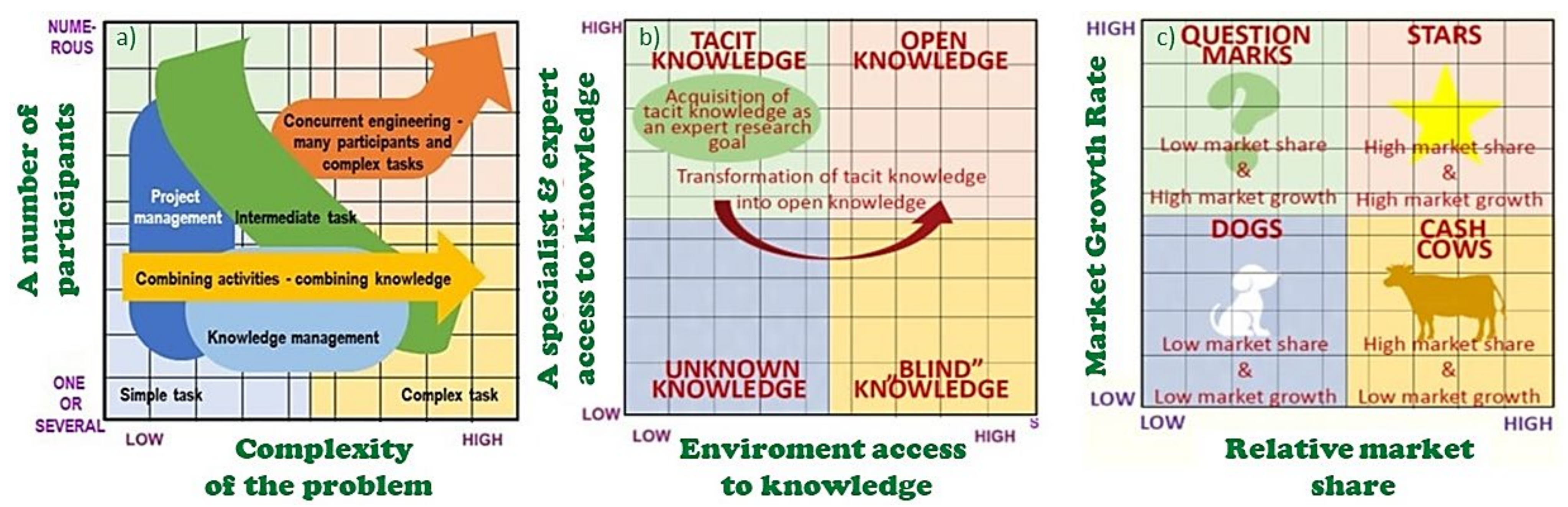
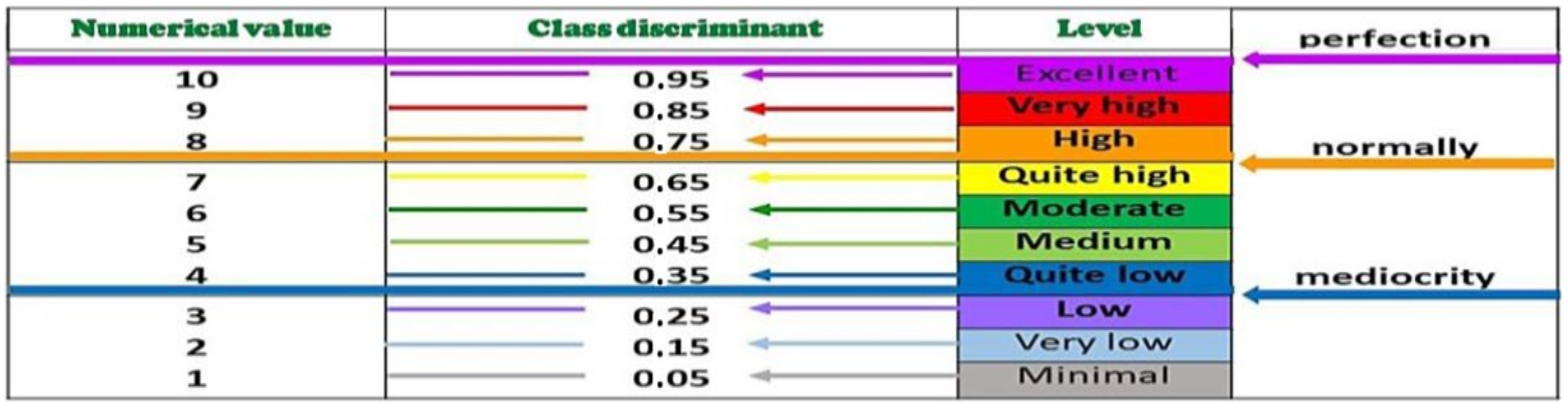
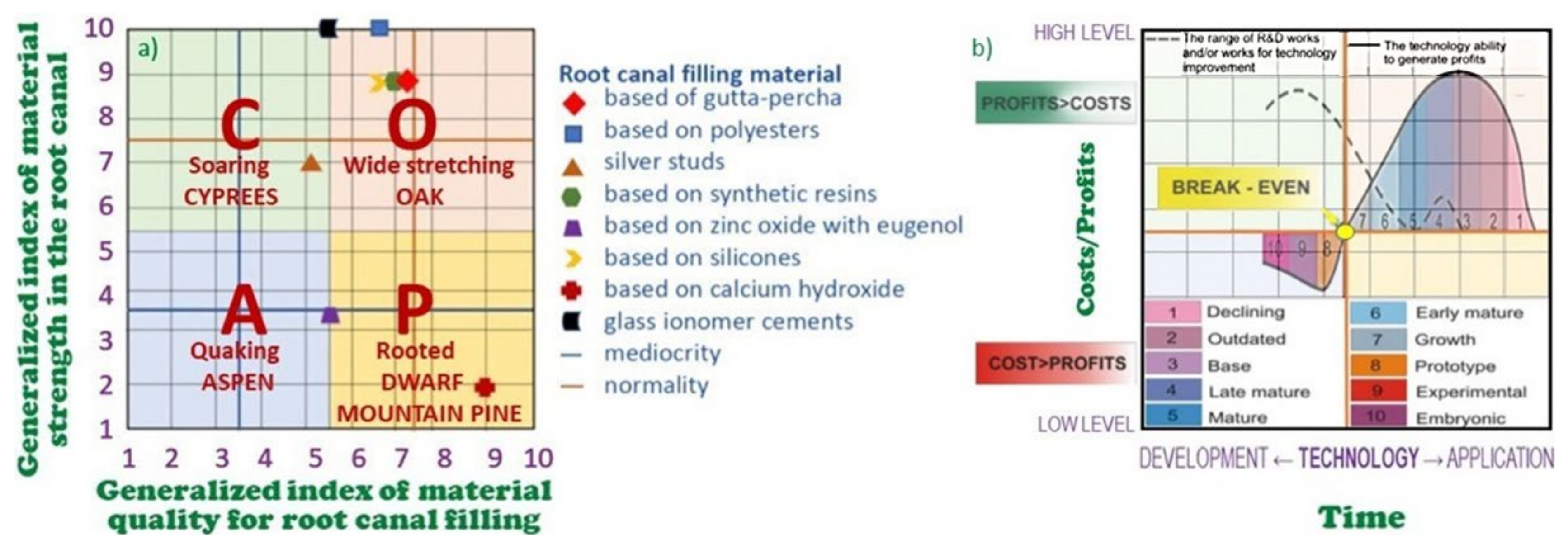
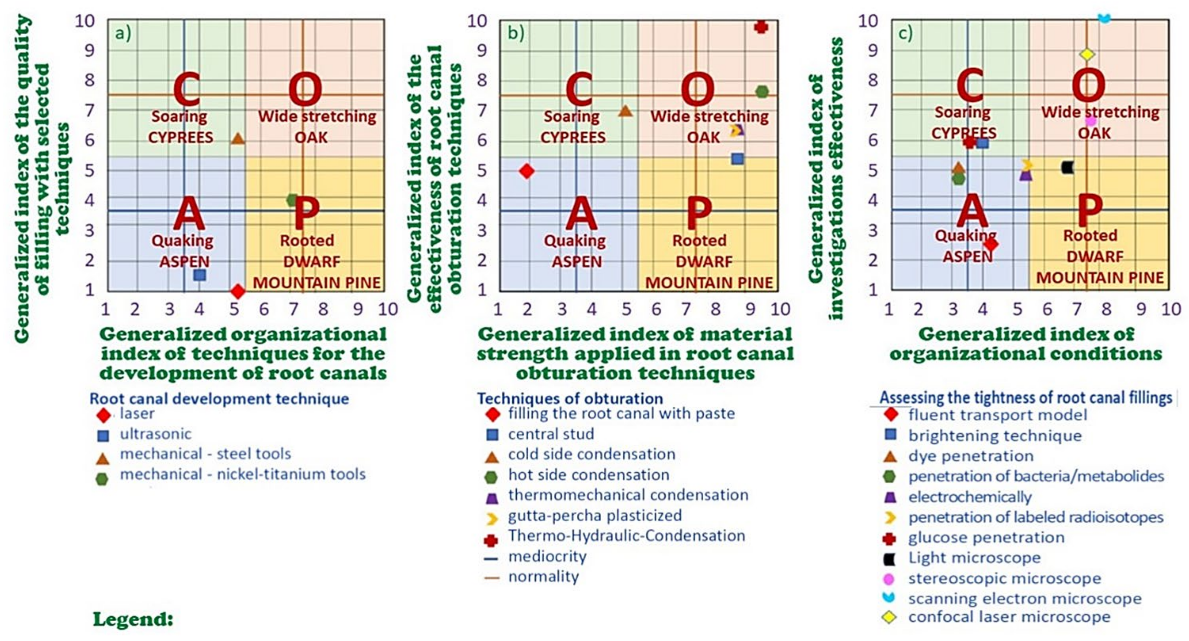
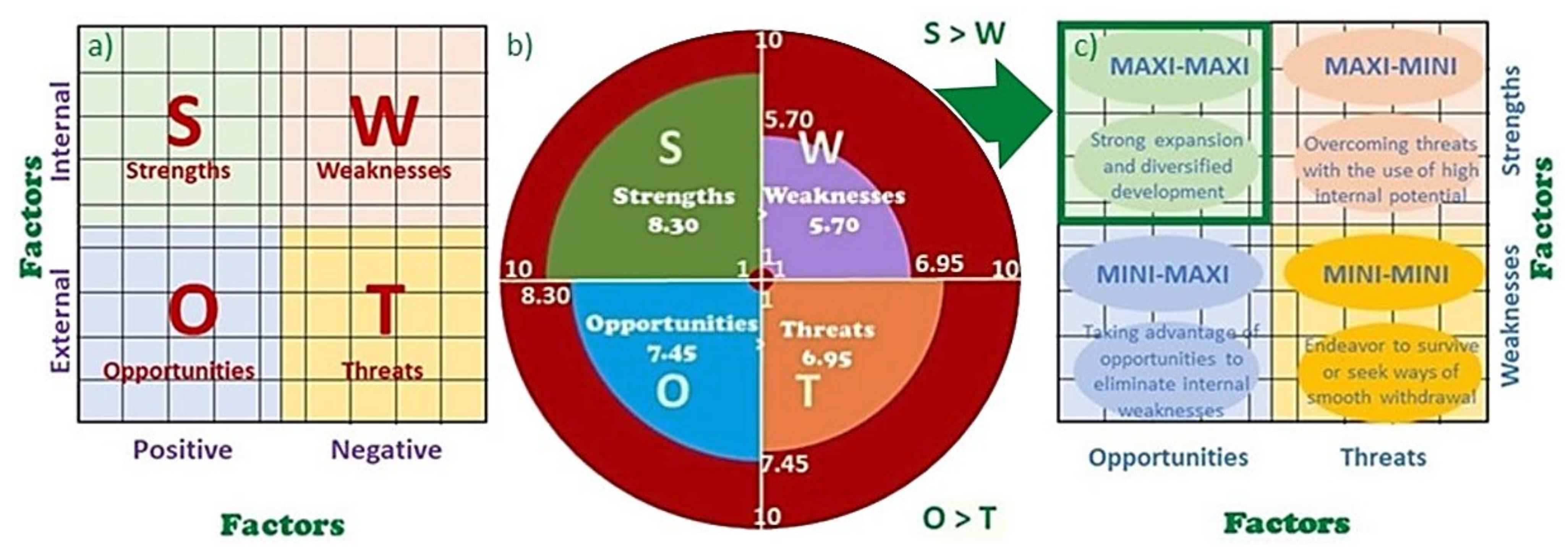
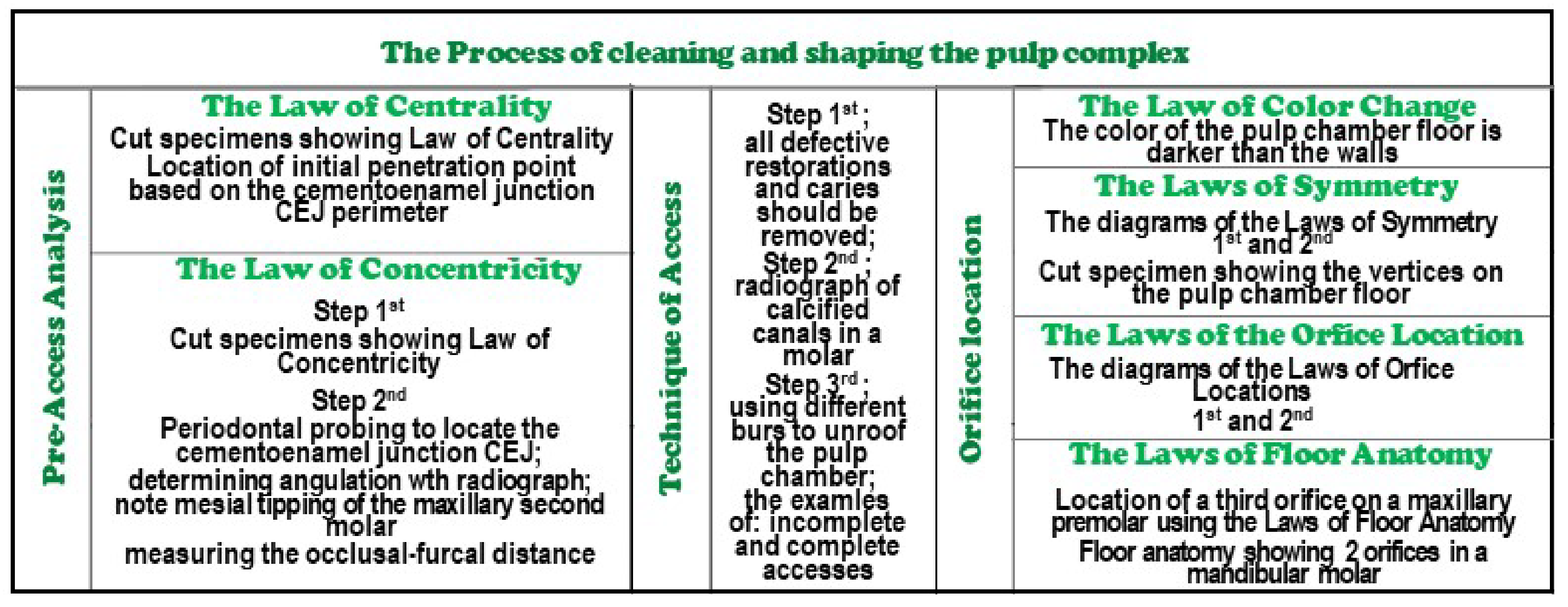
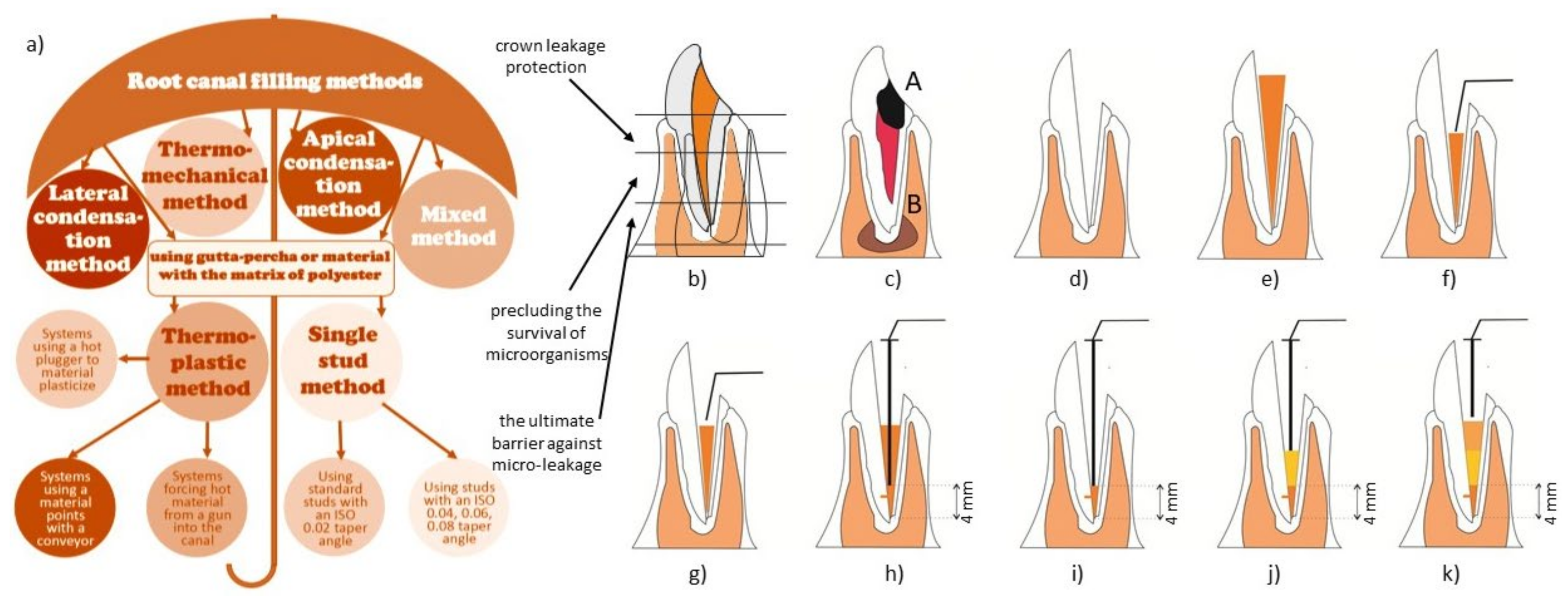
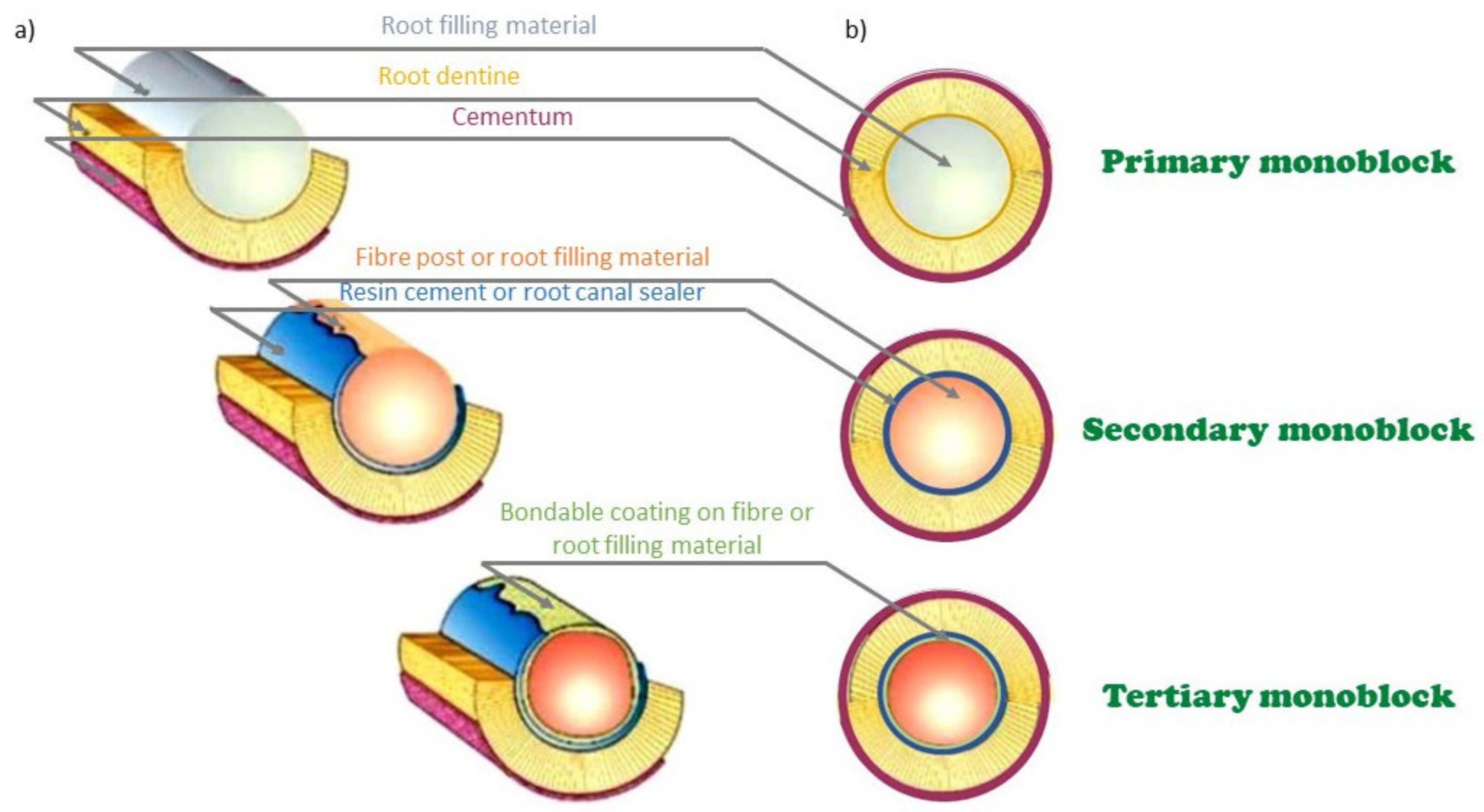
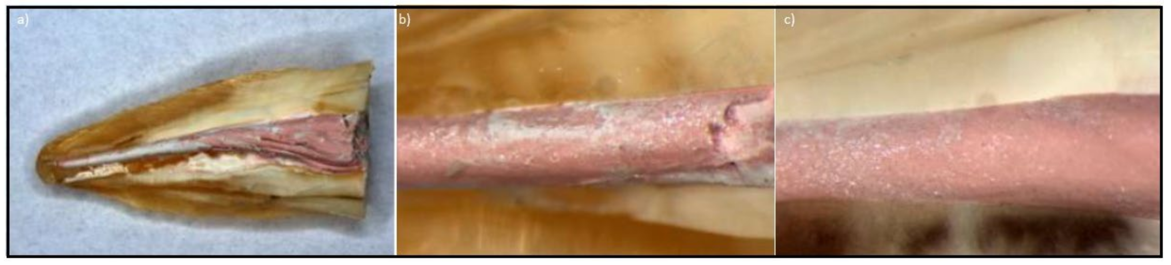
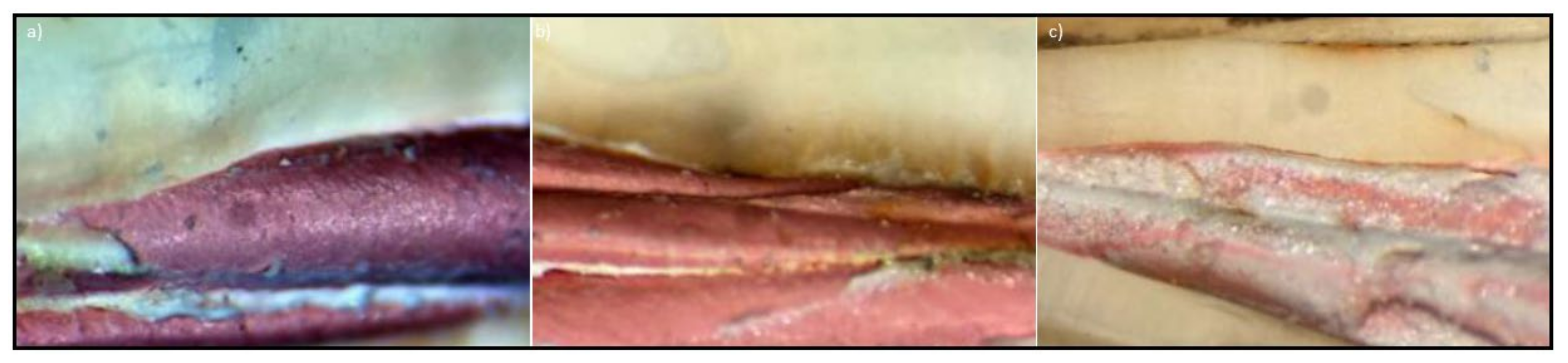
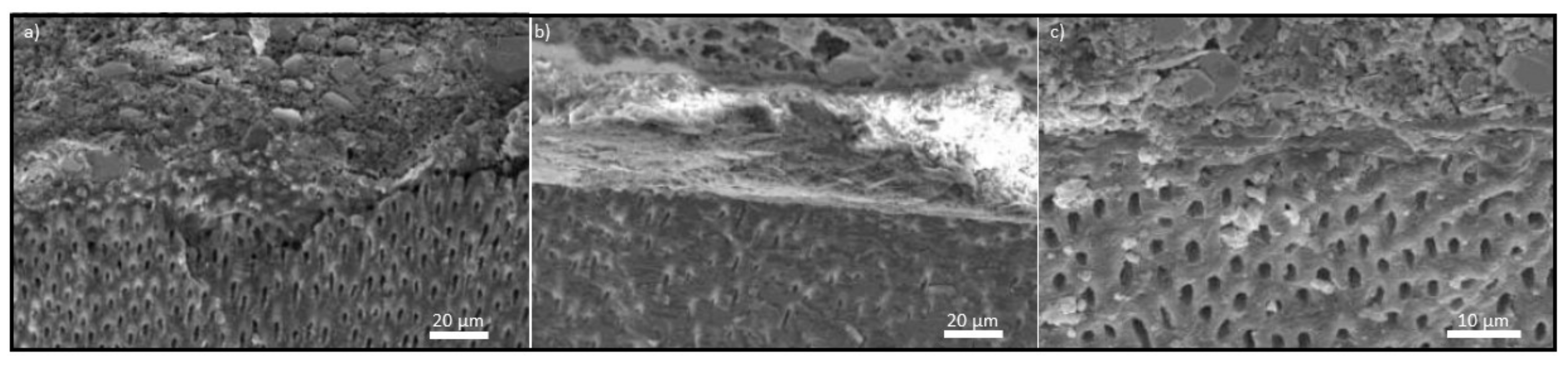
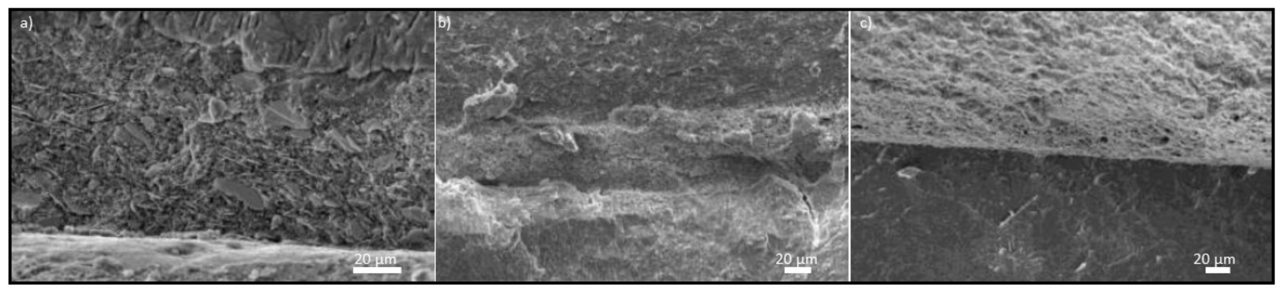
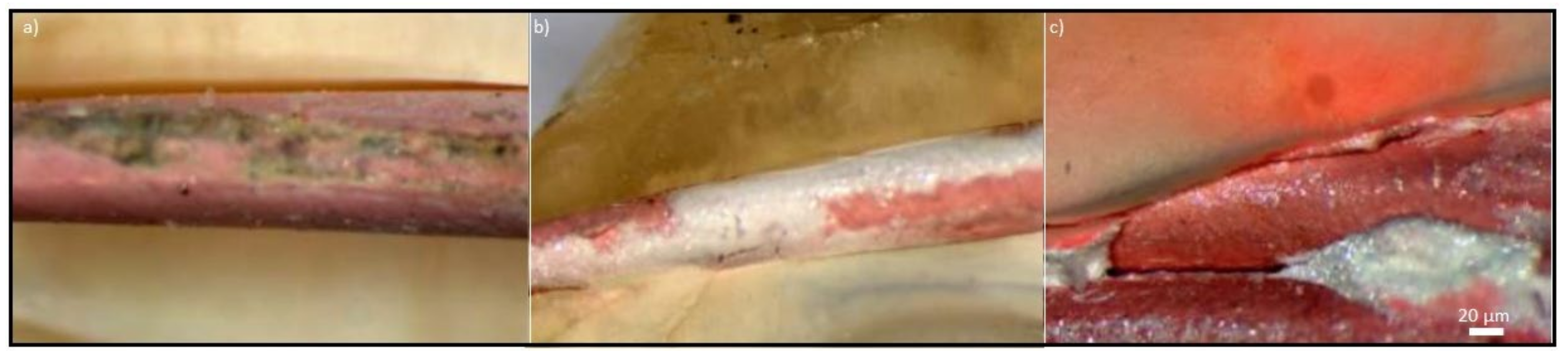
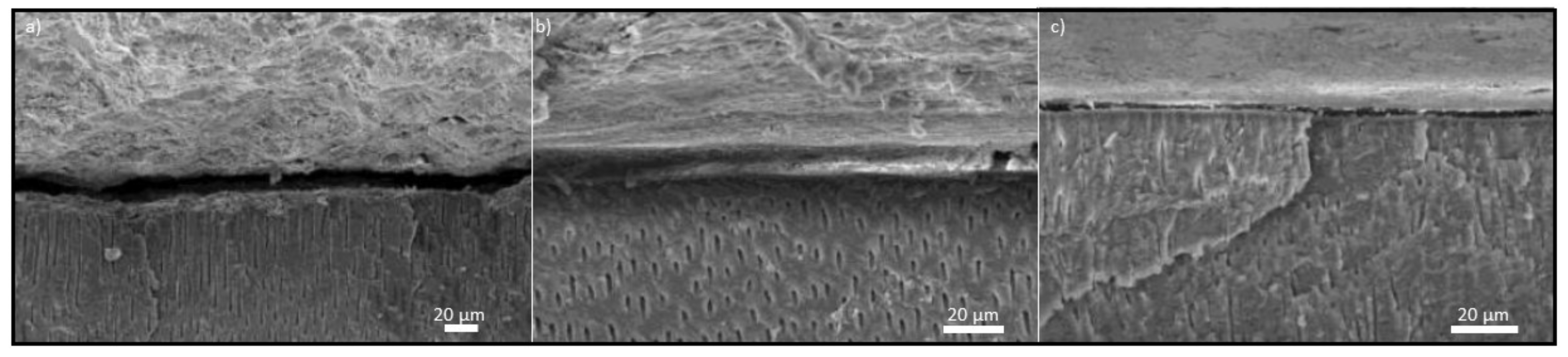
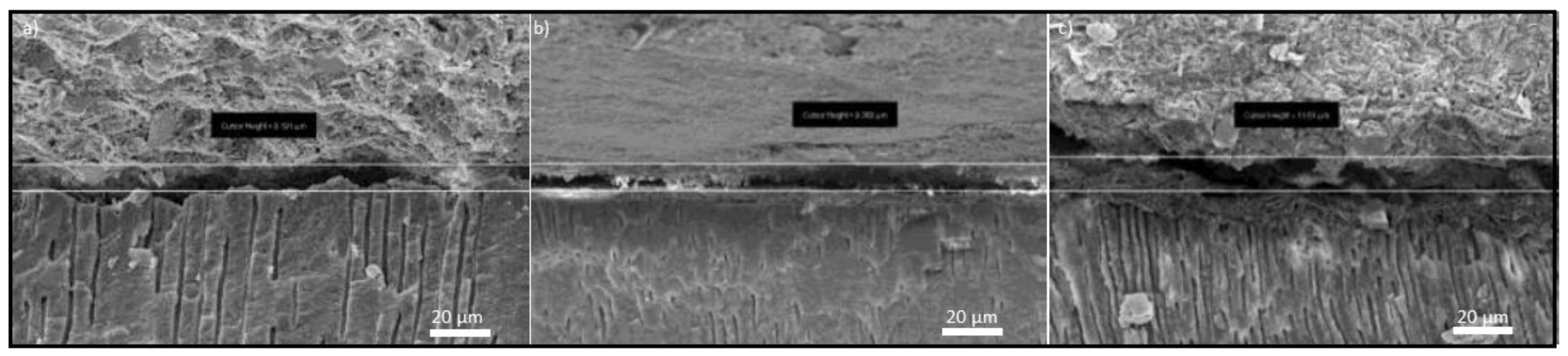
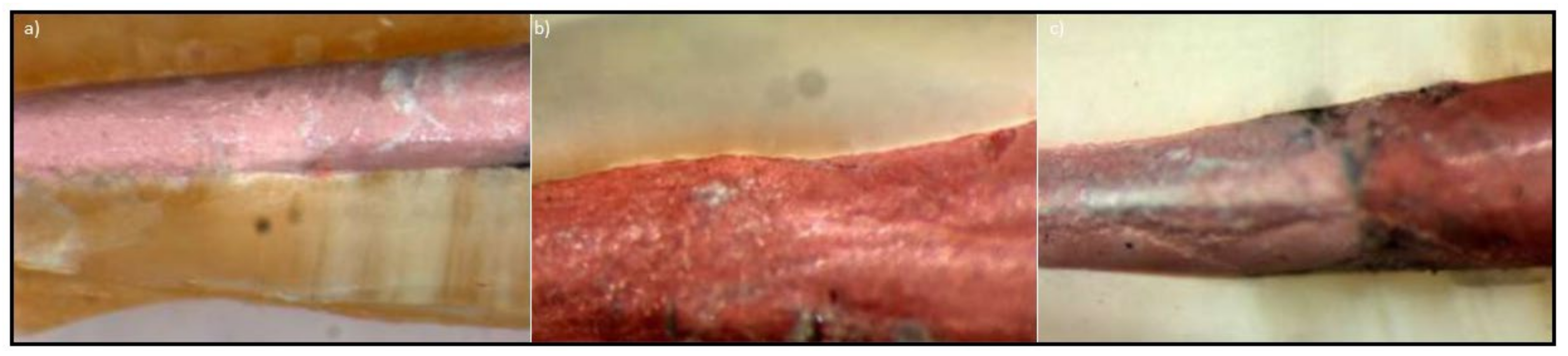
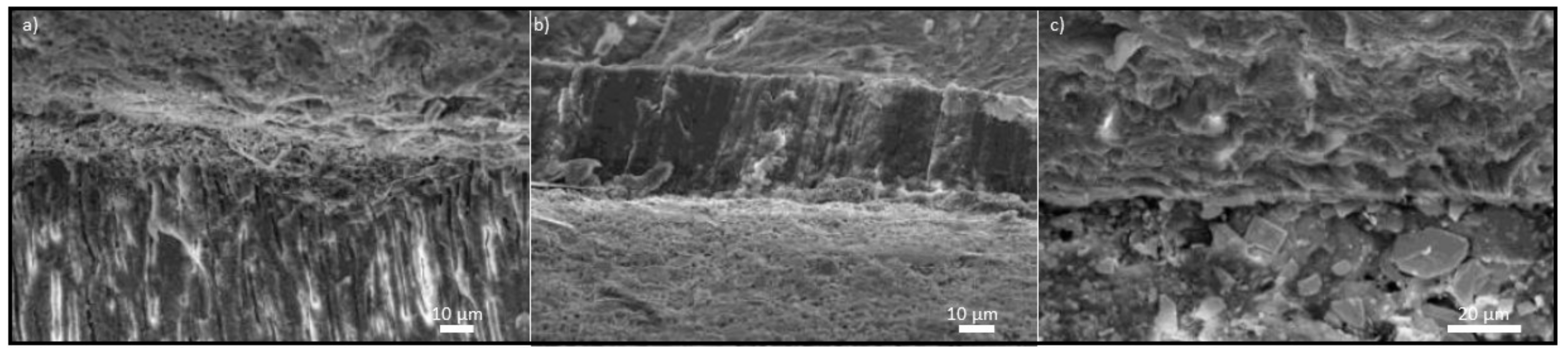
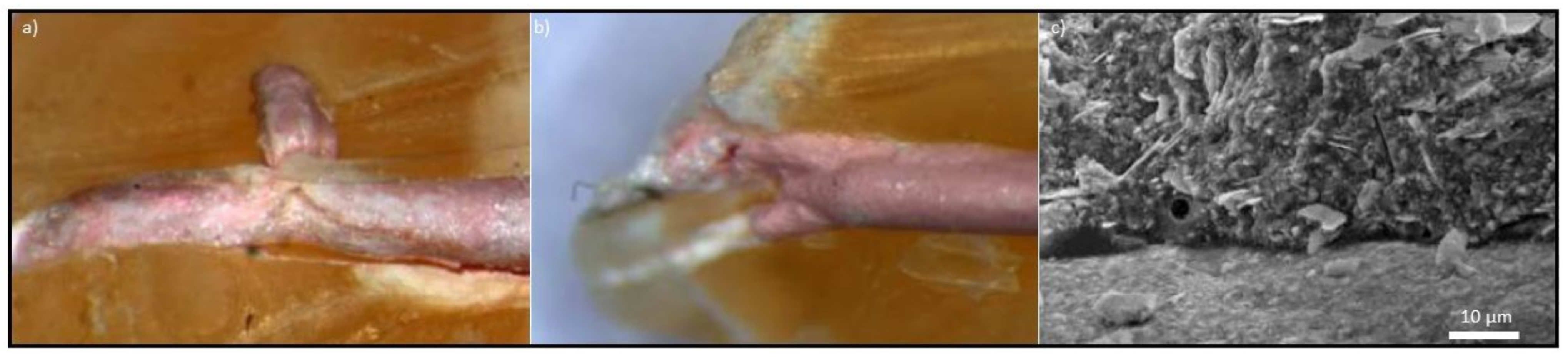
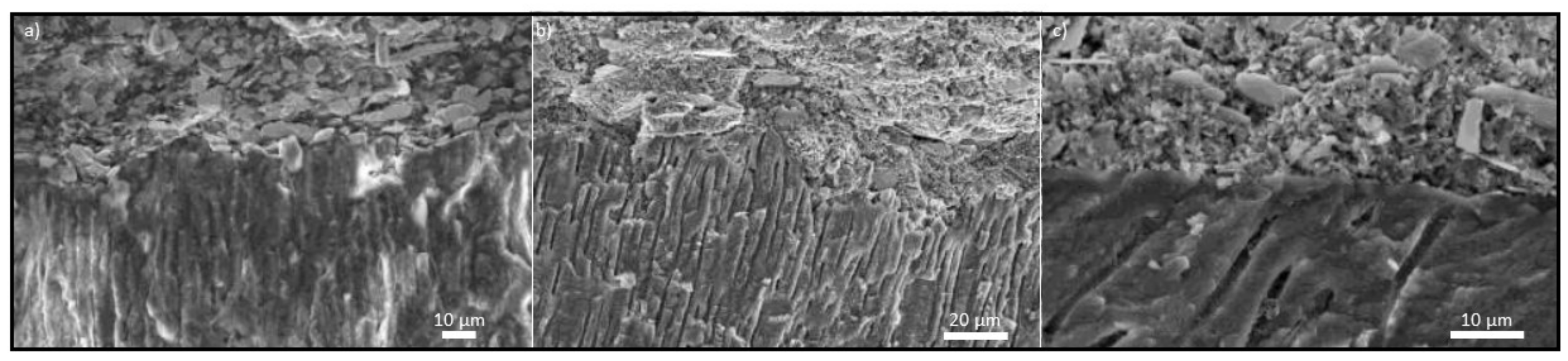
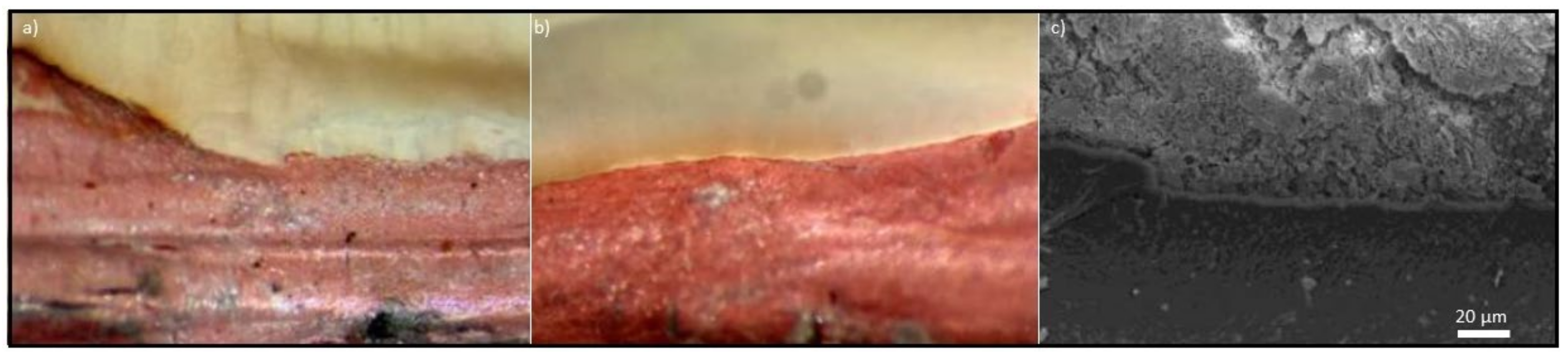
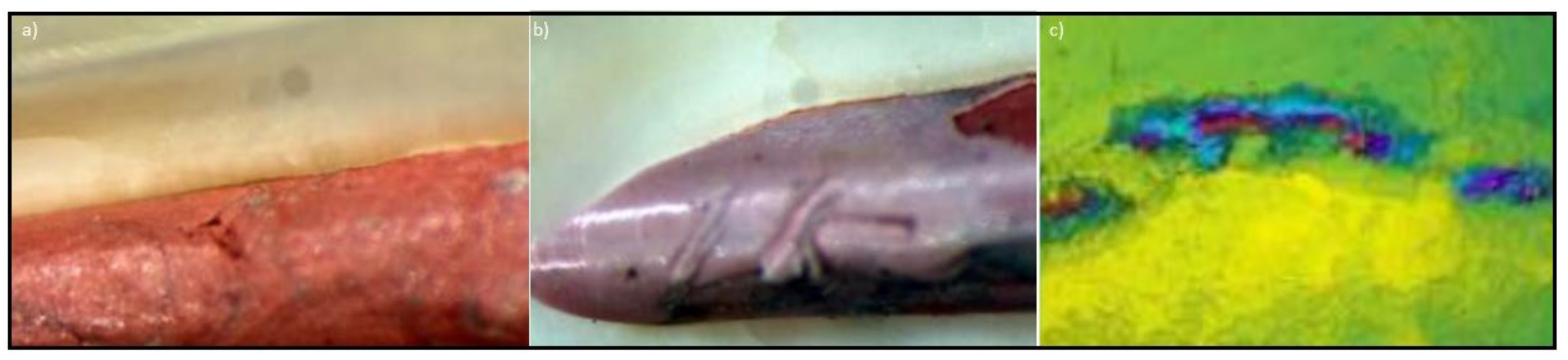
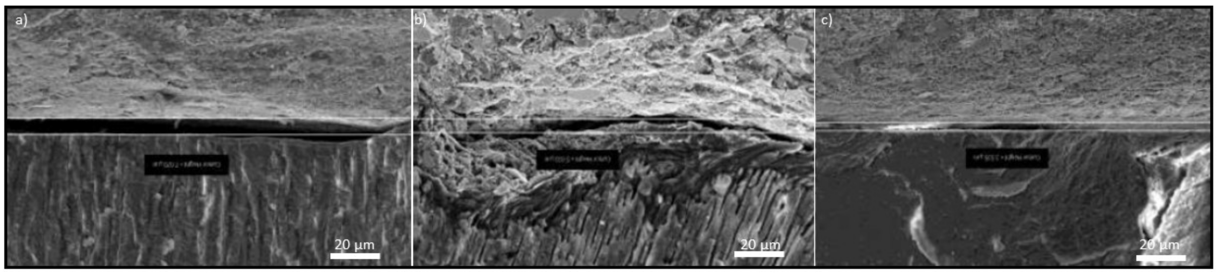
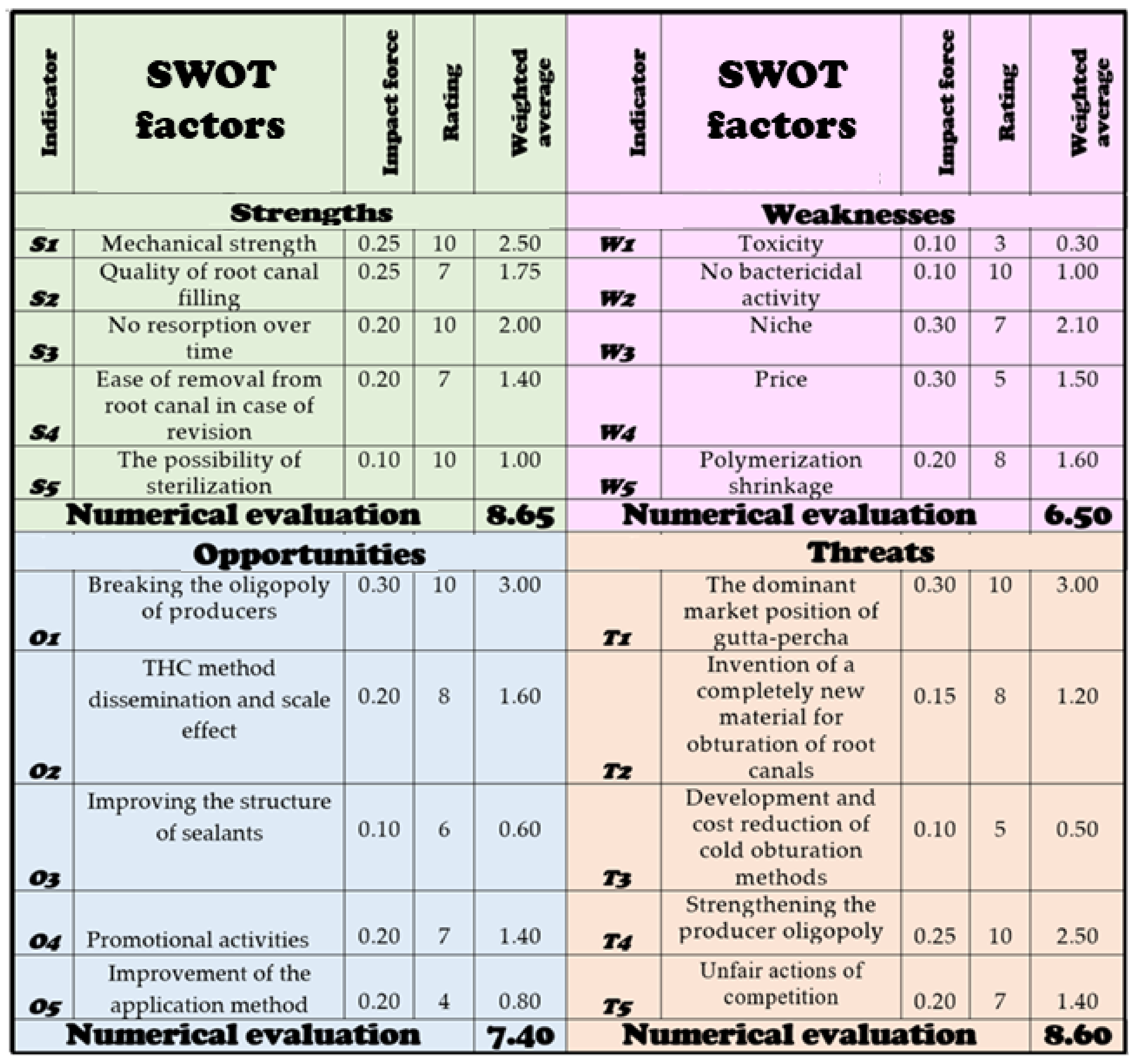
Publisher’s Note: MDPI stays neutral with regard to jurisdictional claims in published maps and institutional affiliations.

