Comparison of the Relationship between Lying and Standing Ultrasonography Measures of Muscle Morphology with Isometric and Dynamic Force Production Capabilities

 ,
,  , ,
, , 
Abstract
:1. Introduction
2. Materials and Methods
2.1. Muscle Size and Architecture
2.2. Lying Cross-Sectional Area Measurement
2.3. Lying Muscle Thickness and Pennation Angle Measurement
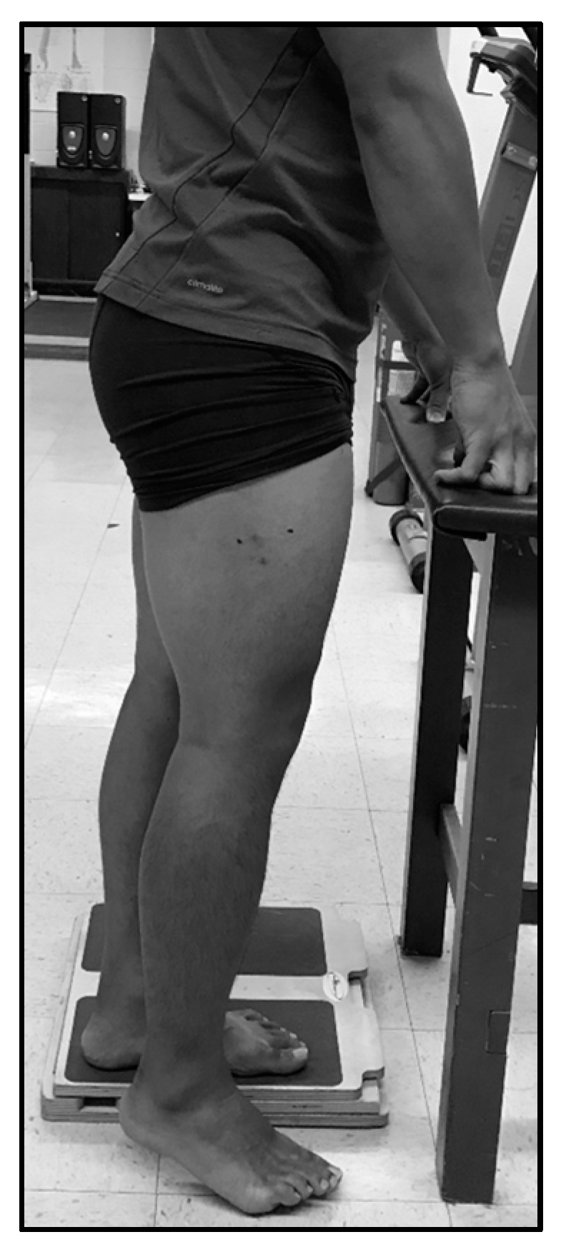
2.4. Standing Ultrasonography Measurement
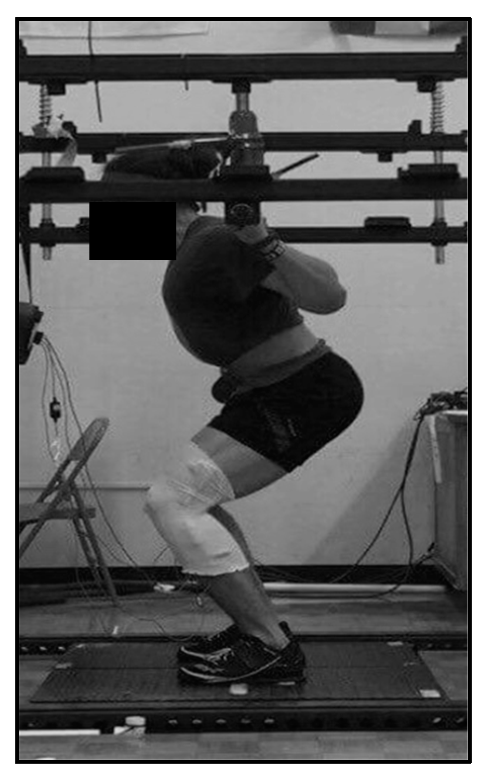
2.5. Isometric Strength Assessment
2.6. Dynamic Strength Assessment
2.7. Statistical Analyses
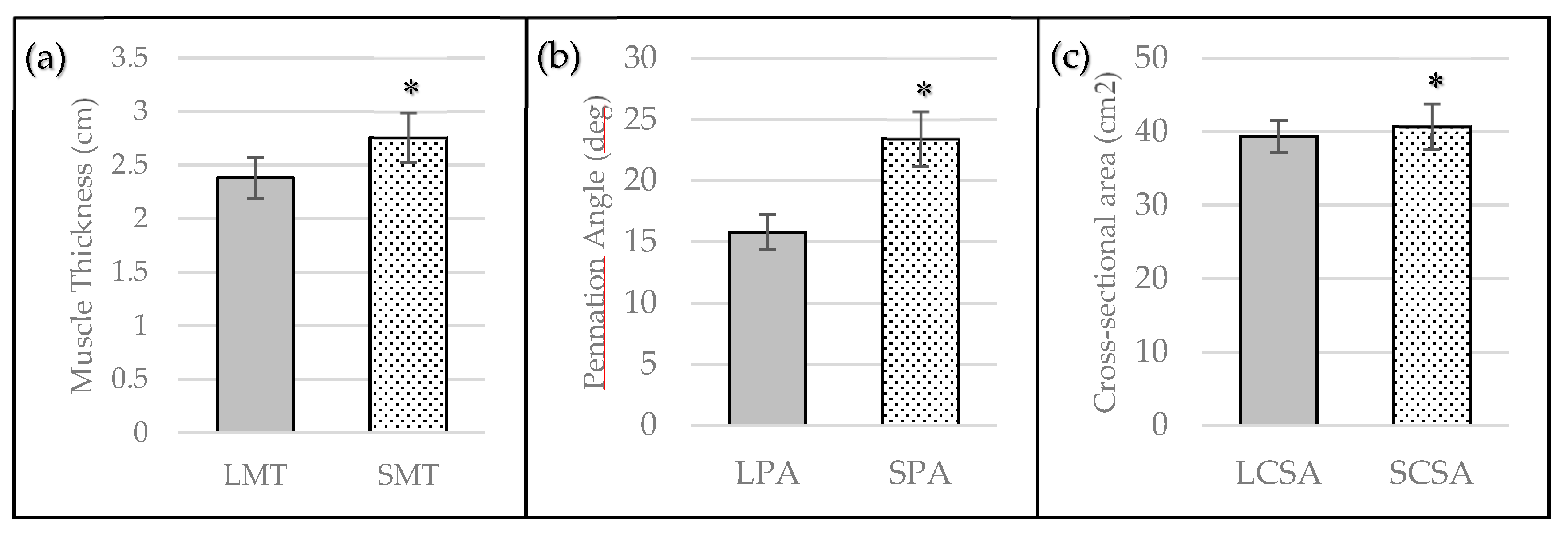
3. Results
4. Discussion
5. Conclusions
Acknowledgments
Author Contributions
Conflicts of Interest
References
- Hodges, P.; Pengel, L.; Herbert, R.; Gandevia, S. Measurement of muscle contraction with ultrasound imaging. Muscle Nerve 2003, 27, 682–692. [Google Scholar] [CrossRef] [PubMed]
- Miyatani, M.; Kanehisa, H.; Ito, M.; Kawakami, Y.; Fukunaga, T. The accuracy of volume estimates using ultrasound muscle thickness measurements in different muscle groups. Eur. J. Appl. Physiol. 2004, 91, 264–272. [Google Scholar] [PubMed]
- Häkkinen, K.; Keskinen, K. Muscle cross-sectional area and voluntary force production characteristics in elite strength-and endurance-trained athletes and sprinters. Eur. J. Appl. Physiol. Occup. Physiol. 1989, 59, 215–220. [Google Scholar] [CrossRef] [PubMed]
- Hides, J.A.; Richardson, C.A.; Jull, G.A. Magnetic resonance imaging and ultrasonography of the lumbar multifidus muscle: Comparison of two different modalities. Spine 1995, 20, 54–58. [Google Scholar] [CrossRef] [PubMed]
- Raadsheer, M.; Van Eijden, T.; Van Spronsen, P.; Van Ginkel, F.; Kiliaridis, S.; Prahl-Andersen, B. A comparison of human masseter muscle thickness measured by ultrasonography and magnetic resonance imaging. Arch. Oral Biol. 1994, 39, 1079–1084. [Google Scholar] [CrossRef]
- Walton, J.; Roberts, N.; Whitehouse, G. Measurement of the quadriceps femoris muscle using magnetic resonance and ultrasound imaging. Br. J. Sports Med. 1997, 31, 59–64. [Google Scholar] [CrossRef] [PubMed]
- Ahtiainen, J.P.; Hoffren, M.; Hulmi, J.J.; Pietikäinen, M.; Mero, A.A.; Avela, J.; Häkkinen, K. Panoramic ultrasonography is a valid method to measure changes in skeletal muscle cross-sectional area. Eur. J. Appl. Physiol. 2010, 108, 273. [Google Scholar] [CrossRef] [PubMed]
- Dupont, A.C.; Sauerbrei, E.E.; Fenton, P.V.; Shragge, P.C.; Loeb, G.E.; Richmond, F.J. Real-time sonography to estimate muscle thickness: Comparison with mri and ct. J. Clin. Ultrasound 2001, 29, 230–236. [Google Scholar] [CrossRef] [PubMed]
- Wells, A.J.; Fukuda, D.H.; Hoffman, J.R.; Gonzalez, A.M.; Jajtner, A.R.; Townsend, J.R.; Mangine, G.T.; Fragala, M.S.; Stout, J.R. Vastus lateralis exhibits non-homogenous adaptation to resistance training. Muscle Nerve 2014, 50, 785–793. [Google Scholar] [CrossRef] [PubMed]
- Bazyler, C.D.; Mizuguchi, S.; Harrison, A.P.; Sato, K.; Kavanaugh, A.A.; Deweese, B.H.; Stone, M.H. Changes in muscle architecture, explosive ability, and track and field throwing performance throughout a competitive season and after a taper. J. Strength Cond. Res. 2017, 31, 2785–2793. [Google Scholar] [CrossRef] [PubMed]
- Cormie, P.; McGuigan, M.R.; Newton, R.U. Developing maximal neuromuscular power. Sports Med. 2011, 41, 17–38. [Google Scholar] [CrossRef] [PubMed]
- Gerstner, G.R.; Thompson, B.J.; Rosenberg, J.G.; Sobolewski, E.J.; Scharville, M.J.; Ryan, E.D. Neural and muscular contributions to the age-related reductions in rapid strength. Med. Sci. Sports Exerc. 2017, 49, 1331–1339. [Google Scholar] [CrossRef] [PubMed]
- Maffiuletti, N.A.; Aagaard, P.; Blazevich, A.J.; Folland, J.; Tillin, N.; Duchateau, J. Rate of force development: Physiological and methodological considerations. Eur. J. Appl. Physiol. 2016, 116, 1091–1116. [Google Scholar] [CrossRef] [PubMed]
- Zaras, N.D.; Stasinaki, A.-N.E.; Methenitis, S.K.; Krase, A.A.; Karampatsos, G.P.; Georgiadis, G.V.; Spengos, K.M.; Terzis, G.D. Rate of force development, muscle architecture, and performance in young competitive track and field throwers. J. Strength Cond. Res. 2016, 30, 81–92. [Google Scholar] [CrossRef] [PubMed]
- Walker, S.; Blazevich, A.J.; Haff, G.G.; Tufano, J.J.; Newton, R.U.; Hakkinen, K. Greater strength gains after training with accentuated eccentric than traditional isoinertial loads in already strength-trained men. Front. Physiol. 2016, 7, 149. [Google Scholar] [CrossRef] [PubMed]
- Oppliger, R.A.; Magnes, S.A.; Popowski, L.A.; Gisolfi, C.V. Accuracy of urine specific gravity and osmolality as indicators of hydration status. Int. J. Sport Nutr. Exerc. Metab. 2005, 15, 236–251. [Google Scholar] [CrossRef] [PubMed]
- Damas, F.; Phillips, S.M.; Lixandrão, M.E.; Vechin, F.C.; Libardi, C.A.; Roschel, H.; Tricoli, V.; Ugrinowitsch, C. Early resistance training-induced increases in muscle cross-sectional area are concomitant with edema-induced muscle swelling. Eur. J. Appl. Physiol. 2016, 116, 49–56. [Google Scholar] [CrossRef] [PubMed]
- Bazyler, C.D.; Mizuguchi, S.; Sole, C.J.; Suchomel, T.J.; Sato, K.; Kavanaugh, A.A.; DeWeese, B.H.; Stone, M.H. Jumping performance is preserved, but not muscle thickness in collegiate volleyball players after a taper. J. Strength Cond. Res. 2017. [Google Scholar] [CrossRef] [PubMed]
- Prestes, J.; Tibana, R.A.; de Araujo Sousa, E.; da Cunha Nascimento, D.; de Oliveira Rocha, P.; Camarço, N.F.; de Sousa, N.M.F.; Willardson, J.M. Strength and muscular adaptations following 6 weeks of rest-pause versus traditional multiple-sets resistance training in trained subjects. J. Strength Cond. Res. 2017. [Google Scholar] [CrossRef] [PubMed]
- McBride, J.M.; Cormie, P.; Deane, R. Isometric squat force output and muscle activity in stable and unstable conditions. J. Strength Cond. Res. 2006, 20, 915–918. [Google Scholar] [PubMed]
- Bazyler, C.D.; Beckham, G.K.; Sato, K. The use of the isometric squat as a measure of strength and explosiveness. J. Strength Cond. Res. 2015, 29, 1386–1392. [Google Scholar] [CrossRef] [PubMed]
- Kraska, J.M.; Ramsey, M.W.; Haff, G.G.; Frthke, N.; Sands, W.A.; Stone, M.E.; Stone, M.H. Relationship between strength characteristics and unweighted and weighted vertical jump height. Int. J. Sports Physiol. Perform. 2009, 4, 461–473. [Google Scholar] [CrossRef] [PubMed]
- Carroll, K.M.; Wagle, J.P.; Sato, K.; DeWeese, B.H.; Mizuguchi, S.; Stone, M.H. Reliability of a commercially available and algorithm-based kinetic analysis software compared to manual-based software. Sports Biomech. 2017, 26. [Google Scholar] [CrossRef] [PubMed]
- Bishop, P.A.; Jones, E.; Woods, A.K. Recovery from training: A brief review: Brief review. J. Strength Cond. Res. 2008, 22, 1015–1024. [Google Scholar] [CrossRef] [PubMed]
- Suchomel, T.J.; Sato, K.; DeWeese, B.H.; Ebben, W.P.; Stone, M.H. Potentiation effects of half-squats performed in a ballistic or nonballistic manner. J. Strength Cond. Res. 2016, 30, 1652–1660. [Google Scholar] [CrossRef] [PubMed]
- Hopkins, W.G. Spreadsheets for analysis of validity and reliability. Sportscience 2017, 21, 36–44. [Google Scholar]
- Hopkins, W.G.; Marshall, S.W.; Batterham, A.M.; Hanin, J. Progressive statistics for studies in sports medicine and exercise science. Med. Sci. Sports Exerc. 2009, 41, 3–13. [Google Scholar] [CrossRef] [PubMed]
- Loram, I.D.; Maganaris, C.N.; Lakie, M. Use of ultrasound to make noninvasive in vivo measurement of continuous changes in human muscle contractile length. J. Appl. Physiol. 2006, 100, 1311–1323. [Google Scholar] [CrossRef] [PubMed]
- Wakeling, J.M.; Randhawa, A. Transverse strains in muscle fascicles during voluntary contraction: A 2d frequency decomposition of b-mode ultrasound images. J. Biomed. Imaging 2014, 2014, 4. [Google Scholar] [CrossRef] [PubMed]
- Drakonaki, E.; Allen, G.; Wilson, D. Ultrasound elastography for musculoskeletal applications. Br. J. Radiol. 2012, 85, 1435–1445. [Google Scholar] [CrossRef] [PubMed]
- Fukunaga, T.; Miyatani, M.; Tachi, M.; Kouzaki, M.; Kawakami, Y.; Kanehisa, H. Muscle volume is a major determinant of joint torque in humans. Acta Physiol. 2001, 172, 249–255. [Google Scholar] [CrossRef] [PubMed]
- Men, Y.; Young, A.; Stokes, M.; Crowe, M. The size and strength of the quadriceps muscles of old. Clin. Physiol. Funct. Imaging 1985, 5, 145–154. [Google Scholar] [CrossRef]
- Miura, A.; Endo, M.; Sato, H.; Sato, H.; Barstow, T.J.; Fukuba, Y. Relationship between the curvature constant parameter of the power-duration curve and muscle cross-sectional area of the thigh for cycle ergometry in humans. Eur. J. Appl. Physiol. 2002, 87, 238–244. [Google Scholar] [CrossRef] [PubMed]
- Aagaard, P.; Andersen, J.L.; Dyhre-Poulsen, P.; Leffers, A.M.; Wagner, A.; Magnusson, S.P.; Halkjær-Kristensen, J.; Simonsen, E.B. A mechanism for increased contractile strength of human pennate muscle in response to strength training: Changes in muscle architecture. J. Physiol. 2001, 534, 613–623. [Google Scholar] [CrossRef] [PubMed]
- Andersen, L.L.; Andersen, J.L.; Zebis, M.K.; Aagaard, P. Early and late rate of force development: Differential adaptive responses to resistance training? Scand. J. Med. Sci. Sports 2010, 20, e162–e169. [Google Scholar] [CrossRef] [PubMed]
- Andersen, L.L.; Aagaard, P. Influence of maximal muscle strength and intrinsic muscle contractile properties on contractile rate of force development. Eur. J. Appl. Physiol. 2006, 96, 46–52. [Google Scholar] [CrossRef] [PubMed]
- Heggelund, J.; Fimland, M.S.; Helgerud, J.; Hoff, J. Maximal strength training improves work economy, rate of force development and maximal strength more than conventional strength training. Eur. J. Appl. Physiol. 2013, 113, 1565–1573. [Google Scholar] [CrossRef] [PubMed]
- Tillin, N.A.; Pain, M.T.G.; Folland, J. Explosive force production during isometric squats correlates with athletic performance in rugby union players. J. Sports Sci. 2013, 31, 66–76. [Google Scholar] [CrossRef] [PubMed]
- Aagaard, P. Training-induced changes in neural function. Exerc. Sport Sci. Rev. 2003, 31, 61–67. [Google Scholar] [CrossRef] [PubMed]
- Alegre, L.M.; Jiménez, F.; Gonzalo-Orden, J.M.; Martín-Acero, R.; Aguado, X. Effects of dynamic resistance training on fascicle lengthand isometric strength. J. Sports Sci. 2006, 24, 501–508. [Google Scholar] [CrossRef] [PubMed]
- Bottinelli, R.; Betto, R.; Schiaffino, S.; Reggiani, C. Unloaded shortening velocity and myosin heavy chain and alkali light chain isoform composition in rat skeletal muscle fibres. J. Physiol. 1994, 478, 341–349. [Google Scholar] [CrossRef] [PubMed]
- Bottinelli, R.; Canepari, M.; Pellegrino, M.; Reggiani, C. Force-velocity properties of human skeletal muscle fibres: Myosin heavy chain isoform and temperature dependence. J. Physiol. 1996, 495, 573–586. [Google Scholar] [CrossRef] [PubMed]
- Bottinelli, R.; Schiaffino, S.; Reggiani, C. Force-velocity relations and myosin heavy chain isoform compositions of skinned fibres from rat skeletal muscle. J. Physiol. 1991, 437, 655–672. [Google Scholar] [CrossRef] [PubMed]
- Harridge, S.; Bottinelli, R.; Canepari, M.; Pellegrino, M.; Reggiani, C.; Esbjörnsson, M.; Saltin, B. Whole-muscle and single-fibre contractile properties and myosin heavy chain isoforms in humans. Pflüg. Arch. 1996, 432, 913–920. [Google Scholar] [CrossRef]
- Aagaard, P.; Simonsen, E.B.; Andersen, J.L.; Magnusson, P.; Dyhre-Poulsen, P. Increased rate of force development and neural drive of human skeletal muscle following resistance training. J. Appl. Physiol. 2002, 93, 1318–1326. [Google Scholar] [CrossRef] [PubMed]
- Blazevich, A.J.; Cannavan, D.; Horne, S.; Coleman, D.R.; Aagaard, P. Changes in muscle force–length properties affect the early rise of force in vivo. Muscle Nerve 2009, 39, 512–520. [Google Scholar] [CrossRef] [PubMed]
- Lieber, R.L.; Ward, S.R. Skeletal muscle design to meet functional demands. Philos. Trans. R. Soc. Lond. B 2011, 366, 1466–1476. [Google Scholar] [CrossRef] [PubMed]
- Edman, K.; Josephson, R. Determinants of force rise time during isometric contraction of frog muscle fibres. J. Physiol. 2007, 580, 1007–1019. [Google Scholar] [CrossRef] [PubMed]
- Hunter, J.P.; Marshall, R.N.; McNair, P.J. Relationships between ground reaction force impulse and kinematics of sprint-running acceleration. J. Appl. Biomech. 2005, 21, 31–43. [Google Scholar] [CrossRef] [PubMed]
- Kawamori, N.; Nosaka, K.; Newton, R.U. Relationships between ground reaction impulse and sprint acceleration performance in team sport athletes. J. Strength Cond. Res. 2013, 27, 568–573. [Google Scholar] [CrossRef] [PubMed]
- Morin, J.-B.; Edouard, P.; Samozino, P. Technical ability of force application as a determinant factor of sprint performance. Med. Sci. Sports Exerc. 2011, 43, 1680–1688. [Google Scholar] [CrossRef] [PubMed]
- Behm, D.G.; Wahl, M.J.; Button, D.C.; Power, K.E.; Anderson, K.G. Relationship between hockey skating speed and selected performance measures. J. Strength Cond. Res. 2005, 19, 326. [Google Scholar] [PubMed]


| Sets × Repetitions × Intensity (% 1RM) | Rest Interval |
|---|---|
| 1% × 5% × 30% | 1 min |
| 1% × 3% × 50% | 1 min |
| 1% × 2% × 70% | 2 min |
| 1% × 1% × 80% | 3 min |
| 1% × 1% × 90% | 3 min |
| 1RM attempts | 3 min |
| Measure | CV | ICC |
|---|---|---|
| LMT | 2.03% | 0.98 |
| SMT | 1.40% | 0.99 |
| LPA | 6.65% | 0.90 |
| SPA | 6.18% | 0.84 |
| LCSA | 1.93% | 0.95 |
| SCSA | 3.63% | 0.91 |
| Measure | Outcome | IPF | RFD50 | RFD100 | RFD200 | IMP50 | IMP100 | IMP200 | 1RM |
|---|---|---|---|---|---|---|---|---|---|
| LMT | Pearson’s r | 0.46 | 0.29 | 0.27 | 0.18 | 0.32 | 0.33 | 0.32 | 0.56 * |
| p-value | 0.10 | 0.31 | 0.35 | 0.55 | 0.26 | 0.25 | 0.26 | 0.04 | |
| Interpretation | Moderate | Small | Small | Small | Moderate | Moderate | Moderate | Large | |
| SMT | Pearson’s r | 0.73 * | 0.59 * | 0.53 * | 0.52 | 0.54 * | 0.58 * | 0.59 * | 0.55 * |
| p-value | <0.01 | 0.03 | 0.05 | 0.06 | 0.04 | 0.03 | 0.03 | 0.04 | |
| Interpretation | Very Large | Large | Large | Large | Large | Large | Large | Large | |
| LPA | Pearson’s r | 0.20 | −0.04 | 0.02 | −0.03 | 0.13 | 0.11 | 0.09 | 0.46 |
| p-value | 0.49 | 0.90 | 0.95 | 0.91 | 0.67 | 0.72 | 0.76 | 0.10 | |
| Interpretation | Small | Trivial | Trivial | Trivial | Small | Small | Trivial | Moderate | |
| SPA | Pearson’s r | 0.49 | 0.59 * | 0.66 * | 0.54 * | 0.38 | 0.47 | 0.53 * | 0.32 |
| p-value | 0.08 | 0.03 | 0.01 | 0.05 | 0.18 | 0.09 | 0.05 | 0.26 | |
| Interpretation | Moderate | Large | Large | Large | Moderate | Moderate | Large | Moderate | |
| LCSA | Pearson’s r | 0.38 | 0.33 | 0.25 | 0.27 | 0.52 | 0.49 | 0.44 | 0.60 * |
| p-value | 0.18 | 0.25 | 0.38 | 0.36 | 0.06 | 0.08 | 0.11 | 0.03 | |
| Interpretation | Moderate | Moderate | Small | Small | Large | Moderate | Moderate | Large | |
| SCSA | Pearson’s r | 0.58 * | 0.50 | 0.48 | 0.46 | 0.62 * | 0.63 * | 0.61 * | 0.77 * |
| p-value | 0.03 | 0.07 | 0.08 | 0.10 | 0.02 | 0.02 | 0.02 | <0.01 | |
| Interpretation | Large | Large | Moderate | Moderate | Large | Large | Large | Very Large |
© 2017 by the authors. Licensee MDPI, Basel, Switzerland. This article is an open access article distributed under the terms and conditions of the Creative Commons Attribution (CC BY) license (http://creativecommons.org/licenses/by/4.0/).
Share and Cite
Wagle, J.P.; Carroll, K.M.; Cunanan, A.J.; Taber, C.B.; Wetmore, A.; Bingham, G.E.; DeWeese, B.H.; Sato, K.; Stuart, C.A.; Stone, M.H. Comparison of the Relationship between Lying and Standing Ultrasonography Measures of Muscle Morphology with Isometric and Dynamic Force Production Capabilities. Sports 2017, 5, 88. https://doi.org/10.3390/sports5040088
Wagle JP, Carroll KM, Cunanan AJ, Taber CB, Wetmore A, Bingham GE, DeWeese BH, Sato K, Stuart CA, Stone MH. Comparison of the Relationship between Lying and Standing Ultrasonography Measures of Muscle Morphology with Isometric and Dynamic Force Production Capabilities. Sports. 2017; 5(4):88. https://doi.org/10.3390/sports5040088
Chicago/Turabian StyleWagle, John P., Kevin M. Carroll, Aaron J. Cunanan, Christopher B. Taber, Alexander Wetmore, Garett E. Bingham, Brad H. DeWeese, Kimitake Sato, Charles A. Stuart, and Michael H. Stone. 2017. "Comparison of the Relationship between Lying and Standing Ultrasonography Measures of Muscle Morphology with Isometric and Dynamic Force Production Capabilities" Sports 5, no. 4: 88. https://doi.org/10.3390/sports5040088
APA StyleWagle, J. P., Carroll, K. M., Cunanan, A. J., Taber, C. B., Wetmore, A., Bingham, G. E., DeWeese, B. H., Sato, K., Stuart, C. A., & Stone, M. H. (2017). Comparison of the Relationship between Lying and Standing Ultrasonography Measures of Muscle Morphology with Isometric and Dynamic Force Production Capabilities. Sports, 5(4), 88. https://doi.org/10.3390/sports5040088