Review

Is There a Progressive Withdrawal of Physiological Protections against High-Intensity Exercise-Induced Fatigue during Puberty?

Clermont Auvergne University, Blaise Pascal University, EA 3533, Laboratory of metabolic adaptations during exercise in physiological and pathological conditions (AME2P), BP 80026, Aubière cedex F-63171, France
*
Author to whom correspondence should be addressed.
Sports 2015, 3(4), 346-357; https://doi.org/10.3390/sports3040346
Submission received: 16 October 2015 / Revised: 27 November 2015 / Accepted: 30 November 2015 / Published: 4 December 2015
(This article belongs to the Special Issue Paediatric Exercise Physiology)

Abstract

:
Puberty is characterized by a large number of physiological modifications that translate into increased neuromuscular fatigue during high-intensity exercise in adolescents compared to prepubertal children. This greater neuromuscular fatigue in adolescents could be attributed to peripheral (i.e., muscular) and central (i.e., nervous) factors that change through puberty. Among the peripheral changes are muscle mass, fiber type composition, energy metabolism and musculo-tendinous stiffness. Among the central modifications are the voluntary activation level, the antagonist co-activation and a differential interplay between central and peripheral fatigue. The objective of this review article will be to underline the importance of these factors on the development of neuromuscular fatigue during high-intensity exercise throughout puberty and to highlight that the adolescents could be physiologically less protected against fatiguing high-intensity exercise than their prepubertal counterparts.

1. Introduction

Physical activity in children (The term “child” will refer to girls and boys prior to the development of secondary sex characteristics, roughly defined as up to the age of 11 years for girls and up to age 13 years for boys.) and adolescents (The term “adolescent” will be applied to girls aged 12–18 years and boys aged 14–18 years.) is most often characterized by repeated high-intensity short-term exercises that can lead to fatigue (The term “fatigue” will be defined as “any exercise-induced reduction in the maximal capacity of the skeletal muscle to generate force or power output” [1].). Although fatigue is often seen as a “negative” response of the neuromuscular system to exercise, an alternative view is to consider fatigue as a protective phenomenon that prevents any energy crisis and preserves the integrity of the body. This state, which is most often reversible after a rest period, has been the subject of a number of scientific studies in children and adolescents over the last 15 years. The growing interest in this topic is due to the fact that the child is continuously developing and is therefore a unique model of plasticity, responding differently to exercise-related physical constraints as a function of its development stage. Scientific evidence indicates that children fatigue less than adolescents during high-intensity exercise. This lower fatigability in children could confer a better “protection” against any extensive homeostasis disturbance, muscle damage, and biological harm, compared to adolescents. Therefore, the purpose of the present review will be to underline the effects of puberty on the development and etiology of neuromuscular fatigue during high-intensity exercise and to highlight that the adolescents could be physiologically less protected against fatiguing high-intensity exercise than their prepubertal counterparts.

2. Fatigue and Maturation

It is now well accepted that children fatigue less than adults during high-intensity exercise. For instance, during repeated cycling sprints separated by short recovery periods, peak power output is maintained longer in children compared to adults [2]. The same is observed during repeated running sprints [3]. This lower fatigability in children declines over the pubertal period. Indeed, from childhood to adolescence, there is a progressive decline in the ability to maintain force or power during repeated high-intensity exercises. For instance, during repeated cycling sprints, the decline of cycling peak power is higher in adolescents compared to children and higher in adults compared to adolescents [4]. This has recently been confirmed during a series of maximal voluntary contractions of the knee extensors and knee flexors [5,6]. Finally, when muscle contractions include an eccentric phase (i.e., a stretch of the active muscle), the decline of force during exercise is higher in adolescents compared to children and higher in adults compared to adolescents [7]. Moreover, the magnitude of symptoms appearing during the days following a series of eccentric contractions (i.e., stiffness, edema, decreased range of motion) is higher in adults compared to adolescents, and higher in adolescents compared to children [7].
In summary, growth and maturation influence the rate and the amount of fatigue during repeated high-intensity exercise. Indeed, adolescents fatigue faster than their prepubertal counterparts. This greater muscle fatigability in adolescents seems to be explained by peripheral (i.e., muscular) and central (i.e., nervous) changes that occur through puberty and that we propose to address in this chapter (Figure 1).
Figure 1. Schematic illustration of the various mechanisms that could explain the lower peripheral fatigue (i.e., muscle fatigue) in children during high-intensity exercise.
Sports 03 00346 g001

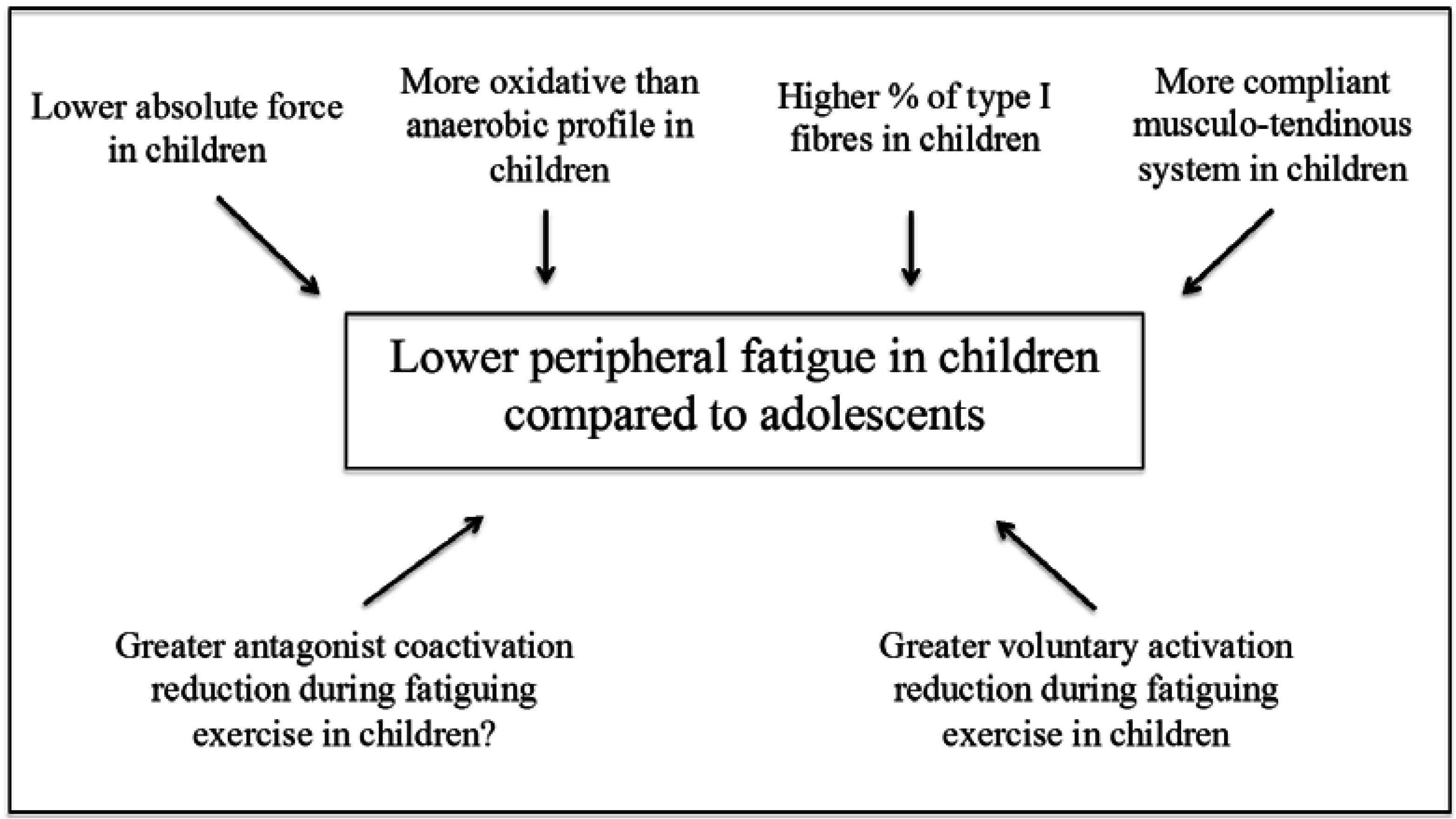
3. Peripheral Factors

3.1. Muscle Mass and Fibre Type Composition

The greater fatigability of adolescents during high-intensity exercise could be explained by the implication of a larger muscle mass during exercise compared to children. Indeed, when adolescents perform a series of sprints or exercises at a given percentage of their maximal power, they produce greater absolute power than children [8]. This greater muscle mass engaged during exercise in adolescents could be the cause of greater metabolic disturbances that are usually observed during high-intensity exercise in adolescents compared to children (see the Section 4.2). An alternative hypothesis, often mentioned when comparing the fatigability between populations that produce different levels of force or power (women vs. men; elderly vs. young adults) is that individuals with higher power or force have higher vascular occlusion during exercise. The resulting decrease in blood flow could contribute to an increased accumulation of muscle by-products (i.e., lactate, H+ ions, inorganic phosphate) and to the reduced supply of nutrients and oxygen. This suggestion is however purely speculative because this has never been measured in adolescents compared to children. However, the removal rate of some muscle by-products (i.e., H+ ions) and the resynthesis rate of energy substrates as phosphocreatine were found to be lower in adolescents compared to children (see the Section 4.2), suggesting that the occlusion may be increased because of the higher force produced by the adolescents. Finally, some studies suggested that the magnitude of muscle damage could be dependent on the absolute power produced during eccentric contractions [9,10]. Therefore, the gradual increase in the force production capacity from childhood to adulthood may explain the gradual increase of the amplitude of post-exercise muscle damage with advancing age. However, this statement has recently been questioned [7] and remains to be verified.
Furthermore, it has been shown during fatigue protocols involving repeated maximal contractions that the fatigability differs between individuals who display a different muscle typology, i.e., a different fiber type composition. More specifically, a high proportion of fast-twitch muscle fibers (type II) may contribute to a greater muscle fatigability [11,12]. The data available on the muscle phenotype during growth indicate that type II fibers reach proportions similar to those in adults during the adolescence period [13,14,15]. The decrease in the percentage of slow-twitch muscle fibers (type I) from childhood to adolescence is probably explained by a transformation of type I fibers into type II fibers [16]. Therefore, the greater percentage of type II fibers in adolescents could partly explain their greater fatigability during high-intensity exercise. These differences in muscle typology could also explain the gradual increase of post-exercise muscle damage during growth. Indeed, the fast-type motor units (MU) generally have an optimal length that is shorter than the optimal length of the muscle, while slow-type MUs have, on average, an optimal length superior to that of the muscle [17]. During eccentric contractions, type II MUs are much more strained over the descending portion of their force-length relationship, which increases the occurrence of micro-lesions within these MUs compared to type I MUs. The greater percentage of type II fibers in adolescents compared to children could therefore partially contribute to the higher muscle damage experienced by adolescents. However, this hypothesis remains to be verified.

3.2. Energy Metabolism

A number of studies have suggested that adolescents accumulate much more muscle by-products during high-intensity exercise than children, which may explain their greater fatigability. In the early 1970s, Eriksson and coworkers were the first researchers to suggest that glycolytic activity was lower in prepubertal children and that it was dependent on their level of maturation. This suggestion was based on a positive correlation between testicular volume (maturation index) and muscle lactate concentrations obtained after maximal exercise [18]. Furthermore, a lower activity of phosphofructokinase (PFK) was reported in prepubertal boys vs. older subjects [19]. Other studies also reported a PFK activity that tended to be similar in 13–15 year-old adolescents and young adults [20]. However, Petersen, Gaul, Stanton and Hamstock [21] showed that during high-intensity exercise, muscle pH values (i.e., characterizing the muscle acidosis) were not different between prepubescent and pubescent female swimmers. The authors concluded that the activity of the anaerobic glycolysis was not attenuated before puberty. This divergence of results may be related to the differences in the protocols used (i.e., subjects’ maturation, training status of individuals, etc.) [21]. Therefore, the potential role of anaerobic glycolysis in the fatigability differences between children and adolescents remains still poorly understood. Further studies are required to clarify this issue.
Studies that have investigated the activity of the aerobic metabolism in children and adolescents provide more consensual results. The data obtained from muscle biopsies indicate that adolescents have a lower activity of the oxidative metabolism compared to prepubertal children. Indeed, it has been shown that the fumarase/PFK ratio is lower in adolescents compared to children [22]. This suggests that the oxidative pathway contributes less than the anaerobic glycolysis to ATP supply during exercise in adolescents compared to children. Therefore, the reliance on oxidative metabolism could be lower in adolescents than in children during exercise. This profile might explain the higher blood/muscle lactate and H+ ion values obtained after maximal exercise in adolescents [2]. It could also be the cause of the slower recovery of some energy substrates (i.e., phosphocreatine) following exercise and the slower elimination of metabolites (i.e., lactate and H+ ions) from the skeletal muscle in adolescents compared to children [23,24,25,26,27,28]. The lower oxidative profile of adolescents could therefore partly explain their slower recovery and their greater fatigability during repeated high-intensity exercise protocols.

3.3. Musculo-Tendinous Stiffness

As mentioned above (see Section 4.1), adolescents develop higher levels of force and power than children. These higher force levels are associated with a higher musculo-tendinous stiffness in adolescents compared to children and in adults compared to adolescents [29] since stiffness is positively related to the level of maximal force. Recent studies have suggested that a low musculo-tendinous stiffness could act as a “mechanical buffer” during muscle contractions, which would limit the development of fatigue [30,31]. This effect could be particularly significant during eccentric contractions, where the elongation of the tendon would limit the stretching of muscle fibers and hence the occurrence of muscle damage. This hypothesis is consistent with studies showing a progressive increase of fatigability and muscle symptoms following eccentric exercise from childhood to adulthood [7]. However, direct experimental evidence for this effect remains to be verified.
The musculo-tendinous stiffness also affects the force-length relationship of the skeletal muscle. Although O’Brien et al. [32] and Kluka et al. [33] have shown no difference of optimal angle (i.e., the angle where force is maximal) between children and adults, Marginson and Eston [34] showed that the optimal angle corresponded to longer muscle lengths in children compared to adults. As all studies used the same angle to compare the fatigability between children and adults, their results could be affected since the children could exercise at a shorter relative length than adults. However, in adults, any exercise at a short muscle length reduces the development of peripheral fatigue [35] and promotes the development of central fatigue [36]. Should these effects hold true in children, this could explain, at least in part, the results of studies reporting a lower peripheral fatigue and a greater central fatigue in prepubertal children and adolescents compared to adults [37,38]. However, the effect of muscle length on the development and etiology of neuromuscular fatigue should be confirmed in children and adolescents and a systematic comparison at the optimal angle should be considered when the effects of fatigue protocols are compared between children, adolescents and adults.

4. Central Factors

Beyond the peripheral factors, central, i.e., nervous, factors could be considered as protective mechanisms in children. Such factors include, in general, the capacity to maximally activate the MUs of agonist muscles, and the level of co-activation of the antagonist muscles.

4.1. Voluntary Activation

A number of studies focused on the activation of motor units to explain the differences of specific force (i.e., normalized to body dimensions) between children, adolescents and adults. Although some studies reported a comparable level of voluntary activation between children and adults [39] or between children and adolescents [40], other studies have reported a lower voluntary activation level in children compared to adolescents [41] and adults [33,42,43]. Overall, children have a reduced ability to maximally activate their MUs, which has often been attributed to a relative immaturity of the cortico-spinal pathway. This conclusion, however, should be qualified since the differences of voluntary activation between children, adolescents and adults could be dependent on sex [38,42] and the muscle length at which the voluntary activation level is evaluated [33]. The evolution of this ability to maximally activate the MUs during growth has also been poorly described. Only Grosset et al. [44] showed an increase of the voluntary activation level of the plantar flexors between seven and 11 years. Therefore, there is currently little data covering the pre-pubertal period, puberty and adulthood. In addition, all the above-mentioned studies evaluated the level of voluntary activation in an isometric condition and it is currently unclear whether these results also apply to dynamic contractions [45].
A low voluntary activation contributes to the development of a low level of force, which may have consequences on the development of fatigability. Indeed, as seen above (see Section 4.1), a low force reduces the development of fatigability. Moreover, according to Henneman’s size principle [46], individuals with a low voluntary activation level recruit a greater relative proportion of slow-twitch motor units than individuals with a similar muscle typology, but a higher voluntary activation level. As mentioned in Section 4.1, this preferential recruitment of slow-twitch motor units, which are more resistant to the development of metabolic fatigue and muscle damage, could reduce the fatigability. Finally, a low voluntary activation level may facilitate the rotation of motor units and/or agonist muscles during maximal exercise, which seems impossible when the motor unit activation level is almost maximal [47]. However, there is currently no evidence to support these assumptions in children. Indeed, the study of the electrical activity of motor units requires invasive investigation methods, which are not applicable in children for ethical reasons.

4.2. Antagonist Co-Activation

The increase in the activation level of the antagonist muscles is also a way to reduce the torque output at the joint, and thus to protect the articular structures against any damage. A number of studies have evaluated the antagonist co-activation level in children, adolescents and adults but have reported conflicting results. Some studies found no difference between children and adults [48] and between adolescents and adults [49] during isokinetic contractions. Similar conclusions have also been reported between children and adults during isometric tasks [33,42,50,51,52]. However, some authors observed higher co-activation levels during submaximal isometric contractions in children compared to adults [44], with a level of antagonist co-activation, which tended to decline between seven and 11 years. Conversely, O’Brien et al. [42] reported higher co-activation levels in adults compared to children, which seems consistent with the greater force production capacity in adults. Finally, higher co-activation levels have been observed in children during poly-articular movements such as walking [53] and jumping [54,55].
The existence of a higher co-activation level in children appears to be mainly observed during complex motor tasks (poly-articular movements and sustained submaximal isometric tasks at a given force). This phenomenon seems to be more related to problems of motor coordination or motor learning in children than to the existence of real protective mechanisms. As such, Armatas et al. [50] showed that the co-activation level did not change in children and adults during fatiguing exercise involving repeated maximal voluntary contractions of the knee extensors.

5. Interplay between Peripheral and Central Factors

In the early 2000s, Tim Noakes proposed a theory called the “central governor”, which suggests that physiological responses are essentially determined and limited by the central nervous system [56]. The level of voluntary activation would be determined as a function of (i) sensory information arising from the physical, thermal and biochemical states of active muscles and joints; and (ii) the prediction of the effects of exercise on the development of fatigue (teleoanticipation). According to Noakes’ theory, the purpose of such regulation would be to protect the body against any biological harm or life-threatening event. This theory was expanded by Amann and Dempsey [57], who suggest that the type III and IV afferents could regulate the level of voluntary activation to confine the development of peripheral fatigue within acceptable limits. There would be, therefore, a peripheral fatigue threshold, and central fatigue would be aimed at regulating the reach of this threshold.
While there is a consensus on the existence of a lower peripheral fatigue in children compared to adults, the importance of central fatigue remains controversial. According to the Amann and Noakes’ theories, this lower peripheral fatigue should result in reduced afferent feedback and therefore a reduced central fatigue in children, if no specific protective mechanism is involved. This is actually what Gorianovas et al. [52] reported following a fatiguing plyometric exercise in children compared to adults. However, the same team also observed a greater central fatigue in adolescents compared to adults after a sustained voluntary maximal contraction of the knee extensors [38]. These findings have recently been confirmed by our research team in children compared to adults after repeated maximal voluntary contractions of the knee extensors [37]. These results support the hypothesis of an increased protective mechanism in prepubertal children and adolescents, which would result in greater central fatigue for a given level of peripheral fatigue. Nevertheless, Hatzikotoulas et al. [39] did not observe any difference in central fatigue between children and adults. However, in this study, children experienced a lower peripheral fatigue than adults, suggesting that at similar peripheral fatigue, children could have developed more central fatigue. The question of specific regulatory mechanisms of central fatigue in children remains therefore open to debate. The problem, however, remains complex to address because it is presently technically difficult to measure the activity of type III and IV afferents in humans during exercise. Only indirect approaches using anesthetic injections are currently implemented [57]. From an ethical point of view, it is unconceivable to use these methods in children and adolescents.
However, the specificity of physiological responses during exercise in prepubertal children leads us to believe that the activity of type III and IV afferents could be different between children and adults. Indeed, if these afferents are involved in mechano- and metabo-sensitivity, they are also involved in nociception and regulation of cardiovascular and ventilatory responses. Given that prepubertal children have a greater sensitivity to pain [58] and exacerbated cardio-respiratory responses to exercise (relative to their body dimensions) [59] that promote aerobic metabolism [2], they could therefore be characterized by a greater sensitivity to type III and IV afferents. This greater sensitivity would then explain the results of studies, which have reported an increased [37,38] or comparable [39] central fatigue and a lower peripheral fatigue in prepubertal children compared to adults. However, this hypothesis remains to be verified.

6. Conclusions

The children and adolescents are characterized by specific physiological responses during exercise, which are distinguished particularly under fatiguing conditions. The specificity of these responses may reflect the existence of protective mechanisms during exercise in children, which could withdraw progressively during adolescence. However, this changing period between childhood and adulthood has scarcely been studied in the literature. Additional research is required to better understand the gradual modification of physiological responses during exercise from childhood to adulthood, and hence appreciate the existence of these potential protective mechanisms. As such, the investigation of nervous regulation of fatigue could offer new insights into the understanding of these protective mechanisms, but this remains difficult to implement technically. From a practical point of view, the puberty-related physiological changes should be considered in the management of training load in adolescent athletes. In particular, the recovery duration between sets or repetitions of high-intensity exercises should be increased during the transition from childhood to adolescence (i.e., from 11–12 years in girls and from 13–14 years in boys) because of the greater fatigability and the slower recovery in adolescents compared to prepubertal children.

Author Contributions

Sébastien Ratel and Vincent Martin drafted, edited and approved the final version of the manuscript.

Conflicts of Interest

The authors declare no conflict of interest.

References

  1. Vollestad, N.K. Measurement of human muscle fatigue. J. Neurosci. Methods 1997, 74, 219–227. [Google Scholar] [CrossRef]
  2. Ratel, S.; Duche, P.; Hennegrave, A.; van Praagh, E.; Bedu, M. Acid-base balance during repeated cycling sprints in boys and men. J. Appl. Physiol. 2002, 92, 479–485. [Google Scholar] [CrossRef] [PubMed]
  3. Ratel, S.; Williams, C.A.; Oliver, J.; Armstrong, N. Effects of age and recovery duration on performance during multiple treadmill sprints. Int. J. Sports Med. 2006, 27, 1–8. [Google Scholar] [CrossRef] [PubMed]
  4. Ratel, S.; Bedu, M.; Hennegrave, A.; Dore, E.; Duche, P. Effects of age and recovery duration on peak power output during repeated cycling sprints. Int. J. Sports Med. 2002, 23, 397–402. [Google Scholar] [CrossRef] [PubMed]
  5. Dipla, K.; Tsirini, T.; Zafeiridis, A.; Manou, V.; Dalamitros, A.; Kellis, E.; Kellis, S. Fatigue resistance during high-intensity intermittent exercise from childhood to adulthood in males and females. Eur. J. Appl. Physiol. 2009, 106, 645–653. [Google Scholar] [CrossRef] [PubMed]
  6. Zafeiridis, A.; Dalamitros, A.; Dipla, K.; Manou, V.; Galanis, N.; Kellis, S. Recovery during high-intensity intermittent anaerobic exercise in boys, teens, and men. Med. Sci. Sports Exerc. 2005, 37, 505–512. [Google Scholar] [CrossRef] [PubMed]
  7. Chen, T.C.; Chen, H.L.; Liu, Y.C.; Nosaka, K. Eccentric exercise-induced muscle damage of pre-adolescent and adolescent boys in comparison to young men. Eur. J. Appl. Physiol. 2014, 114, 1183–1195. [Google Scholar] [CrossRef] [PubMed]
  8. Van Praagh, E.; Dore, E. Short-term muscle power during growth and maturation. Sports Med. 2002, 32, 701–728. [Google Scholar] [PubMed]
  9. McCully, K.K.; Faulkner, J.A. Characteristics of lengthening contractions associated with injury to skeletal muscle fibers. J. Appl. Physiol. 1986, 61, 293–299. [Google Scholar] [PubMed]
  10. Warren, G.L.; Hayes, D.A.; Lowe, D.A.; Armstrong, R.B. Mechanical factors in the initiation of eccentric contraction-induced injury in rat soleus muscle. J. Physiol. 1993, 464, 457–475. [Google Scholar] [CrossRef] [PubMed]
  11. Colliander, E.B.; Dudley, G.A.; Tesch, P.A. Skeletal muscle fiber type composition and performance during repeated bouts of maximal, concentric contractions. Eur. J. Appl. Physiol. Occup. Physiol. 1988, 58, 81–86. [Google Scholar] [CrossRef] [PubMed]
  12. Hamada, T.; Sale, D.G.; MacDougall, J.D.; Tarnopolsky, M.A. Interaction of fibre type, potentiation and fatigue in human knee extensor muscles. Acta Physiol. Scand. 2003, 178, 165–173. [Google Scholar] [CrossRef] [PubMed]
  13. Du Plessis, M.P.; Smit, P.J.; du Plessis, L.A.S.; Geyer, H.J.; Mathews, G.; Louw, H.N.J. The composition of muscle fibers in a group of adolescents. In Children and Exercise xi; Binkhorst, R.A., Kemper, H.C.G., Saris, W.H.M., Eds.; Human Kinetics: Champaign, IL, USA, 1985; pp. 323–328. [Google Scholar]
  14. Fournier, M.; Ricci, J.; Taylor, A.W.; Ferguson, R.J.; Montpetit, R.R.; Chaitman, B.R. Skeletal muscle adaptation in adolescent boys: Sprint and endurance training and detraining. Med. Sci. Sports Exerc. 1982, 14, 453–456. [Google Scholar] [CrossRef] [PubMed]
  15. Glenmark, B.; Hedberg, G.; Kaijser, L.; Jansson, E. Muscle strength from adolescence to adulthood—Relationship to muscle fibre types. Eur. J. Appl. Physiol. Occup. Physiol. 1994, 68, 9–19. [Google Scholar] [CrossRef] [PubMed]
  16. Lexell, J.; Sjostrom, M.; Nordlund, A.S.; Taylor, C.C. Growth and development of human muscle: A quantitative morphological study of whole vastus lateralis from childhood to adult age. Muscle Nerve 1992, 15, 404–409. [Google Scholar] [CrossRef] [PubMed]
  17. Brockett, C.L.; Morgan, D.L.; Gregory, J.E.; Proske, U. Damage to different motor units from active lengthening of the medial gastrocnemius muscle of the cat. J. Appl. Physiol. 2002, 92, 1104–1110. [Google Scholar] [CrossRef] [PubMed]
  18. Eriksson, B.O.; Karlsson, J.; Saltin, B. Muscle metabolites during exercise in pubertal boys. Acta Paediatr. Scand. Suppl. 1971, 217, 154–157. [Google Scholar] [CrossRef] [PubMed]
  19. Eriksson, B.O.; Gollnick, P.D.; Saltin, B. Muscle metabolism and enzyme activities after training in boys 11–13 years old. Acta Physiol. Scand. 1973, 87, 485–497. [Google Scholar] [CrossRef] [PubMed]
  20. Haralambie, G. Enzyme activities in skeletal muscle of 13–15 years old adolescents. Bull. Eur. Physiopathol. Respir. 1982, 18, 65–74. [Google Scholar] [PubMed]
  21. Petersen, S.R.; Gaul, C.A.; Stanton, M.M.; Hanstock, C.C. Skeletal muscle metabolism during short-term, high-intensity exercise in prepubertal and pubertal girls. J. Appl. Physiol. 1999, 87, 2151–2156. [Google Scholar] [PubMed]
  22. Berg, A.; Keul, J. Biochemical changes during exercise in children. In Young Athletes/Biological, Psychological and Educational Perspectives; Malina, R.M., Ed.; Human Kinetics: Champaign, IL, USA, 1988; pp. 61–77. [Google Scholar]
  23. Beneke, R.; Hutler, M.; Jung, M.; Leithauser, R.M. Modeling the blood lactate kinetics at maximal short-term exercise conditions in children, adolescents, and adults. J. Appl. Physiol. 2005, 99, 499–504. [Google Scholar] [CrossRef] [PubMed]
  24. Dotan, R.; Ohana, S.; Bediz, C.; Falk, B. Blood lactate disappearance dynamics in boys and men following exercise of similar and dissimilar peak-lactate concentrations. J. Pediatr. Endocrinol. Metab. 2003, 16, 419–429. [Google Scholar] [CrossRef] [PubMed]
  25. Engel, F.A.; Sperlich, B.; Stockinger, C.; Hartel, S.; Bos, K.; Holmberg, H.C. The kinetics of blood lactate in boys during and following a single and repeated all-out sprints of cycling are different than in men. Appl. Physiol. Nutr. Metab. 2015, 40, 623–631. [Google Scholar] [CrossRef] [PubMed]
  26. Fleischman, A.; Makimura, H.; Stanley, T.L.; McCarthy, M.A.; Kron, M.; Sun, N.; Chuzi, S.; Hrovat, M.I.; Systrom, D.M.; Grinspoon, S.K. Skeletal muscle phosphocreatine recovery after submaximal exercise in children and young and middle-aged adults. J. Clin. Endocrinol. Metab. 2010, 95, E69–E74. [Google Scholar] [CrossRef] [PubMed]
  27. Ratel, S.; Tonson, A.; le Fur, Y.; Cozzone, P.; Bendahan, D. Comparative analysis of skeletal muscle oxidative capacity in children and adults: A 31p-mrs study. Appl. Physiol. Nutr. Metab. 2008, 33, 720–727. [Google Scholar] [CrossRef] [PubMed]
  28. Tonson, A.; Ratel, S.; le Fur, Y.; Vilmen, C.; Cozzone, P.J.; Bendahan, D. Muscle energetics changes throughout maturation: A quantitative 31p-mrs analysis. J. Appl. Physiol. 2010, 109, 1769–1778. [Google Scholar] [CrossRef] [PubMed]
  29. Kubo, K.; Teshima, T.; Hirose, N.; Tsunoda, N. Growth changes in morphological and mechanical properties of human patellar tendon in vivo. J. Appl. Biomech. 2014, 30, 415–422. [Google Scholar] [CrossRef] [PubMed]
  30. Hicks, K.M.; Onambele-Pearson, G.L.; Winwood, K.; Morse, C.I. Gender differences in fascicular lengthening during eccentric contractions: The role of the patella tendon stiffness. Acta Physiol. 2013, 209, 235–244. [Google Scholar] [CrossRef] [PubMed]
  31. Lichtwark, G.A.; Barclay, C.J. A compliant tendon increases fatigue resistance and net efficiency during fatiguing cyclic contractions of mouse soleus muscle. Acta physiol. 2012, 204, 533–543. [Google Scholar] [CrossRef] [PubMed]
  32. O’Brien, T.D.; Reeves, N.D.; Baltzopoulos, V.; Jones, D.A.; Maganaris, C.N. The effects of agonist and antagonist muscle activation on the knee extension moment-angle relationship in adults and children. Eur. J. Appl. Physiol. 2009, 106, 849–856. [Google Scholar] [CrossRef] [PubMed]
  33. Kluka, V.; Martin, V.; Vicencio, S.G.; Jegu, A.G.; Cardenoux, C.; Morio, C.; Coudeyre, E.; Ratel, S. Effect of muscle length on voluntary activation level in children and adults. Med. Sci. Sports Exerc. 2015, 47, 718–724. [Google Scholar] [CrossRef] [PubMed]
  34. Marginson, V.; Eston, R. The relationship between torque and joint angle during knee extension in boys and men. J. Sports Sci. 2001, 19, 875–880. [Google Scholar] [CrossRef] [PubMed]
  35. Fitch, S.; McComas, A. Influence of human muscle length on fatigue. J. Physiol. 1985, 362, 205–213. [Google Scholar] [CrossRef] [PubMed]
  36. Desbrosses, K.; Babault, N.; Scaglioni, G.; Meyer, J.P.; Pousson, M. Neural activation after maximal isometric contractions at different muscle lengths. Med. Sci. Sports Exerc. 2006, 38, 937–944. [Google Scholar] [CrossRef] [PubMed]
  37. Ratel, S.; Kluka, V.; Vicencio, S.G.; Jegu, A.G.; Cardenoux, C.; Morio, C.; Coudeyre, E.; Martin, V. Insights into the mechanisms of neuromuscular fatigue in boys and men. Med. Sci. Sports Exerc. 2015, 47, 2319–2328. [Google Scholar] [CrossRef] [PubMed]
  38. Streckis, V.; Skurvydas, A.; Ratkevicius, A. Children are more susceptible to central fatigue than adults. Muscle Nerve 2007, 36, 357–363. [Google Scholar] [CrossRef] [PubMed]
  39. Hatzikotoulas, K.; Patikas, D.; Ratel, S.; Bassa, E.; Kotzamanidis, C. Central and peripheral fatigability in boys and men during maximal contraction. Med. Sci. Sports Exerc. 2014, 46, 1326–1333. [Google Scholar] [CrossRef] [PubMed]
  40. Belanger, A.Y.; McComas, A.J. Contractile properties of human skeletal muscle in childhood and adolescence. Eur. J. Appl. Physiol. Occup. Physiol. 1989, 58, 563–567. [Google Scholar] [CrossRef] [PubMed]
  41. Blimkie, C.J. Age- and sex-associated variations in strength duing childhood: Anthopometric, morphologic, neurologic, biomechanic, endocrinologic, genetic and physcial activity correlates. In Perspectives in Exercise Science and Sport Medicine: Youth, Exercise and Sport; Gisolfi, C.V., Lamb, D.R., Eds.; Benchmark Press: Indianapolis, IN, USA, 1989; Volume 2, pp. 99–163. [Google Scholar]
  42. O’Brien, T.D.; Reeves, N.D.; Baltzopoulos, V.; Jones, D.A.; Maganaris, C.N. In vivo measurements of muscle specific tension in adults and children. Exp. Physiol. 2010, 95, 202–210. [Google Scholar] [CrossRef] [PubMed]
  43. Martin, V.; Kluka, V.; Garcia Vicencio, S.; Maso, F.; Ratel, S. Children have a reduced maximal voluntary activation level of the adductor pollicis muscle compared to adults. Eur. J. Appl. Physiol. 2015, 115, 1485–1491. [Google Scholar] [CrossRef] [PubMed]
  44. Grosset, J.F.; Mora, I.; Lambertz, D.; Perot, C. Voluntary activation of the triceps surae in prepubertal children. J. Electromyogr. Kinesiol. 2008, 18, 455–465. [Google Scholar] [CrossRef] [PubMed]
  45. Martin, V.; Ratel, S. Determining the muscle voluntary activation characteristics in children: A methodological challenge. Commentary on “child-adult differences in muscle activation—A review”. Pediatr. Exerc. Sci. 2014, 26, 365–368. [Google Scholar] [CrossRef] [PubMed]
  46. Henneman, E.; Somjen, G.; Carpenter, D.O. Excitability and inhibitability of motoneurons of different sizes. J. Neurophysiol. 1965, 28, 599–620. [Google Scholar] [PubMed]
  47. Nordlund, M.M.; Thorstensson, A.; Cresswell, A.G. Central and peripheral contributions to fatigue in relation to level of activation during repeated maximal voluntary isometric plantar flexions. J. Appl. Physiol. 2004, 96, 218–225. [Google Scholar] [CrossRef] [PubMed]
  48. Bassa, E.; Patikas, D.; Kotzamanidis, C. Activation of antagonist knee muscles during isokinetic efforts in prepubertal and adult males. Pediatr. Exerc. Sci. 2005, 17, 171–181. [Google Scholar]
  49. Kellis, E.; Unnithan, V.B. Co-activation of vastus lateralis and biceps femoris muscles in pubertal children and adults. Eur. J. Appl. Physiol. Occup. Physiol. 1999, 79, 504–511. [Google Scholar] [CrossRef] [PubMed]
  50. Armatas, V.; Bassa, E.; Patikas, D.; Kitsas, I.; Zangelidis, G.; Kotzamanidis, C. Neuromuscular differences between men and prepubescent boys during a peak isometric knee extension intermittent fatigue test. Pediatr. Exerc. Sci. 2010, 22, 205–217. [Google Scholar] [PubMed]
  51. Falk, B.; Usselman, C.; Dotan, R.; Brunton, L.; Klentrou, P.; Shaw, J.; Gabriel, D. Child-adult differences in muscle strength and activation pattern during isometric elbow flexion and extension. Appl. Physiol. Nutr. Metab. 2009, 34, 609–615. [Google Scholar] [CrossRef] [PubMed]
  52. Gorianovas, G.; Skurvydas, A.; Streckis, V.; Brazaitis, M.; Kamandulis, S.; McHugh, M.P. Repeated bout effect was more expressed in young adult males than in elderly males and boys. Biomed. Res. Int. 2013, 2013. [Google Scholar] [CrossRef] [PubMed]
  53. Frost, G.; Dowling, J.; Dyson, K.; Bar-Or, O. Cocontraction in three age groups of children during treadmill locomotion. J. Electromyogr. Kinesiol. 1997, 7, 179–186. [Google Scholar] [CrossRef]
  54. Lazaridis, S.; Bassa, E.; Patikas, D.; Giakas, G.; Gollhofer, A.; Kotzamanidis, C. Neuromuscular differences between prepubescents boys and adult men during drop jump. Eur. J. Appl. Physiol. 2010, 110, 67–74. [Google Scholar] [CrossRef] [PubMed]
  55. Lazaridis, S.N.; Bassa, E.I.; Patikas, D.; Hatzikotoulas, K.; Lazaridis, F.K.; Kotzamanidis, C.M. Biomechanical comparison in different jumping tasks between untrained boys and men. Pediatr. Exerc. Sci. 2013, 25, 101–113. [Google Scholar] [PubMed]
  56. Noakes, T.D.; st Clair Gibson, A.; Lambert, E.V. From catastrophe to complexity: A novel model of integrative central neural regulation of effort and fatigue during exercise in humans: Summary and conclusions. Br. J. Sports Med. 2005, 39, 120–124. [Google Scholar] [CrossRef] [PubMed]
  57. Amann, M.; Proctor, L.T.; Sebranek, J.J.; Pegelow, D.F.; Dempsey, J.A. Opioid-mediated muscle afferents inhibit central motor drive and limit peripheral muscle fatigue development in humans. J. Physiol. 2009, 587, 271–283. [Google Scholar] [CrossRef] [PubMed]
  58. Sandrini, G.; Alfonsi, E.; Ruiz, L.; Livieri, C.; Verri, A.P.; Nappi, G. Age-related changes in excitability of nociceptive flexion reflex. An electrophysiological study in school-age children and young adults. Funct. Neurol. 1989, 4, 53–58. [Google Scholar] [PubMed]
  59. Rowland, T.W.; Cunningham, L.N. Development of ventilatory responses to exercise in normal white children. A longitudinal study. Chest 1997, 111, 327–332. [Google Scholar] [CrossRef] [PubMed]

