Article

Periodized Aerobic Training between Thresholds Improves Submaximal Cardiorespiratory Parameters in Octogenarians

by
Cristian Cofre-Bolados
1,
Félix Vidal
2,
Héctor Gutiérrez Espinoza
3,*,
Ignacio Betancourt-Peters
4,
Pedro A. Orihuela
5 and
Mikel Izquierdo
2,6
1
Laboratory of Sciences of Physical Activity, Sport and Health, Faculty of Medical Sciences, Universidad de Santiago de Chile, Santiago 9170022, Chile
2
Navarrabiomed, Hospital Universitario de Navarra (HUN), Navarra Institute for Health Research (IdiSNA), Universidad Pública de Navarra (UPNA), 31008 Pamplona, Spain
3
Escuela de Fisioterapia, Universidad de las Americas, Quito 170504, Ecuador
4
Departamento de Ciencias Exactas, Facultad de Ingeniería, Arquitectura y Diseño, Universidad San Sebastián, Sede Tres Pascualas, Concepción 4081339, Chile
5
Laboratorio de Inmunología de la Reproducción, Facultad de Química y Biología, Universidad de Santiago de Chile, Santiago 9160000, Chile
6
CIBER of Frailty and Healthy Aging (CIBERFES), Instituto de Salud Carlos III, 28029 Madrid, Spain
*
Author to whom correspondence should be addressed.
Sports 2023, 11(11), 219; https://doi.org/10.3390/sports11110219
Submission received: 29 August 2023 / Revised: 18 October 2023 / Accepted: 20 October 2023 / Published: 8 November 2023
(This article belongs to the Special Issue Optimising Interval Training Prescription (Volume II))

Abstract

:
Background and Aims: The worldwide aging population is expanding, with more individuals living into their 80s. Physiological functions decline gradually with age, compounded by sedentary lifestyles. Incorporating physical activity into daily routine is crucial for maintaining independence. This study aimed to assess a periodized high-intensity aerobic training program (PEZO-BT) in octogenarians, focusing on submaximal ergospirometry effects. Methods: A total of 48 non-frail octogenarian subjects (12 females, 36 males) were randomized into control and intervention groups. All subjects underwent submaximal cardiopulmonary exercise testing with gas analysis at baseline, stopping after the respiratory compensation point (RCP). Our intervention group completed a 14-week PEZO-BT aerobic training program. The outcomes were oxygen consumption at first ventilatory threshold (VO2AT), ventilatory efficiency slope (VE/VCO2), oxygen uptake efficiency slope (OUES), cardiorespiratory optimal point (COP), oxygen pulse change (ΔVO2/HR) from anaerobic threshold (AT) to respiratory compensation point (RCP), and power output at anaerobic threshold (POAT). Results: Mixed ANOVA examined time and treatment effects. If significance emerged, post hoc t-tests were used to compare significances between groups. The homogeneity of variance was assessed using Levene’s test. Chi-square tests compared ergospirometry criteria and ventilatory performance within groups. The mean differences at post intervention were significant in VO2AT (p < 0.001), VE/VCO2 (p < 0.001), ΔVO2/HR (p < 0.05), and POAT (p < 0.001), while OUES and COP were not significant (p > 0.05). However, clinical effects were observed in the entire intervention group. Conclusions: Training improved exercise capacity and workload. Overall, this periodic aerobic and high-intensity interval training (HIIT) program yielded significant improvements in cardiorespiratory fitness (CRF) in previously untrained octogenarians with and without comorbidities. The findings suggest implications for promoting long-term healthy aging.

1. Introduction

Population aging is a universal process inherent to human beings. It is one of the four “mega-trends” that are shaping the current world population, along with population growth, urbanization, and international migration. Each of these mega-trends will continue to significantly impact sustainable development in the coming decades [1,2].
The world population is undergoing an epidemiological transition characterized by decreased premature mortality, increased life expectancy, and gradually declining birth rates. The octogenarian proportion has grown from 1.41% of the population in 2002 to around 2.5% a decade later [3,4]. Physiological functions decline gradually with age, and sedentary lifestyles are linked to reduced muscle function and cardiorespiratory fitness (CRF) [5,6,7], impairing the ability to perform daily activities and maintain independence [8]. However, adequate physical exercise (PE) can significantly mitigate these age-related declines in CRF [9,10]. Strategies to increase physical activity (PA) now focus on “lifestyle integration”—incorporating exercise into daily routines to make it more accessible and sustainable. The World Health Organization (WHO) recommends at least 150 min per week of moderate aerobic exercise (MAE) or 75 min of vigorous aerobic exercise (VAE) for adults over 65, plus muscle-strengthening activities at least twice weekly [11,12,13,14]. Moderate-to-vigorous aerobic exercise best improves maximal and submaximal aerobic capacity, peripheral oxygen extraction, stroke volume, and arterial stiffness [15], while high-intensity interval training (HIIT) is very effective depending on the individual’s fitness level [16,17,18,19,20,21,22,23,24,25]. These recommendations promote active, healthy lifestyles in older adults to improve quality of life and prevent chronic diseases [14]. In recent years, various training programs have emerged to be tailored to older individuals’ needs and their abilities. These programs aim to enhance overall wellbeing and functional capacity in both frail and non-frail seniors [5,9,11,13,26,27,28,29,30].
Cardiopulmonary exercise testing (CPET) assessing maximal oxygen consumption (VO2max) is the gold standard for measuring CRF and its clinical and sports applications [31,32,33]. Despite its potential clinical value as a cardiovascular risk factor, CRF is not routinely evaluated due to challenges, risks, costs, and selection bias [34]. Ironically, excluding higher-risk individuals masks the relationship between measured CRF and disease outcomes [35,36]. Therefore, studies have proposed submaximal CPET parameters as alternatives to maximal testing, including oxygen consumption at ventilatory threshold (VO2AT) [37], VE/VECO2 slope (ventilatory class) [35], oxygen uptake efficiency slope (OUES) [38], cardiorespiratory optimal point (COP) [39,40] or minute ventilation equivalents [41,42], and oxygen pulse (VO2/HR) at anaerobic threshold (AT) and respiratory compensation point (RCP) [7,27,43]. Defined cutoffs distinguish normal from abnormal CPET results [7,44]. Additionally, submaximal continuous exercise (SCE) improves respiratory strength, lung function, exercise tolerance, fatigue, and quality of life [45,46,47].
This study will assess a periodized high-intensity aerobic training program (PEZO-BT) in octogenarians, focusing on submaximal ergospirometry effects.

2. Materials and Methods

2.1. Subjects

A total of 48 octogenarian adults, including both men and women, were included in the study through a convenience sampling method (Table 1). Participants were individuals who successfully completed the intervention, demonstrating over 80% attendance across all scheduled sessions (14 weeks of training, 3 sessions per week). This cohort was selected from an initial pool of 54 older adults. The inclusion criteria required participants to be over 80 years old at the outset of the study, have stable cardiometabolic pathologies as determined by medical assessment, and pass the initial medical examination, which included a resting ECG, anamnesis, and the same submaximal ergometry.
They were selected for their participation in PA programs specifically tailored for older adults. These programs were conducted by the Young Men’s Christian Association (YMCA) in Santiago, Chile, with a focus on their enrollment in the Center for Adapted Exercise (CAE) program. This program was designed to assist older adults in resuming exercise after an 18-month period of inactivity resulting from the SARS-CoV-2 pandemic. All participants were assessed as functionally independent and devoid of frailty, having previously engaged in physical exercise programs at the YMCA in Santiago before the pandemic.
Throughout the confinement period, which was characterized by limited physical activity opportunities, none of the participants engaged in structured or supervised exercise sessions. Initial activity, prior to resuming their exercise program, involved a submaximal CPET evaluation to guide their subsequent training regimen based on the assessment outcomes. The subjects were randomly assigned into control (23) and intervention (25) groups. The control group performed concurrent training sessions starting and finishing with 10 min of continuous aerobic training ranging from 50 to 60% of their heart rate reserve (HRR). Strength training was arranged in circuits of 5 exercises (leg press, leg extension, chess press, lat row, and femoral curl) performed on the same day, completing 3 sets of 10 repetitions at 50 to 60% of a repetition maximum (RM) for each exercise, and resting 2 min between sets and 3 to 5 min after completing one lap of the circuit. The intervention group performed a PEZO-BT program. The control and intervention groups trained 3 times per week for a 14-week period. In addition, aerobic training was monitored with a heart rate monitor (Polar M430). The subjects were supervised by a qualified sports scientist coach at CAE facilities.
Informed consent was obtained from all participants, and the study protocol was approved by the Ethics Committee of the University of Santiago. This research adhered to the principles outlined in the Declaration of Helsinki.

2.2. Submaximal CPET Test

The participants underwent a preventive and pre-participation medical evaluation conducted by specialist physicians in Sports Medicine and Physical Activity. This evaluation included the measurement of health parameters such as resting heart rate, blood pressure, and overall musculoskeletal status for the test. Subsequently, they underwent a submaximal CPET using a Cortex Metamax 3B gas analyzer, followed by an adapted incremental protocol performed on a Technogym Excite (MR) cycle ergometer. The test started with an initial load of 30 watts, with 10-watt increments every minute, maintaining a cadence between 50 and 60 cycles per minute (Figure 1), and concluded upon reaching the RCP, determined through graphs 6 and 9, according to the nine-panel proposal by Wasserman (ventilatory equivalents and end-tidal pressures of oxygen and carbon dioxide, respectively). These results allowed us to determine the AT and RCP, specifically using graphs 6 and 9 by Wasserman, as recommended by the equipment manufacturer, along with confirmation by a second expert. Additionally, reference submaximal parameters associated with diagnosis, prognosis, and life expectancy were recorded, including VO2AT, OUES, VE/VCO2, COP, and ∆VO2/HR (RCP vs. AT).

2.3. Training Zones

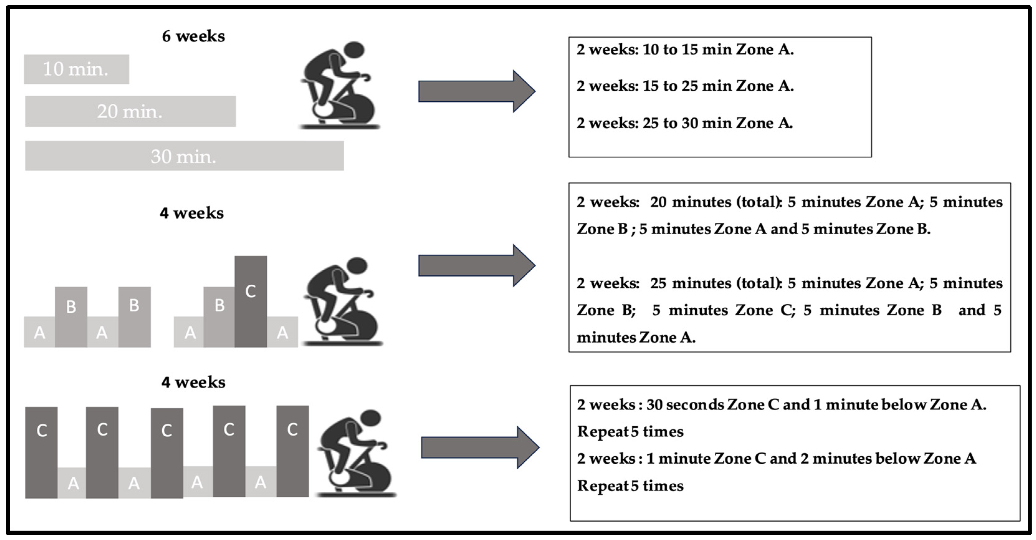
The determined thresholds were used to define PEZO-BT for each participant to implement a progressive and individualized training program. This program encompasses the continuous aerobic (CA) method, the stepped or incremental aerobic (IA) method, and the HIIT method, divided into three phases based on the subdivision of three zones between the thresholds: Zones A, B, and C [16,17,20,48,49,50]. Each third is stated by heart rate to control the intensity of every period (Figure 2).

2.4. Training

During the first period (mesocycle of adaptation), a CA training was conducted for 6 weeks. This involved cycle ergometer sessions at Zone A intensity, with a gradual progression of duration: 10 to 15 min in the first 2 weeks, 15 to 25 min in the following 2 weeks, and finally 25 to 30 min in the last 2 weeks. These sessions were performed 3 times per week.
In the second period (mesocycle of development), lasting 4 weeks, the IA method was implemented. Different intensity zones were alternated during 20 and 25 min of cycle ergometer sessions. For example, during the first 2 weeks, there were intervals of 5 min at Zone A intensity, followed by 5 min at Zone B intensity, and then again at Zone A and B. In the next 2 weeks, the intervals were 5 min at Zone A, 5 min at Zone B, 5 min at Zone C, and again at Zone A. These sessions were performed 3 times per week.
The third period (mesocycle of shock) lasted 4 weeks and involved the HIIT method. The sessions were performed at Zone C intensity and recovery at Zone A intensity. During the first 2 weeks, there were intervals of 30 s at Zone C, followed by 1 min below Zone A, repeated 5 times. In the next 2 weeks, the intervals were 1 min at Zone C, followed by 2 min below Zone A, also repeated 5 times. These sessions were conducted 2 times per week, with a minimum rest period of 72 h (Figure 3).

2.5. Statistical Analysis

Statistical analyses were performed using R (4.1.0), to test the hypotheses of interest. A mixed ANOVA analysis was considered, with time as the within-subject factor, considering 2 levels (2 measurements, pre and post), and treatment (group type) as the between-subject factor, which also had 2 levels (control group and experimental group). It was examined whether the interaction effects, and then the main effects, were significant according to the ANOVA analysis. The main effects of both the treatment and time allowed us to assess if there was an influence of these factors on the dependent variables, while the interaction effect was associated with whether the influence of one factor depended on the level of the other. The size of the effect (SE) associated with each effect was obtained through the results of eta squared. Values between 0.01 and 0.059 are considered small effect, values between 0.06 and 0.0139 are considered medium effect, and values over 0.14 are considered large effect.
If significant effects were found, post hoc tests of means (t-tests) were subsequently conducted, comparing the levels of their respective factors, and exploring the differences. Bonferroni correction was applied for significance, with a significance level set at 5%. Prior to analysis, the homogeneity of variances across groups was checked using Levene’s test separately for the pre and post time points. Additionally, a chi-square test, considering Yates’ continuity correction, was performed to compare ergospirometry criteria and ventilatory performance within the control and intervention groups based on contingency tables.

3. Results

All subjects completed the 14-week intervention, and all subjects of the control and intervention groups were evaluated. The results are presented in box plots to display the minimum, first quartile, median, third quartile, and maximum values of the submaximal variables of the subjects. Also, a contingency table was used to investigate the presence of an association or independence between variables by comparing the observed frequencies with the expected frequencies (Table 2). This analysis facilitated the classification of subjects as either normal or altered, along with the assignment of their corresponding performance scores.

3.1. Ergospirometry Submaximal Variables

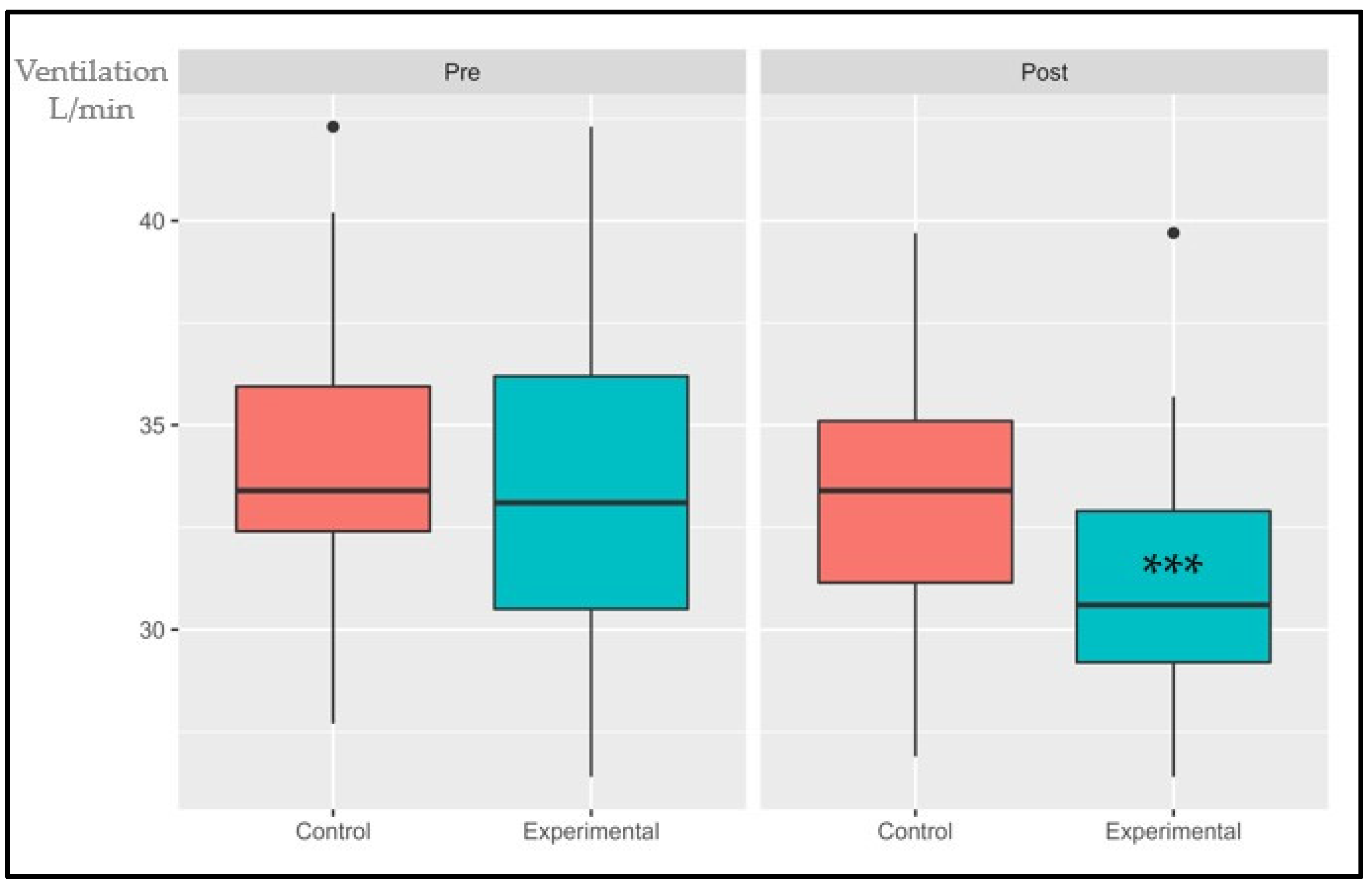
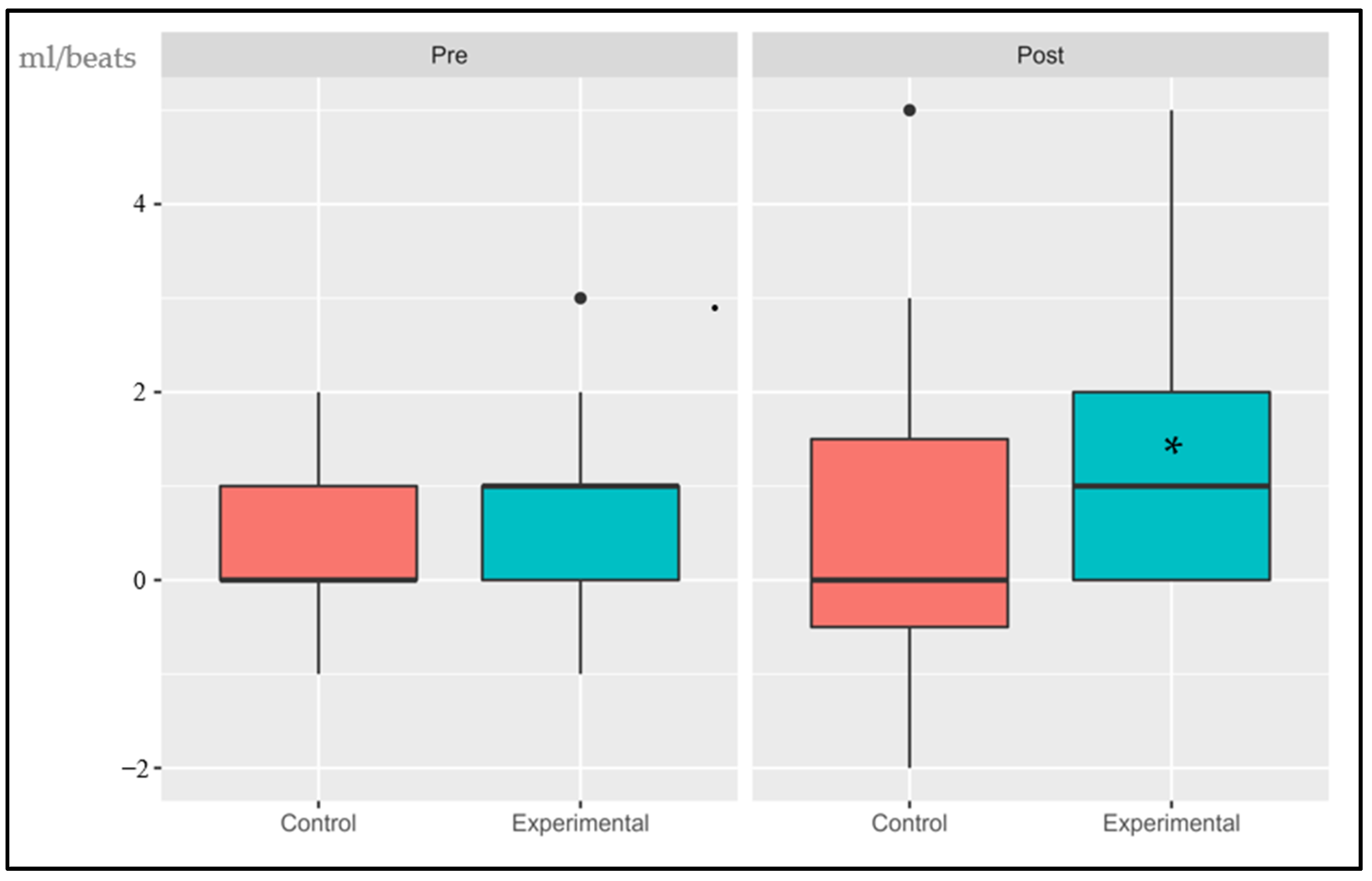
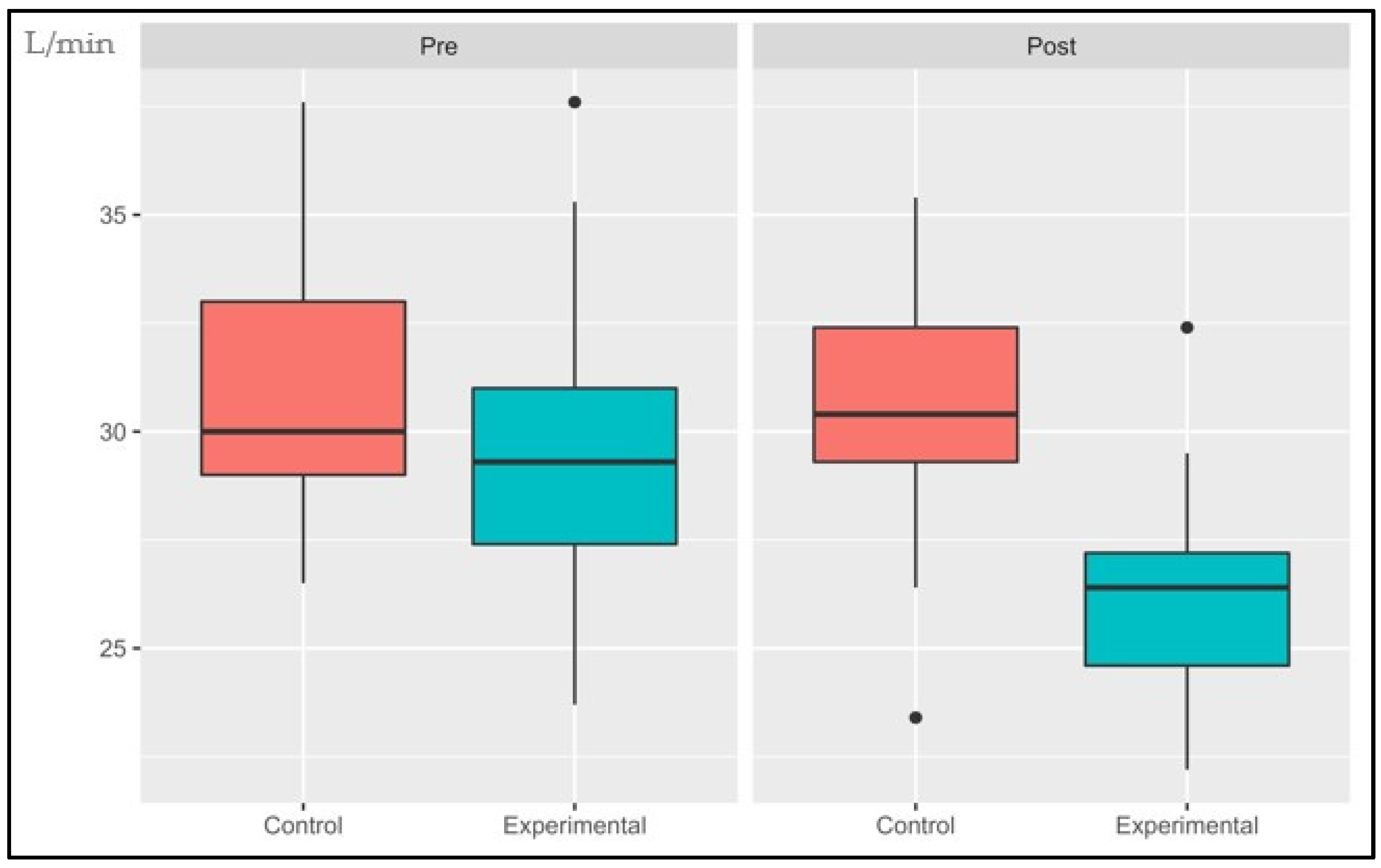
The following data are presented in box plots (Figure 4, Figure 5, Figure 6, Figure 7, Figure 8 and Figure 9), where the interested variables were analyzed. The control and experimental groups were compared in the pre and post period.

3.1.1. VO2AT

The interaction effect, primary effects of treatment, and time were significant: (p < 0.001, SE = 0.04) and (p = 0.006, SE = 0.145); (p < 0.001, SE = 0.055), respectively. There were no statistical differences in the pre period (p = 0.155) while the post period showed significant differences (p < 0.001). The control group had an average increase of 1.67% while the experimental group had an increase of 14.4%.

3.1.2. Ventilatory Class (VE/VCO2)

The interaction effect and primary effects of the treatment were not significant: (p > 0.05, SE = 0.007) and (p > 0.05, SE = 0.05), respectively. Time showed significant changes (p < 0.001, SE = 0.056). There were no statistical differences in the pre period (p = 0.0572) while the post period show significant differences (p < 0.001). The control group had an average decrease of 2.95% while the experimental group had a decrease of 6.39%.

3.1.3. Oxygen Uptake Efficiency Slope (OUES)

The interaction effect and primary effects were not significant: (p > 0.05, SE = 0.054) and (p > 0.05, SE = 0.102), respectively. However, time showed significant changes (p < 0.001, SE = 0.361). There were no statistical differences in the pre or post period: (p = 0.789) and (p = 0.636), respectively. The control group had an average increase of 11.15% while the experimental group had an increase of 5.48%.

3.1.4. Delta Oxygen Pulse (∆VO2/HR RCP vs. AT)

The interaction effect, the primary effects, and time were not significant: (p > 0.05, SE = 0.013), (p > 0.05, SE = 0.102) and (p < 0.001, SE = 0.037), respectively. There were no statistical differences in the pre period (p = 0.1) while the post period showed significant changes (p < 0.05). The control group had an average increase of 1.93% while the experimental group has an increase of 9.85%.

3.1.5. Cardiopulmonary Optimal Point (COP)

The interaction effect was significant (p < 0.05, SE = 0.083). The primary effect was not significant (p > 0.05, SE = 0.218), while time showed significant changes (p < 0.001, SE = 0126). There were no statistical differences in the pre or post period: (p = 0.622) and (p = 0.347), respectively. The control group had an average decrease of 1.08% while the experimental group had a decrease of 12.15%.

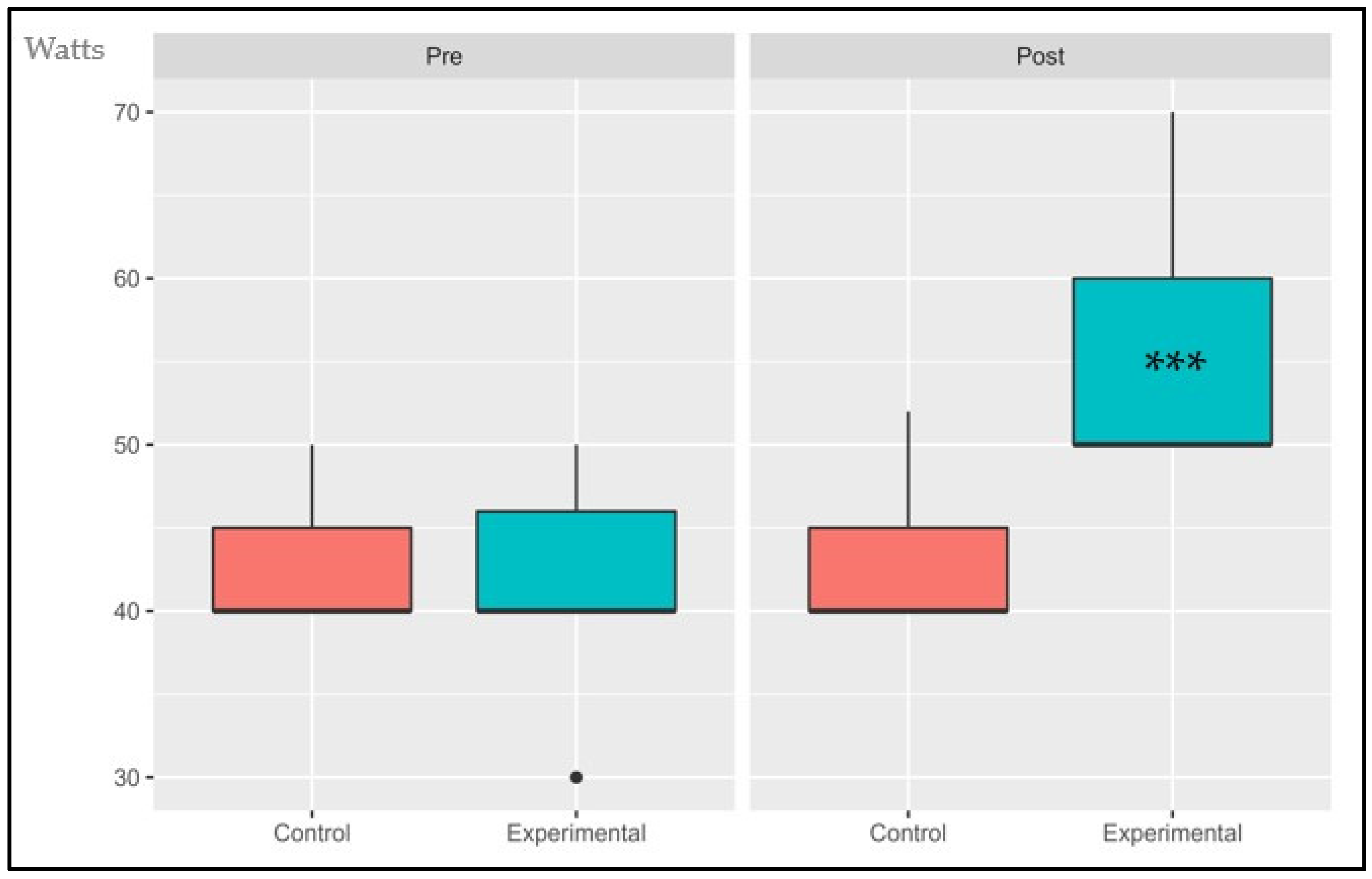
3.1.6. Power Output at AT (POAT)

The interaction effect, the primary effects, and time showed significant changes: (p < 0.001, SE = 0.260), (p = 0.006, SE = 0.181) and (p < 0.001, SE = 0.260), respectively. There were no statistical differences in the pre period (p = 1.4) while the post period presented statistical differences (p < 0.001). The control group had an average decrease of 0.62% while the experimental group has an increase of 31.6%.

3.2. Contingence Table

Comparison of ergospirometry criteria and ventilatory performance within the control and intervention groups.

4. Discussion

No previous study has examined the impact of a periodized aerobic endurance training approach within the PEZO-BT zone on CRF, determined by analyzing submaximal parameters during CPET, stopped at the RCP in elderly individuals of both sexes with comorbidities. Here, we show that 14 weeks of CA, combining IA with integrated HIIT, yielded positive changes in CRF. This was evidenced by enhancements in VO2AT, VE/VCO2, OUES, COP, ∆VO2/HR RCP vs. AT, and POAT, providing a potential strategy to counteract established age-related physiological declines in untrained octogenarians.
CRF can be assessed using submaximal parameters with comparable predictive strength for morbidity and mortality as VO2max [7,40,47,49,51,52,53,54]. Previous studies [55] reported that short-term, low-volume aerobic training with HIIT (2–8 weeks) improved peakVO2 (~4–13%) in sedentary and recreationally active healthy adults, while a 4-week HIIT program increased peak VO2 (9.6%) and anaerobic threshold (13%) in healthy older adults. Similarly, in our study, VO2AT increased by 14%. Values below 11 mL/kg/min are considered high-risk for surgical mortality [37], suggesting potential broader implications for healthy aging, as improved CRF is associated with lower all-cause mortality and cardiovascular events. Although not all subjects in the intervention group showed significant changes, there were notable improvements in their performance scores. The percentage of individuals with no limitation increased from 8% to 60%, those with moderate limitation decreased from 64% to 36%, and those with severe limitation decreased from 28% to 4% (p < 0.05). In contrast, the control group exhibited no change in individuals with no limitation (0% both pre and post assessment), an increase from 47.8% to 69.5% in those with moderate limitation, and a decrease from 52.2% to 30.4% in individuals with severe limitation (p < 0.05).
The VE/VCO2 slope reflects exercise tolerance and predicts mortality in heart failure patients [56]. Regular endurance training can improve ventilatory efficiency during exercise, potentially by reducing peripheral chemoreceptor sensitivity and lowering the VE/VCO2 slope [57]. Slope values exceeding 34 are associated with an increased risk of mortality [58]. In our study, the VE/VCO2 slope significantly decreased from 34.4 to 33.25, falling below the slope threshold of 34. Initially, nine subjects had an abnormal VE/VCO2 slope, but after the training period, only three retained this abnormality. Recently, the VE/VCO2 slope has been proposed as an alternative assessment for research involving individuals with health- or age-related limitations [59].
A meta-analysis in heart failure patients with preserved ejection fraction found that HIIT did not result in improvements in VE/VCO2 slopes [60], which contrasts with our findings. It is important to note that exercise duration varied across studies, which could potentially influence outcomes [61,62]. In our experimental group, the percentage of individuals with an abnormal slope decreased from 36% to 12%. In a 12-week study on HIIT that examined improvements in mitochondrial bioenergetics among heart failure patients, pronounced increases in OUES were observed after the intervention. This variable represents the relationship between ventilation per minute and oxygen uptake during incremental exercise, encompassing both respiratory function and musculoskeletal performance within a single index. In contrast, the control group received only medical counseling without exercise [63]. However, it is important to note that our study did not find a significant difference in OUES changes post training between the experimental and control groups. OUES represents total body efficiency providing a comprehensive view of physiological efficiency, not limited to peripheral efficiency alone. In contrast to the VE/VCO2 ratio, which primarily considers pulmonary conditions, this distinction highlights the differences between these two variables [7].
The oxygen pulse serves as a metabolic surrogate for stroke volume, which typically increases and then stabilizes during exercise. Premature flattening of the VO2/HR curve and a decrease in the oxygen pulse in later stages of exercise indicate inadequate cardiac performance relative to increased effort demands [64]. In a study of U.S. veterans, changes in the oxygen pulse occurred concurrently with alterations in peripheral endothelial function and smooth muscle activity following an aerobic training program [65]. These findings align with our study, where the intervention group improved the oxygen pulse delta between CRP and AT by almost 10% (9.85%), while the control group improved by only 1.85%. The development of myocardial ischemia during exercise may result in a reduction in stroke volume, leading to premature flattening of the O2 pulse curve. A descending O2 curve has been suggested to indicate the presence of exercise-induced myocardial ischemia, assessed through myocardial perfusion imaging [66], thus the association between myocardial fibrosis and an anomaly in the O2 pulse has also been demonstrated [67]. Given the potential presence of many of the pathological conditions studied and their association with older populations, particularly octogenarians, the delta in VO2/HR between RCP and AT has been utilized. The absence of a difference in favor of the RCP value or even a drop in the oxygen pulse at RCP vs. AT is considered abnormal [68].
COP represents the integration of the circulatory and pulmonary systems [69]. A study involving 3331 individuals defined COP categories with cutoffs: <22 L/min, 22–30 L/min, and >30 L/min. COP > 30 L/min strongly and independently predicted all-cause mortality, either alone or in conjunction with lower VO2max [39]. Following the intervention, our experimental group had fewer subjects with COP alterations (reducing from 11 to 4), whereas the control group experienced an increase (from 11 to 13) in individuals with COP > 30 L/min. An earlier study [40] delineated that a high COP, either independently or in conjunction with VO2 max, could serve as a robust predictor for all-cause mortality in community-residing adults, whether apparently healthy or afflicted with chronic conditions. COP, a submaximal prognostic index, offers a convenient adjunct to CPET assessment, particularly valuable for individuals unable or unwilling to attain maximal exertion, as observed in this investigation. Consequently, the COP index within the intervention group exhibited a substantial reduction from an initial 44% impairment (11 subjects) to a mere 4% (1 subject) post-intervention.
Furthermore, we have integrated POAT as an indicator of mechanical efficiency, concomitant with enhancements in the submaximal parameters. A meta-analysis [70] ascertained that variables such as VE/VCO2 slope, OUES, maximal oxygen pulse, and respiratory exchange ratio (RER) responded comparably to both MAE and HIIT. These findings align with a systematic review encompassing patients with coronary disease, both with and without reduced ejection fraction [71].
As far as we know, no prior study has examined the effects of an octogenarian training program on these six selected submaximal parameters. We report that 14 weeks of training had a positive influence on submaximal parameters, likely through cardiorespiratory and mitochondrial bioenergetic adaptations explained by the Wasserman model [33]. Additionally, our findings suggest specific or integrated systemic adaptations of the respiratory, cardiovascular, and muscular systems, as represented in the theoretical model adaptation we propose. This adaptation aims to elucidate the interdependence of muscle tissue, the heart, the lungs, and their respective physiological systems, with each of the selected parameters.
Our findings provide a potential strategy to counteract the physiological age-related declines established in octogenarians with illnesses, aiming to restore parameters within normal ranges and consequently promote efficient individual and integrated functioning of the respiratory, cardiovascular, and muscular systems, as postulated by the model. This would constitute a theoretical contribution from our study.
A specific program of continuous aerobic training in the VO2AT zone [51] and HIIT [72] in the CRP zone has been shown to increase submaximal parameters, offering numerous benefits for cardiorespiratory fitness and overall health. The combination of both methods and their physiological adaptations enhances the overall effectiveness of the training program. Our study reveals that the training program increases VO2AT and improves other submaximal parameters, along with exercise workload capacity.
Previous studies have demonstrated that short-term, 2- to 8-week HIIT programs effectively increased peak VO2 (by approximately 4–13%) in sedentary and recreationally active adults [55,73,74]. These findings hold significant implications for healthy aging, as improved cardiorespiratory fitness is associated with reduced cardiovascular and all-cause mortality, along with enhancements in cardiorespiratory components [75,76].
However, there are limitations to this study. Firstly, the sample size was relatively small. Future research should consider larger sample sizes, as a larger sample may provide a more accurate representation of the differences between the control and experimental groups, especially for variables that did not change significantly during the 14-week intervention. Secondly, we did not control for the nutritional and dietary status of the participants, which could potentially influence the impact of training. Factors such as digestive pathologies, calorie intake, protein percentage, and hydration status should be considered in future investigations.

5. Conclusions

This study examined the effects of a 14-week periodized aerobic training program that incorporated HIIT on octogenarians of both sexes with comorbidities. The training had a positive impact on submaximal cardiopulmonary exercise test parameters, including oxygen consumption at the ventilatory threshold, slope of ventilatory efficiency, slope of oxygen absorption efficiency, cardiorespiratory sweet spot, oxygen pulse change from anaerobic threshold to respiratory compensation point, and output power at anaerobic threshold. These improvements likely resulted from cardiorespiratory and mitochondrial adaptations, providing a strategy to counteract age-related physical declines. The training increased exercise capacity and workload. Overall, this periodic aerobic program and HIIT produced significant improvements in cardiovascular fitness in previously untrained octogenarians with and without comorbidities. Our findings suggest implications for promoting long-term healthy aging.

6. Practical Applications

This study was not conducted at our laboratory facilities; instead, it was performed and controlled at an exercise center dedicated to elderly individuals (CAE), where the subjects trained daily. This demonstrates the real-world applicability of our PEZO-BT program. We utilized ergospirometry to determine the ventilatory thresholds, but it is worth noting that low-cost equipment and alternative methods, such as lactate curve analysis or the talk test, are currently available for threshold determination.

Author Contributions

C.C.-B. was responsible for the conceptualization, funding, project management, methodology, data preservation, writing, original draft, review, and editing. F.V. was responsible for the conceptualization, funding, methodology, administration of the project, the methodology and statistical analysis, the review, and the editing. I.B.-P. was responsible for the statistical analysis. H.G.E. was responsible for conceptualization, revision, and editing. P.A.O. was responsible for revision and editing M.I. was responsible for conceptualization, data preservation, writing, review, and editing. All authors have read and agreed to the published version of the manuscript.

Funding

This research received resources from the Vicerrectoria for Research, Development and Innovation of the University of Santiago de Chile. Dicyt, código 022087CB_DAS, categoría Dicyt Asociativo.

Institutional Review Board Statement

The study was conducted in accordance with the Declaration of Helsinki and approved by the Comté de Ética of the University of Santiago de Chile (293/2020 on 24 July 2020).

Informed Consent Statement

Informed consent was obtained from all subjects involved in the study. Consent has been obtained from the patients to publish this paper.

Data Availability Statement

The datasets generated and/or analyzed during the current study are not publicly available due the terms of consent/assent to which the participants agreed but are available from the corresponding author on reasonable request. Please contact the corresponding author to discuss availability of data and materials.

Acknowledgments

To the Vicerrectoria for Research, Development, and Innovation of the University of Santiago de Chile. Dicyt, código 022087CB_DAS, categoría Dicyt Asociativo. Vicerrectoría de Investigación y Desarrollo, Universidad de Santiago de Chile, USACH, Chile.

Conflicts of Interest

The authors declare no conflict of interest.

References

  1. United Nations. World Population Ageing 2019 (st/esa/ser. a/444); Department of Economic and Social Affairs: New York, NY, USA, 2020. [Google Scholar]
  2. Courel-Ibanez, J.; Buendia-Romero, A.; Pallares, J.G.; Garcia-Conesa, S.; Martinez-Cava, A.; Izquierdo, M. Impact of Tailored Multicomponent Exercise for Preventing Weakness and Falls on Nursing Home Residents’ Functional Capacity. J. Am. Med. Dir. Assoc. 2022, 23, 98–104.e103. [Google Scholar] [CrossRef]
  3. Valdivia, G.; Dominguez, A.L.A. Población de 80 y más años en Chile: Una visión preliminar desde el punto de vista epidemiológico. Revista Médica Clínica Las Condes 2012, 23, 5–12. [Google Scholar] [CrossRef]
  4. Departamento de Estadísticas e Información de Salud. Población ine por Grupo Etario. Available online: https://deis.minsal.cl/ (accessed on 25 December 2021).
  5. Izquierdo, M.; Duque, G.; Morley, J.E. Physical activity guidelines for older people: Knowledge gaps and future directions. Lancet Healthy Longev. 2021, 2, e380–e383. [Google Scholar] [CrossRef] [PubMed]
  6. Naruse, M.; Trappe, S.; Trappe, T.A. Human skeletal muscle-specific atrophy with aging: A comprehensive review. J. Appl. Physiol. 2023, 134, 900–914. [Google Scholar] [CrossRef] [PubMed]
  7. Cofre-Bolados, C.; Ferrari, G.; Valdivia-Moral, P.; Vidal-Diaz, F.; Ramirez-Velez, R.; Izquierdo-Redin, M. Sub Maximal Ergospirometry Parameters in Untrained Non-Frail Octogenarian Subjects. Medicina (Kaunas) 2022, 58, 378. [Google Scholar] [CrossRef] [PubMed]
  8. Fernandez-Garcia, A.I.; Moradell, A.; Navarrete-Villanueva, D.; Subias-Perie, J.; Perez-Gomez, J.; Ara, I.; Gonzalez-Gross, M.; Casajus, J.A.; Vicente-Rodriguez, G.; Gomez-Cabello, A. Effects of Multicomponent Training Followed by a Detraining Period on Frailty Level and Functional Capacity of Older Adults with or at Risk of Frailty: Results of 10-Month Quasi-Experimental Study. Int. J. Environ. Res. Public Health 2022, 19, 12417. [Google Scholar] [CrossRef] [PubMed]
  9. Cadore, E.L.; Casas-Herrero, A.; Zambom-Ferraresi, F.; Idoate, F.; Millor, N.; Gomez, M.; Rodriguez-Manas, L.; Izquierdo, M. Multicomponent exercises including muscle power training enhance muscle mass, power output, and functional outcomes in institutionalized frail nonagenarians. Age (Dordr.) 2014, 36, 773–785. [Google Scholar] [CrossRef]
  10. Trappe, S.; Hayes, E.; Galpin, A.; Kaminsky, L.; Jemiolo, B.; Fink, W.; Trappe, T.; Jansson, A.; Gustafsson, T.; Tesch, P. New records in aerobic power among octogenarian lifelong endurance athletes. J. Appl. Physiol. 2013, 114, 3–10. [Google Scholar] [CrossRef]
  11. Casas-Herrero, A.; Anton-Rodrigo, I.; Zambom-Ferraresi, F.; Saez de Asteasu, M.L.; Martinez-Velilla, N.; Elexpuru-Estomba, J.; Marin-Epelde, I.; Ramon-Espinoza, F.; Petidier-Torregrosa, R.; Sanchez-Sanchez, J.L.; et al. Effect of a multicomponent exercise programme (VIVIFRAIL) on functional capacity in frail community elders with cognitive decline: Study protocol for a randomized multicentre control trial. Trials 2019, 20, 362. [Google Scholar] [CrossRef]
  12. Janssens, J.P.; Pache, J.C.; Nicod, L.P. Physiological changes in respiratory function associated with ageing. Eur. Respir. J. 1999, 13, 197–205. [Google Scholar] [CrossRef]
  13. Petrella, M.; Aprahamian, I.; Mamoni, R.L.; de Vasconcellos Romanini, C.F.; Lima, N.A.; de Cassio Robello, E.; da Costa, D.L.; An, V.N.; Aguirre, B.N.; Galdeano, J.R.; et al. The effect of a multicomponent exercise protocol (VIVIFRAIL(c)) on inflammatory profile and physical performance of older adults with different frailty status: Study protocol for a randomized controlled trial. BMC Geriatr. 2021, 21, 83. [Google Scholar] [CrossRef] [PubMed]
  14. World Health Organization. WHO Guidelines on Physical Activity and Sedentary Behaviour: Web Annex: Evidence Profiles; World Health Organization: Geneva, Switzerland, 2020. [Google Scholar]
  15. Izquierdo, M.; Merchant, R.A.; Morley, J.E.; Anker, S.D.; Aprahamian, I.; Arai, H.; Aubertin-Leheudre, M.; Bernabei, R.; Cadore, E.L.; Cesari, M.; et al. International Exercise Recommendations in Older Adults (ICFSR): Expert Consensus Guidelines. J. Nutr. Health Aging 2021, 25, 824–853. [Google Scholar] [CrossRef] [PubMed]
  16. Billat, L.V. Interval training for performance: A scientific and empirical practice. Special recommendations for middle- and long-distance running. Part II: Anaerobic interval training. Sports Med. 2001, 31, 75–90. [Google Scholar] [CrossRef]
  17. Billat, L.V. Interval training for performance: A scientific and empirical practice. Special recommendations for middle- and long-distance running. Part I: Aerobic interval training. Sports Med. 2001, 31, 13–31. [Google Scholar] [CrossRef] [PubMed]
  18. Laursen, P.B.; Jenkins, D.G. The scientific basis for high-intensity interval training: Optimising training programmes and maximising performance in highly trained endurance athletes. Sports Med. 2002, 32, 53–73. [Google Scholar] [CrossRef]
  19. Buchheit, M.; Laursen, P.B. High-intensity interval training, solutions to the programming puzzle: Part II: Anaerobic energy, neuromuscular load and practical applications. Sports Med. 2013, 43, 927–954. [Google Scholar] [CrossRef]
  20. Buchheit, M.; Laursen, P.B. High-intensity interval training, solutions to the programming puzzle: Part I: Cardiopulmonary emphasis. Sports Med. 2013, 43, 313–338. [Google Scholar] [CrossRef]
  21. Rohmansyah, N.A.; Ka Praja, R.; Phanpheng, Y.; Hiruntrakul, A. High-Intensity Interval Training Versus Moderate-Intensity Continuous Training for Improving Physical Health in Elderly Women. Inquiry 2023, 60, 469580231172870. [Google Scholar] [CrossRef]
  22. Roxburgh, B.H.; Nolan, P.B.; Weatherwax, R.M.; Dalleck, L.C. Is moderate intensity exercise training combined with high intensity interval training more effective at improving cardiorespiratory fitness than moderate intensity exercise training alone? J. Sports Sci. Med. 2014, 13, 702. [Google Scholar]
  23. Kessler, H.S.; Sisson, S.B.; Short, K.R. The potential for high-intensity interval training to reduce cardiometabolic disease risk. Sports Med. 2012, 42, 489–509. [Google Scholar] [CrossRef]
  24. Alvarez, C.; Ramirez-Campillo, R.; Martinez-Salazar, C.; Mancilla, R.; Flores-Opazo, M.; Cano-Montoya, J.; Ciolac, E.G. Low-Volume High-Intensity Interval Training as a Therapy for Type 2 Diabetes. Int. J. Sports Med. 2016, 37, 723–729. [Google Scholar] [CrossRef] [PubMed]
  25. Tjønna, A.E.; Lee, S.J.; Rognmo, Ø.; Stølen, T.O.; Bye, A.; Haram, P.M.; Loennechen, J.P.l.; Al-Share, Q.Y.; Skogvoll, E.; Slørdahl, S.A. Aerobic interval training versus continuous moderate exercise as a treatment for the metabolic syndrome: A pilot study. Circulation 2008, 118, 346–354. [Google Scholar] [CrossRef] [PubMed]
  26. Cadore, E.L.; Saez de Asteasu, M.L.; Izquierdo, M. Multicomponent exercise and the hallmarks of frailty: Considerations on cognitive impairment and acute hospitalization. Exp. Gerontol. 2019, 122, 10–14. [Google Scholar] [CrossRef] [PubMed]
  27. Hosseinzadeh, K. High Intensity Interval Training in Elderly Could Improve Elderly Health Significantly. Int. J. Aging Health Mov. 2019, 1, 10–11. [Google Scholar]
  28. Older, P.; Hall, A. The role of cardiopulmonary exercise testing for preoperative evaluation of the elderly. Mortality 1996, 11, 10. [Google Scholar]
  29. West, M.; Jack, S.; Grocott, M. Perioperative cardiopulmonary exercise testing in the elderly. Best Pract. Res. Clin. Anaesthesiol. 2011, 25, 427–437. [Google Scholar] [CrossRef]
  30. Izquierdo, M. Multicomponent physical exercise program: Vivifrail. Nutr. Hosp. 2019, 36, 50–56. [Google Scholar] [CrossRef]
  31. Lambert, C.P.; Evans, W.J. Adaptations to aerobic and resistance exercise in the elderly. Rev. Endocr. Metab. Disord. 2005, 6, 137–143. [Google Scholar] [CrossRef]
  32. Gonzales, T.I.; Westgate, K.; Strain, T.; Hollidge, S.; Jeon, J.; Christensen, D.L.; Jensen, J.; Wareham, N.J.; Brage, S. Cardiorespiratory fitness assessment using risk-stratified exercise testing and dose–response relationships with disease outcomes. Sci. Rep. 2021, 11, 15315. [Google Scholar] [CrossRef]
  33. Sietsema, K.E.; Stringer, W.W.; Sue, D.Y.; Ward, S. Wasserman & Whipp’s Principles of Exercise Testing and Interpretation, 6th ed.; Lippincott Williams & Wilkins (LWW): Philadelphia, PA, USA, 2020. [Google Scholar]
  34. Ross, R.; Blair, S.N.; Arena, R.; Church, T.S.; Després, J.-P.; Franklin, B.A.; Haskell, W.L.; Kaminsky, L.A.; Levine, B.D.; Lavie, C.J.; et al. Importance of Assessing Cardiorespiratory Fitness in Clinical Practice: A Case for Fitness as a Clinical Vital Sign: A Scientific Statement from the American Heart Association. Circulation 2016, 134, e653–e699. [Google Scholar] [CrossRef]
  35. Arena, R.; Myers, J.; Abella, J.; Peberdy, M.A.; Bensimhon, D.; Chase, P.; Guazzi, M. Development of a ventilatory classification system in patients with heart failure. Circulation 2007, 115, 2410–2417. [Google Scholar] [CrossRef] [PubMed]
  36. Guazzi, M.; Arena, R.; Halle, M.; Piepoli, M.F.; Myers, J.; Lavie, C.J. 2016 focused update: Clinical recommendations for cardiopulmonary exercise testing data assessment in specific patient populations. Eur. Heart J. 2018, 39, 1144–1161. [Google Scholar] [CrossRef] [PubMed]
  37. Gitt, A.K.; Wasserman, K.; Kilkowski, C.; Kleemann, T.; Kilkowski, A.; Bangert, M.; Schneider, S.; Schwarz, A.; Senges, J. Exercise anaerobic threshold and ventilatory efficiency identify heart failure patients for high risk of early death. Circulation 2002, 106, 3079–3084. [Google Scholar] [CrossRef] [PubMed]
  38. Coeckelberghs, E.; Buys, R.; Goetschalckx, K.; Cornelissen, V.A.; Vanhees, L. Prognostic value of the oxygen uptake efficiency slope and other exercise variables in patients with coronary artery disease. Eur. J. Prev. Cardiol. 2016, 23, 237–244. [Google Scholar] [CrossRef] [PubMed]
  39. Ramos, P.S.; Araújo, C.G.S. Cardiorespiratory optimal point during exercise testing as a predictor of all-cause mortality. Rev. Port. De Cardiol. 2017, 36, 261–269. [Google Scholar] [CrossRef]
  40. Dias Ferreira Reis, J.; Goncalves, A.; Bras, P.; Ferreira, V.; Viegas, J.; Rio, P.; Moreira, R.; Pereira Silva, T.; Timoteo, A.; Soares, R. Prognostic value of the cardiorespiratory optimal point during submaximal exercise testing. Eur. Heart J. 2020, 41, ehaa946.0957. [Google Scholar] [CrossRef]
  41. Baba, R.; Tsuyuki, K.; Yano, H.; Ninomiya, K.; Ebine, K. Robustness of the oxygen uptake efficiency slope to exercise intensity in patients with coronary artery disease. Nagoya J. Med. Sci. 2010, 72, 83–89. [Google Scholar]
  42. Barros, C.R.; Monte-Silva, K.; Sales, C.B.; Souza, R.P.; Santos, T.; Pessoa, M.; Viana, R.; Alcoforado, L.; Lima, A.M.J.; Fernandes, J.; et al. Oxygen uptake efficiency slope: A submaximal test evaluation tool that provides cardiopulmonary reserve data in individuals with Parkinson’s disease. Braz. J. Phys. Ther. 2021, 25, 641–647. [Google Scholar] [CrossRef]
  43. Silva, M.; Baptista, L.; Neves, R.; França, E.; Loureiro, H.; Rezende, M.; Ferreira, V.; Veríssimo, M.; Martins, R. High intensity interval training improves health-related quality of life in adults and older adults with diagnosed cardiovascular risk. J. Phys. Educ. Sport 2019, 19, 611–618. [Google Scholar]
  44. Ramírez-Vélez, R.; García-Alonso, N.; Legarra-Gorgoñón, G.; Oscoz-Ochandorena, S.; Oteiza, J.; Izquierdo, M. Ventilatory efficiency in response to maximal exercise in persistent COVID-19 syndrome patients: A cross-sectional study. Rev. Esp. Cardiol. 2023, 76, 206–209. [Google Scholar] [CrossRef]
  45. Rudd, A.; Khan, H.; Gamble, D.; Stephen, P.; Horgan, G.; Dawson, A.; Frenneaux, M.; Dawson, D.K. 76 Oxygen uptake efficiency slope—A valuable substitute for peak vo2? Heart 2022, 108, A56–A57. [Google Scholar] [CrossRef]
  46. Laukkanen, J.A.; Kunutsor, S.K.; Yates, T.; Willeit, P.; Kujala, U.M.; Khan, H.; Zaccardi, F. Prognostic relevance of cardiorespiratory fitness as assessed by submaximal exercise testing for all-cause mortality: A UK Biobank prospective study. Mayo Clin. Proc. 2020, 95, 867–878. [Google Scholar] [CrossRef] [PubMed]
  47. Reed, J.L.; Cotie, L.M.; Cole, C.A.; Harris, J.; Moran, B.; Scott, K.; Terada, T.; Buckley, J.P.; Pipe, A.L. Submaximal Exercise Testing in Cardiovascular Rehabilitation Settings (BEST Study). Front. Physiol. 2019, 10, 1517. [Google Scholar] [CrossRef]
  48. David, R.; Bassett, J. Scientific contributions of A. V. Hill: Exercise physiology pioneer. J. Appl. Physiol. 2002, 93, 1567–1582. [Google Scholar] [CrossRef]
  49. Meyer, T.; Lucia, A.; Earnest, C.P.; Kindermann, W. A conceptual framework for performance diagnosis and training prescription from submaximal gas exchange parameters--theory and application. Int. J. Sports Med. 2005, 26 (Suppl. S1), S38–S48. [Google Scholar] [CrossRef] [PubMed]
  50. Issurin, V.B. New horizons for the methodology and physiology of training periodization. Sports Med. 2010, 40, 189–206. [Google Scholar] [CrossRef] [PubMed]
  51. Araújo, B.T.S.; Barros, A.; Nunes, D.T.X.; Remígio de Aguiar, M.I.; Mastroianni, V.W.; de Souza, J.A.F.; Fernades, J.; Campos, S.L.; Brandão, D.C.; Dornelas de Andrade, A. Effects of continuous aerobic training associated with resistance training on maximal and submaximal exercise tolerance, fatigue, and quality of life of patients post-COVID-19. Physiother. Res. Int. 2023, 28, e1972. [Google Scholar] [CrossRef] [PubMed]
  52. Guazzi, M.; Agostoni, P.G. Monitoring gas exchange during a constant work rate exercise in patients with left ventricular dysfunction treated with carvedilol. Am. J. Cardiol. 2000, 85, 660–664. [Google Scholar] [CrossRef]
  53. Malhotra, R.; Bakken, K.; D’Elia, E.; Lewis, G.D. Cardiopulmonary Exercise Testing in Heart Failure. JACC Heart Fail. 2016, 4, 607–616. [Google Scholar] [CrossRef]
  54. Noonan, V.; Dean, E. Submaximal Exercise Testing: Clinical Application and Interpretation. Phys. Ther. 2000, 80, 782–807. [Google Scholar] [CrossRef]
  55. Sloth, M.; Sloth, D.; Overgaard, K.; Dalgas, U. Effects of sprint interval training on VO2max and aerobic exercise performance: A systematic review and meta-analysis. Scand. J. Med. Sci. Sports 2013, 23, e341–e352. [Google Scholar] [CrossRef] [PubMed]
  56. Francis, D.P.; Shamim, W.; Davies, L.C.; Piepoli, M.F.; Ponikowski, P.; Anker, S.D.; Coats, A.J.S. Cardiopulmonary exercise testing for prognosis in chronic heart failure: Continuous and independent prognostic value from VE/VCO2slope and peak VO2. Eur. Heart J. 2000, 21, 154–161. [Google Scholar] [CrossRef] [PubMed]
  57. McGurk, S.P.; Blanksby, B.A.; Anderson, M.J. The relationship of hypercapnic ventilatory responses to age, gender and athleticism. Sports Med. 1995, 19, 173–183. [Google Scholar] [CrossRef]
  58. Moore, B.; Brubaker, P.H.; Stewart, K.P.; Kitzman, D.W. VE/VCO2 slope in older heart failure patients with normal versus reduced ejection fraction compared with age-matched healthy controls. J. Card. Fail. 2007, 13, 259–262. [Google Scholar] [CrossRef] [PubMed]
  59. Louras, P.; Kamil-Rosenberg, S.; Myers, J.; Fairchild, J.K. Validation of the VE/VCO2 Slope as a Marker of Cognitive Performance in Older Adults. Arch. Phys. Med. Rehabil. 2022, 103, e158. [Google Scholar] [CrossRef]
  60. Lai, P.; Xue, J.-H.; Xie, M.-J.; Ye, J.-H.; Yang, N.; Zhong, Y.-M.; Liao, Y.-L. High-intensity and moderate-intensity interval training in heart failure with preserved ejection fraction: A meta-analysis of randomized controlled trials. Medicine 2023, 102, e33010. [Google Scholar] [CrossRef]
  61. Donelli da Silveira, A.; Beust de Lima, J.; da Silva Piardi, D.; dos Santos Macedo, D.; Zanini, M.; Nery, R.; Laukkanen, J.A.; Stein, R. High-intensity interval training is effective and superior to moderate continuous training in patients with heart failure with preserved ejection fraction: A randomized clinical trial. Eur. J. Prev. Cardiol. 2020, 27, 1733–1743. [Google Scholar] [CrossRef]
  62. Mueller, S.; Winzer, E.B.; Duvinage, A.; Gevaert, A.B.; Edelmann, F.; Haller, B.; Pieske-Kraigher, E.; Beckers, P.; Bobenko, A.; Hommel, J. Effect of high-intensity interval training, moderate continuous training, or guideline-based physical activity advice on peak oxygen consumption in patients with heart failure with preserved ejection fraction: A randomized clinical trial. JAMA 2021, 325, 542–551. [Google Scholar] [CrossRef]
  63. Chou, C.-H.; Fu, T.-C.; Tsai, H.-H.; Hsu, C.-C.; Wang, C.-H.; Wang, J.-S. High-intensity interval training enhances mitochondrial bioenergetics of platelets in patients with heart failure. Int. J. Cardiol. 2019, 274, 214–220. [Google Scholar] [CrossRef]
  64. Palermo, P.; Mapelli, M.; Campodonico, J.; Vignati, C.; Agostoni, P. P310 Cardiopulmonary Exercise Test in the Evaluation of a patient with Hfref: The Importance in the Analysis of Gas Kinetics. Eur. Heart J. Suppl. 2023, 25, D163–D164. [Google Scholar] [CrossRef]
  65. Oliveira, R.B.; Myers, J.; Araújo, C.G.; Abella, J.; Mandic, S.; Froelicher, V. Maximal exercise oxygen pulse as a predictor of mortality among male veterans referred for exercise testing. Eur. J. Cardiovasc. Prev. Rehabil. 2009, 26, 358–364. [Google Scholar] [CrossRef]
  66. Belardinelli, R.; Lacalaprice, F.; Carle, F.; Minnucci, A.; Cianci, G.; Perna, G.; D’Eusanio, G. Exercise-induced myocardial ischaemia detected by cardiopulmonary exercise testing. Eur. Heart J. 2003, 24, 1304–1313. [Google Scholar] [CrossRef] [PubMed]
  67. Munhoz, E.C.; Hollanda, R.; Vargas, J.P.; Silveira, C.W.; Lemos, A.L.; Hollanda, R.; Ribeiro, J.P. Flattening of oxygen pulse during exercise may detect extensive myocardial ischemia. Med. Sci. Sports Exerc. 2007, 39, 1221–1226. [Google Scholar] [CrossRef] [PubMed]
  68. De Lorenzo, A.; da Silva, C.L.; Souza, F.C.C.; Serra, S.; Marino, P.; Sl Lima, R. Clinical, scintigraphic, and angiographic predictors of oxygen pulse abnormality in patients undergoing cardiopulmonary exercise testing. Clin. Cardiol. 2017, 40, 914–918. [Google Scholar] [CrossRef] [PubMed]
  69. Ramos, P.S.; Ricardo, D.R.; Araujo, C.G. Cardiorespiratory optimal point: A submaximal variable of the cardiopulmonary exercise testing. Arq. Bras. Cardiol. 2012, 99, 988–996. [Google Scholar] [CrossRef] [PubMed]
  70. Du, L.; Zhang, X.; Chen, K.; Ren, X.; Chen, S.; He, Q. Effect of high-intensity interval training on physical health in coronary artery disease patients: A meta-analysis of randomized controlled trials. J. Cardiovasc. Dev. Dis. 2021, 8, 158. [Google Scholar] [CrossRef]
  71. Pattyn, N.; Coeckelberghs, E.; Buys, R.; Cornelissen, V.A.; Vanhees, L. Aerobic interval training vs. moderate continuous training in coronary artery disease patients: A systematic review and meta-analysis. Sports Med. 2014, 44, 687–700. [Google Scholar] [CrossRef]
  72. Xie, B.; Yan, X.; Cai, X.; Li, J. Effects of High-Intensity Interval Training on Aerobic Capacity in Cardiac Patients: A Systematic Review with Meta-Analysis. BioMed Res. Int. 2017, 2017, 5420840. [Google Scholar] [CrossRef]
  73. Blackwell, J.E.M.; Gharahdaghi, N.; Brook, M.S.; Watanabe, S.; Boereboom, C.L.; Doleman, B.; Lund, J.N.; Wilkinson, D.J.; Smith, K.; Atherton, P.J.; et al. The physiological impact of high-intensity interval training in octogenarians with comorbidities. J. Cachexia Sarcopenia Muscle 2021, 12, 866–879. [Google Scholar] [CrossRef]
  74. Laukkanen, J.A.; Zaccardi, F.; Khan, H.; Kurl, S.; Rauramaa, R. Abstract 19920: Long-term Change in Cardiorespiratory Fitness and All-cause Mortality: A Follow-up Study. Circulation 2015, 132, A19920. [Google Scholar] [CrossRef]
  75. Boereboom, C.L.; Phillips, B.E.; Williams, J.P.; Lund, J.N. A 31-day time to surgery compliant exercise training programme improves aerobic health in the elderly. Tech. Coloproctol. 2016, 20, 375–382. [Google Scholar] [CrossRef] [PubMed]
  76. Lee, D.-c.; Sui, X.; Artero, E.G.; Lee, I.-M.; Church, T.S.; McAuley, P.A.; Stanford, F.C.; Kohl, H.W.; Blair, S.N. Long-Term Effects of Changes in Cardiorespiratory Fitness and Body Mass Index on All-Cause and Cardiovascular Disease Mortality in Men. Circulation 2011, 124, 2483–2490. [Google Scholar] [CrossRef] [PubMed]
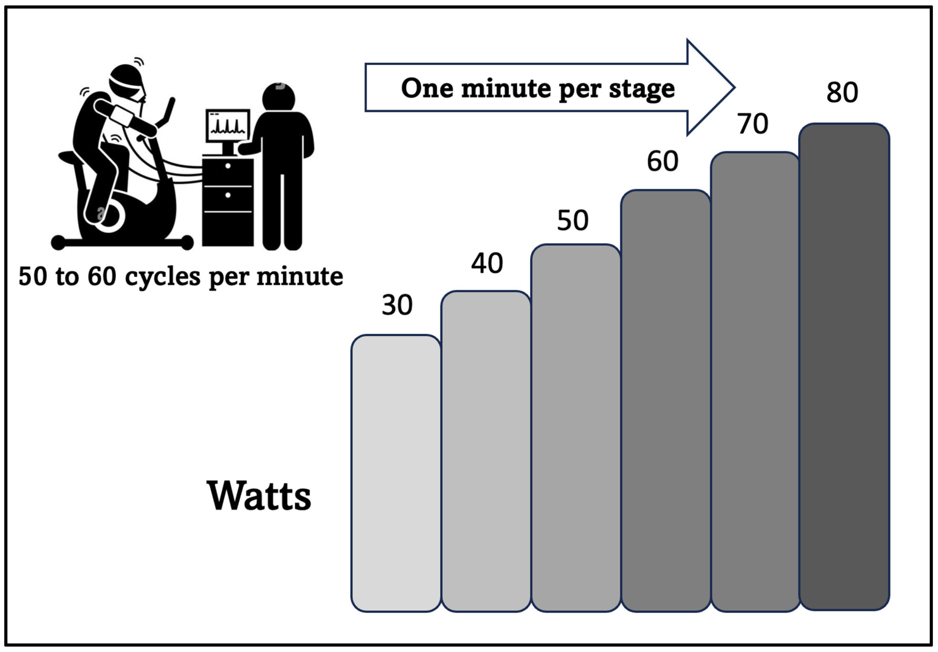
Figure 1. Ramp protocol with a 10-watt load increase per minute, starting at 30 watts (aimed at elderly population with low aerobic power).
Sports 11 00219 g001
Figure 2. Training zones between thresholds. The aerobic training PEZO-BT used the individuals’ HR established for ZONE A, B, and C.
Sports 11 00219 g002
Figure 3. Overview of the training program of Periodization Zone between Thresholds (PEZO-BT).
Sports 11 00219 g003
Figure 4. Absolute changes in VO2 at AT after 14 weeks of intervention; ● represents outlier data. Values are expressed in mL/kg/min. *** p < 0.001.
Sports 11 00219 g004
Figure 5. Absolute changes in ventilatory class after 14 weeks of intervention; ● represents outlier data. Values are expressed in L/min. *** p < 0.001.
Sports 11 00219 g005
Figure 6. Absolute changes in OUES after 14 weeks of intervention; ● represents outlier data. Values are expressed in L/min.
Sports 11 00219 g006
Figure 7. Absolute changes in ∆VO2/HR (RCP vs. AT) after 14 weeks of intervention; ● represents outlier data. Values are expressed as the difference in mL-beats of VO2/HR at RCP and AT. * p < 0.05.
Sports 11 00219 g007
Figure 8. Absolute changes in COP after 14 weeks of intervention; ● represents outlier data. Values are expressed in L/min.
Sports 11 00219 g008
Figure 9. Absolute changes in power output after 14 weeks of intervention; ● represents outlier data. Values are expressed in Watts. *** p < 0.001.
Sports 11 00219 g009
Table 1. Characteristics (n = 48) of the octogenarian sample.
VariableGroup
Control (8M; 15F)Intervention (4M; 21F)
Age (y), mean ± SD83.4 ± 3.583.3 ± 3.4
Weight (kg), mean ± SD64.9 ± 11.166.4 ± 13.7
Height (m), mean ± SD1.58 ± 7.11.58 ± 7.4
BMI (kg/m2), mean ± SD25.9 ± 726.4 ± 4.7
Cardiometabolic disease, number (%)16 (68)18 (72)
Cardiometabolic drugs, number (%)14 (61)16 (64)
Values are presented as mean and standard deviation. Percentages of both groups that present with controlled, non-complex cardiometabolic diseases, such as arterial hypertension, diabetes mellitus, insulin resistance, dyslipidemia, or fatty liver, are included, in addition to the percentage of subjects in the sample who use cardiometabolic drugs. (Data extracted from the medical file of each participant).
Table 2. The table shows the clinical differences between the two groups, classifying the variables as either normal or altered.
Group Differences
VariableCategoryControlχ2pInterventionχ2p
Pre Mean (SD)Post Mean (SD)Pre Mean (SD)Post Mean (SD)
VO2AT (mL/kg/min)<11 altered17 (73.9)16 (69.5)00.9910 (40)4 (16)4.4770.034
≥11 normal6 (26.1)7 (30.5)15 (60)21 (84)
VE/VCO2 (L/min)>34 altered9 (39)8 (34.7)00.999 (36)3 (12)3.3150.069
≤34 normal14 (61)15 (65.3)16 (64)22 (88)
OUES (mL/min)<1550 altered21 (91.3)6 (26)0.010.97115 (60)6 (24)3.2990.069
≥1550 normal2 (8.7)17 (74)10 (40)19 (76)
COP (L/min)>30 altered11 (47.8)13 (56.5)00.9911 (44)1 (4)0.0150.902
≤30 normal12 (52.2)10 (43.5)14 (56)24 (96)
∆VO2/HR RCP vs. AT<0 altered14 (61)13 (57)0.2560.6139 (36)7 (28)00.99
≥0 normal9 (39)10 (43)16 (64)18 (72)
VO2AT (mL/kg/min), VO2 at AT; VE/VCO2 (L/min), ventilatory class (L/min); OUES, oxygen uptake efficiency slope (mL/min); COP, cardiorespiratory optimal point(L/min); ∆VO2/HR RCP vs. AT, difference between oxygen pulse at RCP and AT. The submaximal variables are categorized as altered or normal according with their cutoff points. The ventilatory performance score was derived from the sum of abnormal criteria of each submaximal variable.
Disclaimer/Publisher’s Note: The statements, opinions and data contained in all publications are solely those of the individual author(s) and contributor(s) and not of MDPI and/or the editor(s). MDPI and/or the editor(s) disclaim responsibility for any injury to people or property resulting from any ideas, methods, instructions or products referred to in the content.