Review

Histone Modifications in the Cell Cycle of C. elegans Embryogenesis: A Comparative Review

by
Anati Alyaa Azhar
1 and
Hector Mendoza
2,*
1
Department of Molecular, Cellular and Developmental Biology, University of Michigan, Ann Arbor, MI 48109, USA
2
Department of Biology, Loyola University Chicago, Chicago, IL 60660, USA
*
Author to whom correspondence should be addressed.
Epigenomes 2026, 10(1), 15; https://doi.org/10.3390/epigenomes10010015
Submission received: 12 December 2025 / Revised: 15 February 2026 / Accepted: 26 February 2026 / Published: 27 February 2026

Abstract

Cell division is a highly regulated process that actively involves dynamic changes to the genetic material within the nucleus. DNA is faithfully replicated in the S-Phase of the cell cycle, being converted from loose, relaxed chromatin into tight, condensed chromosomes to be segregated in mitosis. In addition to scaffolding proteins that shape these mitotic chromosomes, post-translational modifications of histones within nucleosomes modulate chromosome dynamics throughout the cell cycle. In this review, we use a comparative approach to highlight some of the major epigenetic marks affected by the cell cycle during embryogenesis of Caenorhabditis elegans: H4K20me1, H3S10ph, H4S1ph, H2AS1ph, and H3T118ph. These five histone post-translational modifications will be specifically highlighted in the context of the mitotic cell cycle, as they are well documented in the C. elegans literature.

1. Introduction

Epigenetic modifications involve stable alterations to gene expression or phenotype achieved without changing the underlying DNA sequence. These modifications are often subject to mitotic inheritance and, in some cases, to transgenerational (meiotic) inheritance. However, many are transient due to their involvement in regulatory processes and more accurately considered features of chromatin dynamics, as opposed to true epigenetic memory. Modifications, like DNA methylation and modifications of histone proteins, are chemical moieties or ‘tags’ that alter DNA accessibility and chromatin structure, which in turn impact gene expression [1]. These processes have been implicated in the regulation of the cell cycle, a coordinated series of events in which gene expression is primarily impacted by chromatin dynamics through covalent changes to DNA and histones [2,3]. The conventional cell cycle begins with an interphase that consists of three phases: the G1-Phase, in which the cell grows, the S-Phase, in which the genetic material is synthesized or replicated, and the G2-Phase, in which the cell prepares for active nuclear division by synthesizing the necessary organelles and proteins. When checkpoint requirements are met, the cell will enter the M-Phase, in which the nucleus actively divides (karyokinesis) and the cytoplasm is split in two (cytokinesis) to produce genetically identical daughter cells [4,5,6].
Epigenetic modifiers regulate the cell cycle by controlling gene expression, as well as chromatin condensation and chromosome segregation [7,8]. Chromatin is highly dynamic in proliferating cells. In the S-Phase, new histones are incorporated with nascent DNA, creating a requirement for the re-establishment of histone modifications. Chromatin remodelers and the transcriptional machinery dissociate from chromatin during mitosis, yet the cell continues to maintain its transcriptional identity post-mitosis, highlighting the importance of histone modifications and their impact on the progression of the cell cycle. Post-translational modification (PTM) of histones involves the covalent attachment of chemical groups to specific residues that can influence the state of chromatin, which consists of repeating nucleosome units in which DNA segments are wrapped around histone proteins to form a “beads-on-a-string” structure that can fit into the nucleus [9,10]. Histone PTMs include methylation, acetylation, and phosphorylation. Specific patterns of histone modifications make up an “epigenetic code” or language that impacts chromatin dynamics and accessibility, ultimately influencing the transcriptional output of specific regions of the DNA [11].
In Caenorhabditis elegans, most active cell division occurs in the first half of embryogenesis, as cells cycle rapidly from the S-Phase to the M-Phase [12]. Apart from two germ cells, the embryonic cells are somatic and divide mitotically. Epigenetic changes vary during this progression, playing a key role in the condensation and segregation of chromosomes, the dissociation of effectors from the chromosomes, and the tight regulation of gene expression [13,14]. Cell cycle regulation in C. elegans has been primarily explored from the perspective of cytoskeletal factors like the mitotic spindle, protein scaffolds like condensins that bind to and segregate chromosomes, and essential regulators such as cyclins and cyclin-dependent kinases (CDKs). The field of C. elegans research lacks a comprehensive summary of the involvement of epigenetic modifications. In this review, we provide a comparative examination of some of the major histone modifications affecting the cell cycle during C. elegans embryogenesis. Based on the available literature, we primarily focus on five epigenetic markers (H4K20me1, H3S10ph, H4S1ph, H2AS1ph, and H3T118ph) and discuss what is known in C. elegans compared to other organismal systems. Particular attention will be given to these five specific histone post-translational modifications, analyzing their roles and regulatory mechanisms during the mitotic cell cycle.

2. Histone Modifications Can Modulate Chromatin

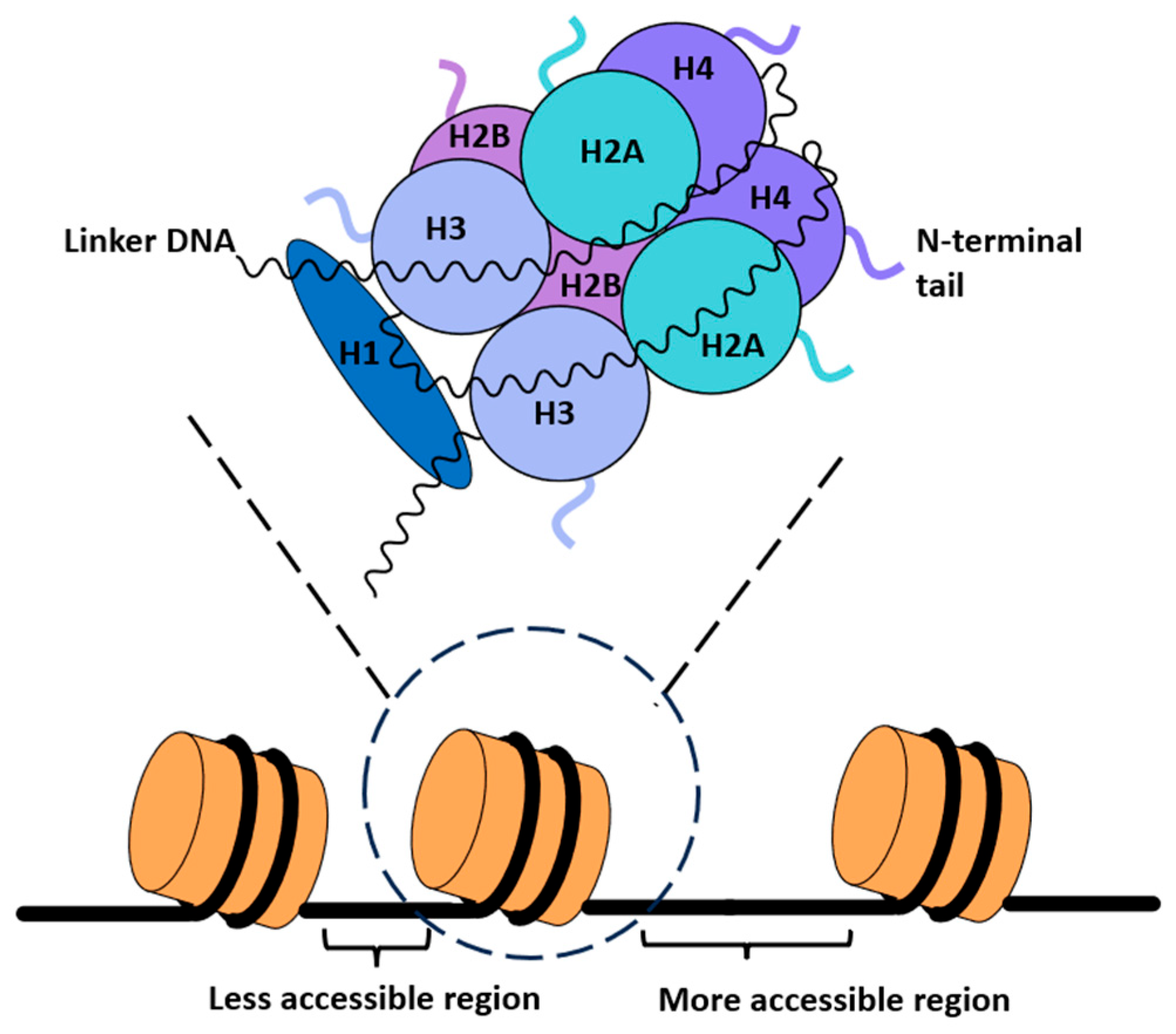
The DNA present in eukaryotic cells is vast in length but is kept packaged neatly in the nucleus. This higher order packaging of the genetic material is due to nucleosomes, the fundamental repetitive units of chromatin [9]. The nucleosome core particle is an octamer complex of proteins which consists of a centrally positioned H3-H4 tetramer flanked by two H2A-H2B dimers [10]. Each nucleosome has 147 bp of DNA wrapped around a core particle, leading to a ~7-fold compaction of the genetic material present in eukaryotic nuclei [9,15,16]. Nucleosomes are also further stabilized by an H1 ‘linker’ histone located at the entry and exit points of the bound DNA [17]. The length of the DNA associated with the linker histone varies widely among organisms, reaching lengths of up to 100 bp, like in the case of echinoderms [18,19,20], and being as short as 7 bp, like in the case of fission yeast [21]. Structural work has previously associated longer linker DNA with a more open chromatin structure that is less thermodynamically stable, while shorter lengths were found in more compact genomic regions (Figure 1) [22,23]. However, the correlation between linker DNA length and chromatin structure is highly complex, as it must also account for factors like the length of nucleosome repeats, nucleosome positioning, histone modifications, and presence of reader proteins that interact with chromatin [24,25].
The four core histones, H2A, H2B, H3, and H4, range from 11 to 15 kDa and are highly conserved amongst eukaryotes [26]. Each core histone consists of a globular core domain within the nucleosome that shares a similar structural motif with each other: a C’ terminal tail and an unstructured N’ terminal tail that extends away from the nucleosome [9]. The residues in this flexible N’ terminal tail can be modified by covalent post-translational modification, including methylation, acetylation, and phosphorylation. There is a cooperative effort between the ‘writer’ proteins that attach a modifier to histones, ‘reader’ proteins that recognize and bind the modifier to elicit a reaction themselves or through the recruitment of other proteins, and ‘eraser’ proteins that will remove this modifier from histones [27]. This pattern of histone modifications produces a ‘histone code’, or a signature that signifies a specific reaction in the cell regarding overall chromatin state and gene expression [11].
Methylation (me), or the attachment of a methyl group (-CH3), occurs predominantly on the basic amino acids lysine (K), arginine (R), and histidine (H). Lysine can be mono (me1), di (me2) or tri (me3) methylated on its ε amine group [28,29,30,31], while arginine can be monomethylated (me1), symmetrically dimethylated (me2s) or asymmetrically dimethylated (me2a) on its guanidine group [32,33]. Histidine, on the other hand, can be monomethylated either at position 1 or 3 of its imidazole ring [34]. The pattern of methylation will determine the functional outcome, allowing for a diverse range of regulatory effects. Methylation is a form of epigenetic regulatory control in many eukaryotes that was once thought to be irreversible due to the stability of this modification compared to others such as phosphorylation [35,36,37]. However, since several lysine demethylases (KDMs) [38,39,40,41,42] and lysine methyltransferases (KMTs) [28,43] have been discovered, it is now recognized that histone methylation can be stably maintained for a long period of time, as in the case of silenced heterochromatin [44,45,46], or can fluctuate during cell differentiation [47,48]. Regulation of histone post-translational modification is directly dependent on the activity of KMTs and KDMs. KMTs are writer proteins that transfer the methyl group from S-adenosyl methionine (SAM) onto lysine residues of histone and non-histone proteins [49]. There are over 30 KMTs fully characterized and while they have impressive target specificity, they are also evolutionarily conserved [50]. KDMs are eraser proteins that remove methyl groups and are distinguished by their highly conserved LSD1 and JmjC domains [38,51,52]. The addition of the methyl group does not modify the charge of the residue and is coined to be the reason that methylation can both promote or repress transcription, depending on the methylation site. Proteins with chromodomains are readers of this methyl modification in which they recognize and bind these methyl groups and carry out the corresponding molecular response.
Phosphorylation (ph) of histones was first described in 1967 [53]. This PTM involves the attachment of a phosphate group (-PO43−) and it occurs on histone amino acids that contain hydroxyl groups (-OH), which are serine (S), threonine (T) and tyrosine (Y). Nuclear protein kinases use ATP to phosphorylate these residues and can be counteracted by nuclear protein phosphatases. The negative charge of the phosphate group added to histones reduces the positive charge of the histones, potentially disrupting histone–histone and histone–DNA electrostatic interactions. Presumably, this would lead to the repelling of the negative phosphorylated histone residue to the negatively charged DNA backbone. However, phosphorylation of a specific site is context-dependent in its outcome. For example, the phosphorylation of histone H3 in nucleosomes that localize to inducible promoters allow for a rapid and transient transcriptional response. In another case, histone H3 phosphorylation is associated with chromatin condensation that allows cells to progress into the M-Phase of the cell cycle, as well as with chromatin decondensation post-mitosis. Thus, histone phosphorylation is dynamic in nature and associated with a myriad of processes, including the DNA damage response, transcription regulation, apoptosis, and, most relevant to this review, chromatin compaction [54].
The acetylation (ac) of histones in vivo was first described in 1964 [55]. In acetylation, acetyl groups (-COCH3) are added to histone proteins. This modification was originally detected in calf thymus cells in arginine-rich histones [56]. It was noted that the lysine-rich histones were highly acetylated compared to the arginine-rich histone fraction [56]. Acetylation involves the transfer of an acetyl group from acetyl-CoA to the ε amine group of lysine (K) or to the guanidium group of arginine (R), neutralizing their positive charges [55]. This process is reversible and mediated by acetylases [57] and deacetylases [58,59]. Through isotope labeling and mass spectrometry, histone acetylation was confirmed to be highly dynamic, with a high degree of turnover despite the existence of stable acetylation sites [60,61]. When acetylation neutralizes the positive charge of the lysine and arginine side chains, histone–DNA and histone–histone electrostatic interactions are altered. This leads to the loosening of DNA, promoting site-specific transcription. Acetylation marks also serve as recognition sites for bromodomain-containing proteins, an evolutionarily conserved protein–protein interaction module present in many ATP-dependent chromatin remodelers [62,63]; and for YEATS domains, which contain immunoglobulin-like folds that regulate gene expression in yeast and other higher eukaryotes [64]. The diversity of acetylation marks is vast, and, to our knowledge, there are no studies investigating the genome-wide effect of this histone modification in C. elegans, making the cell cycle regulation perspective difficult to consider [65].

3. The Cell Cycle During C. elegans Embryogenesis

C. elegans is a microscopic roundworm that exhibits androdioecy, with hermaphrodites determined by two X chromosomes (XX) and males determined by a single X chromosome (XO). This contrasts with species with the XY determination system, in which the Y chromosome triggers male-specific development. C. elegans development starts with the embryo encased by a protective eggshell layer before hatching into a larva (the L1 stage). This larva undergoes three additional larval stages (L2, L3 and L4) before reaching sexual maturity, resulting in an adult hermaphrodite or male.
C. elegans has an invariant cell lineage in which each animal undergoes the same set of cell divisions at specific time points during development [12]. Most cell divisions occur in the first half of embryogenesis, while, in the second half, the embryo undergoes modifications that affect its external appearance to hatch out of the enclosed egg. Very much like in other metazoans, early embryonic development in C. elegans rapidly cycles from the S-Phase to the M-Phase, foregoing gap phases and cell growth to prioritize the increasing cell number [66]. These synchronous early cell divisions can occur due to maternally loaded mRNAs and proteins passed down to the embryo from the hermaphrodite parent. Gap phases are introduced when cell divisions become asynchronous, and their duration is dependent on the cell lineage involved [67]. For instance, the first gap phase introduced in embryonic development is the G2-Phase and is only introduced during the division of intestine primordium cells E2 and E4 once inwards migration of the embryo occurs [66,67]. However, the G2-Phase ends when E4 further divides into E8 cells, showing the tight regulation of these cell cycle phases [67]. Due to the limitations in sensitivity in current assays to establish the different phases of interphase in C. elegans embryos, this warrants further research.
After fertilization, a number of cell divisions occur in the newly fertilized egg to produce founder cells, which divide in relatively fixed timeframes [12]. Most of the founder cells are stem cells that are somatic, serving as the progenitors of a particular tissue or organ. The founder cell, P4, generates the germ line, dividing once at the 88-cell stage to produce two primordial germ cells, Z2 and Z3 [12,68,69]. The precursor cells, Z2 and Z3, do not divide in embryogenesis; instead, the somatic gonad precursors, Z1 and Z4, migrate towards them to generate the gonad primordium. Z2 and Z3 then divide during the mid-L1 stage to produce germ cells within the gonad, while Z1 and Z4 undergo cell division to produce the distal tip center (DTC) and somatic gonad cells [70,71].
A DTC caps each of the two gonad arms in the hermaphrodite adult, serving as a stem cell niche to maintain the population of proliferative germ cells adjacent to it [71,72]. When the germline expands during the L3 and L4 stages, the number of proliferative germ cells rapidly increases. The mitotic zone, also known as the proliferative zone, extends from the distal tip to the transition zone (TZ) in which cells enter meiosis. Proximal germ cells farthest from the DTC enter meiosis during the mid-L3 stage [73,74]. An adult hermaphrodite consists of 959 somatic cells and ~2000 germ cells, while an adult male consists of 1031 somatic cells and ~1000 germ cells [75].

4. Overview of Histone Modifications During C. elegans Embryogenesis

Soon after fertilization, the nucleus of the oocyte undergoes two rounds of meiotic division and then replicates its haploid genome to produce the oocyte-derived pronucleus [76]. The oocyte-derived pronucleus will then fuse with a sperm-derived pronucleus that has also replicated its own haploid genome. Pronuclear fusion occurs as the parental genomes merge at metaphase I, resulting in decondensed chromosomes [77,78]. The single cell embryo transitions from meiotic divisions to mitotic divisions under tight regulation and coordination as meiosis-specific regulators are degraded and replaced by mitosis-specific ones [79,80]. Maternally loaded proteins and RNAs regulate the early cleavages, timing of the first cell divisions, and all events related to the oocyte-to-embryo transition [81]. At the 2-cell stage, chromosomes are positioned randomly, and no epigenetic marks are detected [82]. At the 4-cell stage, zygotic transcription initiates [83]. There is a global reorganization of chromosomes during the 8-cell stage, as epigenetic marks, nuclear domains, and transcriptional activity become evident [82,84].
Histone modifications can also be inherited across generations, establishing an epigenetic ‘memory’ of specific expression patterns in the offspring [85,86,87]. For instance, the acetylation marks H2BK12ac, H3K23ac, H3K27ac, H4K16ac, H4K5ac, H4K8ac and H4K12ac have been previously reported during the 2-cell to 16-cell transition as a result of parental reprogramming pathways [88]. Transient acetylation is also detected during DNA replication as new histones are assembled into chromatin, as observed in Tetrahymena, Drosophila melanogaster and human cells [89]. Active methylation marks such as H3K4me2 [90] and H3K79me2 [88] are also present on the chromosomes of embryos derived from parental chromosomes [78]. There is also inactive chromatin established as the embryo develops, such as H3K9me2/3 that promotes the formation of heterochromatin [91]. However, these methylation marks prove to be more variable throughout development than the acetylation marks.
The epigenetic landscape of a developing embryo is dynamic, constantly changing in response to genetic and environmental factors. For instance, this can be observed by comparing autosomes of early embryos to those of the L3 stage larva. For both the embryo and larva, actively transcribed chromatin is associated with high levels of H3K4me1, H3K27ac, and H3K36me3, and low levels of H3K4me3 on enhancer regions [92]. On the other hand, silent chromatin is enriched with the Polycomb-mediated silencing mark H3K27me3 and the heterochromatin mark H3K9me3 [92]. Differences can also be detected between the early embryo and the L3 larva. For example, there is a sharp increase in H3K36me3 levels in early embryos, while they are kept at lower levels in the L3 stage [92]. Similarly, the L3 larva has high levels of H3K27me1 on active chromatin domains while they are kept at lower levels in the active chromatin domains of early embryos. There are also differences in the epigenetic profile of the X chromosomes from early embryos compared to the L3 larva stage due to dosage compensation (DC), in which the X chromosomes of hermaphrodites (XX) are both downregulated by ~50% to equalize X-specific expression to that of males (XO).
In the C. elegans germline, epigenetic histone modifications play an important role in establishing specific gene expression patterns to ensure proper germline function across generations. For example, the maternal-effect sterile (MES) proteins that are supplied maternally regulate germline development in C. elegans through histone PTMs transmitted to embryos [93,94]. MES-4 is a KMT that generates H3K36 dimethylation (H3K36me2) in the autosomes of early embryos and the C. elegans germline [95,96]. In mes-4 mutants, H3K36me2 is abolished and the F1 progeny are sterile, showing the role the transmission of this epigenetic mark plays in functional germline development [95,97,98]. Indeed, mes-4 mutants have misregulated gene expression in which somatic genes are expressed and germline genes are repressed, contrasting with wild-type [98].
Interestingly, there seems to be an antagonistic relationship between H3K36me3 and H3K27me3 in the germline of C. elegans. While H3K36me3 is associated with active transcription, H3K27me2 deposited by the Polycomb Repressive Complex 2 (PRC2) is associated with repressing gene expression. In wild-type C. elegans, there are alternating clusters of H3K36me3-bound genes and H3K27me3-bound genes on autosomes in the germline [98] creating active and silent domains, respectively, within the germline. Additionally, the repressive H3K27me3 is also enriched on the silent X chromosomes in the germline. In embryos, loss of H3K36me3 was accompanied by an increase in H3K27me3. However, genes that retained H3K36me3 lacked H3K27me3. This result corroborates findings from other studies [99,100,101], leading to an interesting relationship between opposing histone PTMs that cooperate to regulate gene expression.
As previously mentioned, the epigenetic landscape can also be impacted by environmental factors. Such is the case of worms that are fed a high-fat diet, which have high levels of H3K4me3 near the promoter regions of genes related to lipid metabolism, increasing their transcription [87]. This shows that nutrition can also affect the epigenome by altering chromatin state. Indeed, the impact of environmental stressors on the epigenome, ranging from toxic chemicals to microbial pathogens, represents a trending research field that directly benefits from the use of C. elegans as a model organism and warrants its own separate review.
Chromatin is also highly dynamic during the cell cycle. Transcription is generally inhibited during the mitotic phase and this phenomenon is highly conserved across eukaryotes [102]. The epigenome plays a pivotal role as the cells exit mitosis, as it carries the necessary information to restart transcription as the cell continues to grow and develop. Specifically, chromatin is restructured during the S-Phase through the incorporation of newly synthesized histones into nascent DNA, producing a template that is blank from any histone PTMs [103]. Formation of active chromatin is dependent on the reinstatement of the histone modifications transmitted from parent cells to daughter cells. Interestingly, there are histone marks, such as H3K9me2, that are not affected by the cell cycle and are present on both interphase and mitotic chromatin [104]. Contrastingly, other marks like H3S10ph are present in mitosis, specifically during the prophase to telophase transition, before decreasing to undetectable levels as the cell enters the interphase [104,105]. Both H3K9me2 and H3S10ph function as ‘phospho-methyl switches’ that modulate the binding of effector proteins to histone H3, which ultimately impacts the chromatin state [106].
The regulation of histone PTMs during the cell cycle of C. elegans is highly complex, since it can originate from diverse inputs like genetic factors, environmental stressors, cell lineage and developmental status. Coupled with the emergence of other overlapping fields, like RNA interference and aging, this further complicates the interpretation of the roles of histone PTMs in mitosis. In this review, we summarize the histone modifications that are affected by the cell cycle in C. elegans, with special attention to embryogenesis since somatic proliferation ceases as the animal transitions to adulthood. For this reason, the epigenetic marks we review are H4K20me1, H3S10ph, H4S1ph, H2AS1ph, and H3T118ph. Table 1 highlights the function during the cell cycle of each epigenetic mark in C. elegans in contrast to other model organisms. Moreover, we take a comparative approach, highlighting first what is known in other classical organismal systems with the hopes of motivating more research projects centered around epigenetic regulation of the cell cycle.

5. H4K20me1 Peaks in Mitosis and Is Enriched on Dosage-Compensated X Chromosomes

Histone H4 methylation is evolutionarily conserved from simple eukaryotes, like yeast [120], to more complex eukaryotic cells, like humans [120,121]. Most of the methylation on histone H4 occurs on lysine 20 (H4K20) [122,123]. H4K20 can be mono (me1) [29], di (me2) [124] or tri(me3) methylated [33,122]. The research field has attempted to establish a downstream effect clearly associated with H4K20me1/2/3, but it has been found that this modification impacts a variety of processes, including transcriptional silencing [125] and activation [108], heterochromatin formation [126], chromatin compaction [120], and mitosis [127]. Since the signal induced in the cell by this modification can be contradictory, the different methylation states of H4K20 are context dependent.
It is reported that H4K20me2 is the most abundant modified form of H4K20 in eukaryotic cells, with ~80% of histones occupied by this mark [128,129]. H4K20me3 lies on the opposite side of this spectrum, being the least abundant form of methylated H4K20 [130,131,132]. Interestingly, both H4K20me2/3 levels do not alter dramatically throughout the cell cycle compared to H4K20me1 [121,133,134]. H4K20me1 also plays an essential role in the establishment of H4K20me2/3, as it has been shown that the abolishment of H4K20me1 leads to the reduction in levels of H4K20me2/3 [135]. In the late G1-Phase, H4K20me1 levels decrease and are lowest during the S-Phase. This may be due to the fact that during the replication of DNA, new unmodified histones are incorporated into the DNA, which dilutes the concentration of the existing histone modifications [136]. After the replication of DNA, H4K20me1 levels begin to increase and reach a peak during mitosis [133,134,135]. Post-mitosis, H4K20me1 is converted into H4K20me2/3, which leads to the production of quiescent cells in the G0/G1 of the cell cycle [133].
Although H4K20 methylation has been detected in several organisms since the 1970s, the protein that writes H4K20me1 was first uncovered in the 2000s. Human PR-Set7 [137] and its D. melanogaster ortholog, SET8 [137,138], are KMTs with SET domains (Su(var)3-9, Enhancer-of-zeste and Trithorax). X-ray crystallography experiments confirmed that these KMTs methylate H4K20 and produce H4K20me1 [139,140]. In a similar manner, Suv4-20h1 and Suv4-20h2 were found to di- and tri-methylate H4K20me1 in the mouse model [121], with their counterparts discovered later in humans [129] and D. melanogaster [141].
As mentioned earlier, methylation of histones was largely considered a stable process. However, the discovery of a histone demethylase specific to H3K4, LSD1 [38], which is also conserved in both yeast and humans, proved that there is a turnover of histone methylation. In terms of erasers of H4K20 methylation, PHF8 functions as an H4K20me1 demethylase, with a role in the cell cycle in addition to its capability of demethylating H3K9me1/2 and H3K27me2 [142,143,144]. Recently, two human homologous proteins, hHR23A and hHR23B, have been found to demethylate H4K20me1/2/3 in vivo in eukaryotic cells and in vitro through a bulk histone demethylase assay followed by mass spectrometry [145].
In C. elegans, SET-1 is one of the smallest SET domain-containing proteins and was found to be an essential gene, as RNAi-mediated gene silencing led to early embryonic death [146]. A decade later, the association between SET-1 and H4K20 methylation was made. Through the RNAi-mediated gene silencing of set-1, it was reported that H4K20me1 was abolished in the intestinal nuclei of adult C. elegans [110]. This finding was further supported by work with homozygous set-1 mutant animals, which have no detectable H4K20me [109]. These mutants also developed into sterile adults [109], highlighting the importance of establishing the first methyl moiety on H4K20 to serve as the foundation for H4K20me2/3.
The C. elegans ortholog of Suv4-20 is SET-4 and it is the major KMT that converts H4K20me1 into H4K20me2/3. This conclusion was derived from evidence in both homozygous set-4 mutant embryos and adults, in which H4K20me1 levels increase throughout all chromosomes while H4K20me2/3 levels are reduced [109,110]. This led to an elegant model which summarizes the H4K20 writers in C. elegans; SET-1 places the first methyl mark on H4K20, followed by SET-4 that converts it to H4K20me2/3. However, the eraser proteins of H4K20 methylation in this organism are yet to be discovered.
Although the H4K20me1 demethylase in humans have been identified to be PHF8, a homologous counterpart has yet to be found in C. elegans. However, a H4K20-specific demethylase that converts H4K20me2/3 to H4K20me1 in this animal has been identified, DPY-21. Originally, DPY-21 was found to be involved in the X chromosome dosage compensation (DC) mechanism of hermaphrodites [147]. This process equalizes gene expression stemming from the sex chromosomes of the differing biological sexes [148,149]. In C. elegans, hermaphrodites downregulate both X chromosomes by half to equalize the transcriptional output to that of the single X chromosome present in their male counterparts [150]. This process is conducted primarily by the dosage compensation complex (DCC) [147,151,152,153,154,155], a multimeric complex of proteins that work in tandem to bind and repress the X chromosomes in hermaphrodites.
DPY-21 is one of the DCC subunits [147,156] and has been shown to function as a H4K20-specific demethylase, specifically targeting H4K20me2/3 both in vivo, through immunofluorescent imaging of intestinal nuclei and embryos [110,157], and in vitro, through a bulk histone demethylase assay [157]. DPY-21 removes the methyl groups from H4K20me2/3, leading to the enrichment of H4K20me1 on the X chromosomes and the transcriptional downregulation and compaction of these X chromosomes in the somatic cells of hermaphrodites [110,157,158]. DPY-21’s H4K20me2/3 demethylase activity is limited, as it localizes to the X chromosomes, although weak levels of H4K20me1 on autosomes have been reported [109,110], hinting at the existence of uncharacterized demethylases in C. elegans.
As previously mentioned, in eukaryotic cells, H4K20me1 levels decrease during the transition from the G1-Phase to the S-Phase, before increasing again after DNA replication is complete and reaching a peak in mitosis. In post-mitotic cells, H4K20me1 is converted to H4K20me2/3, where it may lay quiescent as the cell prepares to divide again. In C. elegans embryonic nuclei, H4K20me1 is low in distribution throughout all chromosomes in interphase but increases when it associates with condensed chromosomes in mitosis, as proven by the high levels of H4K20me1 detected on prometaphase and metaphase chromosomes [109].
H4K20me1 remains evenly distributed throughout all chromosomes as the embryo develops until it reaches the comma stage, in which there are nuclei with H4K20me1 enrichment on the X chromosomes in hermaphrodite embryos due to the DC-silencing mechanism [109]. By the late 3-fold stage, all nuclei in the hermaphrodite embryo display H4K20me1 enrichment on the X chromosomes while H4K20me2/3 is distributed in a relatively uniform manner throughout all chromosomes [109,159]. This enrichment of H4K20me1 on X chromosomes is dependent on the presence of a functional DCC as it is abolished in DC mutants [109]. In male embryos, there is no requirement for DC and immunofluorescent experiments showed uniform distribution of H4K20me1 throughout all chromosomes [109]. Interestingly, H4K20me1 is also present on the inactivated X in female mammalian cells, although the abolishment of this mark does not affect the downregulation of gene expression as it does in C. elegans [107]. Instead, it is proposed that H4K20me1 may be associated with the compaction of the inactivated X chromosome.

6. H3S10ph Is Associated with Mitotic Chromosome Condensation and Modulates the Binding of Proteins to Chromatin

Early reports on the involvement of histone phosphorylation in the regulation of the eukaryotic cell cycle have been crucial for the development of chromatin remodeling models [160,161,162]. This modification can occur on all four core histones and on the linker histone; however, H3 phosphorylation is of particular importance due to its participation during cell division. These studies provided evidence that H3 phosphorylation was not observed during the interphase periods of the cell cycle but increased during mitosis, specifically in prophase, metaphase, and anaphase, when chromosomes are fully condensed [160]. When cells exit anaphase and chromosomes begin to decondense, H3 dephosphorylation occurs, further supporting that H3 phosphorylation is associated with mitotic chromosome condensation.
Initially, studying phosphorylated histones was troublesome, as the nature of in vitro techniques led to the loss of phosphorylation in isolated chromosomes [163]. This major obstacle was conquered by coupling the isolation of chromosomes to the addition of ATP to ensure maintenance of phosphorylation by endogenous kinases [164]. As a result, H3 phosphorylation was confirmed in mitotic chromosomes, with the major site of phosphorylation being serine 10 (H3S10ph) through tryptic peptide mapping [164]. The discovery of serine 10 as the major phosphorylation site of histone H3 triggered a cascade of studies to characterize this modification, and in terms of mitosis, H3S10ph was found to be correlated to chromosome condensation [111,112,114], while its dephosphorylation was associated with chromosome decondensation [165,166]. These patterns have been observed in both simple [105,111,112] and complex eukaryotic systems [105,160,166]. Exceptions exist, as in the case with D. melanogaster, in which strong reduction in H3S10ph only leads to slight decondensation of chromatin [167].
H3S10ph initiates on pericentromeric heterochromatin during the G2-Phase and, as chromosomes condense, the mark spreads throughout all chromosomes [114]. This modification occurs primarily in late-replicating chromatin in which maximal chromosome condensation is obtained during the G2/early prophase transition [168]. In mammals, centromeres are the primary site for the initiation of chromosome condensation [169]. In the G2-Phase, H3S10ph initiates on centromeres before increasing on pericentric heterochromatin, prekinetochores and advancing to the chromosome arms [114]. When premature chromosome condensation is induced in cells, pericentric H3S10 is phosphorylated. However, prekinetochores that are not in the G2-Phase are not fully replicated and lack H3 phosphorylation [169,170]. These results point to the relationship between H3S10 phosphorylation and the condensing of mitotic chromosomes, as well as the replication of genetic material for the cell to undergo division. When cells arrested in the early S-Phase are induced to undergo mitosis and forego the G2-Phase, less than 1% of cells exhibit proper mitotic chromosome condensation [168]. The few cells that enter mitosis exhibit H3S10ph, highlighting that the accumulation of this modification in G2-Phase is essential for mitotic entry [168]. The dephosphorylation of H3S10 begins in late anaphase and early telophase and is completed just before chromosomal decondensation post-mitosis [114,168,171]. Thus, H3S10ph is required for mitotic chromosome condensation, mitotic entry, and its dephosphorylation is associated with the decondensation of chromosomes post-mitosis.
Following the discovery of H3S10 phosphorylation and its association with mitosis in eukaryotic cells, H3S10ph was then established to be a mitotic mark in C. elegans. In embryos, as well as in L1 larvae, H3S10ph is observed on mitotic chromosomes from prometaphase to telophase [113]. Since then, H3S10ph has been used as a mitotic mark in several C. elegans studies [105,172,173]. Interestingly, H3S10ph is also present in the mitotic region of the hermaphrodite gonads, located between the distal tip cells (DTC) and the transition zone to meiosis. In the nuclei of this mitotic region, H3S10ph is present on metaphase and telophase chromosomes, parallel to what is observed in embryos, before becoming undetectable in meiosis [105].
In addition to its role for proper mitotic chromosome condensation, H3S10ph is tightly linked to H3K9me2. H3K9me2 is adjacent to H3S10ph, and these two residues have been hypothesized to function as a ‘phospho-methyl switch’ to regulate the binding of histone H3 to effector proteins [174,175,176]. H3K9me2 is required for the nuclear periphery localization of heterochromatin [48,104]. CEC-4 is a chromodomain-containing protein in C. elegans that anchors H3K9 methylation-bearing chromatin to the nuclear periphery in interphase [177,178,179]. When H3K9 is di-methylated, its reader protein CEC-4 tethers it at the nuclear lamina [106]. However, the mutation H3S10E, which mimics a constitutively phosphorylated state, displaces H3K9me2-marked chromatin from the nuclear periphery [104]. Contrastingly, the mutation H3S10A which is non-phosphorylatable had no effect on the localization of H3K9me2-marked chromatin to the nuclear lamina [104]. Indeed, it is reported that the binding of recombinant, purified CEC-4 to methylated H3K9 peptides decreases significantly when the adjacent H3S10 is phosphorylated [106]. From these in vitro biochemical assays, the phosphorylation of H3S10 can prevent reader proteins, including CEC-4 from reading and binding to H3K9me2. However, in vivo assays are required to establish that this is translatable within a model organism as well.
Before cells enter mitosis, H3K9me2-localized chromatin detaches from the nuclear periphery and remains in the nucleoplasm. Once the cell enters prophase, the nuclear lamina is disassembled and H3S10ph increases. H3K9me2 remains present on both interphase and mitotic chromosomes while H3S10ph is localized only to mitotic chromosomes. Thus, it is proposed that when H3S10ph increases in mitosis, it inhibits the binding of effector proteins to H3K9me2, preventing H3K9me2 readers/tethers to anchor H3K9me2-marked chromatin to the nuclear periphery [180,181]. As the cell enters telophase, H3K9me2-marked chromatin is reestablished on the nuclear lamina. H3K9me2S10ph-marked chromatin remained in the nucleoplasm, suggesting that dephosphorylation of serine 10 occurs before reassociation of the H3K9me2-marked chromatin with the nuclear lamina [104].
In addition to the H3K9me2/H3S10ph ‘switch’, H3S10ph also prevents the binding of heterochromatin protein 1 (HP1) to heterochromatin [182]. During interphase, HP1 binds to H3K9me3-marked heterochromatin domains before it decreases in prophase until it becomes undetectable in prometaphase as observed through immunofluorescent microscopy of mouse embryonic fibroblasts [181]. This research group found that H3S10ph and HP1 localization to chromosomes occur at alternating times. When H3S10 phosphorylation is inhibited, HP1 remains enriched on pericentric heterochromatin during mitosis. In an in vitro assay, HP1 binds to H3K9meS10ph less efficiently than it does to H3K9me3 peptides [177,181]. It is proposed that the negative charge of phosphorylated serine 10 repels the negatively charged glutamate 52 residue in HP1. Thus, H3S10ph is able to regulate the binding of proteins to chromatin as in the case of CEC-4 to methylated H3K9 and HP1 to H3K9me3-marked heterochromatin in vitro, providing a foundation for further in vivo studies.
H3S10 phosphorylation is associated with mitosis, and this modification modulates effector proteins that bind to chromatin. Additionally, this mitotic marker also plays a role in the maintenance of a stable genome, as observed in studies with Saccharomyces cerevisiae that was then extended to human cells and C. elegans. R-loops are an indicator of genomic instability [183,184]. In yeast mutants, R-loop accumulations exhibit increased H3S10ph [183]. When this research group expanded their study to include C. elegans, R-loop-accumulating mutant worms also had elevated levels of H3S10ph and, interestingly, H3K9me2 in both mitotic and meiotic regions of the hermaphrodite germline. Since H3S10ph is associated with chromosome condensation, the research group also reported that the nuclei in these mutants were highly condensed compared to wild-type. Thus, H3S10ph is associated with chromosome compaction in not only eukaryotic cell lines but also in the C. elegans mitotic and meiotic germline.
The H3S10ph ‘writer’ also plays an important role in cell cycle regulation. When kinase activity for H3S10ph is outcompeted with the transfection of H3S10 peptides into cells, entry to mitosis is prevented. Cells that managed to enter mitosis despite the transfection exhibited H3S10ph, proving the importance of the phosphorylation state of H3S10 [168]. Preliminary in vitro assays identified a cAMP-dependent kinase as the enzyme responsible for H3S10ph [185], paving the way for subsequent in vivo studies that further characterized the pathways involved [186,187]. In 2002, the kinase IpI1p and the phosphatase Glc7p were identified in S. cerevisiae as the writer and eraser proteins for H3S10 phosphorylation through genetic assays and in vitro biochemical assays [105]. This same research group expanded their studies into C. elegans, discovering that AIR-2, an Aurora B kinase involved in cell division, serves as the kinase that phosphorylates H3S10. This was observed when the RNAi-mediated silencing of air-2 abolished H3S10ph in embryos, leading to embryonic arrest at the 1-cell stage [105]. This same silencing also affected mitotic nuclei in hermaphrodite gonads, leading to aberrant mitotic divisions with decondensed chromatin [105]. These results show that genetically, AIR-2 is required for the phosphorylation of H3S10. However, in vitro biochemical assays are necessary to determine if AIR-2 is able to directly phosphorylate H3S10.
The discovery of AIR-2 as a kinase involved in H3S10 phosphorylation in C. elegans led to the identification of its H3 phosphatase counterpart. In budding yeast, Glc7p is a type 1 protein phosphatase (PP1) that removes the phosphate group from H3S10 [105]. In C. elegans, two isoforms encode for PP1, Ceglc-7α and Ceglc-7β. In worms with a loss of H3S10ph, disruption of either isoform through RNAi led to a rescue of H3S10ph [105]. Thus, Ceglc-7α and Ceglc-7β are both able to dephosphorylate H3S10. However, there are differences amongst these two isoforms. The silencing of Ceglc-7α led to 100% embryonic lethality in which embryos are arrested at late stages, exhibiting misshapen nuclei consisting of abnormal amounts of DNA due to defective chromosome segregation. On the other hand, the silencing of Ceglc-7β leads to ~50% embryonic lethality, with all surviving embryos arrested at the 50–100 cell stage. When the silencing of both Ceglc-7α and Ceglc-7β was coupled, there was 100% embryonic lethality and arrest at the 50–100 cell stage [105]. Taken together, these results suggest that both isoforms may be involved in additional pathways.
Additionally, various studies have expanded on PP1, identifying several of its protein interactors, such as the human factor C1 (HCF) implicated in viral transcription [188]. The C. elegans hcf-1 deletion mutant has small brood sizes and embryonic lethality. Interestingly, the hcf-1 mutants are defective in cell division due to mitotic spindle defects and misalignment [172]. In these hcf-1 mutant embryos, H3S10ph is significantly reduced. This loss of H3S10ph is reproducible in mammalian cells with depleted HCF-1 [172]. However, when Ceglc-7α and Ceglc-7β were silenced through RNAi in hcf-1 mutant C. elegans embryos, two contrasting phenotypes are observed: embryos with elevated H3S10ph as well as embryos that lacked H3S10ph. The research group proposes that HCF-1 may play a role in the maintenance of H3S10ph in a pathway different than that of the Ceglc-7α/Ceglc-7β phosphatase. However, this could also be due to knockdown inefficiency through RNAi-mediated silencing. Thus, more research is needed to expand the relationship between HCF-1 and H3S10ph.
To summarize, H3S10ph is associated with mitotic chromosome condensation and its dephosphorylation with chromosome decondensation. The accumulation of this histone PTM also helps the cell cycle progress from the G2-Phase to the M-Phase. Additionally, it can regulate the binding of proteins including CEC-4 and HP1 to its neighboring residue, methylated H3K9, at least in vitro. Studies in C. elegans have thus far utilized H3S10ph widely as a marker of mitosis and there is research that shows it is correlated to the condensing of chromosomes in both the mitotic and meiotic regions of the hermaphrodite germline. The multifaceted roles of H3S10 phosphorylation, as revealed by current research, underscore its biological significance and highlight the need for further in-depth studies to fully elucidate its functions.

7. H4S1ph Is Biphasic Throughout the Cell Cycle and Peaks in Mitosis

H4S1 phosphorylation was initially detected in newly synthesized histones, in which the phosphorylation and acetylation of residues on H4 were determined to be transient in dividing cells [189]. The first five amino acids of the N’ terminal tail of H4 are conserved amongst most eukaryotic organisms, with a few exceptions like in Tetrahymena [115]. This N’ terminus area also exhibits frequent modifications, in which arginine 3 (H4R3) is methylated during transcriptional activation [190] and lysine 5 (H4K5) is acetylated on newly synthesized histones [89,189]. Moreover, phosphorylation of H4 occurs on serine 1 (H4S1) according to mass spectrometry data [115].
Phosphorylated H4S1 was found to be present in asynchronous eukaryotic cells. When studied further, there was a drastic increase in phosphorylated H4S1/H2AS1 in cells arrested in mitosis, denoting the link of this mark with mitosis [115]. The specific timing of H4S1 phosphorylation was established through the immunofluorescent imaging of synchronous HeLa cells at specific time points [115]. H4S1ph increases at the G1/S-Phase checkpoint and in early S-Phase but decreased to basal levels during the mid S-Phase to the early G2-Phase. Interestingly, H4S1 is once again phosphorylated nearing mitosis, where it reaches a peak. It is maintained at high levels until the end of telophase and the early G1-Phase [115]. Thus, H4S1ph exhibits a biphasic pattern of distribution in which it increases in G1-Phase/early S-Phase, falls to basal levels in mid S-Phase/early G2-Phase, before reaching a peak in mitosis. This biphasic distribution of H4S1ph is compelling; however, it has only been shown in HeLa cells. Since the distribution of histone PTMs may vary between cell lines as well as organisms, the pattern of H4S1ph reported is limited and warrants further research to establish if this is indeed true for other systems.
Interestingly, in non-mitotic cells, there is a low level of H4S1ph detected due to phosphorylation of H4S1 in newly synthesized and deposited histones [115,189]. Although histone modifications are more commonly nuclear in nature, there are also modifications that occur in the cytoplasm, including acetylation and phosphorylation. Presence of H4S1ph in non-mitotic cells may be due to the phosphorylation of the residue occurring early in histone biosynthesis while the nascent polypeptide chains are still attached to polysomes in the cytoplasm [115,189,191]. Through the labeling of purified erythrocyte histones, it was found that in newly synthesized histones, H4S1 is modified by phosphorylation or acetylation before later removal [189]. This finding has been limited to in vitro biochemical assays and would benefit from more in vivo studies to establish if H4S1 is indeed phosphorylated in the cytoplasm during synthesis.
As previously mentioned, H3S10ph is considered a hallmark of mitosis. In mitotic chromosomes of C. elegans, H3S10ph was detected at high levels in early embryos undergoing [115]. In C. elegans embryos, phosphorylated H4S1 is present on the condensed chromosomes of prophase and metaphase, colocalizing with H3S10ph. This mitotic-specific pattern of H4S1 phosphorylation is reproducible in other organisms such as Physarum polycephalum [115]. There are exceptions to this cell cycle-specific pattern of H4S1ph. For instance, in D. melanogaster, the first residue of H4 is a threonine (H4T1) instead of a serine [115]. Both serine and threonine are polar amino acids with neutral charges, but threonine has an additional methyl group that makes it bulkier. H4T1ph was detected in chromosomes in metaphase as well as in the S-Phase [115]. This low level of H4T1 phosphorylation in the S-Phase may be due to the phosphorylation of newly synthesized histones that are necessary for the stable incorporation of histones into nascent chromatin [89,189]. This deduction requires further experimentation in order to provide more insights on the role of this phosphorylation mark in cell cycle regulation.
In yeast, H4S1 is conserved but there is no mitotic phosphorylation of this mark [115]. Instead, it is present in basal levels throughout the cell cycle of this eukaryote [192]. Since its discovery in mitosis, H4S1ph has been associated with DNA damage repair, the regulation of chromatin acetylation [193], transcription [194] and yeast sporulation [194]. Casein Kinase II (CKII) has been shown to phosphorylate H4S1 in vitro [195]. Additional work is needed to prove CKII activity in vivo. Interestingly, research has revealed that there is a meiotic role associated with H4S1ph in which it is conserved during gametogenesis in several organisms including yeast, D. melanogaster, and mice [196]. In terms of meiosis, H4S1ph promotes chromosomal compaction and transcriptional downregulation [194,196]. Since the only research done in C. elegans concerning H4S1ph is its localization in mitotic nuclei of young embryos, exploring this modification during germline development and maintenance could shed light on potential ‘writers’, ‘readers’, and ‘erasers’.

8. H2AS1ph Increases in Mitosis and Is Implicated in Transcription Regulation In Vitro

Amongst the four core histones, H2A has the largest sequence divergence, creating multiple H2A family proteins and a myriad of variants amongst organisms [197]. Like H4, the first five residues of H2A are sequentially conserved across various eukaryotes and maintained throughout evolution [115,198]. The number of phosphorylation sites on H2A have been detected and multiple functions are associated with these modifications. For example, phosphorylation of H2A at serine 129 occurs in response to DNA damage to facilitate DNA repair in S. cerevisiae [199,200]. In S. pombe, H2A serine 121 is phosphorylated for the tethering of the protein Shugoshin to centromeres, preventing chromosomal instability [201].
H2A serine 1 has been found to be the residue most commonly phosphorylated based of mass spectrometric data in human cells [202]. The distribution of H2AS1 phosphorylation throughout the cell cycle has been characterized in several systems, including C. elegans, D. melanogaster, and HeLa cells [115]. H2AS1ph exhibits the same biphasic pattern as described earlier in H4S1ph in which levels in the G1-Phase/early S-Phase transition, fall to basal levels in mid S-Phase/early G2-Phase and peak in mitosis [115]. Since the first five residues of the amino terminal in both histone H4 and histone H2A are conserved, this may explain the same levels of distribution displayed by both. H2AS1ph is high in mitosis but there are low levels of it detected in S-Phase, which could also be due to H2AS1ph on newly synthesized histones. However, this has only been reported for H4S1ph and not H2AS1ph, which warrants more research [189,203,204]. Interestingly, mouse embryonic fibroblasts do not exhibit a strong biphasic pattern of H2AS1 as previously reported in HeLa cells [115]; however, H2AS1ph levels appear to be highest in mitotic cells, which corroborates previous findings [204].
In addition to the distribution of H2AS1 phosphorylation throughout the cell cycle, research has also uncovered some of its protein regulators. HMGN1 is a nuclear protein that binds nucleosomes and reduces the compaction of chromatin fiber [205], which alters transcription, replication, and repair [205,206,207]. Using mouse embryonic fibroblasts, as well as through in vitro biochemical assays, the loss of HMGN1 increases H2AS1 phosphorylation, while HGMN1 binding to nucleosomes inhibits it [203,204]. Mitogen- and stress-induced kinase (MSK1) phosphorylates H2A, and this negatively regulates transcription in vitro [203]. However, since this finding is restricted to in vitro reconstitution assays, more research needs to be conducted to demonstrate that MSK1 phosphorylates H2AS and if this modification results in reduced gene expression. Protein Phosphatase 2Cγ (PP2Cγ) has been shown to bind H2AS1ph [208]. However, this assay is limited to the physical interaction during the co-immunoprecipitation of PP2Cγ and H2AS1ph. More experiments, such as H2AS1 peptide phosphorylation assays and mutational screens, need to be conducted in order to determine if PP2Cγ directly phosphorylates H2AS1.
Histone H2A has many variants, which are non-allelic isoforms with different primary sequences and temporal expression, such as H2A.X and H2A.Z [209]. This adds another layer of complexity in studying H2AS1ph. Moreover, this epigenetic mark has not been explored extensively in C. elegans and in the context of the regulation of the cell cycle. The characterization of H2AS1ph regulators, like HGMN1 and MSK1, represent excellent starting points for C. elegans work. Interestingly, in Xenopus, H2 Serine 1 is phosphorylated concomitant with germinal vesicle breakdown, which is when meiosis resumes in oocytes but the modification is independent of DNA replication and cell cycle transitions [210]. This finding may also serve as the foundation of studies regarding maintenance of the C. elegans germline by H2AS1ph.

9. H3 T118ph Ensures Chromosome Alignment and Proper Shaping of Mitotic Chromosomes

The phosphorylation of histone H3 on threonine 118 (H3T118ph) was initially detected through mass spectrometry of mammalian core histones [211,212]. This residue is located within the globular domain of H3, positioned at the interface of the histone and bound DNA. H3T118 is conserved amongst many eukaryotic species [118] and in the last decade, has been implicated to have ties to the cell cycle [119,211].
Initially, H3T118 was studied in the context of SIN (Swi-INdependent) mutants in yeast. SWI/SNF complexes reorganize chromatin structure, enabling binding of other proteins to nucleosomal DNA to influence transcription [213,214,215]. SIN mutants allow transcription while foregoing the SWI/SNF complex. One of the SIN mutants, SIN2, was later identified as a mutation of histone H3 in yeast [216,217], converting threonine 118 to isoleucine 118 [116,117]. Threonine 118 is positioned on the surface of the H3-H4 tetramer and is oriented close to the bound DNA and can form hydrogen bonds. When H3T118 is mutated to H3I118, protein-DNA interactions are disrupted, leading to nucleosome dissociation [218,219]. This shows that H3T118 plays an important role within the nucleosome structure. Not only is the H3T118 residue itself important, but its phosphorylation is also an essential component. In yeast, the phosphomimetic mutation H3E118 and the non-phosphorylatable mutation H3A118 are lethal [118]. H3 T118ph enhances nucleosome mobility, which allows proper repositioning of nucleosomes in response to physiological temperature fluctuations [220]. However, these results are limited to in vitro biochemical assays, limiting their translational potential in a more biological context.
Many studies on H3T118 phosphorylation are related to nucleosome stability and mobility within chromatin. At present, only one research group has studied H3T118ph in relations to the mitotic cell cycle, with a large focus on cell lines [7,119]. During the cell cycle, there is an increase in H3T118ph levels in mitotic eukaryotic cells [119]. Immunofluorescence experiments demonstrate that H3T118ph levels increase from prophase to anaphase but are largely reduced during interphase [119]. In mitosis, H3T118ph forms discrete foci on chromatin in prophase, pro-metaphase and on the spindle midbody during anaphase [119]. In both D. melanogaster and C. elegans, H3T118ph localizes to chromatin and centrosomes in mitosis [119]. Interestingly, in C. elegans embryos during pro-metaphase, H3T118ph is also detectable on the periphery of the holocentric chromosomes where centromeres are located [119].
When H3T118ph is mutated, there are mitotic defects. For instance, in human cells transfected with mutant H3T118ph, there is improper chromosome alignment in metaphase, lagging chromosomes that fail to move to the cell poles in anaphase, and delayed cytokinesis [119]. Additionally, these H3T118ph mutations cause the loss of sister chromatid arm cohesion and produce shorter chromosomes [119]. Since the shaping of mitotic chromosomes involves chromosome scaffolding proteins, the research group focused on how altering H3T118ph affects any of these scaffolding proteins. Indeed, when H3T118ph is mutated, there is a loss of Condensin I occupancy on chromatin followed by longer chromatin loops observed in SEM. Interestingly, the study also reports that the localizations of H3T118ph and Condensin I are distinct although they appear on chromosomes at similar periods [119]. Additionally, in vivo biochemical assays show that Condensin I binds to nucleosomes regardless of the phosphorylation state of H3T118. When the kinase responsible for H3T118 phosphorylation was overexpressed, it was reported that Condensin I occupancy on mitotic chromosomes was significantly reduced. From these results, the research group hypothesizes that H3T118ph regulates Condensin I binding to chromatin, thereby influencing chromosome packing. However, these findings are limited and specific to HeLa cells, probing the question if this is reproducible in other cell lines and/or in other species.
In terms of physiological importance, the lethality of H3T118ph yeast mutants is reproducible in D. melanogaster. All H3 gene copies were deleted from D. melanogaster and replaced with transgenes containing mutated H3T118ph [119]. Animals bearing the wild-type H3 transgenes survived until adulthood while animals expressing the mutant H3T118ph transgenes all died as embryos after depletion of the maternal contribution of histones. This shows that H3T118ph is also crucial for the development of both yeast and D. melanogaster. Since H3T118ph is conserved in various other eukaryotic species including C. elegans, these results compel the question of whether this histone PTM plays a crucial role in the embryonic development of other organisms.
The same research group also attempted to identify the main kinase responsible for the phosphorylation of H3T118 through both in vitro and in vivo assays [119]. In a phosphorylation screen, Aurora A was the only cell cycle kinase that exhibited high phosphorylation activity of the H3T118 peptide [119]. When aurora A kinase was inhibited or knocked down, H3T118ph was diminished from mitotic chromosomes [119]. However, it was also reported that the inhibition of Aurora B kinase ultimately prevents Aurora A kinase from localizing to chromatin in order to phosphorylate H3T118 [119]. Thus, the research group successfully showed that Aurora A kinase is able to directly phosphorylate the H3T118 residue; however, the role of Aurora B kinase remains unclear [119]. Taken together, these results show the complexity of phosphoregulation, as kinases often have multiple substrates.
As stated above, research on H3T188ph has largely been focused on yeast and mammalian cell lines. In C. elegans, H3T118ph is observed on mitotic chromosomes of young embryos, specifically from prophase to anaphase before it diminishes in interphase [119]. Since studies have shown that H3T118 phosphorylation is also involved in chromosome alignment as well as the proper shaping of mitotic chromosomes, it would be interesting to explore the relationship of H3T118ph and Condensin I in C. elegans. Additionally, since aurora A is a kinase that phosphorylates H3T118, its C. elegans orthologue, AIR-1, should stimulate further experimentation.

10. Concluding Remarks

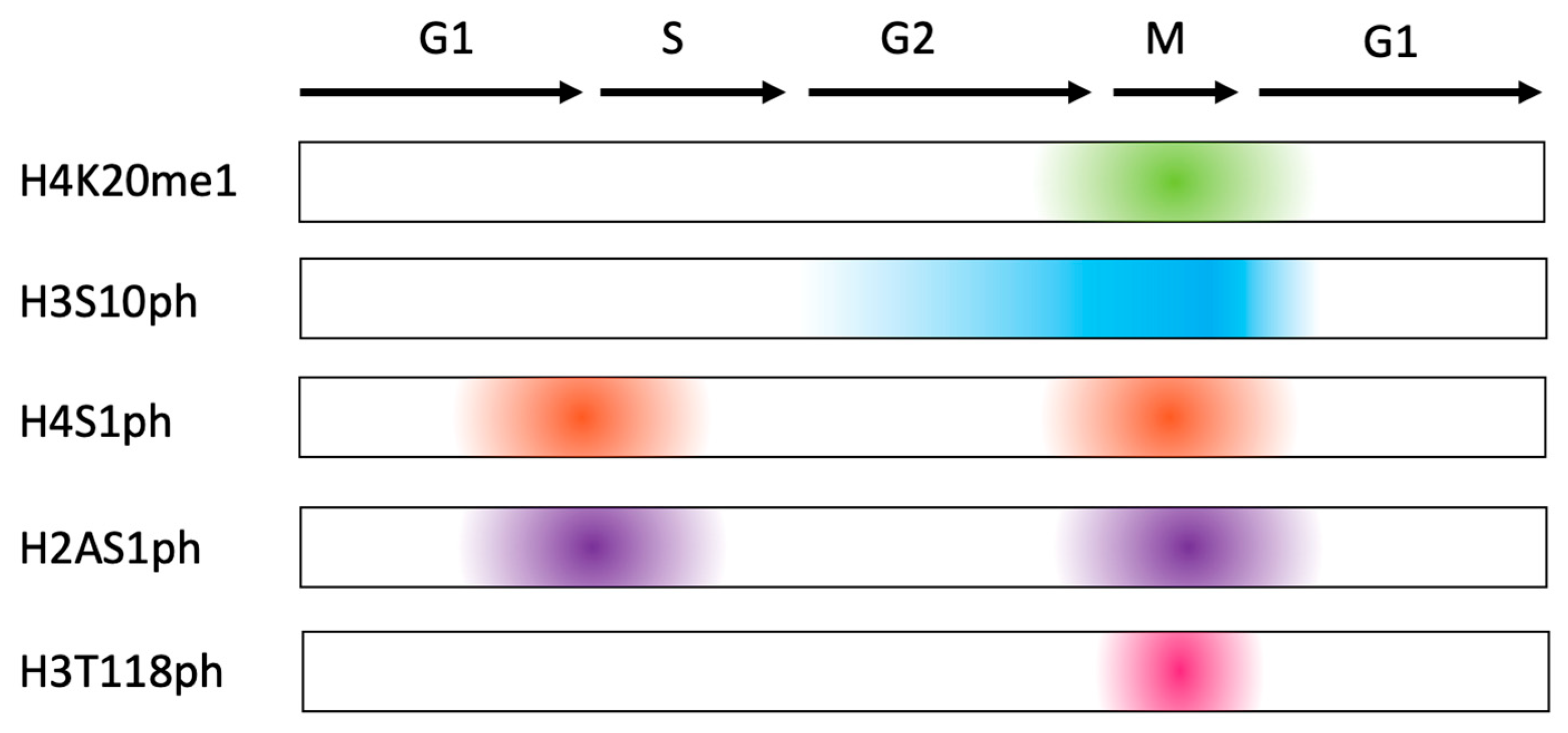
Post-translational modification of histones play an important role in the regulation of the cell cycle of C. elegans embryos. However, as illustrated in Figure 2 and based on the available literature, most of what is known about these epigenetic modifiers focuses on mitosis. Although H4K20me2/3 are stably distributed throughout the cell cycle, H4K20me1 levels are low in interphase and peak in mitosis. In C. elegans, hermaphrodites (denoted with ‘XX’) downregulate both X chromosomes to equalize to the single X chromosome present in their male counterparts (denoted with ‘XO’) [150]. The dosage-compensated X chromosomes in hermaphrodite somatic cells of C. elegans are enriched with the repressive mark H4K20me1, which plays a role in chromosome compaction and gene downregulation [110,157,158]. Contrastingly, for other organisms such as female mammals with two X chromosomes, H4K20me1 decorates the inactivated X chromosome but does not play a role in gene downregulation [107]. Instead, it is proposed that H4K20me1 is associated with the compaction of the silenced X chromosome in female mammalian cells, similar to that of C. elegans hermaphrodite cells [107].
In eukaryotic cells, H3S10ph is associated with a variety of functions in which it is essential for mitotic chromosome condensation, mitotic entry, maintenance of a stable genome, dissociation of effector proteins from chromatin and, along with H3K9me2, acts as a ‘phospho-methyl switch’. H3S10ph is a hallmark of mitosis and thus has been used as a mitotic mark in many eukaryotic systems including human cells, Xenopus, and C. elegans. In eukaryotic cells, the role of phosphorylation of H3S10 in mitosis has been linked to chromosome condensation, while its dephosphorylation leads to chromosome decondensation following the end of mitosis. This epigenetic mark has also been shown to be essential for mitotic entry, as cells are arrested at the G2/M checkpoint when H3S10ph is abolished in eukaryotic cell systems. However, there are exceptions to the role of H3S10ph in organisms such as D. melanogaster, in which the removal of H3S10ph does not lead to significant decompaction in chromosomes. This brings to question whether H3S10ph is also involved in mitotic entry and chromosome condensation in C. elegans. However, the relationship of this mark to condensed mitotic chromosomes in C. elegans has not fully been explored as assays with H3S10ph have been limited to the localization of this mark to pericentromeric chromosomes, similar to eukaryotic cells, before it spreads throughout the chromosomes as the cell cycle progresses.
In HeLa cells and mouse fibroblasts, H4S1ph and H2AS1ph exhibit a biphasic pattern in the cell cycle [115]. H4S1ph/H2AS1ph increases in G1-Phase/early S-Phase, falls to basal levels in mid S-Phase/early G2-Phase, before reaching a peak in mitosis and being dephosphorylated again [115]. However, there are still differences, as mouse fibroblasts do not exhibit a strong biphasic pattern as observed in HeLa cells [204]. Additionally, although H4S1ph is conserved across organisms, D. melanogaster has H4T1ph instead, and in fly embryos, H4T1ph is detected during both mitosis and the S-Phase [115]. Research regarding H4S1ph/H2AS1ph is limited to the localization of the modifications to mitotic chromosomes in C. elegans embryos. Additionally, the biphasic nature of these two marks has yet to be confirmed in C. elegans. More research is needed in additional eukaryotic model systems to have a more complete picture of how these marks are involved in the regulation of the cell cycle.
In several cell lines including HeLa cells, cancer cells and human breast and lung cells, H3T118ph increases in mitosis and localizes to centrosomes and the spindle midbody. H3T118ph work has been done primarily in eukaryotic cells, linking it to chromosome alignment and the proper shaping of mitotic chromosomes. It has also been shown to be crucial in development, as its disruption is lethal in yeast and D. melanogaster. In C. elegans, H3T118ph increases in mitosis and localizes to centromeres on the holocentric chromosomes of C. elegans [119]. However, this modification’s role throughout the C. elegans cell cycle in terms of chromosome alignment and condensing of chromosomes has not been further explored. Since H3T118ph plays a role in the proper development of yeast [118] and flies [119], it would be interesting to determine if this mitotic-restricted histone PTM is also essential for the survival of C. elegans embryos.
As summarized in this review, H4K20me1, H3S10ph, H4S1ph, H2AS1ph, and H3T118ph are histone PTMs that are affected by the cell cycle in distribution. These modifications each exhibit different patterns of distribution but increase most during the M-Phase of the cell cycle. Moreover, not all histone PTMs are influenced by the cell cycle, as seen from the perspectives of H4K20me2/3 and H3K9me2. Thus, the role of histone PTMs in the cell cycle is highly complex. C. elegans represents a promising model system to elucidate and examine the chromatin landscape throughout the eukaryotic cell cycle.

Author Contributions

A.A.A. conceived this project. A.A.A. and H.M. wrote and edited the manuscript. A.A.A. created the artwork. All authors have read and agreed to the published version of the manuscript.

Funding

This research received no external funding.

Data Availability Statement

No new data were created or analyzed in this study. Data sharing is not applicable to this article.

Conflicts of Interest

The authors declare no conflicts of interest.

References

  1. Berger, S.L.; Kouzarides, T.; Shiekhattar, R.; Shilatifard, A. An operational definition of epigenetics. Genes Dev. 2009, 23, 781–783. [Google Scholar] [CrossRef] [PubMed]
  2. Ding, Q.; MacAlpine, D.M. Defining the replication program through the chromatin landscape. Crit. Rev. Biochem. Mol. Biol. 2011, 46, 165–179. [Google Scholar] [CrossRef] [PubMed][Green Version]
  3. Blomen, V.A.; Boonstra, J. Stable transmission of reversible modifications: Maintenance of epigenetic information through the cell cycle. Cell. Mol. Life Sci. 2011, 68, 27–44. [Google Scholar] [CrossRef] [PubMed]
  4. Hartwell, L.H.; Weinert, T.A. Checkpoints: Controls that ensure the order of cell cycle events. Science 1989, 246, 629–634. [Google Scholar] [CrossRef]
  5. Evans, T.; Rosenthal, E.T.; Youngblom, J.; Distel, D.; Hunt, T. Cyclin: A protein specified by maternal mRNA in sea urchin eggs that is destroyed at each cleavage division. Cell 1983, 33, 389–396. [Google Scholar] [CrossRef]
  6. Durkacz, B.; Carr, A.; Nurse, P. Transcription of the cdc2 cell cycle control gene of the fission yeast Schizosaccharomyces pombe. EMBO J. 1986, 5, 369–373. [Google Scholar] [CrossRef]
  7. Andonegui-Elguera, M.A.; Caceres-Gutierrez, R.E.; Lopez-Saavedra, A.; Cisneros-Soberanis, F.; Justo-Garrido, M.; Diaz-Chavez, J.; Herrera, L.A. The Roles of Histone Post-Translational Modifications in the Formation and Function of a Mitotic Chromosome. Int. J. Mol. Sci. 2022, 23, 8704. [Google Scholar] [CrossRef]
  8. Gibney, E.R.; Nolan, C.M. Epigenetics and gene expression. Heredity 2010, 105, 4–13. [Google Scholar] [CrossRef]
  9. Luger, K.; Mader, A.W.; Richmond, R.K.; Sargent, D.F.; Richmond, T.J. Crystal structure of the nucleosome core particle at 2.8 A resolution. Nature 1997, 389, 251–260. [Google Scholar] [CrossRef]
  10. Arents, G.; Burlingame, R.W.; Wang, B.C.; Love, W.E.; Moudrianakis, E.N. The nucleosomal core histone octamer at 3.1 A resolution: A tripartite protein assembly and a left-handed superhelix. Proc. Natl. Acad. Sci. USA 1991, 88, 10148–10152. [Google Scholar] [CrossRef]
  11. Jenuwein, T.; Allis, C.D. Translating the histone code. Science 2001, 293, 1074–1080. [Google Scholar] [CrossRef] [PubMed]
  12. Sulston, J.E.; Schierenberg, E.; White, J.G.; Thomson, J.N. The embryonic cell lineage of the nematode Caenorhabditis elegans. Dev. Biol. 1983, 100, 64–119. [Google Scholar] [CrossRef] [PubMed]
  13. Dormann, H.L.; Tseng, B.S.; Allis, C.D.; Funabiki, H.; Fischle, W. Dynamic regulation of effector protein binding to histone modifications: The biology of HP1 switching. Cell Cycle 2006, 5, 2842–2851. [Google Scholar] [CrossRef] [PubMed]
  14. Valls, E.; Sanchez-Molina, S.; Martinez-Balbas, M.A. Role of histone modifications in marking and activating genes through mitosis. J. Biol. Chem. 2005, 280, 42592–42600. [Google Scholar] [CrossRef]
  15. Arents, G.; Moudrianakis, E.N. Topography of the histone octamer surface: Repeating structural motifs utilized in the docking of nucleosomal DNA. Proc. Natl. Acad. Sci. USA 1993, 90, 10489–10493. [Google Scholar] [CrossRef]
  16. White, C.L.; Suto, R.K.; Luger, K. Structure of the yeast nucleosome core particle reveals fundamental changes in internucleosome interactions. EMBO J. 2001, 20, 5207–5218. [Google Scholar] [CrossRef]
  17. Thoma, F.; Koller, T.; Klug, A. Involvement of histone H1 in the organization of the nucleosome and of the salt-dependent superstructures of chromatin. J. Cell Biol. 1979, 83, 403–427. [Google Scholar] [CrossRef]
  18. Woodcock, C.L.; Skoultchi, A.I.; Fan, Y. Role of linker histone in chromatin structure and function: H1 stoichiometry and nucleosome repeat length. Chromosome Res. 2006, 14, 17–25. [Google Scholar] [CrossRef]
  19. Athey, B.D.; Smith, M.F.; Rankert, D.A.; Williams, S.P.; Langmore, J.P. The diameters of frozen-hydrated chromatin fibers increase with DNA linker length: Evidence in support of variable diameter models for chromatin. J. Cell Biol. 1990, 111, 795–806. [Google Scholar] [CrossRef]
  20. Zalenskaya, I.A.; Pospelov, V.A.; Zalensky, A.O.; Vorob’ev, V.I. Nucleosomal structure of sea urchin and starfish sperm chromatin. Histone H2B is possibly involved in determining the length of linker DNA. Nucleic Acids Res. 1981, 9, 473–487. [Google Scholar] [CrossRef]
  21. Lantermann, A.B.; Straub, T.; Stralfors, A.; Yuan, G.C.; Ekwall, K.; Korber, P. Schizosaccharomyces pombe genome-wide nucleosome mapping reveals positioning mechanisms distinct from those of Saccharomyces cerevisiae. Nat. Struct. Mol. Biol. 2010, 17, 251–257. [Google Scholar] [CrossRef]
  22. Grigoryev, S.A. Nucleosome spacing and chromatin higher-order folding. Nucleus 2012, 3, 493–499. [Google Scholar] [CrossRef] [PubMed]
  23. Correll, S.J.; Schubert, M.H.; Grigoryev, S.A. Short nucleosome repeats impose rotational modulations on chromatin fibre folding. EMBO J. 2012, 31, 2416–2426. [Google Scholar] [CrossRef] [PubMed]
  24. Luque, A.; Ozer, G.; Schlick, T. Correlation among DNA Linker Length, Linker Histone Concentration, and Histone Tails in Chromatin. Biophys. J. 2016, 110, 2309–2319. [Google Scholar] [CrossRef] [PubMed]
  25. Wakim, J.G.; Spakowitz, A.J. Physical modeling of nucleosome clustering in euchromatin resulting from interactions between epigenetic reader proteins. Proc. Natl. Acad. Sci. USA 2024, 121, e2317911121. [Google Scholar] [CrossRef]
  26. Baxevanis, A.D.; Landsman, D. Histone Sequence Database: A compilation of highly-conserved nucleoprotein sequences. Nucleic Acids Res. 1996, 24, 245–247. [Google Scholar] [CrossRef]
  27. Gillette, T.G.; Hill, J.A. Readers, writers, and erasers: Chromatin as the whiteboard of heart disease. Circ. Res. 2015, 116, 1245–1253. [Google Scholar] [CrossRef]
  28. Rea, S.; Eisenhaber, F.; O’Carroll, D.; Strahl, B.D.; Sun, Z.W.; Schmid, M.; Opravil, S.; Mechtler, K.; Ponting, C.P.; Allis, C.D.; et al. Regulation of chromatin structure by site-specific histone H3 methyltransferases. Nature 2000, 406, 593–599. [Google Scholar] [CrossRef]
  29. Murray, K. The Occurrence of Epsilon-N-Methyl Lysine in Histones. Biochemistry 1964, 3, 10–15. [Google Scholar] [CrossRef]
  30. Greer, E.L.; Beese-Sims, S.E.; Brookes, E.; Spadafora, R.; Zhu, Y.; Rothbart, S.B.; Aristizabal-Corrales, D.; Chen, S.; Badeaux, A.I.; Jin, Q.; et al. A histone methylation network regulates transgenerational epigenetic memory in C. elegans. Cell Rep. 2014, 7, 113–126. [Google Scholar] [CrossRef]
  31. Paik, W.K.; Kim, S. Protein Methylation. Science 1971, 174, 114–119. [Google Scholar] [CrossRef]
  32. Byvoet, P.; Shepherd, G.R.; Hardin, J.M.; Noland, B.J. The distribution and turnover of labeled methyl groups in histone fractions of cultured mammalian cells. Arch. Biochem. Biophys. 1972, 148, 558–567. [Google Scholar] [CrossRef]
  33. Borun, T.W.; Pearson, D.; Paik, W.K. Studies of histone methylation during the HeLa S-3 cell cycle. J. Biol. Chem. 1972, 247, 4288–4298. [Google Scholar] [CrossRef] [PubMed]
  34. Hayashi, T.; Daitoku, H.; Uetake, T.; Kako, K.; Fukamizu, A. Histidine Ntau-methylation identified as a new posttranslational modification in histone H2A at His-82 and H3 at His-39. J. Biol. Chem. 2023, 299, 105131. [Google Scholar] [CrossRef] [PubMed]
  35. Strahl, B.D.; Ohba, R.; Cook, R.G.; Allis, C.D. Methylation of histone H3 at lysine 4 is highly conserved and correlates with transcriptionally active nuclei in Tetrahymena. Proc. Natl. Acad. Sci. USA 1999, 96, 14967–14972. [Google Scholar] [CrossRef] [PubMed]
  36. Kouzarides, T. Histone methylation in transcriptional control. Curr. Opin. Genet. Dev. 2002, 12, 198–209. [Google Scholar] [CrossRef]
  37. Shilatifard, A. Chromatin modifications by methylation and ubiquitination: Implications in the regulation of gene expression. Annu. Rev. Biochem. 2006, 75, 243–269. [Google Scholar] [CrossRef]
  38. Shi, Y.; Lan, F.; Matson, C.; Mulligan, P.; Whetstine, J.R.; Cole, P.A.; Casero, R.A.; Shi, Y. Histone demethylation mediated by the nuclear amine oxidase homolog LSD1. Cell 2004, 119, 941–953. [Google Scholar] [CrossRef]
  39. Cloos, P.A.; Christensen, J.; Agger, K.; Maiolica, A.; Rappsilber, J.; Antal, T.; Hansen, K.H.; Helin, K. The putative oncogene GASC1 demethylates tri- and dimethylated lysine 9 on histone H3. Nature 2006, 442, 307–311. [Google Scholar] [CrossRef]
  40. Fodor, B.D.; Kubicek, S.; Yonezawa, M.; O’Sullivan, R.J.; Sengupta, R.; Perez-Burgos, L.; Opravil, S.; Mechtler, K.; Schotta, G.; Jenuwein, T. Jmjd2b antagonizes H3K9 trimethylation at pericentric heterochromatin in mammalian cells. Genes. Dev. 2006, 20, 1557–1562. [Google Scholar] [CrossRef]
  41. Tsukada, Y.; Fang, J.; Erdjument-Bromage, H.; Warren, M.E.; Borchers, C.H.; Tempst, P.; Zhang, Y. Histone demethylation by a family of JmjC domain-containing proteins. Nature 2006, 439, 811–816. [Google Scholar] [CrossRef] [PubMed]
  42. Whetstine, J.R.; Nottke, A.; Lan, F.; Huarte, M.; Smolikov, S.; Chen, Z.; Spooner, E.; Li, E.; Zhang, G.; Colaiacovo, M.; et al. Reversal of histone lysine trimethylation by the JMJD2 family of histone demethylases. Cell 2006, 125, 467–481. [Google Scholar] [CrossRef] [PubMed]
  43. Gray, Z.H.; Honer, M.A.; Ghatalia, P.; Shi, Y.; Whetstine, J.R. 20 years of histone lysine demethylases: From discovery to the clinic and beyond. Cell 2025, 188, 1747–1783. [Google Scholar] [CrossRef] [PubMed]
  44. Saksouk, N.; Barth, T.K.; Ziegler-Birling, C.; Olova, N.; Nowak, A.; Rey, E.; Mateos-Langerak, J.; Urbach, S.; Reik, W.; Torres-Padilla, M.E.; et al. Redundant mechanisms to form silent chromatin at pericentromeric regions rely on BEND3 and DNA methylation. Mol. Cell 2014, 56, 580–594. [Google Scholar] [CrossRef]
  45. Yu, M.C.; Lamming, D.W.; Eskin, J.A.; Sinclair, D.A.; Silver, P.A. The role of protein arginine methylation in the formation of silent chromatin. Genes Dev. 2006, 20, 3249–3254. [Google Scholar] [CrossRef]
  46. Rowbotham, S.P.; Barki, L.; Neves-Costa, A.; Santos, F.; Dean, W.; Hawkes, N.; Choudhary, P.; Will, W.R.; Webster, J.; Oxley, D.; et al. Maintenance of silent chromatin through replication requires SWI/SNF-like chromatin remodeler SMARCAD1. Mol. Cell 2011, 42, 285–296. [Google Scholar] [CrossRef]
  47. Spracklin, G.; Abdennur, N.; Imakaev, M.; Chowdhury, N.; Pradhan, S.; Mirny, L.A.; Dekker, J. Diverse silent chromatin states modulate genome compartmentalization and loop extrusion barriers. Nat. Struct. Mol. Biol. 2023, 30, 38–51. [Google Scholar] [CrossRef]
  48. Padeken, J.; Methot, S.P.; Gasser, S.M. Establishment of H3K9-methylated heterochromatin and its functions in tissue differentiation and maintenance. Nat. Rev. Mol. Cell Biol. 2022, 23, 623–640. [Google Scholar] [CrossRef]
  49. Smith, B.C.; Denu, J.M. Chemical mechanisms of histone lysine and arginine modifications. Biochim. Biophys. Acta 2009, 1789, 45–57. [Google Scholar] [CrossRef]
  50. Mentch, S.J.; Locasale, J.W. One-carbon metabolism and epigenetics: Understanding the specificity. Ann. N. Y. Acad. Sci. 2016, 1363, 91–98. [Google Scholar] [CrossRef]
  51. Takeuchi, T. A gene trap approach to identify genes that control development. Dev. Growth Differ. 1997, 39, 127–134. [Google Scholar] [CrossRef] [PubMed]
  52. Anand, R.; Marmorstein, R. Structure and mechanism of lysine-specific demethylase enzymes. J. Biol. Chem. 2007, 282, 35425–35429. [Google Scholar] [CrossRef] [PubMed]
  53. Gutierrez, R.M.; Hnilica, L.S. Tissue specificity of histone phosphorylation. Science 1967, 157, 1324–1325. [Google Scholar] [CrossRef] [PubMed]
  54. Bradbury, E.M.; Inglis, R.J.; Matthews, H.R.; Sarner, N. Phosphorylation of Very Lysine-Rich Histone in Physarum-Polycephalum—Correlation with Chromosome Condensation. Eur. J. Biochem. 1973, 33, 131–139. [Google Scholar] [CrossRef]
  55. Allfrey, V.G.; Faulkner, R.; Mirsky, A.E. Acetylation and Methylation of Histones and Their Possible Role in the Regulation of Rna Synthesis. Proc. Natl. Acad. Sci. USA 1964, 51, 786–794. [Google Scholar] [CrossRef]
  56. Johns, E.W.; Phillips, D.M.; Simson, P.; Butler, J.A. Improved fractionations of arginine-rich histones from calf thymus. Biochem. J. 1960, 77, 631–636. [Google Scholar] [CrossRef]
  57. Kuo, M.H.; Brownell, J.E.; Sobel, R.E.; Ranalli, T.A.; Cook, R.G.; Edmondson, D.G.; Roth, S.Y.; Allis, C.D. Transcription-linked acetylation by Gcn5p of histones H3 and H4 at specific lysines. Nature 1996, 383, 269–272. [Google Scholar] [CrossRef]
  58. Taunton, J.; Hassig, C.A.; Schreiber, S.L. A mammalian histone deacetylase related to the yeast transcriptional regulator Rpd3p. Science 1996, 272, 408–411. [Google Scholar] [CrossRef]
  59. Yao, Y.L.; Yang, W.M. Beyond histone and deacetylase: An overview of cytoplasmic histone deacetylases and their nonhistone substrates. J. Biomed. Biotechnol. 2011, 2011, 146493. [Google Scholar] [CrossRef]
  60. Byvoet, P.; Morris, H.P. N-acetylation of arginine-rich hepatoma histones. Cancer Res. 1971, 31, 468–470. [Google Scholar]
  61. Zheng, Y.; Thomas, P.M.; Kelleher, N.L. Measurement of acetylation turnover at distinct lysines in human histones identifies long-lived acetylation sites. Nat. Commun. 2013, 4, 2203. [Google Scholar] [CrossRef]
  62. Ladurner, A.G.; Inouye, C.; Jain, R.; Tjian, R. Bromodomains mediate an acetyl-histone encoded antisilencing function at heterochromatin boundaries. Mol. Cell 2003, 11, 365–376. [Google Scholar] [CrossRef] [PubMed]
  63. Haynes, S.R.; Dollard, C.; Winston, F.; Beck, S.; Trowsdale, J.; Dawid, I.B. The bromodomain: A conserved sequence found in human, Drosophila and yeast proteins. Nucleic Acids Res. 1992, 20, 2603. [Google Scholar] [CrossRef] [PubMed]
  64. Li, Y.; Wen, H.; Xi, Y.; Tanaka, K.; Wang, H.; Peng, D.; Ren, Y.; Jin, Q.; Dent, S.Y.; Li, W.; et al. AF9 YEATS domain links histone acetylation to DOT1L-mediated H3K79 methylation. Cell 2014, 159, 558–571. [Google Scholar] [CrossRef] [PubMed]
  65. Emerson, F.J.; Lee, S.S. Chromatin: The old and young of it. Front. Mol. Biosci. 2023, 10, 1270285. [Google Scholar] [CrossRef]
  66. Edgar, L.G.; McGhee, J.D. DNA synthesis and the control of embryonic gene expression in C. elegans. Cell 1988, 53, 589–599. [Google Scholar] [CrossRef]
  67. Wong, M.K.; Ho, V.W.S.; Huang, X.; Chan, L.Y.; Xie, D.; Li, R.; Ren, X.; Guan, G.; Ma, Y.; Hu, B.; et al. Initial characterization of gap phase introduction in every cell cycle of C. elegans embryogenesis. Front. Cell Dev. Biol. 2022, 10, 978962. [Google Scholar] [CrossRef]
  68. Harrell, J.R.; Goldstein, B. Internalization of multiple cells during C. elegans gastrulation depends on common cytoskeletal mechanisms but different cell polarity and cell fate regulators. Dev. Biol. 2011, 350, 1–12. [Google Scholar] [CrossRef]
  69. Wang, J.T.; Seydoux, G. Germ cell specification. Adv. Exp. Med. Biol. 2013, 757, 17–39. [Google Scholar] [CrossRef]
  70. Pazdernik, N.; Schedl, T. Introduction to germ cell development in Caenorhabditis elegans. Adv. Exp. Med. Biol. 2013, 757, 1–16. [Google Scholar] [CrossRef]
  71. Kimble, J.; Hirsh, D. The postembryonic cell lineages of the hermaphrodite and male gonads in Caenorhabditis elegans. Dev. Biol. 1979, 70, 396–417. [Google Scholar] [CrossRef] [PubMed]
  72. Roy, D.; Michaelson, D.; Hochman, T.; Santella, A.; Bao, Z.; Goldberg, J.D.; Hubbard, E.J.A. Cell cycle features of C. elegans germline stem/progenitor cells vary temporally and spatially. Dev. Biol. 2016, 409, 261–271. [Google Scholar] [CrossRef]
  73. Hansen, D.; Hubbard, E.J.; Schedl, T. Multi-pathway control of the proliferation versus meiotic development decision in the Caenorhabditis elegans germline. Dev. Biol. 2004, 268, 342–357. [Google Scholar] [CrossRef] [PubMed]
  74. Hirsh, D.; Oppenheim, D.; Klass, M. Development of the reproductive system of Caenorhabditis elegans. Dev. Biol. 1976, 49, 200–219. [Google Scholar] [CrossRef] [PubMed]
  75. Sulston, J.E.; Horvitz, H.R. Post-embryonic cell lineages of the nematode, Caenorhabditis elegans. Dev. Biol. 1977, 56, 110–156. [Google Scholar] [CrossRef]
  76. Begasse, M.L.; Hyman, A.A. The first cell cycle of the Caenorhabditis elegans embryo: Spatial and temporal control of an asymmetric cell division. Results Probl. Cell Differ. 2011, 53, 109–133. [Google Scholar] [CrossRef]
  77. Rahman, M.; Chang, I.Y.; Harned, A.; Maheshwari, R.; Amoateng, K.; Narayan, K.; Cohen-Fix, O. C. elegans pronuclei fuse after fertilization through a novel membrane structure. J. Cell Biol. 2020, 219, e201909137. [Google Scholar] [CrossRef]
  78. Jash, E.; Csankovszki, G. Chromatin Organization during C. elegans Early Development. DNA 2024, 4, 64–83. [Google Scholar] [CrossRef]
  79. DeRenzo, C.; Seydoux, G. A clean start: Degradation of maternal proteins at the oocyte-to-embryo transition. Trends Cell Biol. 2004, 14, 420–426. [Google Scholar] [CrossRef]
  80. Bowerman, B.; Kurz, T. Degrade to create: Developmental requirements for ubiquitin-mediated proteolysis during early C. elegans embryogenesis. Development 2006, 133, 773–784. [Google Scholar] [CrossRef]
  81. Robertson, S.; Lin, R. The oocyte-to-embryo transition. Adv. Exp. Med. Biol. 2013, 757, 351–372. [Google Scholar] [CrossRef] [PubMed]
  82. Arai, R.; Sugawara, T.; Sato, Y.; Minakuchi, Y.; Toyoda, A.; Nabeshima, K.; Kimura, H.; Kimura, A. Reduction in chromosome mobility accompanies nuclear organization during early embryogenesis in Caenorhabditis elegans. Sci. Rep. 2017, 7, 3631. [Google Scholar] [CrossRef] [PubMed]
  83. Seydoux, G.; Mello, C.C.; Pettitt, J.; Wood, W.B.; Priess, J.R.; Fire, A. Repression of gene expression in the embryonic germ lineage of C. elegans. Nature 1996, 382, 713–716. [Google Scholar] [CrossRef] [PubMed]
  84. Edgar, L.G.; Wolf, N.; Wood, W.B. Early transcription in Caenorhabditis elegans embryos. Development 1994, 120, 443–451. [Google Scholar] [CrossRef]
  85. Burton, A.; Torres-Padilla, M.E. Epigenetic reprogramming and development: A unique heterochromatin organization in the preimplantation mouse embryo. Brief. Funct. Genom. 2010, 9, 444–454. [Google Scholar] [CrossRef]
  86. Hales, B.F.; Grenier, L.; Lalancette, C.; Robaire, B. Epigenetic programming: From gametes to blastocyst. Birth Defects Res. A Clin. Mol. Teratol. 2011, 91, 652–665. [Google Scholar] [CrossRef]
  87. Wan, Q.L.; Meng, X.; Wang, C.; Dai, W.; Luo, Z.; Yin, Z.; Ju, Z.; Fu, X.; Yang, J.; Ye, Q.; et al. Histone H3K4me3 modification is a transgenerational epigenetic signal for lipid metabolism in Caenorhabditis elegans. Nat. Commun. 2022, 13, 768. [Google Scholar] [CrossRef]
  88. Samson, M.; Jow, M.M.; Wong, C.C.; Fitzpatrick, C.; Aslanian, A.; Saucedo, I.; Estrada, R.; Ito, T.; Park, S.K.; Yates, J.R., 3rd; et al. The specification and global reprogramming of histone epigenetic marks during gamete formation and early embryo development in C. elegans. PLoS Genet. 2014, 10, e1004588. [Google Scholar] [CrossRef]
  89. Sobel, R.E.; Cook, R.G.; Perry, C.A.; Annunziato, A.T.; Allis, C.D. Conservation of deposition-related acetylation sites in newly synthesized histones H3 and H4. Proc. Natl. Acad. Sci. USA 1995, 92, 1237–1241. [Google Scholar] [CrossRef]
  90. Arico, J.K.; Katz, D.J.; van der Vlag, J.; Kelly, W.G. Epigenetic patterns maintained in early Caenorhabditis elegans embryos can be established by gene activity in the parental germ cells. PLoS Genet. 2011, 7, e1001391. [Google Scholar] [CrossRef]
  91. Mutlu, B.; Chen, H.M.; Moresco, J.J.; Orelo, B.D.; Yang, B.; Gaspar, J.M.; Keppler-Ross, S.; Yates, J.R., 3rd; Hall, D.H.; Maine, E.M.; et al. Regulated nuclear accumulation of a histone methyltransferase times the onset of heterochromatin formation in C. elegans embryos. Sci. Adv. 2018, 4, eaat6224. [Google Scholar] [CrossRef] [PubMed]
  92. Evans, K.J.; Huang, N.; Stempor, P.; Chesney, M.A.; Down, T.A.; Ahringer, J. Stable Caenorhabditis elegans chromatin domains separate broadly expressed and developmentally regulated genes. Proc. Natl. Acad. Sci. USA 2016, 113, E7020–E7029. [Google Scholar] [CrossRef] [PubMed]
  93. Capowski, E.E.; Martin, P.; Garvin, C.; Strome, S. Identification of grandchildless loci whose products are required for normal germ-line development in the nematode Caenorhabditis elegans. Genetics 1991, 129, 1061–1072. [Google Scholar] [CrossRef] [PubMed]
  94. Xu, L.; Fong, Y.; Strome, S. The Caenorhabditis elegans maternal-effect sterile proteins, MES-2, MES-3, and MES-6, are associated in a complex in embryos. Proc. Natl. Acad. Sci. USA 2001, 98, 5061–5066. [Google Scholar] [CrossRef]
  95. Bender, L.B.; Suh, J.; Carroll, C.R.; Fong, Y.; Fingerman, I.M.; Briggs, S.D.; Cao, R.; Zhang, Y.; Reinke, V.; Strome, S. MES-4: An autosome-associated histone methyltransferase that participates in silencing the X chromosomes in the C. elegans germ line. Development 2006, 133, 3907–3917. [Google Scholar] [CrossRef]
  96. Fong, Y.; Bender, L.; Wang, W.; Strome, S. Regulation of the different chromatin states of autosomes and X chromosomes in the germ line of C. elegans. Science 2002, 296, 2235–2238. [Google Scholar] [CrossRef]
  97. Xu, L.; Paulsen, J.; Yoo, Y.; Goodwin, E.B.; Strome, S. Caenorhabditis elegans MES-3 is a target of GLD-1 and functions epigenetically in germline development. Genetics 2001, 159, 1007–1017. [Google Scholar] [CrossRef]
  98. Gaydos, L.J.; Rechtsteiner, A.; Egelhofer, T.A.; Carroll, C.R.; Strome, S. Antagonism between MES-4 and Polycomb repressive complex 2 promotes appropriate gene expression in C. elegans germ cells. Cell Rep. 2012, 2, 1169–1177. [Google Scholar] [CrossRef]
  99. Kreher, J.; Takasaki, T.; Cockrum, C.; Sidoli, S.; Garcia, B.A.; Jensen, O.N.; Strome, S. Distinct Roles of Two Histone Methyltransferases in Transmitting H3K36me3-Based Epigenetic Memory Across Generations in Caenorhabditis elegans. Genetics 2018, 210, 969–982. [Google Scholar] [CrossRef]
  100. Kaneshiro, K.R.; Rechtsteiner, A.; Strome, S. Sperm-inherited H3K27me3 impacts offspring transcription and development in C. elegans. Nat. Commun. 2019, 10, 1271. [Google Scholar] [CrossRef]
  101. Tabuchi, T.M.; Rechtsteiner, A.; Jeffers, T.E.; Egelhofer, T.A.; Murphy, C.T.; Strome, S. Caenorhabditis elegans sperm carry a histone-based epigenetic memory of both spermatogenesis and oogenesis. Nat. Commun. 2018, 9, 4310. [Google Scholar] [CrossRef]
  102. Contreras, A.; Perea-Resa, C. Transcriptional repression across mitosis: Mechanisms and functions. Biochem. Soc. Trans. 2024, 52, 455–464. [Google Scholar] [CrossRef]
  103. Ma, Y.; Kanakousaki, K.; Buttitta, L. How the cell cycle impacts chromatin architecture and influences cell fate. Front. Genet. 2015, 6, 19. [Google Scholar] [CrossRef] [PubMed]
  104. Poleshko, A.; Smith, C.L.; Nguyen, S.C.; Sivaramakrishnan, P.; Wong, K.G.; Murray, J.I.; Lakadamyali, M.; Joyce, E.F.; Jain, R.; Epstein, J.A. H3K9me2 orchestrates inheritance of spatial positioning of peripheral heterochromatin through mitosis. Elife 2019, 8, e49278. [Google Scholar] [CrossRef] [PubMed]
  105. Hsu, J.Y.; Sun, Z.W.; Li, X.; Reuben, M.; Tatchell, K.; Bishop, D.K.; Grushcow, J.M.; Brame, C.J.; Caldwell, J.A.; Hunt, D.F.; et al. Mitotic phosphorylation of histone H3 is governed by Ipl1/aurora kinase and Glc7/PP1 phosphatase in budding yeast and nematodes. Cell 2000, 102, 279–291. [Google Scholar] [CrossRef] [PubMed]
  106. Gonzalez-Sandoval, A.; Towbin, B.D.; Kalck, V.; Cabianca, D.S.; Gaidatzis, D.; Hauer, M.H.; Geng, L.; Wang, L.; Yang, T.; Wang, X.; et al. Perinuclear Anchoring of H3K9-Methylated Chromatin Stabilizes Induced Cell Fate in C. elegans Embryos. Cell 2015, 163, 1333–1347. [Google Scholar] [CrossRef]
  107. Tjalsma, S.J.D.; Hori, M.; Sato, Y.; Bousard, A.; Ohi, A.; Raposo, A.C.; Roensch, J.; Le Saux, A.; Nogami, J.; Maehara, K.; et al. H4K20me1 and H3K27me3 are concurrently loaded onto the inactive X chromosome but dispensable for inducing gene silencing. EMBO Rep. 2021, 22, e51989. [Google Scholar] [CrossRef]
  108. Shoaib, M.; Chen, Q.; Shi, X.; Nair, N.; Prasanna, C.; Yang, R.; Walter, D.; Frederiksen, K.S.; Einarsson, H.; Svensson, J.P.; et al. Histone H4 lysine 20 mono-methylation directly facilitates chromatin openness and promotes transcription of housekeeping genes. Nat. Commun. 2021, 12, 4800. [Google Scholar] [CrossRef]
  109. Vielle, A.; Lang, J.; Dong, Y.; Ercan, S.; Kotwaliwale, C.; Rechtsteiner, A.; Appert, A.; Chen, Q.B.; Dose, A.; Egelhofer, T.; et al. H4K20me1 contributes to downregulation of X-linked genes for C. elegans dosage compensation. PLoS Genet. 2012, 8, e1002933. [Google Scholar] [CrossRef]
  110. Wells, M.B.; Snyder, M.J.; Custer, L.M.; Csankovszki, G. Caenorhabditis elegans dosage compensation regulates histone H4 chromatin state on X chromosomes. Mol. Cell. Biol. 2012, 32, 1710–1719. [Google Scholar] [CrossRef]
  111. Wei, Y.; Mizzen, C.A.; Cook, R.G.; Gorovsky, M.A.; Allis, C.D. Phosphorylation of histone H3 at serine 10 is correlated with chromosome condensation during mitosis and meiosis in Tetrahymena. Proc. Natl. Acad. Sci. USA 1998, 95, 7480–7484. [Google Scholar] [CrossRef] [PubMed]
  112. Allis, C.D.; Gorovsky, M.A. Histone phosphorylation in macro- and micronuclei of Tetrahymena thermophila. Biochemistry 1981, 20, 3828–3833. [Google Scholar] [CrossRef] [PubMed]
  113. Hendzel, M.J.; Wei, Y.; Mancini, M.A.; Van Hooser, A.; Ranalli, T.; Brinkley, B.R.; Bazett-Jones, D.P.; Allis, C.D. Mitosis-specific phosphorylation of histone H3 initiates primarily within pericentromeric heterochromatin during G2 and spreads in an ordered fashion coincident with mitotic chromosome condensation. Chromosoma 1997, 106, 348–360. [Google Scholar] [CrossRef] [PubMed]
  114. Lieb, J.D.; Albrecht, M.R.; Chuang, P.T.; Meyer, B.J. MIX-1: An essential component of the C. elegans mitotic machinery executes X chromosome dosage compensation. Cell 1998, 92, 265–277. [Google Scholar] [CrossRef]
  115. Barber, C.M.; Turner, F.B.; Wang, Y.; Hagstrom, K.; Taverna, S.D.; Mollah, S.; Ueberheide, B.; Meyer, B.J.; Hunt, D.F.; Cheung, P.; et al. The enhancement of histone H4 and H2A serine 1 phosphorylation during mitosis and S-phase is evolutionarily conserved. Chromosoma 2004, 112, 360–371. [Google Scholar] [CrossRef]
  116. Kruger, W.; Peterson, C.L.; Sil, A.; Coburn, C.; Arents, G.; Moudrianakis, E.N.; Herskowitz, I. Amino-Acid Substitutions in the Structured Domains of Histones H3 and H4 Partially Relieve the Requirement of the Yeast Swi/Snf Complex for Transcription. Gene Dev. 1995, 9, 2770–2779. [Google Scholar] [CrossRef]
  117. Prelich, G.; Winston, F. Mutations That Suppress the Deletion of an Upstream Activating Sequence in Yeast—Involvement of a Protein-Kinase and Histone-H3 in Repressing Transcription In-Vivo. Genetics 1993, 135, 665–676. [Google Scholar] [CrossRef]
  118. Hyland, E.M.; Cosgrove, M.S.; Molina, H.; Wang, D.; Pandey, A.; Cottee, R.J.; Boeke, J.D. Insights into the role of histone H3 and histone H4 core modifiable residues in Saccharomyces cerevisiae. Mol. Cell. Biol. 2005, 25, 10060–10070. [Google Scholar] [CrossRef]
  119. Wike, C.L.; Graves, H.K.; Hawkins, R.; Gibson, M.D.; Ferdinand, M.B.; Zhang, T.; Chen, Z.; Hudson, D.F.; Ottesen, J.J.; Poirier, M.G.; et al. Aurora-A mediated histone H3 phosphorylation of threonine 118 controls condensin I and cohesin occupancy in mitosis. Elife 2016, 5, e11402. [Google Scholar] [CrossRef]
  120. Edwards, C.R.; Dang, W.; Berger, S.L. Histone H4 lysine 20 of Saccharomyces cerevisiae is monomethylated and functions in subtelomeric silencing. Biochemistry 2011, 50, 10473–10483. [Google Scholar] [CrossRef][Green Version]
  121. Schotta, G.; Sengupta, R.; Kubicek, S.; Malin, S.; Kauer, M.; Callen, E.; Celeste, A.; Pagani, M.; Opravil, S.; De La Rosa-Velazquez, I.A.; et al. A chromatin-wide transition to H4K20 monomethylation impairs genome integrity and programmed DNA rearrangements in the mouse. Genes Dev. 2008, 22, 2048–2061. [Google Scholar] [CrossRef] [PubMed]
  122. Thomas, G.; Lange, H.W.; Hempel, K. Kinetics of histone methylation in vivo and its relation to the cell cycle in Ehrlich ascites tumor cells. Eur. J. Biochem. 1975, 51, 609–615. [Google Scholar] [CrossRef] [PubMed]
  123. DeLange, R.J.; Fambrough, D.M.; Smith, E.L.; Bonner, J. Calf and pea histone IV. I. Amino acid compositions and the identical COOH-terminal 19-residue sequence. J. Biol. Chem. 1968, 243, 5906–5913. [Google Scholar] [CrossRef] [PubMed]
  124. Hempel, K.; Lange, H.W.; Birkofer, L. Epsilon-N-Trimethyllysin a New Amino Acid in Histones. Naturwissenschaften 1968, 55, 37. [Google Scholar] [CrossRef]
  125. Schotta, G.; Lachner, M.; Sarma, K.; Ebert, A.; Sengupta, R.; Reuter, G.; Reinberg, D.; Jenuwein, T. A silencing pathway to induce H3-K9 and H4-K20 trimethylation at constitutive heterochromatin. Genes Dev. 2004, 18, 1251–1262. [Google Scholar] [CrossRef]
  126. Segal, T.; Salmon-Divon, M.; Gerlitz, G. The Heterochromatin Landscape in Migrating Cells and the Importance of H3K27me3 for Associated Transcriptome Alterations. Cells 2018, 7, 205. [Google Scholar] [CrossRef]
  127. Julien, E.; Herr, W. A switch in mitotic histone H4 lysine 20 methylation status is linked to M phase defects upon loss of HCF-1. Mol. Cell 2004, 14, 713–725. [Google Scholar] [CrossRef]
  128. Young, N.L.; DiMaggio, P.A.; Plazas-Mayorca, M.D.; Baliban, R.C.; Floudas, C.A.; Garcia, B.A. High throughput characterization of combinatorial histone codes. Mol. Cell. Proteom. 2009, 8, 2266–2284. [Google Scholar] [CrossRef]
  129. Yang, H.; Pesavento, J.J.; Starnes, T.W.; Cryderman, D.E.; Wallrath, L.L.; Kelleher, N.L.; Mizzen, C.A. Preferential dimethylation of histone H4 lysine 20 by Suv4-20. J. Biol. Chem. 2008, 283, 12085–12092. [Google Scholar] [CrossRef]
  130. Kourmouli, N.; Jeppesen, P.; Mahadevhaiah, S.; Burgoyne, P.; Wu, R.; Gilbert, D.M.; Bongiorni, S.; Prantera, G.; Fanti, L.; Pimpinelli, S.; et al. Heterochromatin and tri-methylated lysine 20 of histone H4 in animals. J. Cell Sci. 2004, 117, 2491–2501. [Google Scholar] [CrossRef]
  131. Martens, J.H.; O’Sullivan, R.J.; Braunschweig, U.; Opravil, S.; Radolf, M.; Steinlein, P.; Jenuwein, T. The profile of repeat-associated histone lysine methylation states in the mouse epigenome. EMBO J. 2005, 24, 800–812. [Google Scholar] [CrossRef]
  132. Phalke, S.; Nickel, O.; Walluscheck, D.; Hortig, F.; Onorati, M.C.; Reuter, G. Retrotransposon silencing and telomere integrity in somatic cells of Drosophila depends on the cytosine-5 methyltransferase DNMT2. Nat. Genet. 2009, 41, 696–702. [Google Scholar] [CrossRef] [PubMed]
  133. Evertts, A.G.; Manning, A.L.; Wang, X.; Dyson, N.J.; Garcia, B.A.; Coller, H.A. H4K20 methylation regulates quiescence and chromatin compaction. Mol. Biol. Cell 2013, 24, 3025–3037. [Google Scholar] [CrossRef] [PubMed]
  134. Pesavento, J.J.; Yang, H.; Kelleher, N.L.; Mizzen, C.A. Certain and progressive methylation of histone H4 at lysine 20 during the cell cycle. Mol. Cell. Biol. 2008, 28, 468–486. [Google Scholar] [CrossRef] [PubMed]
  135. Oda, H.; Okamoto, I.; Murphy, N.; Chu, J.; Price, S.M.; Shen, M.M.; Torres-Padilla, M.E.; Heard, E.; Reinberg, D. Monomethylation of histone H4-lysine 20 is involved in chromosome structure and stability and is essential for mouse development. Mol. Cell. Biol. 2009, 29, 2278–2295. [Google Scholar] [CrossRef]
  136. Zee, B.M.; Britton, L.M.; Wolle, D.; Haberman, D.M.; Garcia, B.A. Origins and formation of histone methylation across the human cell cycle. Mol. Cell. Biol. 2012, 32, 2503–2514. [Google Scholar] [CrossRef]
  137. Nishioka, K.; Rice, J.C.; Sarma, K.; Erdjument-Bromage, H.; Werner, J.; Wang, Y.M.; Chuikov, S.; Valenzuela, P.; Tempst, P.; Steward, R.; et al. PR-Set7 is a nucleosome-specific methyltransferase that modifies lysine 20 of histone H4 and is associated with silent chromatin. Mol. Cell 2002, 9, 1201–1213. [Google Scholar] [CrossRef]
  138. Fang, J.; Feng, Q.; Ketel, C.S.; Wang, H.; Cao, R.; Xia, L.; Erdjument-Bromage, H.; Tempst, P.; Simon, J.A.; Zhang, Y. Purification and functional characterization of SET8, a nucleosomal histone H4-lysine 20-specific methyltransferase. Curr. Biol. 2002, 12, 1086–1099. [Google Scholar] [CrossRef]
  139. Couture, J.F.; Collazo, E.; Brunzelle, J.S.; Trievel, R.C. Structural and functional analysis of SET8, a histone H4 Lys-20 methyltransferase. Genes Dev. 2005, 19, 1455–1465. [Google Scholar] [CrossRef]
  140. Xiao, B.; Jing, C.; Kelly, G.; Walker, P.A.; Muskett, F.W.; Frenkiel, T.A.; Martin, S.R.; Sarma, K.; Reinberg, D.; Gamblin, S.J.; et al. Specificity and mechanism of the histone methyltransferase Pr-Set7. Genes Dev. 2005, 19, 1444–1454. [Google Scholar] [CrossRef]
  141. Sakaguchi, A.; Karachentsev, D.; Seth-Pasricha, M.; Druzhinina, M.; Steward, R. Functional characterization of the Drosophila Hmt4-20/Suv4-20 histone methyltransferase. Genetics 2008, 179, 317–322. [Google Scholar] [CrossRef] [PubMed]
  142. Liu, W.; Tanasa, B.; Tyurina, O.V.; Zhou, T.Y.; Gassmann, R.; Liu, W.T.; Ohgi, K.A.; Benner, C.; Garcia-Bassets, I.; Aggarwal, A.K.; et al. PHF8 mediates histone H4 lysine 20 demethylation events involved in cell cycle progression. Nature 2010, 466, 508–512. [Google Scholar] [CrossRef] [PubMed]
  143. Zhu, Z.; Wang, Y.; Li, X.; Wang, Y.; Xu, L.; Wang, X.; Sun, T.; Dong, X.; Chen, L.; Mao, H.; et al. PHF8 is a histone H3K9me2 demethylase regulating rRNA synthesis. Cell Res. 2010, 20, 794–801. [Google Scholar] [CrossRef] [PubMed]
  144. Qi, H.H.; Sarkissian, M.; Hu, G.Q.; Wang, Z.B.; Bhattacharjee, A.; Gordon, D.B.; Gonzales, M.; Lan, F.; Ongusaha, P.P.; Huarte, M.; et al. Histone H4K20/H3K9 demethylase PHF8 regulates zebrafish brain and craniofacial development. Nature 2010, 466, 503–511. [Google Scholar] [CrossRef]
  145. Cao, X.; Chen, Y.; Wu, B.; Wang, X.; Xue, H.; Yu, L.; Li, J.; Wang, Y.; Wang, W.; Xu, Q.; et al. Histone H4K20 Demethylation by Two hHR23 Proteins. Cell Rep. 2020, 30, 4152–4164.e6. [Google Scholar] [CrossRef]
  146. Terranova, R.; Pujol, N.; Fasano, L.; Djabali, M. Characterisation of set-1, a conserved PR/SET domain gene in Caenorhabditis elegans. Gene 2002, 292, 33–41. [Google Scholar] [CrossRef]
  147. Hodgkin, J. X-Chromosome Dosage and Gene-Expression in Caenorhabditis elegans—2 Unusual Dumpy Genes. Mol. Gen. Genet. 1983, 192, 452–458. [Google Scholar] [CrossRef]
  148. Lyon, M.F. Gene Action in X-Chromosome of Mouse (Mus musculus L). Nature 1961, 190, 372–373. [Google Scholar] [CrossRef]
  149. Muller, H. Further studies on the nature and causes of gene mutations. In Proceedings of the Sixth International Congress of Genetics, Ithaca, NY, USA, 24–31 August 1932; Volume 1, pp. 213–255. [Google Scholar]
  150. Meyer, B.J.; Casson, L.P. Caenorhabditis elegans compensates for the difference in X chromosome dosage between the sexes by regulating transcript levels. Cell 1986, 47, 871–881. [Google Scholar] [CrossRef]
  151. Csankovszki, G.; Collette, K.; Spahl, K.; Carey, J.; Snyder, M.; Petty, E.; Patel, U.; Tabuchi, T.; Liu, H.; McLeod, I.; et al. Three distinct condensin complexes control C. elegans chromosome dynamics. Curr. Biol. 2009, 19, 9–19. [Google Scholar] [CrossRef]
  152. Plenefisch, J.D.; DeLong, L.; Meyer, B.J. Genes that implement the hermaphrodite mode of dosage compensation in Caenorhabditis elegans. Genetics 1989, 121, 57–76. [Google Scholar] [CrossRef] [PubMed]
  153. Davis, T.L.; Meyer, B.J. SDC-3 coordinates the assembly of a dosage compensation complex on the nematode X chromosome. Development 1997, 124, 1019–1031. [Google Scholar] [CrossRef] [PubMed]
  154. Nusbaum, C.; Meyer, B.J. The Caenorhabditis elegans gene sdc-2 controls sex determination and dosage compensation in XX animals. Genetics 1989, 122, 579–593. [Google Scholar] [CrossRef] [PubMed]
  155. Villeneuve, A.M.; Meyer, B.J. The role of sdc-1 in the sex determination and dosage compensation decisions in Caenorhabditis elegans. Genetics 1990, 124, 91–114. [Google Scholar] [CrossRef]
  156. Yonker, S.A.; Meyer, B.J. Recruitment of C. elegans dosage compensation proteins for gene-specific versus chromosome-wide repression. Development 2003, 130, 6519–6532. [Google Scholar] [CrossRef]
  157. Brejc, K.; Bian, Q.; Uzawa, S.; Wheeler, B.S.; Anderson, E.C.; King, D.S.; Kranzusch, P.J.; Preston, C.G.; Meyer, B.J. Dynamic Control of X Chromosome Conformation and Repression by a Histone H4K20 Demethylase. Cell 2017, 171, 85–102.e23. [Google Scholar] [CrossRef]
  158. Kramer, M.; Kranz, A.L.; Su, A.; Winterkorn, L.H.; Albritton, S.E.; Ercan, S. Correction: Developmental Dynamics of X-Chromosome Dosage Compensation by the DCC and H4K20me1 in C. elegans. PLoS Genet. 2016, 12, e1005899. [Google Scholar] [CrossRef]
  159. Custer, L.M.; Snyder, M.J.; Flegel, K.; Csankovszki, G. The onset of C. elegans dosage compensation is linked to the loss of developmental plasticity. Dev. Biol. 2014, 385, 279–290. [Google Scholar] [CrossRef]
  160. Gurley, L.R.; D’Anna, J.A.; Barham, S.S.; Deaven, L.L.; Tobey, R.A. Histone phosphorylation and chromatin structure during mitosis in Chinese hamster cells. Eur. J. Biochem. 1978, 84, 1–15. [Google Scholar] [CrossRef]
  161. Gurley, L.R.; Walters, R.A.; Tobey, R.A. The metabolism of histone fractions. Phosphorylation and synthesis of histones in late G1-arrest. Arch. Biochem. Biophys. 1974, 164, 469–477. [Google Scholar] [CrossRef]
  162. Gurley, L.R.; Walters, R.A.; Tobey, R.A. Cell cycle-specific changes in histone phosphorylation associated with cell proliferation and chromosome condensation. J. Cell Biol. 1974, 60, 356–364. [Google Scholar] [CrossRef]
  163. D’Anna, J.A.; Gurley, L.R.; Deaven, L.L. Dephosphorylation of histones H1 and H3 during the isolation of metaphase chromosomes. Nucleic Acids Res. 1978, 5, 3195–3207. [Google Scholar] [CrossRef] [PubMed][Green Version]
  164. Paulson, J.R.; Taylor, S.S. Phosphorylation of histones 1 and 3 and nonhistone high mobility group 14 by an endogenous kinase in HeLa metaphase chromosomes. J. Biol. Chem. 1982, 257, 6064–6072. [Google Scholar] [CrossRef] [PubMed]
  165. Ajiro, K.; Yasuda, H.; Tsuji, H. Vanadate triggers the transition from chromosome condensation to decondensation in a mitotic mutant (tsTM13) inactivation of p34cdc2/H1 kinase and dephosphorylation of mitosis-specific histone H3. Eur. J. Biochem. 1996, 241, 923–930. [Google Scholar] [CrossRef] [PubMed]
  166. Ajiro, K.; Yoda, K.; Utsumi, K.; Nishikawa, Y. Alteration of cell cycle-dependent histone phosphorylations by okadaic acid. Induction of mitosis-specific H3 phosphorylation and chromatin condensation in mammalian interphase cells. J. Biol. Chem. 1996, 271, 13197–13201. [Google Scholar] [CrossRef]
  167. Hanson, K.K.; Kelley, A.C.; Bienz, M. Loss of Drosophila borealin causes polyploidy, delayed apoptosis and abnormal tissue development. Development 2005, 132, 4777–4787. [Google Scholar] [CrossRef][Green Version]
  168. Van Hooser, A.; Goodrich, D.W.; Allis, C.D.; Brinkley, B.R.; Mancini, M.A. Histone H3 phosphorylation is required for the initiation, but not maintenance, of mammalian chromosome condensation. J. Cell Sci. 1998, 111, 3497–3506. [Google Scholar] [CrossRef]
  169. He, D.; Brinkley, B.R. Structure and dynamic organization of centromeres/prekinetochores in the nucleus of mammalian cells. J. Cell Sci. 1996, 109, 2693–2704. [Google Scholar] [CrossRef]
  170. Brenner, S.; Pepper, D.; Berns, M.W.; Tan, E.; Brinkley, B.R. Kinetochore structure, duplication, and distribution in mammalian cells: Analysis by human autoantibodies from scleroderma patients. J. Cell Biol. 1981, 91, 95–102. [Google Scholar] [CrossRef]
  171. Sauve, D.M.; Anderson, H.J.; Ray, J.M.; James, W.M.; Roberge, M. Phosphorylation-induced rearrangement of the histone H3 NH2-terminal domain during mitotic chromosome condensation. J. Cell Biol. 1999, 145, 225–235. [Google Scholar] [CrossRef]
  172. Lee, S.; Horn, V.; Julien, E.; Liu, Y.; Wysocka, J.; Bowerman, B.; Hengartner, M.O.; Herr, W. Epigenetic regulation of histone H3 serine 10 phosphorylation status by HCF-1 proteins in C. elegans and mammalian cells. PLoS ONE 2007, 2, e1213. [Google Scholar] [CrossRef]
  173. Petty, E.L.; Collette, K.S.; Cohen, A.J.; Snyder, M.J.; Csankovszki, G. Restricting dosage compensation complex binding to the X chromosomes by H2A.Z/HTZ-1. PLoS Genet. 2009, 5, e1000699. [Google Scholar] [CrossRef] [PubMed]
  174. Varier, R.A.; Outchkourov, N.S.; de Graaf, P.; van Schaik, F.M.; Ensing, H.J.; Wang, F.; Higgins, J.M.; Kops, G.J.; Timmers, H.T. A phospho/methyl switch at histone H3 regulates TFIID association with mitotic chromosomes. EMBO J. 2010, 29, 3967–3978. [Google Scholar] [CrossRef] [PubMed]
  175. Fischle, W.; Wang, Y.; Allis, C.D. Binary switches and modification cassettes in histone biology and beyond. Nature 2003, 425, 475–479. [Google Scholar] [CrossRef]
  176. Wang, F.; Higgins, J.M. Histone modifications and mitosis: Countermarks, landmarks, and bookmarks. Trends Cell Biol. 2013, 23, 175–184. [Google Scholar] [CrossRef] [PubMed]
  177. Gonzalez-Sandoval, A.; Gasser, S.M. Mechanism of chromatin segregation to the nuclear periphery in C. elegans embryos. Worm 2016, 5, e1190900. [Google Scholar] [CrossRef]
  178. DasGupta, A.; Lee, T.L.; Li, C.; Saltzman, A.L. Emerging Roles for Chromo Domain Proteins in Genome Organization and Cell Fate in C. elegans. Front. Cell Dev. Biol. 2020, 8, 590195. [Google Scholar] [CrossRef]
  179. Trombley, J.; Rakozy, A.I.; McClear, C.A.; Jash, E.; Csankovszki, G. Condensin IDC, DPY-21, and CEC-4 maintain X chromosome repression in C. elegans. PLoS Genet. 2025, 21, e1011247. [Google Scholar] [CrossRef]
  180. Eberlin, A.; Grauffel, C.; Oulad-Abdelghani, M.; Robert, F.; Torres-Padilla, M.E.; Lambrot, R.; Spehner, D.; Ponce-Perez, L.; Wurtz, J.M.; Stote, R.H.; et al. Histone H3 tails containing dimethylated lysine and adjacent phosphorylated serine modifications adopt a specific conformation during mitosis and meiosis. Mol. Cell. Biol. 2008, 28, 1739–1754. [Google Scholar] [CrossRef]
  181. Hirota, T.; Lipp, J.J.; Toh, B.H.; Peters, J.M. Histone H3 serine 10 phosphorylation by Aurora B causes HP1 dissociation from heterochromatin. Nature 2005, 438, 1176–1180. [Google Scholar] [CrossRef]
  182. Nielsen, S.J.; Schneider, R.; Bauer, U.M.; Bannister, A.J.; Morrison, A.; O’Carroll, D.; Firestein, R.; Cleary, M.; Jenuwein, T.; Herrera, R.E.; et al. Rb targets histone H3 methylation and HP1 to promoters. Nature 2001, 412, 561–565. [Google Scholar] [CrossRef]
  183. Castellano-Pozo, M.; Santos-Pereira, J.M.; Rondon, A.G.; Barroso, S.; Andujar, E.; Perez-Alegre, M.; Garcia-Muse, T.; Aguilera, A. R loops are linked to histone H3 S10 phosphorylation and chromatin condensation. Mol. Cell 2013, 52, 583–590. [Google Scholar] [CrossRef] [PubMed]
  184. Castellano-Pozo, M.; Garcia-Muse, T.; Aguilera, A. The Caenorhabditis elegans THO complex is required for the mitotic cell cycle and development. PLoS ONE 2012, 7, e52447. [Google Scholar] [CrossRef] [PubMed]
  185. Taylor, S.S. The in vitro phosphorylation of chromatin by the catalytic subunit of cAMP-dependent protein kinase. J. Biol. Chem. 1982, 257, 6056–6063. [Google Scholar] [CrossRef] [PubMed]
  186. DeManno, D.A.; Cottom, J.E.; Kline, M.P.; Peters, C.A.; Maizels, E.T.; Hunzicker-Dunn, M. Follicle-stimulating hormone promotes histone H3 phosphorylation on serine-10. Mol. Endocrinol. 1999, 13, 91–105. [Google Scholar] [CrossRef][Green Version]
  187. Schmitt, A.; Gutierrez, G.J.; Lenart, P.; Ellenberg, J.; Nebreda, A.R. Histone H3 phosphorylation during Xenopus oocyte maturation: Regulation by the MAP kinase/p90Rsk pathway and uncoupling from DNA condensation. FEBS Lett. 2002, 518, 23–28. [Google Scholar] [CrossRef]
  188. Ajuh, P.M.; Browne, G.J.; Hawkes, N.A.; Cohen, P.T.; Roberts, S.G.; Lamond, A.I. Association of a protein phosphatase 1 activity with the human factor C1 (HCF) complex. Nucleic Acids Res. 2000, 28, 678–686. [Google Scholar] [CrossRef][Green Version]
  189. Ruiz-Carrillo, A.; Wangh, L.J.; Allfrey, V.G. Processing of newly synthesized histone molecules. Science 1975, 190, 117–128. [Google Scholar] [CrossRef]
  190. Strahl, B.D.; Briggs, S.D.; Brame, C.J.; Caldwell, J.A.; Koh, S.S.; Ma, H.; Cook, R.G.; Shabanowitz, J.; Hunt, D.F.; Stallcup, M.R.; et al. Methylation of histone H4 at arginine 3 occurs in vivo and is mediated by the nuclear receptor coactivator PRMT1. Curr. Biol. 2001, 11, 996–1000. [Google Scholar] [CrossRef]
  191. Liew, C.C.; Haslett, G.W.; Allfrey, V.G. N-acetyl-seryl-tRNA and polypeptide chain initiation during histone biosynthesis. Nature 1970, 226, 414–417. [Google Scholar] [CrossRef]
  192. Goh, P.Y.; Lim, H.H.; Surana, U. Cdc20 protein contains a destruction-box but, unlike Clb2, its proteolysisis not acutely dependent on the activity of anaphase-promoting complex. Eur. J. Biochem. 2000, 267, 434–449. [Google Scholar] [CrossRef]
  193. Utley, R.T.; Lacoste, N.; Jobin-Robitaille, O.; Allard, S.; Côté, J. Regulation of NuA4 histone acetyltransferase activity in transcription and DNA repair by phosphorylation of histone H4. Mol. Cell. Biol. 2005, 25, 8179–8190. [Google Scholar] [CrossRef]
  194. Govin, J.; Schug, J.; Krishnamoorthy, T.; Dorsey, J.; Khochbin, S.; Berger, S.L. Genome-wide mapping of histone H4 serine-1 phosphorylation during sporulation in. Nucleic Acids Res. 2010, 38, 4599–4606. [Google Scholar] [CrossRef] [PubMed]
  195. Cheung, W.L.; Turner, F.B.; Krishnamoorthy, T.; Wolner, B.; Ahn, S.H.; Foley, M.; Dorsey, J.A.; Peterson, C.L.; Berger, S.L.; Allis, C.D. Phosphorylation of histone H4 serine 1 during DNA damage requires casein kinase II in S. cerevisiae. Curr. Biol. 2005, 15, 656–660. [Google Scholar] [CrossRef] [PubMed]
  196. Krishnamoorthy, T.; Chen, X.; Govin, J.; Cheung, W.L.; Dorsey, J.; Schindler, K.; Winter, E.; Allis, C.D.; Guacci, V.; Khochbin, S.; et al. Phosphorylation of histone H4 Ser1 regulates sporulation in yeast and is conserved in fly and mouse spermatogenesis. Genes Dev. 2006, 20, 2580–2592. [Google Scholar] [CrossRef] [PubMed]
  197. Wratting, D.; Thistlethwaite, A.; Harris, M.; Zeef, L.A.; Millar, C.B. A conserved function for the H2A.Z C terminus. J. Biol. Chem. 2012, 287, 19148–19157. [Google Scholar] [CrossRef]
  198. Galasinski, S.C.; Louie, D.F.; Gloor, K.K.; Resing, K.A.; Ahn, N.G. Global regulation of post-translational modifications on core histones. J. Biol. Chem. 2002, 277, 2579–2588. [Google Scholar] [CrossRef]
  199. Downs, J.A.; Lowndes, N.F.; Jackson, S.P. A role for Saccharomyces cerevisiae histone H2A in DNA repair. Nature 2000, 408, 1001–1004. [Google Scholar] [CrossRef]
  200. van Attikum, H.; Fritsch, O.; Hohn, B.; Gasser, S.M. Recruitment of the INO80 complex by H2A phosphorylation links ATP-dependent chromatin remodeling with DNA double-strand break repair. Cell 2004, 119, 777–788. [Google Scholar] [CrossRef]
  201. Kawashima, S.A.; Yamagishi, Y.; Honda, T.; Ishiguro, K.; Watanabe, Y. Phosphorylation of H2A by Bub1 prevents chromosomal instability through localizing shugoshin. Science 2010, 327, 172–177. [Google Scholar] [CrossRef]
  202. Bonenfant, D.; Coulot, M.; Towbin, H.; Schindler, P.; van Oostrum, J. Characterization of histone H2A and H2B variants and their post-translational modifications by mass spectrometry. Mol. Cell Proteom. 2006, 5, 541–552. [Google Scholar] [CrossRef] [PubMed]
  203. Zhang, Y.; Griffin, K.; Mondal, N.; Parvin, J.D. Phosphorylation of histone H2A inhibits transcription on chromatin templates. J. Biol. Chem. 2004, 279, 21866–21872. [Google Scholar] [CrossRef] [PubMed]
  204. Postnikov, Y.V.; Belova, G.I.; Lim, J.H.; Bustin, M. Chromosomal protein HMGN1 modulates the phosphorylation of serine 1 in histone H2A. Biochemistry 2006, 45, 15092–15099. [Google Scholar] [CrossRef] [PubMed][Green Version]
  205. Lim, J.H.; Catez, F.; Birger, Y.; West, K.L.; Prymakowska-Bosak, M.; Postnikov, Y.V.; Bustin, M. Chromosomal protein HMGN1 modulates histone H3 phosphorylation. Mol. Cell 2004, 15, 573–584. [Google Scholar] [CrossRef]
  206. Bustin, M. Chromatin unfolding and activation by HMGN(*) chromosomal proteins. Trends Biochem. Sci. 2001, 26, 431–437. [Google Scholar] [CrossRef]
  207. Birger, Y.; West, K.L.; Postnikov, Y.V.; Lim, J.H.; Furusawa, T.; Wagner, J.P.; Laufer, C.S.; Kraemer, K.H.; Bustin, M. Chromosomal protein HMGN1 enhances the rate of DNA repair in chromatin. EMBO J. 2003, 22, 1665–1675. [Google Scholar] [CrossRef]
  208. Kimura, H.; Takizawa, N.; Allemand, E.; Hori, T.; Iborra, F.J.; Nozaki, N.; Muraki, M.; Hagiwara, M.; Krainer, A.R.; Fukagawa, T.; et al. A novel histone exchange factor, protein phosphatase 2Cgamma, mediates the exchange and dephosphorylation of H2A-H2B. J. Cell Biol. 2006, 175, 389–400. [Google Scholar] [CrossRef]
  209. Bonisch, C.; Hake, S.B. Histone H2A variants in nucleosomes and chromatin: More or less stable? Nucleic Acids Res. 2012, 40, 10719–10741. [Google Scholar] [CrossRef]
  210. Wang, W.L.; Anderson, L.C.; Nicklay, J.J.; Chen, H.; Gamble, M.J.; Shabanowitz, J.; Hunt, D.F.; Shechter, D. Phosphorylation and arginine methylation mark histone H2A prior to deposition during Xenopus laevis development. Epigenetics Chromatin 2014, 7, 22. [Google Scholar] [CrossRef]
  211. Olsen, J.V.; Vermeulen, M.; Santamaria, A.; Kumar, C.; Miller, M.L.; Jensen, L.J.; Gnad, F.; Cox, J.; Jensen, T.S.; Nigg, E.A.; et al. Quantitative phosphoproteomics reveals widespread full phosphorylation site occupancy during mitosis. Sci. Signal. 2010, 3, ra3. [Google Scholar] [CrossRef]
  212. Zhang, L.; Eugeni, E.E.; Parthun, M.R.; Freitas, M.A. Identification of novel histone post-translational modifications by peptide mass fingerprinting. Chromosoma 2003, 112, 77–86. [Google Scholar] [CrossRef]
  213. Imbalzano, A.N.; Kwon, H.; Green, M.R.; Kingston, R.E. Facilitated binding of TATA-binding protein to nucleosomal DNA. Nature 1994, 370, 481–485. [Google Scholar] [CrossRef]
  214. Kwon, H.; Imbalzano, A.N.; Khavari, P.A.; Kingston, R.E.; Green, M.R. Nucleosome disruption and enhancement of activator binding by a human SW1/SNF complex. Nature 1994, 370, 477–481. [Google Scholar] [CrossRef] [PubMed]
  215. Cote, J.; Quinn, J.; Workman, J.L.; Peterson, C.L. Stimulation of GAL4 derivative binding to nucleosomal DNA by the yeast SWI/SNF complex. Science 1994, 265, 53–60. [Google Scholar] [CrossRef] [PubMed]
  216. Sternberg, P.W.; Stern, M.J.; Clark, I.; Herskowitz, I. Activation of the yeast HO gene by release from multiple negative controls. Cell 1987, 48, 567–577. [Google Scholar] [CrossRef] [PubMed]
  217. Kruger, W.; Herskowitz, I. A negative regulator of HO transcription, SIN1 (SPT2), is a nonspecific DNA-binding protein related to HMG1. Mol. Cell. Biol. 1991, 11, 4135–4146. [Google Scholar] [CrossRef]
  218. Muthurajan, U.M.; Bao, Y.; Forsberg, L.J.; Edayathumangalam, R.S.; Dyer, P.N.; White, C.L.; Luger, K. Crystal structures of histone Sin mutant nucleosomes reveal altered protein-DNA interactions. EMBO J. 2004, 23, 260–271. [Google Scholar] [CrossRef]
  219. Kurumizaka, H.; Wolffe, A.P. Sin mutations of histone H3: Influence on nucleosome core structure and function. Mol. Cell. Biol. 1997, 17, 6953–6969. [Google Scholar] [CrossRef]
  220. North, J.A.; Javaid, S.; Ferdinand, M.B.; Chatterjee, N.; Picking, J.W.; Shoffner, M.; Nakkula, R.J.; Bartholomew, B.; Ottesen, J.J.; Fishel, R.; et al. Phosphorylation of histone H3(T118) alters nucleosome dynamics and remodeling. Nucleic Acids Res. 2011, 39, 6465–6474. [Google Scholar] [CrossRef]
Figure 1. Schematic diagram of nucleosomes and the corresponding core particle. Eight histone proteins (two of each H2A, H2B, H3, and H4) constitute the core particle. Generally, wider spacing between nucleosomes leads to more accessible regions of DNA, while narrower spacing leads to nucleosomes packing closer together, limiting DNA accessibility. However, this relationship is also dependent on many other factors and the impact on gene expression may deviate in species- and tissue-specific contexts.
Epigenomes 10 00015 g001
Figure 2. Histone modifications throughout the cell cycle of C. elegans embryogenesis. Arrows indicate cell cycle progression. Colors represent presence of the histone mark in the corresponding stage of the cell cycle. These modifications are predominantly found in mitosis based on the available bibliography.
Epigenomes 10 00015 g002
Table 1. Mitotic histone marks as reported in multiple species with associated functions and distribution in the C. elegans model system.
Histone ModificationSpeciesAssociated FunctionsDistribution in C. elegans
H4K20me1MammalianPresent on inactivated X chromosomes in female somatic cells [107] and transcription regulation [108]Decreased in G1-Phase to mid S-Phase [109]; increased on Pro-Metaphase and Metaphase [109]; enriched on dosage-compensated X chromosomes of hermaphrodites [109,110]
H3S10phTetrahymenaMitotic chromosome condensation [111,112]Prophase to Anaphase [105,113]; Metaphase and Telophase in mitotic region of hermaphrodite gonads [105]
HeLa cellsMitosis [114]
YeastMitosis and Meiosis [105]
H4S1phMammalianMitosis [115]Increased in Prophase and Metaphase [115]
D. melanogasterMitosis and Interphase [115]
H2AS1phMammalianMitosis [115]Increased in Prophase and Metaphase [115]
D. melanogasterMitosis and Interphase [115]
H3 T118phYeastTranscription regulation [116,117] and development [118]Decreased in interphase [119]; increased in Prophase, Pro-Metaphase, Anaphase [119]
HeLa cellsMitotic chromosome segregation [119]
D. melanogasterDevelopment [119] and mitosis [119]
Disclaimer/Publisher’s Note: The statements, opinions and data contained in all publications are solely those of the individual author(s) and contributor(s) and not of MDPI and/or the editor(s). MDPI and/or the editor(s) disclaim responsibility for any injury to people or property resulting from any ideas, methods, instructions or products referred to in the content.