Article

Filling the Gap: Establishing a Statewide Tick and Tick-Borne Pathogen Surveillance Program
by
Kyndall C. Dye-Braumuller
1,2,
Lídia Gual-Gonzalez
1,2,3,
Emily Owens Pickle
1,2,
Christopher Lee
1,2,
Madeleine M. Meyer-Torelli
1,2,
Chris L Evans
4,
Jennifer G. Chandler
5,
Rebecca T. Trout Fryxell
5 and
Melissa S. Nolan
1,2,*
1
Institute for Infectious Disease Translational Research, University of South Carolina, Columbia, SC 29208, USA
2
Department of Epidemiology and Biostatistics, Arnold School of Public Health, University of South Carolina, Columbia, SC 29208, USA
3
Department of Public Health Sciences, Clemson University, Clemson, SC 29634, USA
4
South Carolina Department of Public Health, Bureau of Environmental Health Services, 8500 Farrow Road, State Park Building 5 Room 509, Columbia, SC 29203, USA
5
Department of Entomology and Plant Pathology, The University of Tennessee, Knoxville, TN 37996, USA
*
Author to whom correspondence should be addressed.
Insects 2026, 17(4), 414; https://doi.org/10.3390/insects17040414
Submission received: 27 February 2026 / Revised: 1 April 2026 / Accepted: 3 April 2026 / Published: 12 April 2026
(This article belongs to the Section Medical and Livestock Entomology)

Simple Summary

Ticks are becoming more common in the southeastern United States, and they may carry pathogens that make people and animals sick. In South Carolina, there has been limited information about where ticks are found and what diseases they may spread. To address this, we created a statewide program that brings together researchers, public health officials, and community partners like state parks and animal shelters to collect and study ticks. In the first year of the collaborative, statewide program, we found that the lone star tick was the most common species across the state, especially in coastal areas. Many ticks carried bacteria that can cause illness, although some detected pathogens are not yet fully understood. By building this collaborative program, we were able to gather important information about tick populations and disease risk that can inform future public health programs.

Abstract

Individuals in the southeastern United States of America (USA) have an increasing risk of contracting a tick-borne disease. Land use changes, changing climate, and redistribution of both ticks and their hosts make systematic tick and tick-borne pathogen investigation crucial for public health protection. Prior to 2020, South Carolina had limited data on tick species distribution and tick infection rates. In this work, we describe establishment of a sustainable tick and tick-borne pathogen collaborative network for South Carolina. A major determinant of program success was sharing work effort between the University of South Carolina, the South Carolina Department of Public Health, and key partners including state park employees, local veterinarians, students, and volunteers. The program collected questing ticks from public lands and host-attached ticks from animal shelters. Amblyomma americanum was the most commonly collected tick, with highest density in South Carolina’s southern coastal region. A greater tick species diversity was seen in animal shelter collected versus questing ticks. Pathogen testing results yielded a high presence of Rickettsia amblyommatis among Am. americanum ticks with several other Rickettsia spp. detected including Rickettsia parkeri, Candidatus R. andeanae, R. montanensis, and R. asembonensis. Additional Rickettsiales detected included multiple Ehrlichia and Anaplasma species, with higher presence in the state’s northern region. Borrelia burgdorferi sensu stricto was detected in one questing Ixodes keiransi from the southern coastal region. The current report presents the initial steps for pathogen and tick species surveillance in South Carolina, providing successes and pitfalls as a model for other states and regions to establish similar efforts to improve national tick surveillance.

1. Introduction

From 2004 to 2016, more than 75% of all human vector-borne disease cases in the United States of America (USA) were caused by tick-borne pathogens [1]. Distribution and range changes of medically relevant tick species, such as Ixodes scapularis Say, Haemaphysalis longicornis Neumann, Amblyomma americanum Linnaeus, and Amblyomma maculatum Koch, raise concerns about potential undetected disease transmission and increased public health risk [1,2,3,4]. Despite rising incidence for tick-borne disease in the USA, surveillance remains fragmented and often siloed [4], with most vector control funding in the southeastern USA supporting mosquito surveillance and abatement [5].
There has been recognition of the presence of medically important tick vectors and their public health significance in South Carolina (SC), but no effort to formalize state-wide surveillance. Prior work in the state has primarily focused on narrow objectives, with fewer studies incorporating longitudinal data collection [6,7,8,9,10]. In 1978, the Bureau of Laboratories at the South Carolina Department of Health and Environmental Control conducted a study on spotted fever group Rickettsia (SFGR) tick species distribution and prevalence, and epidemiology in response to a Rocky Mountain Spotted Fever (RMSF) outbreak [9]. Public submissions for this effort yielded over 20,000 ticks collected during a 7-year period, with Dermacentor variabilis (Say), Am. americanum, Am. maculatum, Rhiphicephalus sanguineus (Latreille), and Ix. scapularis reported as the most common species submitted [11]. Another study from 1990–95 showed high Am. americaum abundance in the state [8].
Several multi-state cross-sectional studies were performed in the southern USA which included specimens from SC. One study evaluated several Am. americanum for Ehrlichia spp. and did not find any positives [12]. Another study tested Am. americanum ticks from multiple states and identified a single Borrelia lonestari positive tick and 45.6% R. amblyommatis prevalence in ticks collected from Hunting Island, SC [13], while sampling of ectoparasite presence and pathogen infection rates in free-roaming and captive animals in two SC zoological parks identified an Anaplasma phagocytophilum positive Ix. scapularis pool [14]. A multi-state study that included 11 specimens collected in SC found no evidence of B. burgdorferi in collected Am. americanum (a controversial debate at the time) [15]. A more recent study found no evidence of B. miyamotoi in Ix. scapularis ticks collected in SC [16].
Reported human tick-borne disease incidence in SC shows rising case burdens, similar to national reports but without a high Lyme disease burden. Between 2013 and 2017, 74% of human cases of vector-borne disease were caused by tick-borne pathogens. Approximately half (47%) of SC human cases were caused by spotted fever group rickettsioses (SFGR) [17,18]. According to publicly available data from the SC Department of Public Health, incidence of Lyme disease in the SC Lowcountry, the southernmost public health region of the state, has risen from 1.1 per 100,000 persons (2014–18) to 4.1 cases per 100,000 persons (2019–23) (SC DPH, 2025). Even with a rise in reported cases, these data categorize SC as a low-incidence Lyme disease state [19].
In addition to tick and pathogen surveillance gaps, clinicians and medical providers face challenges in tick-borne disease diagnosis—a global challenge not limited to our state. The non-specific clinical presentation, variable provider awareness of regional tick-borne disease risks, and poor species-specific laboratory testing can lead to delayed diagnosis and underestimation of disease burden [20,21,22,23]. Ultimately, this may reduce the uptake of public prevention measures, and yields little data to support expansion of surveillance, tick intervention work, or policy efforts to address tick-borne disease threats.
This paper describes the establishment of a tick surveillance program for the state of South Carolina spearheaded by the University of South Carolina (USC) and the South Carolina Department of Public Health (SC DPH, formerly SC DHEC). Joint efforts between academic institutions and local or state health departments have demonstrated regional successes in standardization of tick collection, real-time data sharing, and increased program capacity in the northeast and Pacific Coast states; however, tailoring these approaches to southeastern USA’s tick ecologies has yet to be performed [24,25]. South Carolina’s tick program initial goals were to: (1) establish a statewide collaborative network to conduct tick-borne disease research, (2) describe tick-borne pathogen distribution using active surveillance, (3) use these data to identify regions of increased tick-borne disease risk, and (4) improve tick-borne disease data quality at the agency, state, and national level.

2. Materials and Methods

2.1. Collaboration

Melissa Nolan (Associate Professor, USC) and Christopher Evans (State Entomologist, SC DPH) worked closely on project conceptualization, collection methods, scientific and logistical planning for pathogen testing, and data reporting for this effort. Former pre-doctoral student, Kyndall Dye-Braumuller, led field collections, morphological identification, tick processing, and provided data management and administrative support. Early in implementation, joint project meetings were held to discuss standardization of collection methods, data management, and logistics.
Both sites recruited volunteers for tick collecting through public advertisement (SC DPH) and student recruitment (USC). Undergraduate, graduate, and medical students were recruited through posted and circulated advertisements via the USC Office of Undergraduate Affairs. Student volunteers (n = 7) were offered course credit (e.g., independent study, honors thesis or masters practicum hours) and/or graduate school letters of recommendation to ensure benefit to the trainee. Interns, experiential learning opportunities or volunteer positions were offered throughout the spring, summer and/or fall semesters, with students asked to commit to one full academic semester. Students were trained on CDC collection methods by USC and SC DPH staff. A shared collection calendar was made with input from both teams. SC DPH-collected ticks were stored at the state medical entomology lab and transferred to USC for processing at year’s end.
Systematic collection of questing ticks was made possible through collaboration with the South Carolina State Park Service—a state-supported organization that manages and protects >90,000 acres of natural resources. Early phone calls with the state park service’s resource management office established the importance and need for routine tick sampling on public land. In December 2019, a state park research permit was secured for collecting ticks (permit #N-1-20). Due to the COVID-19 pandemic shutdown, state parks were closed for one month (April 2020); to continue tick collections, collaborations with the South Carolina Audubon Society provided alternative sampling locations for this period.
Convenience sampling of host-attached ticks from animal shelters was initiated by USC personnel who created a list of potential shelter collaborators located in key geographic regions. Shelters were ‘cold called’ by students and asked to participate in convenience sampling. Students traveled to interested shelters to provide collection supplies and answer questions. Monthly follow-up calls with each shelter were conducted to assess the need for additional collection supplies and/or coordinate sample transfer to USC.

2.2. Tick Collections

Ten sites were chosen based on documented tick–human contact and to make sure all four SC public health regions (Upstate, Midlands, Pee Dee, Lowcountry) were represented. At least two parks for each public health region were sampled approximately every other week to establish baseline tick species composition, distribution, and density: Paris Mountain and Caesars Head (Upstate); Kings Mountain, Sesquicentennial, and Dreher Island (Midlands); Woods Bay and Myrtle Beach (Pee Dee); and McAlhany Nature Preserve, Edisto Beach, Hunting Island, and Charleston County (Lowcountry).
Collections were conducted from March to October 2020 through active (tick dragging) and passive (CO2-baited traps and ticks found on the collectors) methods following CDC guidelines [26,27] (Figure 1). Drags were constructed from a 1.22 × 1.52 m white duck canvas attached to a 1.22 m wooden dowel, with 6 small zinc washers as weights on the bottom [26,27]. Dragging was conducted along nature trails in parks; each site was dragged for 30 min and drags were checked for ticks every 30 s; this ensured the recommended density-sampling surface area for host-seeking ticks was sampled at every collection site [26,27]. Tick collection teams of two students (graduate and undergraduates paired together) were deployed for each collection. Ticks were removed from the drag cloth and placed in a labeled vial filled with 75% ethanol. Traps baited with CO2 consisted of 0.61 m2 white muslin squares with 0.5–1 kg of dry ice placed directly in the center of the cloth square, and the cloth was set off nature trails in leaf litter or grass with 10 traps placed per collection. No adhesive was used to trap ticks; traps were left undisturbed for 1.5 to 2 h in the environment. Ticks were removed from the trap cloth and placed in 75% ethanol vials if there were 10 or fewer ticks left on the trap and folded into a plastic zip bag if there were over 10 ticks on the trap. Plastic bags with live ticks were placed in a −20 °C freezer overnight and dead ticks were then transferred to vials with 75% ethanol. Any ticks found on the collectors’ body were placed in vials and identified as “on body” or “biting”.
Ten animal humane shelters across the four public health regions agreed to collect ticks from stray dogs brought into their facilities from July to October 2020. Since tick removal was a part of routine animal veterinary health checks, this study was determined to be exempt of animal care research. Labeled 250 mL glass jars filled with 15 mL 75% ethanol were given to each animal shelter at the beginning of each month, and specimen-filled jars were returned for analysis. Collected glass jars contained a month’s worth of collected ticks from all animals brought into the shelter during that period. Contact with collaborating animal shelters was maintained monthly to answer questions and maintain active shelter participation.

2.3. Tick Processing

Within one week of collection, all ticks were identified to species, sex, life stage, and engorgement status. Morphological identifications were conducted with multiple dichotomous keys [28,29,30,31,32,33,34]. Suspected Haemaphysalis longicornis ticks were sent to the United States Department of Agriculture National Veterinary Services Laboratories (USDA NVSL) in Ames, Iowa for morphological confirmation, as these represented the first specimens of this invasive tick in South Carolina.
After taxonomic identification, flat/unfed adult Ixodes ticks were organized and shipped to the CDC Bacterial Diseases Branch, Division of Vector-Borne Diseases in Fort Collins, CO by USC as a single, statewide submission. Due to the COVID-19 pandemic, Dr. Nolan’s molecular laboratory was elicited for pandemic response testing, requiring an unanticipated need to collaborate with another medical entomology diagnostic laboratory for tick-testing support. Metastriate ticks were submitted to the Medical and Veterinary Entomology Laboratory at the University of Tennessee Knoxville (MVE UTK), led by Dr. Rebecca Trout-Fryxell.

2.4. Pathogen Testing and Sequencing

All adult host-seeking, unengorged Ixodes spp. were submitted to CDC for pathogen testing using real-time PCR to detect presence of Borrelia spp. and Anaplasma spp. Please note, ticks were not tested for Babesia microti due to a lack of prior molecular detection in southeastern USA Ixodes spp. ticks [35]. Methodologies used for CDC pathogen testing are described elsewhere [26,36]. Ixodes spp. collected from animal shelters (engorged) were tested at MVE UTK, using the methods described below. Specimens were bisected longitudinally with a sterilized scalpel blade; one half was stored in 80% ethanol as a voucher specimen, and the other half underwent nucleic acid extraction for pathogen testing. Ticks were individually homogenized in lysis buffer using a TissueLyser (Qiagen, Hilden, Germany). Total DNA was then extracted using the QIAamp 96 DNA Kit on a QIAcube HT (Qiagen, Hilden, Germany) according to the manufacturer’s protocol.
After extraction, specimens were pooled (up to 15 ticks per pool) by collection, species, sex, and life stage (adult and nymph). Standard PCR was used to screen each tick for up to three SFGR genes. All metastriate ticks were screened for the first SFGR gene, outer membrane protein gene ompA, using previously published methods [37]. All metastriate ticks were screened for a second SFGR gene, citrate synthase gene gltA, which was amplified through standard PCR. Reactions of 30 µL reaction volume were set up as follows: 15 µL DreamTaqTM Hot Start Green PCR Master Mix (2X) (Thermo Fisher Scientific, Waltham, MA, USA), 11 µL nuclease-free water, 2 µL of sample DNA, and 1 µL of each 0.25 µM forward (Rr CS.372 TTT GTA GCT CTT CTC ATC CTA TGG C) and reverse (Rr CS.989 CCC AAG TTC CTT TAA TAC TTC TTT GC) primers [38]. Conditions were set as described by Kollars and Kengluecha (2001) [38] in a Veriti 96-Well Thermacycler (Thermo Fisher Scientific, Waltham, MA, USA). All PCR products were identified via gel electrophoresis (1.5% agarose gel: 1xTAE buffer stained with ethidium bromide for 1.5 h at 100 V).
A third SFGR gene, intergenic spacer gene 23S-5S, was used for standard PCR in a subset of blood-fed and engorged ticks submitted from animal shelters. This was chosen to help clean up PCR results from the ompA and gltA screening as bands are difficult to read from engorged ticks. Reactions of 20 µL total volume were set up as follows: 10 µL DreamTaqTM Hot Start Clear PCR Master Mix (2X), 6 µL nuclease-free water, 2 µL template DNA, and 1 µL of each 0.5 µM forward (RCK/23-5-F GAT AGG TCG GGT GTG GAA GCA C) and reverse (RCK/23-5-R GGG ATG GGA TCG TGT GTT TCA C) primers [39]. Conditions were set as described by Jado et al. (2006) [39]. For quality control, we used a R. parkeri-positive tick (positive control) and two negative controls: (1) no template control of nuclease-free water and (2) a confirmed Rickettsia-negative tick. Gel electrophoresis was conducted to identify products (1.5% agarose gel: 1xTAE buffer stained with ethidium bromide for 1.5 h at 100 V).
Due to the anticipated heavy burden of endosymbiotic Rickettsia amblyommatis in collected Am. americanum ticks, a restriction fragment length polymorphism assay (RFLP assay) was conducted to specifically screen for R. amblyommatis in this individual tick species [40]. Detailed screening methods for RFLP assay methods are also published [37]. A subset of 38 tick samples with positive Rickettsia amplicons were selected for further sequencing based on the following criteria: (1) when the RFLP suggested a different species than R. amblyommatis (n = 12), or (2) when a tick species other than Am. americanum screened positive for gltA and negative for ompA (n = 26).
Nested PCR reactions were conducted to detect Ehrlichia and Anaplasma spp. through screening for the conserved heat shock gene groEL. Detailed primers, reaction makeup, and conditions are described elsewhere [41,42]. Sequencing was attempted for all positive groEL amplicons (n = 37). Positive amplicons based on the defined criteria were sent to Eurofins Genomics (Louisville, KY, USA) for bi-directional Sanger sequencing. Resulting sequences of 376 to 378 bp lengths were aligned in Sequencher 5.1 (Gene Codes Corporation, Ann Arbor, MI, USA) and compared to GenBank deposits via NCBI Basic Local Alignment Search Tool (BLAST version 5.1) using default conditions [43,44].
To account for bacterial co-infection [45] and to improve testing sensitivity, individual ticks or pooled samples were considered positive if at least two genes were detected. For questing ticks, this meant detection of both ompA and gltA, and for animal shelter ticks, this meant detection of two of the three genes (ompA, gltA, or 23S-5S).

2.5. Tick and Pathogen Distribution

For questing ticks, tick density and pathogen results were geocoded by collection site coordinates. A Level IV ecoregion (United States Environmental Protection Agency, US EPA) layer was added to these maps from the ArcGIS Living Atlas. Per shelter report, all animals originated from South Carolina, so ticks from shelters were geocoded to the originating county. The exact location of tick-infested stray animal collection was not available for analysis. Pathogen, tick species, and collection data were aggregated by location and displayed using choropleth imagery to represent density; spatial density was calculated using ArcGIS Pro’s density analysis tool. Maps were produced using ArcGIS Pro 3.1.3 (ESRI Corp, Redlands, CA, USA).

3. Results

Pathogen testing results were analyzed, cleaned, and reported to ArboNET, the national arbovirus surveillance system. Established in response to West Nile virus, ArboNET is managed by the CDC and state health departments; it was expanded in 2018 to include tick surveillance [24]. A total of 4520 ticks were collected from state parks (questing) and shelters (host-attached) (Table 1). Among questing ticks, five species were identified, comprising 3674 ticks: Am. americanum (98.1%), Ix. scapularis (1.4%), Ix. keiransi (<0.01%), De. variabilis (<0.01%), and Am. maculatum (<0.01%). All motile life stages were collected; the majority were adults (40.7%), followed by larvae (31.0%), and nymphs (28.3%). More female (56.6%) than male ticks (43.4%) were collected. Carbon dioxide traps yielded the greatest number of ticks (n = 1802), followed closely by tick drags (n = 1739), and crawling on or biting collectors’ bodies (n = 132). Ticks found attached to collector’s skin were identified within a few hours of a collection effort, and were not engorged. Tick dragging methods yielded slightly higher species diversity than carbon dioxide traps (5 species vs. 4 species).
For animal shelter ticks, eight species (n = 846) were identified, with 41% classified as unknown due to integument damage (could not make a morphological identification). Species distribution was: Am. maculatum (20.9%), De. variabilis (17.4%), Am. americanum (15.6%), Ix. scapularis (3.1%), Ix. keiransi Neumann (0.8%), Rh. sanguineus (0.6%), Ixodes spp. (0.5%), Ha. longicornis (0.2%), and Ha. leporispalustris (Packard) (0.2%). Host animal species and travel history from shelters were not documented. Anecdotally, veterinary staff reported most ticks were collected from stray dogs, with few from feral cats. Most ticks from animal shelters were adults, with females representing 51%. Ticks collected from animal shelters were both flat (44%) and fed (engorged or somewhat engorged, 56%).

3.1. Pathogen Testing: Metastriate Ticks

A total of 1303 ticks were at MVE UTK. Five hundred and thirty-nine (n = 539) Am. americanum were tested for both ompA and gltA genes (Rickettsia spp. genes—97.6% positive); ten of those ticks were screened for the 23S-5S gene, and all ten were positive for both the ompA/23S-5S and gltA/23S-5S combinations (Rickettsia spp. genes). From the SFGR-positive ticks, 512 were confirmed R. amblyommatis-positive by RFLP. A total of 966 Am. americanum were tested for the groEL gene (1.9% positive), indicating presence of either Ehrlichia or Anaplasma spp. bacteria. Amblyomma americanum was the only species from parks that tested positive for both Rickettsia spp. and Ehrlichia or Anaplasma spp.
Amblyomma americanum ticks submitted from animal shelters were the only tick species with confirmed R. amblyommatis presence (30.8% were confirmed by RFLP), and Ehrlichia or Anaplasma spp. (2.6% were positive for the groEL gene). Among the 88 Am. maculatum ticks tested, only one was collected from a state park; this specimen was negative for Rickettsia, Ehrlichia, and Anaplasma spp. Of the remaining 87 ticks collected from animal shelters, 40 (87.0%) tested positive for the ompA/23S–5S gene combination, and 45 (97.8%) tested positive for gltA/23S–5S. Nearly half (47.1%) of ticks tested for both ompA and gltA were positive. None of the SFGR gene-positive ticks were R. amblyommatis-positive. Eighteen (20.7%) Am. maculatum ticks from animal shelters were positive for the groEL gene, indicating possible infection with Ehrlichia or Anaplasma spp.
All seven questing De. variabilis ticks collected from state parks were negative for Rickettsia, Ehrlichia, and Anaplasma spp. Among the 86 De. variabilis ticks from animal shelters, 12 (14.0%) tested positive for the ompA/gltA gene combination. Of 70 tested for ompA/23S–5S and gltA/23S–5S, 8 (11.4%) and 13 (18.6%) were positive, respectively. None of the SFGR-positive ticks were positive for R. amblyommatis or the groEL gene.
Of the five Rh. sanguineus ticks (shelter collected only), fewer than half were positive for SFGR genes, with no evidence of R. amblyommatis. One tick tested positive for the groEL gene, suggesting infection with Ehrlichia or Anaplasma spp. Neither of the two Ha. leporispalustris ticks tested from animal shelters were positive. The two Ha. longicornis collected ticks were not pathogen tested as they were preserved as voucher specimens by the USDA NVSL.

3.2. Pathogen Testing: Ixodes Ticks

Of the flat/unfed adult Ixodes spp. tested at the CDC (n = 45), two Ix. keiransi ticks were positive: one for an unknown Borrelia spp. and B. burgdorferi sensu stricto and one for Anaplasma phagocytophilum (Table 2). All Ix. scapularis ticks were negative for Borrelia spp. and Anaplasma phagocytophilum; however, Ixodes scapularis collected from animal shelters consistently tested positive for SFGR genes, with over 70% positive for all three gene combinations and none were positive for R. amblyommatis. Two (9.1%) Ix. scapularis tested positive for the groEL gene. All Ix. keiransi (100%) were positive for the three SFGR gene combinations and none were positive for R. amblyommatis or groEL.

3.3. Sanger Sequencing

Six Am. americanum, two De. variabilis, and one Am. maculatum tick aligned with R. amblyommatis (percent alignment ranging within 99.45–100% to KY273595 or KJ796417) (Table 3). Seven De. variabilis, five Am. maculatum, and three Am. americanum ticks showed 98.63–100% alignment with R. parkeri (KJ796435 or MG574939). Three Am. maculatum and two De. variabilis ticks had 100% alignment with Candidatus Rickettsia andeanae (KT153033); two Am. maculatum ticks showed 97.6–98.63% alignment with Rickettsia montanensis (KJ796427). Finally, one Am. maculatum and one De. variabilis tick showed 100% alignment with Rickettsia asembonenesis (OR523793).
One Am. americanum tick had 98–99% alignment with Anaplasma odocoilei (JX876642), and one Ix. scapularis had 98% alignment with An. phagocytophilum (MG570466) (Table 3). Five Am. americanum ticks had 99–100% alignment with Ehrlichia ewingii (KJ907744 or AF195273), and one Ix. scapularis had 99% alignment with E. ewingii (KJ907744). Three Am. americanum ticks had 98–99% alignment with E. chaffeensis (KJ907753), and one Am. maculatum had 91% alignment with E. chaffeensis (KJ907753). Three Am. americanum ticks had 91–100% alignment with Panola Mountain Ehrlichia (HQ658904). Lastly, one Am. maculatum tick had 96% alignment with Ehrlichia sp. strain Córdoba (KY425416).

3.4. Seasonality

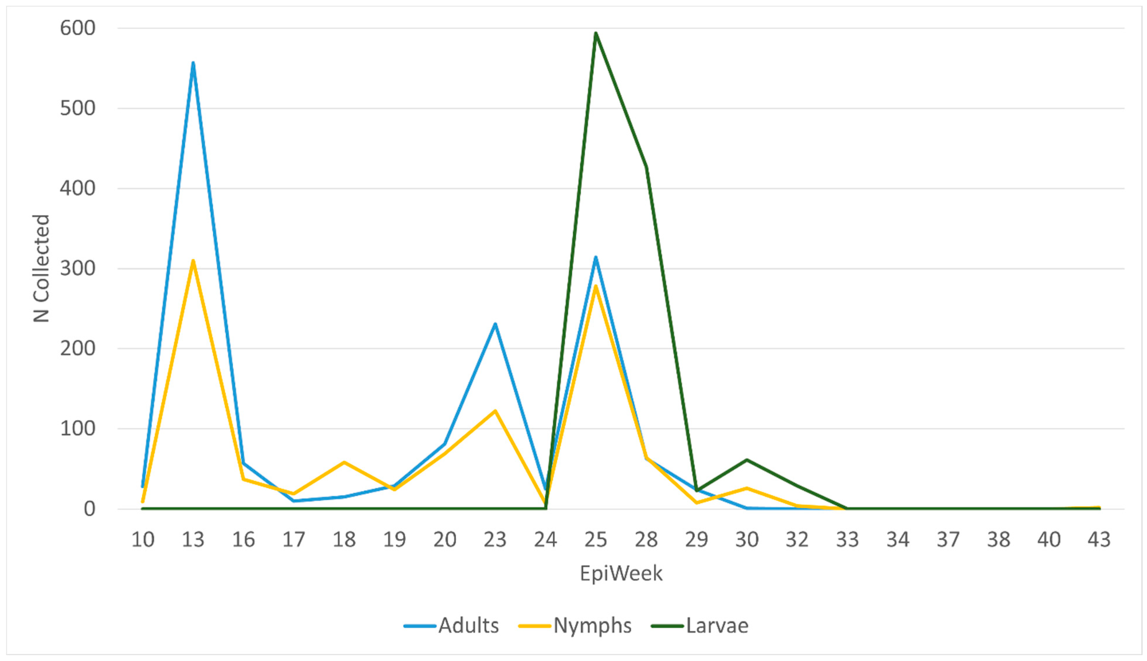
Three collection peaks were observed: EpiWeek 13 (22–28 March 2020), EpiWeek 23 (31 May–6 June 2020), and EpiWeek 25 (14–20 June 2020) (Figure 2). The EpiWeek 25 peak yielded the highest volume of Am. americanum ticks, with approximately 1200 ticks collected. Adult Am. americanum comprised most ticks collected until EpiWeek 25, when we observed a shift to large numbers of larvae collected. Throughout the collection period, nymphal activity peaks typically followed adult patterns. Ixodes scapularis peaked at EpiWeek 13 (22–28 March 2020) (Figure S1). All other species’ seasonality cannot be accurately interpreted due to low collection counts.

3.5. Geographic Distribution

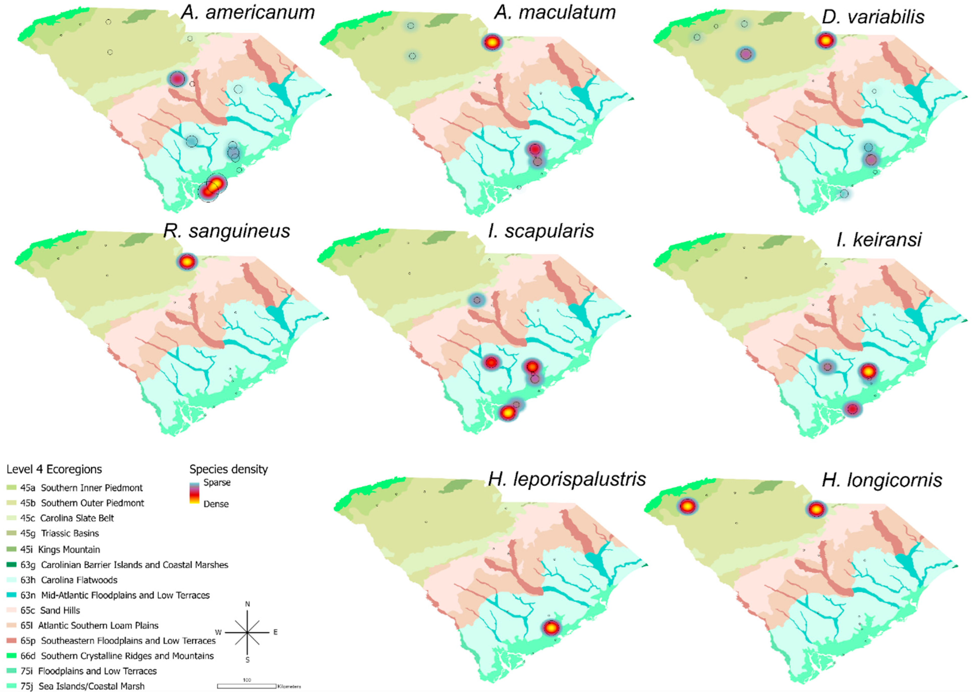
The highest volume of collected ticks was in the southern coastal area, where a large proportion of Am. americanum, Ix. scapularis, and Ix. keiransi were collected (Figure 3). Most of the ticks from the Upstate or Pee Dee regions were submitted from animal shelters (Figure S2). In contrast, the northern part of the state had a greater volume of Am. maculatum, De. variabilis, Rh. sanguineus, and Ha. longicornis.
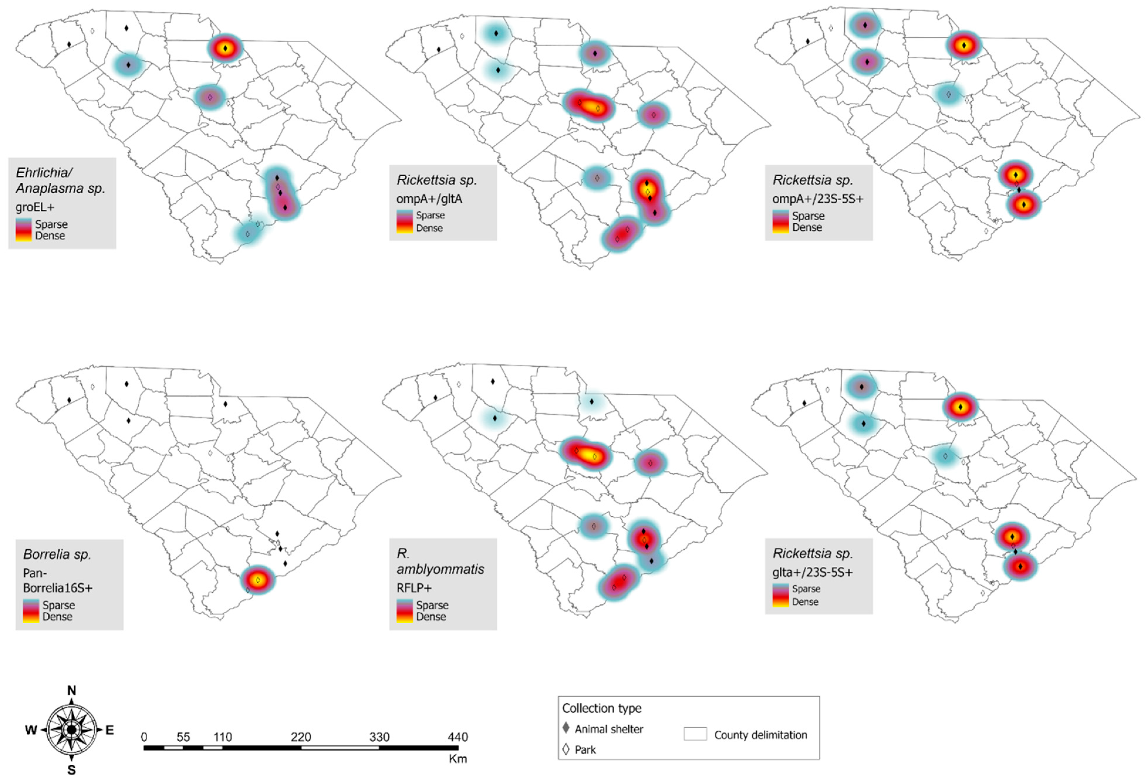
SFGR ompA+/gltA+ distribution and R. amblyommatis-positive tick geographies overlapped, with diffuse presence across the state. Ehrlichia and Anaplasma spp.-positive ticks were distributed along the northern and coastal regions, and one B. burgdorferi sensu stricto-positive tick was from a state park in the southern coastal region (Figure 4).

4. Discussion

This surveillance approach established a scalable, statewide, interdisciplinary network for tick research, integrating trainees into field and laboratory workflows to expand capacity and support workforce development. Sequencing confirmed the presence of multiple Rickettsia, Ehrlichia, and Anaplasma spp. and only one tick infected with B. burgdorferi s.s. Pathogen results from Am. americanum ticks revealed a high R. amblyommatis infection rate (53.2%), in line with a previous report from the SC coast [13], and a trend we also observed in ticks collected between 2021 and 2022 [46]. R. amblyommatis is widely distributed across the southeastern USA [47]. Its role in human disease remains debated, with no direct evidence supporting clinical pathogenicity beyond laboratory models [23,47,48,49]. Candidatus R. andeanae, also detected in the region, has no known human pathogenicity though may play an exclusionary role for other SFGR species in ticks [50,51,52]. Infection was not detected in any of the 2020 questing De. variabilis, Am. maculatum, or Ix. scapularis ticks and only two Ix. keiransi ticks were positive (one for B. burgdorferi s.s. and one for A. phagocytophilum).
R. parkeri, considered the most pathogenic species identified, is thought to be the second most prevalent tick-borne SFG rickettsiae in the Americas and is widely distributed across the southeastern USA [50,51,53,54]. Two Am. maculatum were positive for R. montanensis, a species historically considered non-pathogenic but increasingly implicated in misdiagnosed cases in the mid-Atlantic and southeastern USA [49,55,56], and previously reported in Am. maculatum in this region [54,55,57,58].
In animal shelter-submitted ticks, every tested species was positive for a rickettsial agent except Ha. leporispalustris. Ix. scapularis was positive for all three rickettsial genes tested, and approximately 9% were positive for either Ehrlichia or Anaplasma spp. Ix. keiransi was positive for all three SFGR genes tested, whereas De. variabilis had a lower SFGR-positive percentage than anticipated. Am. maculatum ticks had 20.7% positivity for Ehrlichia or Anaplasma spp. Notably, one De. variabilis and one Am. maculatum amplified sequences with 100% alignment to R. asembonensis, a flea-associated species rarely reported in ticks [59,60,61]. Although previously considered non-pathogenic, emerging evidence suggests potential involvement in human and animal disease [62,63,64,65,66]. Detection in these ticks may reflect co-feeding of infected fleas and naïve ticks on shared hosts, though evidence for this mechanism remains limited [61,67,68].
Most human ehrlichiosis cases in the USA are caused by E. chaffeensis, E. ewingii, E. muris eauclairensis, and, rarely, Panola Mountain Ehrlichia [69,70,71], primarily transmitted by Am. americanum [23,43,54,72]. The increasing incidence of ehrlichiosis nationwide underscores the clinical relevance of detecting multiple Ehrlichia spp. in ticks collected in SC [69,73]. Human anaplasmosis incidence is also increasing annually [74,75]. Although Ix. scapularis is the primary vector, A. phagocytophilum has also been reported in Am. americanum [54], suggesting that inclusion of multiple tick species in surveillance may help identify additional transmission pathways. South Carolina has reported 26 cases of undetermined ehrlichiosis/anaplasmosis, with the highest number occurring in southern coastal counties, though the causative agents and tick species involved remain unknown [76].
Historically, SC has reported RMSF outbreaks associated with SFGR-infected ticks [9]; the majority of SFGR-positive ticks in this study were positive for R. amblyommatis, and none were infected with R. rickettsii. Previous outbreak investigations collected higher numbers of De. variabilis, the primary vector of R. rickettsii, whereas this study predominantly collected Am. americanum [6], which may reflect differences in sampling methods or shifts in tick distribution.
This program aimed to establish a sustainable surveillance network and generate pilot data to support expansion of tick-borne disease research, outreach, and education [77]. The South Carolina tick surveillance program is now recognized among state veterinarians, local health systems, the public, and academic partners as a resource for tick-borne disease expertise. Our sustained collaborations have positioned us to address emerging tick-related public health threats. Programmatically, we met key milestones by building capacity for tick-borne disease research, generating baseline data, and supporting training of a future medical entomology workforce.
The program has expanded from bimonthly collections at 10 sites to year-round surveillance at more than 45 sites, each sampled every six weeks. Resulting data have contributed to peer-reviewed publications, successful grant funding, methodological innovation, and increased policy engagement at state and national levels [46,78,79]. These data have also informed intervention studies using innovative approaches to detect, control, and mitigate the impact of tick-borne disease (studies are ongoing).
There were several important lessons learned throughout this process. Despite efforts to harmonize data collection, differences across sites led to time-intensive cleaning and reconciliation prior to analysis. Establishing a standardized set of shared data points a priori supports the generation of reliable, high-quality datasets. The volume of data generated is substantial, and participating sites should determine early how data will be stored, managed, and shared. For our work, Microsoft Excel served as the primary platform for data collection, allowing alignment with CDC data points for Ixodes submissions. As data volume increased, organization into three categories—(1) collection data, (2) pathogen testing results, and (3) aggregated summary data—by year of surveillance proved critical and required coordinated support from multiple team members.
Given that these data represent collections from a single year, findings should be interpreted cautiously. Shelters did not record the date of tick collection, host animal species or travel history, and samples were stored in a single container. Our primary goal was to engage and establish consistent collection with shelters, which we accomplished. Methods of collection since 2020 have been refined to include more detailed data collection and follow-up with shelters. Animal shelter sites are also prone to selection bias, since the number of ticks submitted may not correspond to true regional tick abundance or distribution. Variability in staff engagement and interest likely influenced participation levels, with some shelters submitting substantially more specimens than others.

5. Conclusions

As Burgdorfer et al. emphasized in their study of RMSF in South Carolina, “…only through educational programs and availability of tick examination services, vigorously pursued, can a significant decrease be brought about in the incidence and mortality from this disease” [6]. Data-driven public health preparedness is a fulcrum of adequately addressing emerging and re-emerging infectious diseases. Together, these findings demonstrate that coordinated surveillance can generate region-specific risk profiles and broader insights into evolving tick-borne disease ecology using a cooperative framework and support national goals in the fight against vector-borne disease.

Supplementary Materials

The following supporting information can be downloaded at: https://www.mdpi.com/article/10.3390/insects17040414/s1, Figure S1: All tick species (except A. americanum) collected by EpiWeek in SC State Parks; Figure S2: Total tick collections distribution across animal shelter and state park collection sites.

Author Contributions

Conceptualization, C.L.E. and M.S.N.; Methodology, K.C.D.-B., M.M.M.-T., J.G.C. and E.O.P.; Validation, M.M.M.-T. and J.G.C.; Formal Analysis, K.C.D.-B., L.G.-G., E.O.P., M.M.M.-T. and C.L.; Resources, R.T.T.F. and M.S.N., Writing—Original draft preparation, K.C.D.-B., L.G.-G. and E.O.P.; Writing—Review and Editing, M.M.M.-T., C.L.E., J.G.C., R.T.T.F. and M.S.N. All authors have read and agreed to the published version of the manuscript.

Funding

The University of South Carolina Arnold School of Public Health Norman J. Arnold Fellowship and Office of the Vice President of Research’s ASPIRE grant mechanism partially funded this project.

Data Availability Statement

The raw data supporting the conclusions of this article will be made available by the authors upon request.

Acknowledgments

We would like to thank the late Paul Nolan and colleagues for providing access to the McAlhaney Nature Preserve. We appreciate the collaboration from our 10 partnering animal shelters for their support of the project: Spartanburg Humane Society, Pickens County Humane Society, Laurens County Humane Society, Lancaster County Animal Shelter, SPCA Humane Organization, Charleston Animal Society, Berkeley Animal Center, Pet Helpers Adoption Center, Palmetto Animal Hospital, and the Humane Society of North Myrtle Beach. We also thank the CDC Division of Vector-Borne Diseases for pathogen testing submitted Ixodes specimens. We would like to thank Matthew Lawson and the South Carolina State Park Service for providing access to state parks for tick collections. We would also like to thank the many student volunteers at the University of South Carolina that made this project a reality—specifically Aidan Warner, Tyler Bunting, Danielle Johnson, Chloe Rodriguez Ramos, McKenzi Norris, and Connor Ross who helped coordinate other students’ efforts.

Conflicts of Interest

The authors declare no conflicts of interest.

References

  1. Rosenberg, R.; Lindsey, N.P.; Fischer, M.; Gregory, C.J.; Hinckley, A.F.; Mead, P.S.; Paz-Bailey, G.; Waterman, S.H.; Drexler, N.A.; Kersh, G.J.; et al. Vital signs: Trends in reported vectorborne disease cases—United States and Territories, 2004–2016. Morb. Mortal. Wkly. Rep. 2018, 67, 496. [Google Scholar] [CrossRef] [Scilit] [PubMed]
  2. Eisen, L. Control of Ixodid ticks and prevention of tick-borne diseases in the United States: The prospect of a new Lyme disease vaccine and the continuing problem with tick exposure on residential properties. Ticks Tick Borne Dis. 2021, 12, 101649. [Google Scholar] [CrossRef] [Scilit]
  3. Eisen, R.J.; Eisen, L. The Blacklegged Tick, Ixodes scapularis: An Increasing Public Health Concern. Trends Parasitol. 2018, 34, 295–309. [Google Scholar] [CrossRef] [Scilit]
  4. Stromdahl, E.; Hickling, G. Beyond Lyme: Aetiology of tick-borne human diseases with emphasis on the South-Eastern United States. Zoonoses Public Health 2012, 59, 48–64. [Google Scholar] [CrossRef] [Scilit]
  5. Dye-Braumuller, K.C.; Gordon, J.R.; Johnson, D.; Morrissey, J.; McCoy, K.; Dinglasan, R.R.; Nolan, M.S. Needs Assessment of Southeastern United States Vector Control Agencies: Capacity Improvement Is Greatly Needed to Prevent the Next Vector-Borne Disease Outbreak. Trop. Med. Infect. Dis. 2022, 7, 73. [Google Scholar] [CrossRef] [Scilit]
  6. Burgdorfer, W.; Adkins, T.R., Jr.; Priester, L.E. Rocky Mountain spotted fever (tick-borne typhus) in South Carolina: An educational program and tick/rickettsial survey in 1973 and 1974. Am. J. Trop. Med. Hyg. 1975, 24, 866–872. [Google Scholar] [CrossRef] [Scilit] [PubMed]
  7. Felz, M.W.; Durden, L.A. Attachment sites of four tick species (Acari: Ixodidae) parasitizing humans in Georgia and South Carolina. J. Med. Entomol. 1999, 36, 361–364. [Google Scholar] [CrossRef] [Scilit]
  8. Felz, M.W.; Durden, L.A.; Oliver, J.H., Jr. Ticks parasitizing humans in Georgia and South Carolina. J. Parasitol. 1996, 82, 505–508. [Google Scholar] [CrossRef] [Scilit] [PubMed]
  9. Loving, S.; Smith, A.; DiSalvo, A.; Burgdorfer, W. Distribution and prevalence of spotted fever group rickettsiae in ticks from South Carolina, with an epidemiological survey of persons bitten by infected ticks. Am. J. Trop. Med. Hyg. 1978, 27, 1255–1260. [Google Scholar] [CrossRef] [Scilit] [PubMed]
  10. Clark, K.L.; Oliver, J.H.; James, A.M.; Durden, L.A.; Banks, C.W. Prevalence of Borrelia burgdorferi sensu lato infection among rodents and host-seeking ticks in South Carolina. J. Med. Entomol. 2002, 39, 198–206. [Google Scholar] [CrossRef] [Scilit]
  11. DiSalvo, A. Tick distribution in South Carolina. J. S. C. Med. Assoc. 1989, 494–495. [Google Scholar]
  12. Loftis, A.D.; Mixson, T.R.; Stromdahl, E.Y.; Yabsley, M.J.; Garrison, L.E.; Williamson, P.C.; Fitak, R.R.; Fuerst, P.A.; Kelly, D.J. Geographic distribution and genetic diversity of the Ehrlichia sp. from Panola Mountain in Amblyomma americanum. BMC Infect. Dis. 2008, 8, 54. [Google Scholar] [CrossRef] [Scilit]
  13. Mixson, T.R.; Campbell, S.R.; Gill, J.S.; Ginsberg, H.S.; Reichard, M.V.; Schulze, T.L.; Dasch, G.A. Prevalence of Ehrlichia, Borrelia, and Rickettsial agents in Amblyomma americanum (Acari: Ixodidae) collected from nine states. J. Med. Entomol. 2006, 43, 1261–1268. [Google Scholar] [CrossRef] [Scilit]
  14. Nelder, M.P.; Reeves, W.K.; Adler, P.H.; Wozniak, A.; Wills, W. Ectoparasites and associated pathogens of free-roaming and captive animals in zoos of South Carolina. Vector-Borne Zoonotic Dis. 2009, 9, 469–477. [Google Scholar] [CrossRef] [Scilit]
  15. Stromdahl, E.Y.; Nadolny, R.M.; Gibbons, J.A.; Auckland, L.D.; Vince, M.A.; Elkins, C.E.; Murphy, M.P.; Hickling, G.J.; Eshoo, M.W.; Carolan, H.E.; et al. Borrelia burgdorferi not confirmed in human-biting Amblyomma americanum ticks from the southeastern United States. J. Clin. Microbiol. 2015, 53, 1697–1704. [Google Scholar] [CrossRef] [Scilit]
  16. Xu, G.; Luo, C.-Y.; Ribbe, F.; Pearson, P.; Ledizet, M.; Rich, S.M. Borrelia miyamotoi in human-biting ticks, United States, 2013–2019. Emerg. Infect. Dis. 2021, 27, 3193. [Google Scholar] [CrossRef] [Scilit] [PubMed]
  17. DADE. The South Carolina Annual Morbidity Report on Reportable Conditions—2018; South Carolina Department of Public Health Bureau of Communicable Disease Prevention and Control: Columbia, SC, USA, 2019.
  18. South Carolina Department of Public Health. The South Carolina Annual Morbidity Report on Reportable Conditions, 2021; DADE Bureau of Communicable Disease Prevention and Control: Atlanta, GA, USA, 2021.
  19. Schwartz, A.M.; Hinckley, A.F.; Mead, P.S.; Hook, S.A.; Kugeler, K.J. Surveillance for Lyme Disease—United States, 2008–2015. MMWR Surveill. Summ. 2017, 66, 1–12. [Google Scholar] [CrossRef] [Scilit] [PubMed]
  20. Aucott, J.; Morrison, C.; Munoz, B.; Rowe, P.C.; Schwarzwalder, A.; West, S.K. Diagnostic challenges of early Lyme disease: Lessons from a community case series. BMC Infect. Dis. 2009, 9, 79. [Google Scholar] [CrossRef] [Scilit]
  21. Berger, J.J.; Hayes, B.K.; The Tick-Borne Disease Working Group. Tick-Borne Disease Working Group 2020 Report to Congress; U.S. Department of Health and Human Services: Washington, DC, USA, 2020.
  22. Marusiak, A.B.; Hollingsworth, B.D.; Abernathy, H.; Alejo, A.; Arahirwa, V.; Mansour, O.; Giandomenico, D.; Schmitz, J.; Williams, C.; Barbarin, A.M.; et al. Patterns of testing for Tick-Borne diseases and implications for Surveillance in the Southeastern US. JAMA Netw. Open 2022, 5, e2212334. [Google Scholar] [CrossRef] [Scilit] [PubMed]
  23. Gleim, E.R.; Garrison, L.E.; Vello, M.S.; Savage, M.Y.; Lopez, G.; Berghaus, R.D.; Yabsley, M.J. Factors associated with tick bites and pathogen prevalence in ticks parasitizing humans in Georgia, USA. Parasites Vectors 2016, 9, 125. [Google Scholar] [CrossRef] [Scilit]
  24. Eisen, R.J.; Foster, E.; Kjemtrup, A.; Saunders, M.E.; Brown, J.; Green, L.; Cervantes, K.; Prusinski, M.A.; White, J.; Barbarin, A.M.; et al. Perspectives from federal and state public health departments on their participation in and the utility of Ixodes scapularis (Acari: Ixodidae) and Ixodes pacificus tick and tick-borne pathogen surveillance in the United States. J. Med. Entomol. 2024, 62, 289–300. [Google Scholar] [CrossRef] [Scilit]
  25. Eisen, R.J.; Paddock, C.D. Tick and Tickborne Pathogen Surveillance as a Public Health Tool in the United States. J. Med. Entomol. 2021, 58, 1490–1502. [Google Scholar] [CrossRef] [Scilit]
  26. CDC. Surveillance for Ixodes scapularis and Pathogens Found in This Tick Species in the United States; Division of Vector Borne Diseases: Fort Collins, CO, USA, 2019.
  27. CDC. Guide to the Surveillance of Metastriate Ticks (Acari: Ixodidae) and Their Pathogens in the United States; Division of Vector Borne Diseases: Fort Collins, CO, USA, 2020.
  28. Keirans, J.E.; Litwak, T.R. Pictorial key to the adults of hard ticks, family Ixodidae (Ixodida: Ixodoidea), east of the Mississippi River. J. Med. Entomol. 1989, 26, 435–448. [Google Scholar] [CrossRef] [Scilit]
  29. Keirans, J.E.; Durden, L.A. Illustrated key to nymphs of the tick genus Amblyomma (Acari: Ixodidae) found in the United States. J. Med. Entomol. 1998, 35, 489–495. [Google Scholar] [CrossRef] [Scilit]
  30. Egizi, A.M.; Robbins, R.G.; Beati, L.; Nava, S.; Evans, C.R.; Occi, J.L.; Fonseca, D.M. A pictorial key to differentiate the recently detected exotic Haemaphysalis longicornis Neumann, 1901 (Acari, Ixodidae) from native congeners in North America. ZooKeys 2019, 818, 117–128. [Google Scholar] [CrossRef] [Scilit] [PubMed]
  31. Durden, L.A.; Keirans, J.E. Nymphs of the Genus Ixodes (Acari: Ixodidae) of the United States: Taxonomy, Identification Key, Distribution, Hosts, and Medical/Veterinary Importance; Entomological Society of America: Lanham, MD, USA, 1996. [Google Scholar]
  32. Cooley, R.A.; Kohls, G.M. The genus Amblyomma (ixodidae) in the United States. J. Parasitol. 1944, 30, 77–111. [Google Scholar] [CrossRef] [Scilit]
  33. Clifford, C.M.; Anastos, G.; Van der Borght-Elbl, A. The Larval Ixodid Ticks of the Eastern United States (Acarina-Ixodidae); Entomological Society of America: Lanham, MD, USA, 1961. [Google Scholar]
  34. Clifford, C.M.; Anastos, G. The use of chaetotaxy in the identification of larval ticks (Acarina: Ixodidae). J. Parasitol. 1960, 46, 567–578. [Google Scholar] [CrossRef] [Scilit]
  35. Vannier, E.G.; Diuk-Wasser, M.A.; Ben Mamoun, C.; Krause, P.J. Babesiosis. Infect. Dis. Clin. N. Am. 2015, 29, 357–370. [Google Scholar] [CrossRef] [Scilit]
  36. Graham, C.B.; Maes, S.E.; Hojgaard, A.; Fleshman, A.C.; Sheldon, S.W.; Eisen, R.J. A molecular algorithm to detect and differentiate human pathogens infecting Ixodes scapularis and Ixodes pacificus (Acari: Ixodidae). Ticks Tick-Borne Dis. 2018, 9, 390–403. [Google Scholar] [CrossRef] [Scilit]
  37. Huang, M.; Demarais, S.; Strickland, B.; Brookshire, W.; Chandler, J.; Butler, R.; Fryxell, R.T. Supplemental feeding of deer reduces tick abundance in Mississippi, USA. J. Vector Ecol. 2022, 47, 29–37. [Google Scholar] [CrossRef] [Scilit] [PubMed]
  38. Kollars, T.M.; Kengluecha, A. Spotted fever group Rickettsia in Dermacentor variabilis (Acari: Ixodidae) infesting raccoons (Carnivora: Procyonidae) and opossums (Marsupialia: Didelphimorphidae) in Tennessee. J. Med. Entomol. 2001, 38, 601–602. [Google Scholar] [CrossRef] [Scilit]
  39. Jado, I.; Escudero, R.; Gil, H.; Jiménez-Alonso, M.I.; Sousa, R.; García-Pérez, A.L.; Rodríguez-Vargas, M.; Lobo, B.; Anda, P. Molecular method for identification of Rickettsia species in clinical and environmental samples. J. Clin. Microbiol. 2006, 44, 4572–4576. [Google Scholar] [CrossRef] [Scilit] [PubMed]
  40. Killmaster, L.; Loftis, A.; Zemtsova, G.; Levin, M. Detection of bacterial agents in Amblyomma americanum (Acari: Ixodidae) from Georgia, USA, and the use of a multiplex assay to differentiate Ehrlichia chaffeensis and Ehrlichia ewingii. J. Med. Entomol. 2014, 51, 868–872. [Google Scholar] [CrossRef] [Scilit]
  41. Dye-Braumuller, K.C.; Lynn, M.K.; Rivas, P.M.C.; Lee, C.; Aquino, M.S.R.; Chandler, J.G.; Fryxell, R.R.T.; Self, S.C.; Kanyangarara, M.; Nolan, M.S. First report of multiple Rickettsia sp., Anaplasma sp., and Ehrlichia sp. in the San Miguel Department of El Salvador from zoonotic tick vectors. Acta Trop. 2023, 242, 106909. [Google Scholar] [CrossRef] [Scilit]
  42. Tabara, K.; Arai, S.; Kawabuchi, T.; Itagaki, A.; Ishihara, C.; Satoh, H.; Okabe, N.; Tsuji, M. Molecular survey of Babesia microti, Ehrlichia species and Candidatus Neoehrlichia mikurensis in wild rodents from Shimane Prefecture, Japan. Microbiol. Immunol. 2007, 51, 359–367. [Google Scholar] [CrossRef] [Scilit]
  43. Trout Fryxell, R.T.; Hendricks, B.M.; Pompo, K.; Mays, S.E.; Paulsen, D.J.; Operario, D.J.; Houston, A.E. Investigating the adult ixodid tick populations and their associated Anaplasma, Ehrlichia, and Rickettsia bacteria at a Rocky Mountain spotted fever hotspot in Western Tennessee. Vector-Borne Zoonotic Dis. 2017, 17, 527–538. [Google Scholar] [CrossRef] [Scilit]
  44. Shah, N.; Altschul, S.F.; Pop, M. Outlier detection in BLAST hits. Algorithms Mol. Biol. 2018, 13, 7. [Google Scholar] [CrossRef] [Scilit]
  45. Moutailler, S.; Valiente Moro, C.; Vaumourin, E.; Michelet, L.; Tran, F.H.; Devillers, E.; Cosson, J.-F.; Gasqui, P.; Van, V.T.; Mavingui, P.; et al. Co-infection of ticks: The rule rather than the exception. PLoS Neglected Trop. Dis. 2016, 10, e0004539. [Google Scholar] [CrossRef] [Scilit]
  46. Gual-Gonzalez, L.; Self, S.C.; Zellars, K.; Meyer, M.; Dye-Braumuller, K.C.; Evans, C.L.; Cantillo-Barraza, O.; Torres, M.W.; Nolan, M.S. Eco-epidemiology of Rickettsia amblyommatis and Rickettsia parkeri in naturally infected ticks (Acari: Ixodida) from South Carolina. Parasites Vectors 2024, 17, 33. [Google Scholar] [CrossRef] [Scilit] [PubMed]
  47. Richardson, E.A.; Roe, R.M.; Apperson, C.S.; Ponnusamy, L. Rickettsia amblyommatis in ticks: A review of distribution, pathogenicity, and diversity. Microorganisms 2023, 11, 493. [Google Scholar] [CrossRef] [Scilit] [PubMed]
  48. Billeter, S.A.; Blanton, H.L.; Little, S.E.; Levy, M.G.; Breitschwerdt, E.B. Detection of “Rickettsia amblyommii” in association with a tick bite rash. Vector-Borne Zoonotic Dis. 2007, 7, 607–610. [Google Scholar] [CrossRef] [Scilit]
  49. Snellgrove, A.N.; Krapiunaya, I.; Scott, P.; Levin, M.L. Assessment of the pathogenicity of Rickettsia amblyommatis, Rickettsia bellii, and Rickettsia montanensis in a guinea pig model. Vector-Borne Zoonotic Dis. 2021, 21, 232–241. [Google Scholar] [CrossRef] [Scilit]
  50. Paddock, C.D.; Denison, A.M.; Dryden, M.W.; Noden, B.H.; Lash, R.R.; Abdelghani, S.S.; Evans, A.E.; Kelly, A.R.; Hecht, J.A.; Karpathy, S.E.; et al. High prevalence of “Candidatus Rickettsia andeanae” and apparent exclusion of Rickettsia parkeri in adult Amblyomma maculatum (Acari: Ixodidae) from Kansas and Oklahoma. Ticks Tick-Borne Dis. 2015, 6, 297–302. [Google Scholar] [CrossRef] [Scilit]
  51. Allerdice, M.E.; Hecht, J.A.; Lash, R.R.; Karpathy, S.E.; Paddock, C.D. Rickettsia parkeri and “Candidatus Rickettsia andeanae” in Amblyomma maculatum (Acari: Ixodidae) collected from the Atlanta metropolitan area, Georgia, United States. Ticks Tick-Borne Dis. 2019, 10, 1066–1069. [Google Scholar] [CrossRef] [Scilit]
  52. Pickle, E.O.; Dye-Braumuller, K.; Gual-Gonzalez, L.; Trout Fryxell, R.T.; Chandler, J.; Qurollo, B.; Nolan, M.S. First identification of Candidatus Rickettsia andeanae in host-attached Dermacentor variabilis and across a large geographic sampling region, South Carolina, U.S.A. J. Vector Ecol. 2025, 50, S4–S7. [Google Scholar] [CrossRef] [Scilit]
  53. Silva-Ramos, C.R.; Hidalgo, M.; Faccini-Martínez, Á.A. Clinical, epidemiological, and laboratory features of Rickettsia parkeri rickettsiosis: A systematic review. Ticks Tick-Borne Dis. 2021, 12, 101734. [Google Scholar] [CrossRef] [Scilit]
  54. Lee, S.; Kakumanu, M.L.; Ponnusamy, L.; Vaughn, M.; Funkhouser, S.; Thornton, H.; Meshnick, S.R.; Apperson, C.S. Prevalence of Rickettsiales in ticks removed from the skin of outdoor workers in North Carolina. Parasites Vectors 2014, 7, 607. [Google Scholar] [CrossRef] [Scilit] [PubMed]
  55. St John, H.K.; Adams, M.L.; Masuoka, P.M.; Flyer-Adams, J.G.; Jiang, J.; Rozmajzl, P.J.; Stromdahl, E.Y.; Richards, A.L. Prevalence, distribution, and development of an ecological niche model of Dermacentor variabilis ticks positive for Rickettsia montanensis. Vector-Borne Zoonotic Dis. 2016, 16, 253–263. [Google Scholar] [CrossRef] [Scilit] [PubMed]
  56. McQuiston, J.H.; Zemtsova, G.; Perniciaro, J.; Hutson, M.; Singleton, J.; Nicholson, W.L.; Levin, M.L. Afebrile spotted fever group Rickettsia infection after a bite from a Dermacentor variabilis tick infected with Rickettsia montanensis. Vector-Borne Zoonotic Dis. 2012, 12, 1059–1061. [Google Scholar] [CrossRef] [Scilit] [PubMed]
  57. Stanley, H.; Rhodes, D.V. Presence of Rickettsia species in ticks collected from companion animals in northeastern Georgia, United States. Vet. Sci. 2021, 8, 37. [Google Scholar] [CrossRef] [Scilit]
  58. Lippi, C.A.; Gaff, H.D.; Nadolny, R.M.; Ryan, S.J. Newer Surveillance Data Extends our Understanding of the Niche of Rickettsia montanensis (Rickettsiales: Rickettsiaceae) Infection of the American Dog Tick (Acari: Ixodidae) in the United States. Vector-Borne Zoonotic Dis. 2023, 23, 316–323. [Google Scholar] [CrossRef] [Scilit]
  59. Troyo, A.; Moreira-Soto, R.D.; Calderon-Arguedas, Ó.; Mata-Somarribas, C.; Ortiz-Tello, J.; Barbieri, A.R.; Avendaño, A.; Vargas-Castro, L.E.; Labruna, M.B.; Hun, L.; et al. Detection of rickettsiae in fleas and ticks from areas of Costa Rica with history of spotted fever group rickettsioses. Ticks Tick-Borne Dis. 2016, 7, 1128–1134. [Google Scholar] [CrossRef] [Scilit] [PubMed]
  60. Dall’Agnol, B.; Souza, U.; Webster, A.; Weck, B.; Stenzel, B.; Labruna, M.; Klafke, G.; Martins, J.R.; Ferreira, C.A.S.; Reck, J. “Candidatus Rickettsia asemboensis” in Rhipicephalus sanguineus ticks, Brazil. Acta Trop. 2017, 167, 18–20. [Google Scholar] [CrossRef] [Scilit]
  61. Low, V.L.; Prakash, B.K.; Tan, T.K.; Sofian-Azirun, M.; Anwar, F.H.K.; Vinnie-Siow, W.Y.; AbuBakar, S. Pathogens in ectoparasites from free-ranging animals: Infection with Rickettsia asembonensis in ticks, and a potentially new species of Dipylidium in fleas and lice. Vet. Parasitol. 2017, 245, 102–105. [Google Scholar] [CrossRef] [Scilit]
  62. Maina, A.N.; Jiang, J.; Luce-Fedrow, A.; St John, H.K.; Farris, C.M.; Richards, A.L. Worldwide presence and features of flea-borne Rickettsia asembonensis. Front. Vet. Sci. 2019, 5, 334. [Google Scholar] [CrossRef] [Scilit] [PubMed]
  63. Loyola, S.; Palacios-Salvatierra, R.; Cáceres-Rey, O.; Richards, A.L. Rickettsia asembonensis Isolated from Four Human Cases with Acute Undifferentiated Febrile Illness in Peru. Pathogens 2024, 13, 489. [Google Scholar] [CrossRef] [Scilit]
  64. Tay, S.T.; Koh, F.X.; Kho, K.L.; Sitam, F.T. Rickettsial infections in monkeys, Malaysia. Emerg. Infect. Dis. 2015, 21, 545. [Google Scholar] [CrossRef] [Scilit]
  65. Kho, K.L.; Koh, F.X.; Singh, H.K.L.; Zan, H.A.M.; Kukreja, A.; Ponnampalavanar, S.; Tay, S.T. Spotted fever group rickettsioses and murine typhus in a Malaysian teaching hospital. Am. J. Trop. Med. Hyg. 2016, 95, 765–768. [Google Scholar] [CrossRef] [Scilit]
  66. Kolo, A.O.; Sibeko-Matjila, K.P.; Maina, A.N.; Richards, A.L.; Knobel, D.L.; Matjila, P.T. Molecular detection of zoonotic rickettsiae and Anaplasma spp. in domestic dogs and their ectoparasites in Bushbuckridge, South Africa. Vector-Borne Zoonotic Dis. 2016, 16, 245–252. [Google Scholar] [CrossRef] [Scilit] [PubMed]
  67. Marquez, A.R.A.; Eamens, K.; Westman, M.; Šlapeta, J. Vector-borne pathogens in ticks and fleas of client-owned dogs in Metro Manila, Philippines. Parasitologia 2021, 1, 247–256. [Google Scholar] [CrossRef] [Scilit]
  68. Fongsaran, C.; Jirakanwisal, K.; Tongluan, N.; Latour, A.; Healy, S.; Christofferson, R.C.; Macaluso, K.R. The role of cofeeding arthropods in the transmission of Rickettsia felis. PLoS Neglected Trop. Dis. 2022, 16, e0010576. [Google Scholar] [CrossRef] [Scilit] [PubMed]
  69. Heitman, K.N.; Dahlgren, F.S.; Drexler, N.A.; Massung, R.F.; Behravesh, C.B. Increasing incidence of ehrlichiosis in the United States: A summary of national surveillance of Ehrlichia chaffeensis and Ehrlichia ewingii infections in the United States, 2008–2012. Am. J. Trop. Med. Hyg. 2016, 94, 52. [Google Scholar] [CrossRef] [Scilit]
  70. Reeves, W.K.; Loftis, A.D.; Nicholson, W.L.; Czarkowski, A.G. The first report of human illness associated with the Panola Mountain Ehrlichia species: A case report. J. Med. Case Rep. 2008, 2, 139. [Google Scholar] [CrossRef] [Scilit] [PubMed]
  71. Xu, G.; Foster, E.; Ribbe, F.; Hojgaard, A.; Eisen, R.J.; Paull, S.; Rich, S.M. Detection of Ehrlichia muris eauclairensis in blacklegged ticks (Ixodes scapularis) and white-footed mice (Peromyscus leucopus) in Massachusetts. Vector-Borne Zoonotic Dis. 2023, 23, 311–315. [Google Scholar] [CrossRef] [Scilit]
  72. Allerdice, M.E.; Hecht, J.A.; Karpathy, S.E.; Paddock, C.D. Evaluation of gulf coast ticks (acari: Ixodidae) for Ehrlichia and Anaplasma species. J. Med. Entomol. 2017, 54, 481–484. [Google Scholar] [PubMed]
  73. Harris, R.M.; Couturier, B.A.; Sample, S.C.; Coulter, K.S.; Casey, K.K.; Schlaberg, R. Expanded geographic distribution and clinical characteristics of Ehrlichia ewingii infections, United States. Emerg. Infect. Dis. 2016, 22, 862. [Google Scholar] [CrossRef] [Scilit]
  74. Russell, A.; Prusinski, M.; Sommer, J.; O’Connor, C.; White, J.; Falco, R.; Kokas, J.; Vinci, V.; Gall, W.; Tober, K.; et al. Epidemiology and spatial emergence of anaplasmosis, New York, USA, 2010–2018. Emerg. Infect. Dis. 2021, 27, 2154. [Google Scholar] [CrossRef] [Scilit]
  75. Baker, A.; Wang, H.-H.; Mogg, M.; Derouen, Z.; Borski, J.; Grant, W.E. Increasing incidence of anaplasmosis in the United States, 2012 through 2016. Vector-Borne Zoonotic Dis. 2020, 20, 855–859. [Google Scholar] [CrossRef] [Scilit]
  76. CDC. Reported Tickborne Disease Cases by County of Residence 2019–2022; Centers for Disease Control and Prevention: Atlanta, GA, USA, 2024.
  77. Mader, E.M.; Ganser, C.; Geiger, A.; Harrington, L.C.; Foley, J.; Smith, R.L.; Mateus-Pinilla, N.; Teel, P.D.; Eisen, R.J. A survey of tick surveillance and control practices in the United States. J. Med. Entomol. 2020, 58, 1503–1512. [Google Scholar] [CrossRef] [Scilit]
  78. Case, B.; Dye-Braumuller, K.C.; Evans, C.; Li, H.; Rustin, L.; Nolan, M.S. Adapting vector surveillance using Bayesian experimental design: An application to an ongoing tick monitoring program in the southeastern United States. Ticks Tick-Borne Dis. 2024, 15, 102329. [Google Scholar] [CrossRef] [Scilit]
  79. Dye-Braumuller, K.C.; Gual-Gonzalez, L.; Abiodun, T.; Rustin, L.P.; Evans, C.L.; Meyer, M.M.; Zellars, K.; Neault, M.J.; Nolan, M.S. Invasive Haemaphysalis longicornis (Acari: Ixodidae) investigation in South Carolina: New records of establishment, pathogen prevalence, and blood meal analyses. J. Med. Entomol. 2023, 60, 1398–1405. [Google Scholar] [CrossRef] [Scilit] [PubMed]
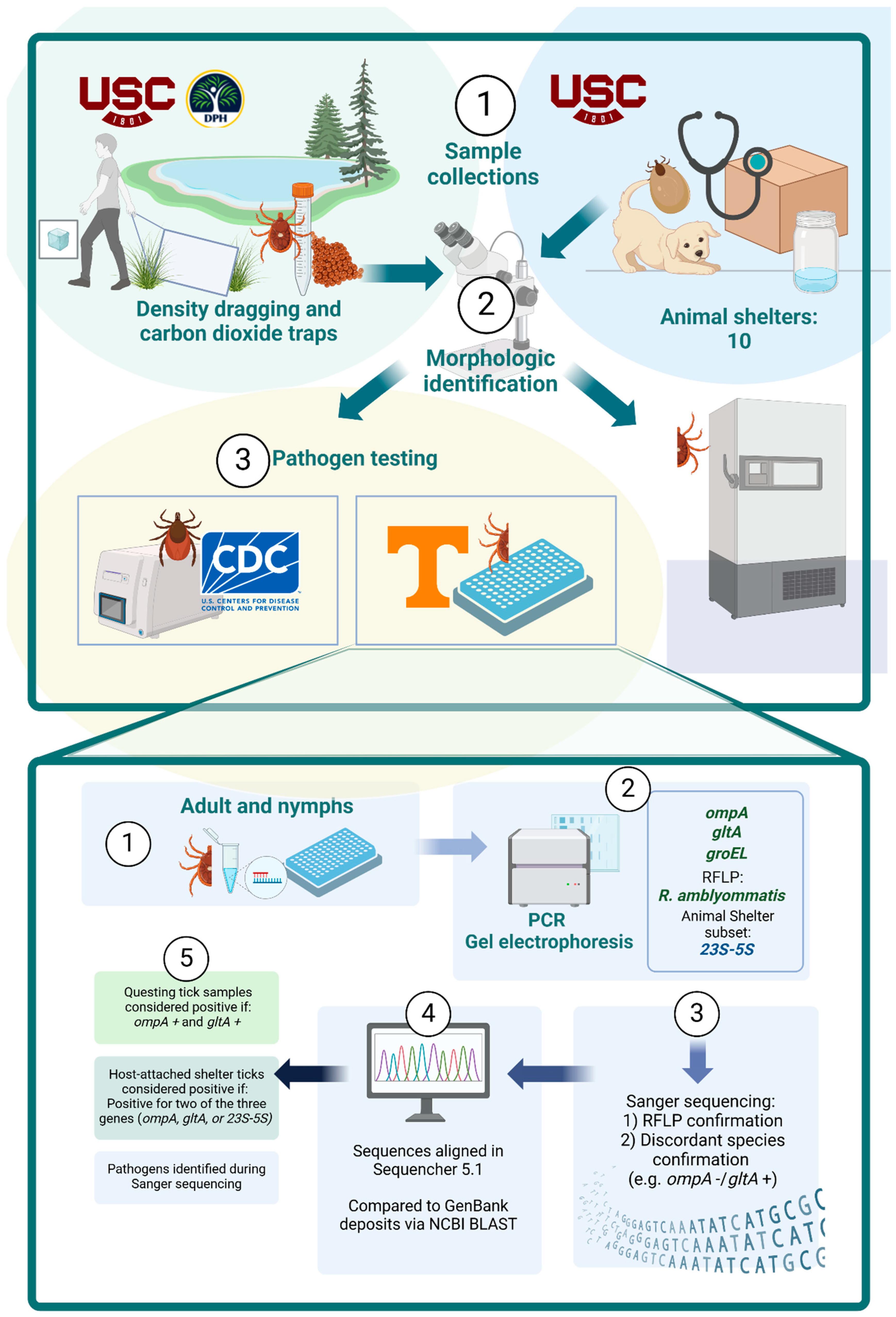
Figure 1. Schematic flow diagram of tick collection, processing, and testing.
Insects 17 00414 g001
Figure 2. Frequency of Amblyomma americanum collected by EpiWeek in SC State Parks, by life stage.
Insects 17 00414 g002
Figure 3. Map of species distribution spatial density by ecoregions.
Insects 17 00414 g003
Figure 4. Geographic distribution of pathogen test results, including density of positives for each collection site (animal shelters and parks).
Insects 17 00414 g004
Table 1. Tick species collected by sex, collection method, and life stage.
Ticks Collected from South Carolina Parks (n = 3674)
Life StageSex (Adults Only)Collection MethodTotal (%)
AdultNymphLarvaMaleFemaleDragTrapBody *Bite **
Amblyomma americanum14351037113461881716901784109233606 (98.1)
Amblyomma maculatum1001010001 (0)
Ixodes scapularis4805222640121053 (1.4)
Ixodes keiransi7004325007 (0.2)
Dermacentor variabilis7005261007 (0.2)
Ticks submitted from South Carolina animal shelters (n = 846)
Life stageSex (adults only)Fed statusTotal (%)
AdultNymphLarvaMaleFemaleFedFlat
Amblyomma americanum8052024568349132 (15.6)
Amblyomma maculatum1621501045833144177 (20.9)
Ixodes scapularis260062081826 (3.1)
Ixodes keiransi70007707 (0.8)
Ixodes unknown spp.40004404 (0.5)
Dermacentor variabilis14700767143104147 (17.4)
Haemaphysalis longicornis20002202 (0.2)
Haemaphysalis leporispalustris20002022 (0.2)
Rhipicephalus sanguineus23002055 (0.6)
Unknown spp. 2862560029648344 (40.7)
Adult unknown species were too crushed or missing too many body parts to be confidently morphologically identified to species or sex. * Ticks found on the collectors’ bodies, but not biting, were counted as ‘body’. ** Ticks found on the collectors’ bodies actively biting were counted as ‘bite’. These were not included in the ‘body’ tick count.
Table 2. Pathogens detected in tested ticks, by species.
Ticks Collected from South Carolina State Parks
N+/N Tested (%)
Tested at MVE UTKTested at CDC Bacterial Diseases Branch
Rickettsia spp.Ehrlichia/Anaplasma spp.Borrelia spp.Borrelia burgdorferi ss (Bbss)Anaplasma phagocytophilum (Ap)
ompA+ and gltA+ompA+ and 23S-5S+gltA+ and 23S-5S+RFLP confirmed (R. amblyommatis)groEL+Pan-Borrelia 16S+Pan-Borrelia 16S+, Bbss fliD+, and Bbss oppA2+Ap p44+ and Ap msp4+
Amblyomma americanum526/539 (97.6)10/10
(100)
10/10
(100)
512/962
(53.2)
18/966
(1.9)
---
Amblyomma maculatum 0/1 (0)--0/1 (0)0/1 (0)---
Ixodes scapularis-----0/38 (0)0/38 (0)0/38 (0)
Ixodes keiransi-----1/7
(14.3)
1/7
(14.3)
1/7
(14.3)
Dermacentor variabilis 0/7 (0)--0/7 (0)0/7 (0)---
Ticks submitted from South Carolina animal shelters *
N+/N tested (%)
Tested at MVE UTK
Rickettsia spp.Ehrlichia/Anaplasma spp.
ompA+ and gltA+ompA+ and 23S-5S+gltA+ and 23S-5S+RFLP confirmed (R. amblyommatis)groEL+
Amblyomma americanum46/117
(39.3)
26/72
(36.1)
30/72
(41.7)
36/117
(30.8)
3/117
(2.6)
Amblyomma maculatum41/87
(47.1)
40/46
(87.0)
45/46
(97.8)
0/87 (0)18/87
(20.7)
Ixodes scapularis18/22
(81.8)
7/10
(70.0)
9/10
(90.0)
0/22 (0)2/22 (9.1)
Ixodes keiransi6/6
(100)
6/6
(100)
6/6
(100)
0/6 (0)0/6 (0)
Ixodes spp. unknown4/4
(100)
4/4
(100)
4/4
(100)
0/4 (0)0/4 (0)
Dermacentor variabilis12/86
(14.0)
8/70
(11.4)
13/70
(18.6)
0/86 (0)0/86 (0)
Haemaphysalis leporispalustris 0/2 (0)--0/2 (0)0/2 (0)
Rhipicephalus sanguineus2/5
(40.0)
2/5
(40.0)
1/5
(20.0)
0/5 (0)1/5
(20.0)
* Both Haemaphysalis longicornis ticks were sent to the National Veterinary Diagnostic Laboratory and retained as voucher specimens; these and all unknown species were not tested for pathogens.  Amblyomma maculatum and Dermacentor variabilis ticks collected in state parks were not tested for the 23S-5S gene.  Haemaphysalis leporispalustris ticks collected from animal shelters were not tested for the 23S-5S gene. Testing of all three Rickettsia spp. genes was not available for all ticks. The algorithm for determining true positive ticks was if a tick tested positive for at least two of the three genes tested. This was to increase the sensitivity of the testing.
Table 3. Sequencing results from Rickettsia gltA or 23S-5S positive ticks or Ehrlichia/Anaplasma groEL-positive ticks.
Pathogen Sequencing ArchetypeSpeciesSample IDCollection DateCollection TypeEngorgement StatusLife StageSexSpecies Identity% Alignment and GenBank Accession NumberPositive on Other Rickettsia Gene Target Test
Sequencing results from gltA productsAmblyomma americanumAAF07756/19/2020parkflatadultFR. amblyommatis100%N
KY273595
Amblyomma americanumAAF08867/10/2020parkflatadultFR. amblyommatis100%N
KY273595
Amblyomma americanumAAF0981 *8/21/2020animalengorgedadultFR. parkeri100%N
MG574939
Amblyomma americanumAAM1033 8/21/2020animalflatadultMR. amblyommatis100%N
KY273595
Amblyomma americanumAAN1066 8/21/2020animalengorgednymphIR. amblyommatis100%Y
KY273595
Dermacentor variabilisDVF1010 *8/21/2020animalengorgedadultFR. parkeri100%N
MG574939
Dermacentor variabilisDVF1012 *8/21/2020animalengorgedadultFR. parkeri100%N
MG574939
Dermacentor variabilisDVF1013 *8/21/2020animalengorgedadultFR. amblyommatis99.62%N
KY273595
Dermacentor variabilisDVF1054 8/21/2020animalflatadultFR. parkeri100%N
MG574939
Amblyomma americanumAAM1129 8/24/2020animalflatadultMR. parkeri100%N
MG574939
Amblyomma americanumAAF1154 8/24/2020animalengorgedadultFR. amblyommatis100%Y
KY273595
Amblyomma maculatumAMF1160 8/24/2020animalflatadultFR. parkeri100%Y
MG574939
Amblyomma maculatumAMF1295 8/24/2020animalengorgedadultFR. parkeri100%Y
MG574939
Amblyomma maculatumAMF1298 8/24/2020animalengorgedadultFCa. R. andeanae100%Y
KT153033
Amblyomma maculatumAMM1173 8/24/2020animalflatadultMCa. R. andeanae100%Y
KT153033
Dermacentor variabilisDMF1214 8/24/2020animalengorgedadultFR. parkeri100%Y
MG574939
Dermacentor variabilisDVF1215 8/24/2020animalengorgedadultFCa. R. andeanae100%Y
KT153033
Dermacentor variabilisDVF1223 8/24/2020animalengorgedadultFR. amblyommatis99.79%N
KY273595
Dermacentor variabilisDVM1227 8/24/2020animalflatadultMR. parkeri100%Y
MG574939
Dermacentor variabilisDVM1232 8/24/2020animalflatadultMR. parkeri99.62%N
MG57439
Dermacentor variabilisDVM1233 8/24/2020animalflatadultMR. parkeri100%Y
MG574939
Dermacentor variabilisDVM1236 8/24/2020animalflatadultMCa. R. andeanae100%Y
KT153033
Amblyomma maculatumAMF1303 9/18/2020animalengorgedadultFR. parkeri100%Y
MG574939
Amblyomma maculatumAMM1082 9/18/2020animalflatadultMR. parkeri100%Y
MG574939
Amblyomma maculatumAMM1090 9/18/2020animalflatadultMCa. R. andeanae100%Y
KT153033
Sequencing results from 23S-5S productsAmblyomma americanumAAF13008/11/2020animalengorgedadultFR. amblyommatis100%Y
KJ796417
Amblyomma americanumAAN1189 8/24/2020animalengorgednymphIR. parkeri98.63%N
KJ796435
Amblyomma maculatumAMM1183 8/24/2020animalflatadultMR. montanensis98.63%Y
KJ796427
Amblyomma maculatumAMF1299 8/24/2020animalengorgedadultFR. amblyommatis99.45%N
KJ796417
Amblyomma maculatumAMN1191 8/24/2020animalengorgednymphIR. asembonensis100%N
OR523793
Dermacentor variabilisDVF1206 8/24/2020animalengorgedadultFR. asembonensis100%Y
OR523793
Amblyomma maculatumAMF13039/18/2020animalengorgedadultFR. parkeri99.72%Y
KJ796435
Amblyomma maculatumAMM120811/3/2020animalflatadultMR. montanensis97.60%Y
KJ796427
Sequencing results from groEL productsAmblyomma americanumAAN00123/6/2020parkflatnymphIA. odocoilei98%N/A
JX876642
Amblyomma americanumAAF00833/27/2020parkflatadultFA. odocoilei99%
JX876642
Amblyomma americanumAAF01053/27/2020parkflatadultFE. ewingii99%
KJ907744
Amblyomma americanumAAM04275/15/2020parkflatadultME. ewingii100%
KJ907744
Amblyomma americanumAAM04856/1/2020parkflatadultME. chaffeensis99%
KJ907753
Amblyomma americanumAAM04816/1/2020parkflatadultME. chaffeensis99%
KJ907753
Amblyomma americanumAAN05436/1/2020parkflatnymphIPanola Mtn Ehrlichia100%
HQ658904
Amblyomma americanumAAM05536/1/2020parkflatadultMPanola Mtn Ehrlichia91%
HQ658904
Amblyomma americanumAAN06436/17/2020parkflatnymphIA. odocoilei99%
JX876642
Amblyomma americanumAAM07036/17/2020parkflatadultME. chaffeensis99.40%
KJ907753
Amblyomma americanumAAF08187/10/2020parkflatadultFE. chaffeensis98%
KJ907753
Amblyomma americanumAAM08577/10/2020parkflatadultMPanola Mtn Ehrlichia100%
HQ658904
Amblyomma americanumAAF09768/21/2020animalengorgedadultFE. ewingii99%
KJ907744
Amblyomma americanumAAF11338/21/2020animalflatadultFE. ewingii99%
KJ907744
Ixodes scapularisISF1052 8/21/2020animalengorgedadultFE. ewingii99%
KJ907744
Ixodes scapularisISM1053 8/21/2020animalflatadultMA. phagocytophilum98%
MG570466
Amblyomma maculatumAMM11978/24/2020animalengorgedadultME. chaffeensis91%
KJ907753
Amblyomma maculatumAMM10899/18/2020animalflatadultMEhrlichia Cordoba96%
KY425416
Matching symbols indicate which ticks were collected from the same animal shelter from the same date: * All collected from the same animal shelter during one month. All collected from the same animal shelter during one month. All collected from the same animal shelter during one month. All collected from the same animal shelter during one month.
Disclaimer/Publisher’s Note: The statements, opinions and data contained in all publications are solely those of the individual author(s) and contributor(s) and not of MDPI and/or the editor(s). MDPI and/or the editor(s) disclaim responsibility for any injury to people or property resulting from any ideas, methods, instructions or products referred to in the content.