Article

Integrative Morphological and Molecular Diagnostics for Euseius nicholsi and Euseius oolong (Acari: Phytoseiidae)

Guangdong Key Laboratory of Animal Conservation and Resource Utilization, Guangdong Public Laboratory of Wild Animal Conservation and Utilization, Institute of Zoology, Guangdong Academy of Sciences, Guangzhou 510260, China
*
Author to whom correspondence should be addressed.
Insects 2025, 16(9), 950; https://doi.org/10.3390/insects16090950
Submission received: 21 July 2025 / Revised: 31 August 2025 / Accepted: 6 September 2025 / Published: 10 September 2025
(This article belongs to the Special Issue Revival of a Prominent Taxonomy of Insects)

Simple Summary

Specimens of a phytoseiid mite were collected from Bajiaozhai National Forest Park, Guilin, China, and showed close resemblance to Euseius nicholsi and Euseius oolong. Despite similarities, some morphological traits, such as body dimensions, number of dorsal solenostomes, spermathecal shape, and setae length differed. To determine whether these variations indicated distinct species or fell within natural variation, we performed molecular analysis using mitochondrial COI, 12S rRNA, and nuclear ITS markers. The genetic divergence among the unidentified specimens, E. nicholsi, and E. oolong was minimal, and all samples clustered together in a single clade in the phylogenetic tree. This suggested that they were not separate species. We therefore concluded that the unidentified specimens are E. nicholsi, and E. oolong should be considered its junior synonym. These findings underscore the value of integrating morphological and molecular tools for accurate mite identification.

Abstract

In a survey of Bajiaozhai National Forest Park (Guilin, China), several specimens of an Euseius sp. were collected. These specimens were very similar to Euseius nicholsi and Euseius oolong, based on morphological observations. However, some morphological characters, such as the body size, number of solenostomes on the dorsal plate, calyx shape of the spermatheca, the shape and number of metapodal platelet, teeth number on the fixed digit, length of setae j3, and macroseta Seg IV, Sti IV, and St IV were different between these specimens and E. nicholsi and E. oolong. To ascertain whether these morphological differences were interspecific or intraspecific variations, molecular analyses were conducted using mitochondrial DNA COI, 12S rRNA, and nuclear ITS markers. Based on the three molecular markers, minimal genetic distances were observed (COI 0–4%, 12S rRNA 0–2%, and ITS 0%) among the putative Euseius sp., E. nicholsi (collected from Bauhinia purpurea in IZGAS and from Eurya macartneyi and Ficus hispida in Shaoguan City), and E. oolong (collected from B. purpurea in IZGAS). Amblyseius swirskii was used as the outgroup. Using the maximum likelihood method, the phylogenetic tree showed that these specimens of Euseius sp., E. nicholsi, and E. oolong clustered in a single clade. Therefore, we propose that this putative Euseius sp. is E. nicholsi, and E. oolong is a junior synonym of E. nicholsi. This study demonstrates the importance of integrative taxonomy for the proper identification of phytoseiid mites.

1. Introduction

Phytoseiid mites are important natural enemies of pest mites and small arthropods [1,2,3]. Up to now, more than 2707 species and 94 genera have been described [4]. Phytoseiid mites are very tiny, about 300–600 μm in length [1]. Some morphological characteristics are very difficult to observe and their values for species discrimination are sometimes subject to discussion [5,6,7]. Furthermore, accurate observation of morphological characteristics is highly dependent on the quality of the mounting preparations. Some features, such as the number of teeth on the cheliceral digits or the presence of solenostomes, are sometimes difficult to observe, and reliable species descriptions require the examination of multiple individuals and preparations to avoid misidentification and confusion [8].
Euseius nicholsi (Ehara and Lee, 1971) (Acari: Phytoseiidae) was first described from a grass in Hong Kong by Ehara and Lee [9]. This species is widely distributed in China and also reported in Thailand [4]. It is a species of great economic value, having been successfully used to control Panonychus citri (McGregor) (Acari: Tetranychidae), Eotetranychus kankitus Ehara (Acari: Tetranychidae), and Polyphagotarsonemus latus (Banks) (Acari: Tarsonemidae) on crops such as citrus and strawberry [2]. Liao et al. [10] described a new species, Euseius oolong Liao and Ho, collected on tea trees (Theaceae) at the Tea Research and Extension Station (Taiwan) and on Bauhinia purpurea (Fabaceae) at the Guangdong Institute of Applied Biological Resources (now named the Institute of Zoology, Guangdong Academy of Sciences, IZGAS) and Sun Yat-sen University (Guangzhou, Guangdong) [10]. E. oolong is morphologically very similar to E. nicholsi, but it differs based on its cup-shaped calyx of the spermatheca, which is funnel-shaped in E. nicholsi. Also, there are five solenostomes in E. oolong but seven in E. nicholsi. Differences in dorsal plate, teeth number on the fixed digit, and setae length also exist between the two species [10,11].
A survey in Bajiaozhai National Forest Park (Guilin, Guangxi) revealed that several specimens were morphologically similar to E. nicholsi and E. oolong, but some differences were observed, such as a smaller body size; dorsal shield reticulation, mainly on the anterior and lateral region; the spermathecal calyx was tubular, whereas it is cup-shaped for E. oolong; one/two pairs of metapodal platelets; four teeth on the fixed digit; and some setae were shorter than in E. nicholsi and E. oolong. As such, two questions arose: (i) To which species does this putative Euseius sp. belong? Many characteristics of the genus Euseius are difficult to see and interpret, especially for the shape of the spermathecal apparatus and setae length on the dorsal shield [5,6,12,13,14]. (ii) Are E. oolong and E. nicholsi synonyms? Synonymy is considered a problem for diversity characterization and correct identification of natural enemies is required for efficient biological control [15]. The combination of molecular markers with morphological analyses has allowed the identification of morphologically similar species, i.e., cryptic species or various synonyms within the family Phytoseiidae [5,15,16,17,18,19,20,21,22,23,24,25,26]. Therefore, molecular methods based on mitochondrial DNA COI, 12S rRNA, and ITS nuclear markers were applied for discrimination of the Euseius species herein considered.

2. Materials and Methods

2.1. Species Considered

The plant-beating method was used to collect specimens, which were then transferred into vials filled with 100% alcohol. Putative Euseius sp. were collected from Cyclobalanopsis glauca (Thunb.) Oerst. (Fagaceae), Sabina squamata (Buch.-Hamilt.) Ant. (Cupressaceae), etc. in Bajiaozhai National Forest Park (Guilin, Guangxi, China) (E 110.720375°, N 26.221212°). Specimens of Euseius oolong were collected from Bauhinia purpurea L. (Fabaceae) at IZGAS [Institute of Zoology, Guangdong Academy of Sciences, Guangzhou, China], at the same locality as paratypes (E 113.300552°, N 23.09173°). E. nicholsi were collected from three host plants in two places, including the same host and place as E. oolong above, and also from Eurya macartneyi Champion (Pentaphylacaceae) and Ficus hispida L. f. (Moraceae) in Shaoguan, Guangdong, respectively.

2.2. Morphological Analysis

A total of 15 specimens of Euseius sp. from different host plants in Bajiaozhai National Forest Park were considered for morphological examination. Mites were mounted in Hoyer’s medium and examined, measured, and illustrated under a phase and DIC (differential interference contrast) microscope (Zeiss® Axio Imager A2, ZEISS Group, Oberkochen, Germany) and image pick-up system (ZEN 2.3 (Blue edition)© Carl Zeiss Microscopy GmbH, 2011). Measurements are presented in micrometers (μm).
The type material (one female holotype, collected from Chai Wan, Hong Kong, 18-X-1970 on a grass, NSMT Ac-13078) of E. nicholsi was checked. Type materials of E. oolong, including two females of paratypes (no. 2029-1, 2) from Bauhinia purpurea at Guangdong Institute of Applied Biological Resources Guangzhou City, Guangdong Province, 23.II.2017, China, collected by J. R. Liao, and deposited at our Institute, were re-measured and redrawn. Three newly collected E. oolong specimens and three newly collected E. nicholsi specimens after DNA extraction were retrieved and their morphological characteristics were verified.
Idiosomal seta terminology follows that of Lindquist and Evans [27] as applied to phytoseiids by Rowell et al. [28] and Chant and Yoshida-Shaul [29], respectively, for dorsum and venter; adenotaxy and poroidotaxy terminology follows that of Athias-Henriot [30]. Morphological identification of newly collected E. oolong specimens was based on Liao et al. [10], and morphological identification of E. nicholsi was based on Ehara and Lee [9]. All specimens were deposited at IZGAS. Their characteristics were observed and measured (Table 1).

2.3. Molecular Experiments and Data Analysis

Three specimens from each species were used for molecular experiments. Total genomic DNA was individually extracted using the HiPure Tissue DNA Micro Kit (Item No. D3125, Guangzhou Magen Biotechnology Co., Ltd., Guangzhou, China) according to the manufacturer’s protocol. After extracting the DNA, voucher specimens were retrieved and morphologically observed following Tixier et al. [19] to confirm molecular assignments. Three molecular markers (COI mtDNA, 12S rRNA, and ITS) were applied. The primers used to amplify these fragments were as follows: COI 5′-3′ GGTCAACAAATCATAAAGATATTGG and 3′-5′ TAAACTTCTGGATGTCCAAAAAATC [31,32]; 12S rRNA 5′-3′ TACTATGTTACGACTTAT and 3′-5′ TACCAGATTAAAATCTCAC [33]; and ITS 5′-3′ AGAGGAAGTAAAAGTCGTAACAAG and 3′-5′ ATATGCTTAAATTCAGGGGG [34]. For 12S rRNA and ITS, because a very low amount of DNA was obtained, an additional PCR was carried out, and the nested primers were designed based on the previous first PCR product, as follows: 12S rRNA 5′-3′ TGAACTAAATTAATTGGCGGCAA and 3′-5′ CTAAGGAGAGTGACGGGCAA; and ITS 5′-3′ GCGAATGGTGCGTGTATGAT and 3′-5′ CAGAAGTGTCCGTGCTGAAA. The PCRs were performed in a 20 μL volume, containing 1 μL of mite DNA, 10 μL of buffer 2 × 0.1 μL of each primer, and 8.8 μL of water. The thermal cycling conditions were for the COI marker: 95 °C for 3 min, followed by 45 cycles of 95 °C for 15 s, 45 °C for 15 s, 72 °C for 15 s, and 72 °C for 1 min; for the 12S rRNA marker: 95 °C for 3 min, followed by 45 cycles of 95 °C for 15 s, 42 °C for 15 s, 72 °C for 15 s, and 72 °C for 1 min, and 1 μL of PCR product was used for the second PCR: 95 °C for 3 min, followed by 45 cycles of 95 °C for 15 s, 52 °C for 15 s 72 °C for 15 s, and 72 °C for 1 min; and for the ITS marker: 95 °C for 3 min, followed by 45 cycles of 95 °C for 15 s, 50.5 °C for 15 s, 72 °C for 15 s, and 72 °C for 1 min, and 1 μL of product was used for the second PCR: 95 °C for 3 min, followed by 45 cycles of 95 °C for 15 s, 52 °C for 15 s and 72 °C for 15s, and 72 °C for 1 min.
Electrophoresis was carried out on 1.5% agarose gel in 1 × TBE buffer for 30 min at 120 volts. PCR products were sequenced using kits (Tsingke Biotechnology Co., Ltd., Beijing, China). The sequences were analyzed for both forward and reverse amplification fragments and aligned using ClustalW®.
Genetic distances (using the Kimura 2-parameter model) were calculated to compare DNA sequences. Maximum likelihood phylogenetic trees (using CART model) were built in MEGA 7.0, using the bootstrap method with 1000 replications to assess relationships. Molecular sequences of outgroup species Amblyseius swirskii Athias-Henriot were retrieved from the NCBI (National Center for Biotechnology Information) (Table 2). Morphological characteristics of all voucher specimens after DNA extracting were checked carefully.

3. Results

3.1. Morphological Analysis

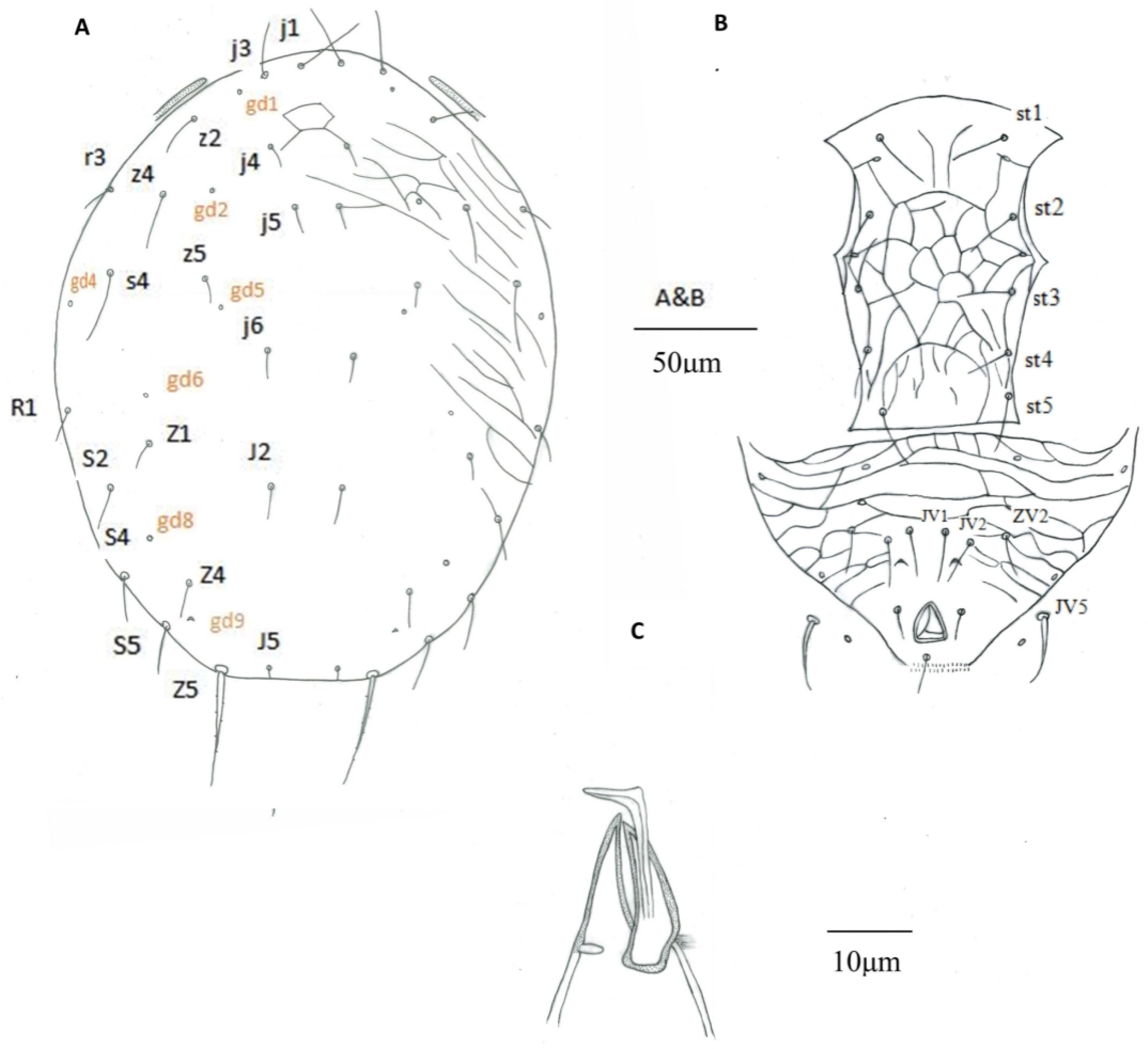
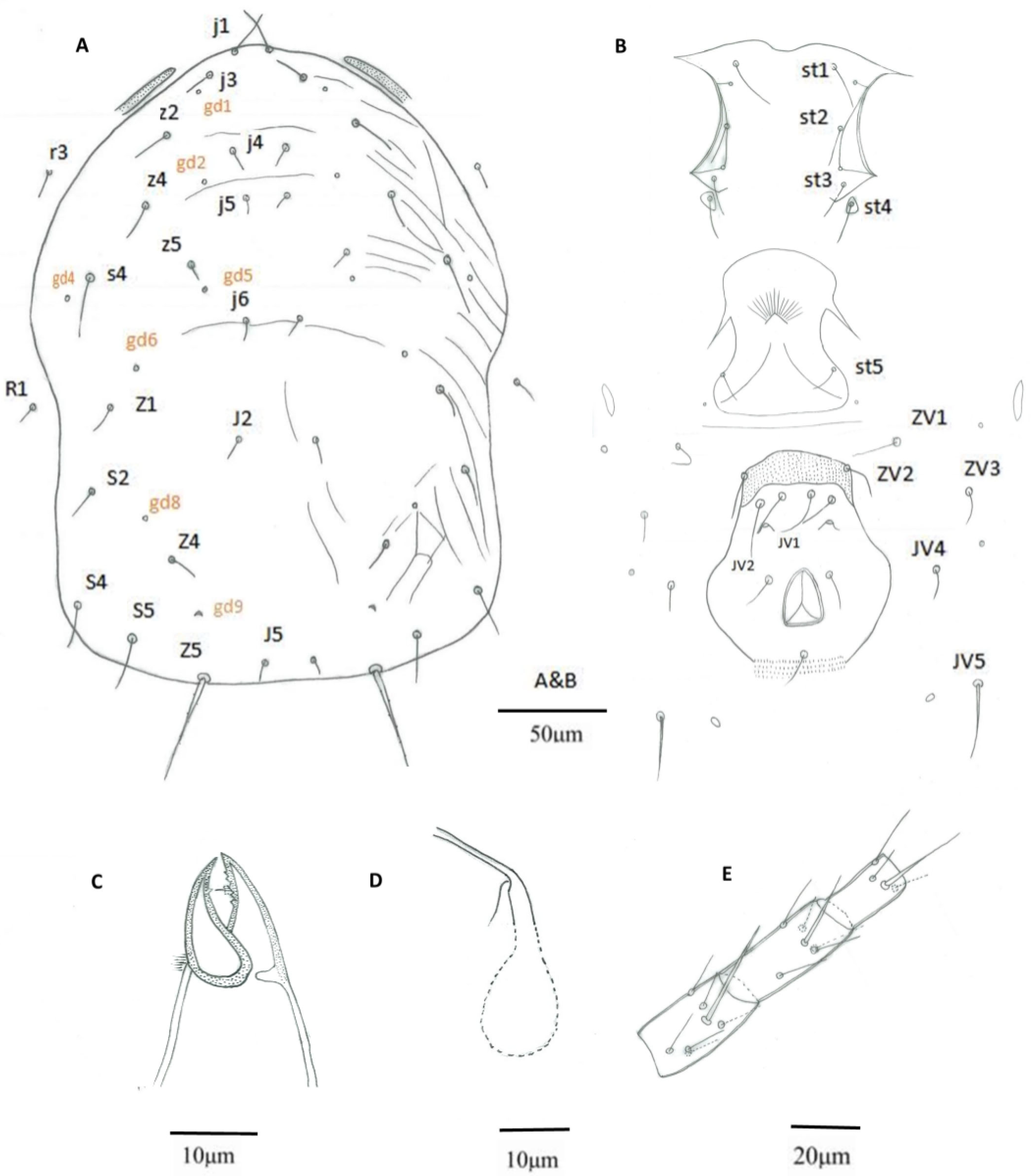
Based on the specimens from Bajiaozhai National Forest Park, morphological characters of this putative Euseius sp. female are described as follows (Figure 1A–E and Figure 2A–C). While some distinguished morphological characteristics of putative Euseius sp., E. nicholsi, and E. oolong are listed in Table 1.
Euseius sp.
Females (n = 12) Dorsum (Figure 1A). Dorsal setal pattern 10A: 9B. Dorsal shield smooth, with anterior reticulation and lateral striation, 316 (299–335) long and 239 (223–254) wide, distances between setae j1–J5 307 (290–327) and s4–s4 183 (170–198), shield nearly oval, constricted at level of R1; r3 and R1 out of the dorsal shield, r3 at level of z4, R1 at level of Z1. All dorsal shield setae smooth and setiform, except Z5 slightly serrated. Seven pairs of solenostomes on dorsal shield (gd1, gd2, gd4, gd5, gd6, gd8, and gd9). Length of setae: j1 29 (25–33), j3 25 (22–31), j4 10 (8–11), j5 10 (8–12), j6 11 (9–14), J2 13 (10–15), J5 5 (4–6), z2 17 (15–22), z4 18 (16–24), z5 10 (7–12), Z1 12 (10–14), Z4 14 (12–17), Z5 59 (45–63), s4 28 (25–34), S2 18 (15–21), S4 24 (19–27), S5 26 (21–30), r3 14 (10–16), and R1 13 (10–15).
Venter (Figure 1B). Ventral setal pattern JV-3: ZV. All ventral setae smooth. Sternal shield lightly reticulated, posterior margin not obvious, 79 (75–84) wide with three pairs of setae st1 29 (24–32), st2 28 (23–32), and st3 27 (24–30), and two pairs of poroids (pst1–pst2), distance between st1–st3 54 (50–59), st2–st2 62 (58–65) and st3–st3 73 (68–77). Metasternal platelets drop-shaped, each with one metasternal seta, st4 25 (22–28) and a poroid (pst3). Genital shield smooth, 80 (76–85) wide, with one pair of thin genital setae st5 29 (27–32), distance between st5–st5 76 (70–86); one pair of associated poroids on soft cuticle near to the posterior corners of the shield. Ventrianal shield smooth, vase-shaped, anterior part strongly sclerotized with a hat-shaped structure, 93 (83–102) long, 47 (42–56) wide at level of ZV2, 75 (70–81) wide at level of anus, with three pairs of thin pre-anal setae JV1 29 (26–33), JV2 27 (25–32), and ZV2 21 (18–24); Pa 14 (12–17) and Pst 15 (13–17) long. Solenostomes gv3 crescentic, posteromesad JV2, distance between them 26 (24–29). Opisthogastric soft cuticle with four pairs of setae, ZV1 23 (20–29), ZV3 12 (9–14), JV4 12 (9–15), and JV5 30 (23–34) long. All ventral setae thin, except JV5, thick. Two pairs of metapodal platelets, primary ones 21 (16–24) long, 4 (3–5) wide, and secondary ones 9 (8–12) long, 1 (1–2) wide.
Peritreme. Peritreme extending to the level between setae j3 and z2.
Chelicera (Figure 1C). Fixed digit 23 (20–26) long, with four teeth and pilus dentilis; movable digit 22 (20–24) long, with one tooth.
Spermatheca (Figure 1D). Calyx tubular, with distal half lightly sclerotized, 11 (10–12) long, distal flaring, 3 (3–4) wide at the opening, atrium C-shaped, 2 (1–2) in depth, incorporated within the calyx; major duct without neck, narrow, and minor duct visible.
Legs. Genua formula for leg I 2-2/2, 1/1-2, leg II 2-2/2, 0/0-1, leg III 1-2/2, 1/0-1, and leg IV 1-2/2, 1/0-1. Tibia III with one macroseta, Sti III 24 (19–27). Genu, tibia, and basitarsus IV each with one macroseta (Figure 1E), Sge IV 41 (33–48), Sti IV 32 (27–37), and St IV 51 (43–62).
Males (n = 3) Dorsum (Figure 2A). Dorsal shield with anterior reticulation and lateral striation, 243 (242–243) long and 190 (167–205) wide at level of s6, shield nearly oval, 19 pairs of dorsal setae, all smooth, except Z5 slightly serrated. Seven pairs of solenostomes on dorsal shield (gd1, gd2, gd4, gd5, gd6, gd8, and gd9). Length of setae: j1 27 (26–28), j3 19 (18–22), j4 10 (9–11), j5 10 (9–13), j6 12 (11–13), J2 13 (12–13), J5 5 (4–5), z2 18 (16–22), z4 22 (18–25), z5 11 (10–14), Z1 13 (11–14), Z4 14 (12–16), Z5 48 (46–51), s4 30 (27–34), S2 19 (18–20), S4 22 (21–24), S5 22 (20–24), r3 14 (12–16), and R1 13 (12–14).
Venter (Figure 2B). Sternogenital shield 119 (114–126) long, reticulated. Wider 81 (78–84) between coxae II–III than at posterior corners 59 (58–59). Five pairs of sternogenital setae (st1–st5), st1 26 (24–30), st2 23 (22–24), st3 21 (19–23), st4 21 (20–22), and st5 22 (21–22), and three pairs of poroids (pst1–pst3). Ventrianal shield subtriangular, 92 (90–97) long, 154 (140–167) wide at level of anterior corners; transversally reticulated; reticulation on anterior part of shield more accentuated; with three pairs of thin pre-anal setae JV1 24 (22–26), JV2 21 (20–22), and ZV2 19 (17–21); Pa 13 (12–13) and Pst 12 (10–14) long. Solenostome gv3 crescentic, posteromedian to JV2, distance between solenostomes 23 (21–25). Opisthogastric soft cuticle with one pair of setae, JV5 25 (20–28) long.
Peritreme. Peritreme extending at level between setae j3 and z2. Peritrematal shield fused with dorsal shield.
Chelicera and spermatodactyl (Figure 2C). Cheliceral dentition not discernible in the examined specimens. Fixed digit 20 (19–20) long, movable digit 20 (18–22) long. Spermatodactyl L-shaped; shaft 25 (24–27), foot 7 (5–9) long.
Legs. Chaetotaxy of genua similar to female. Macroseta on genu I not differentiated. Macrosetae on tibia III 21 (17–24), genu IV 39 (38–40), tibia IV 30 (25–33), and tarsus IV 48 (42–53).
Morphological characteristics of E. oolong based on paratypes were re-drawn for comparison (Figure 3) and are summarized in Table 1.

3.2. Material Measured

Seven ♀♀, Bajiaozhai National Forest Park, Ziyuan County, Guilin City, Guangxi Province (slides no. GL-0591, GL-0592, GL-0841, GL-0561, GL-0562, GL-0951, and GL-0952); two ♀♀ (slides no. GL-0953 and GL-0601) on Cyclobalanopsis glauca (Fagaceae), 30 October 2020, Fang X.D. coll.; two ♀♀ (slides no. GL-0611 and GL-0612) on Sabina squamata (Cupressaceae), same locality, date, and collector; one ♀ (slide no. GL-0681) on Smilax glaucochina (Liliaceae), same locality, date, and collector; two ♂♂ (slides no. GL-0641 and GL-0671) and one ♂ (slide no. GL-0672 ) on Bambusa sp. (Poaceae), same locality, date, and collector.

3.3. Molecular Analysis

Genetic distances between the putative Euseius sp. and two morphologically similar species (E. nicholsi and E. oolong) ranged between 0 and 4% for COI mtDNA, between 0 and 2% for 12S rRNA, and were 0 for ITS. These values were markedly lower than those observed between Euseius sp. specimens and E. finlandicus (23–24% for COI mtDNA, 21–22% for 12S rRNA, and 2% for ITS) or A. swirskii (30–33% for COI mtDNA, 34–39% for 12S rRNA, and 18% for ITS) (Table 3, Table 4, and Table 5).
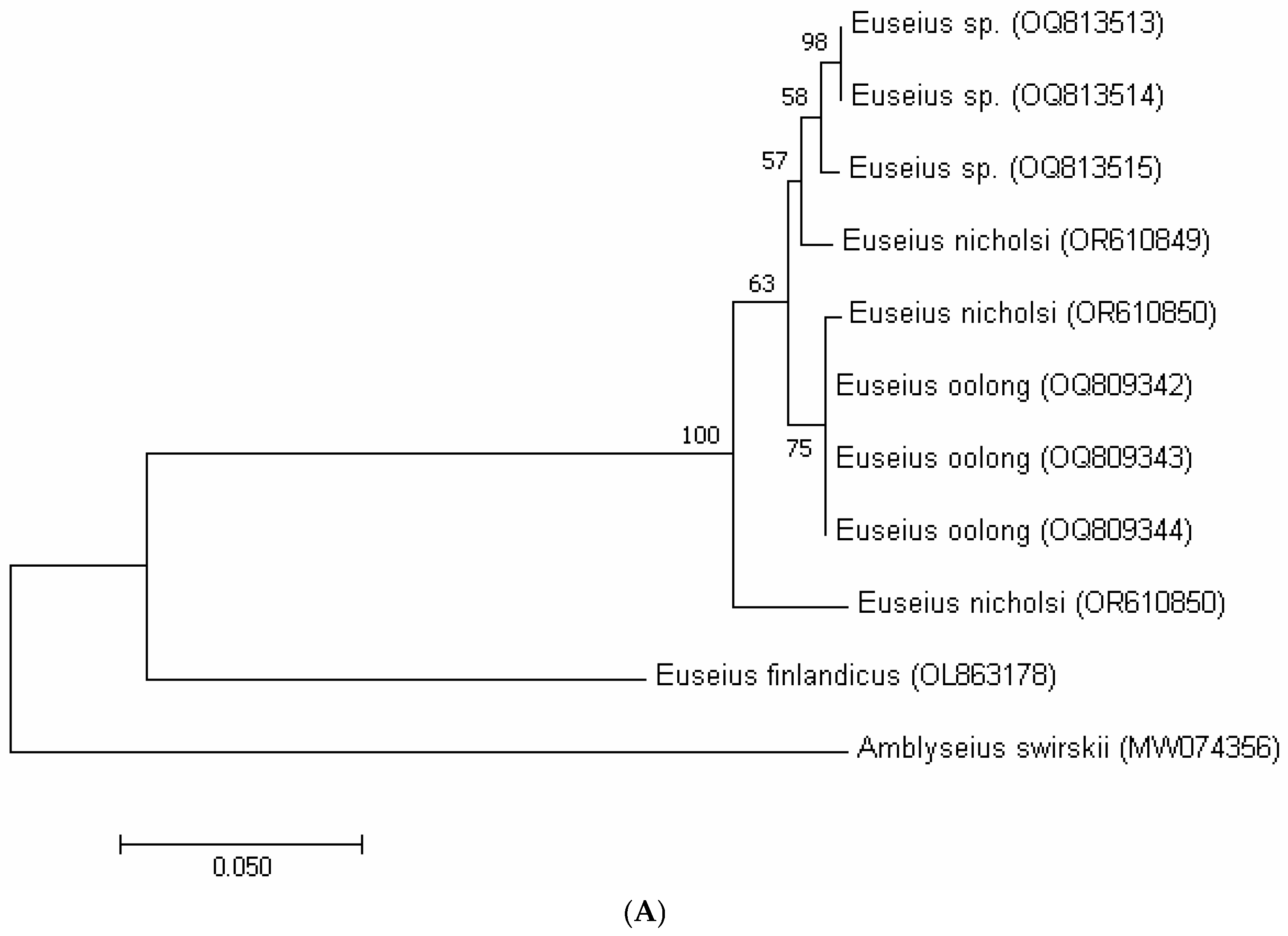
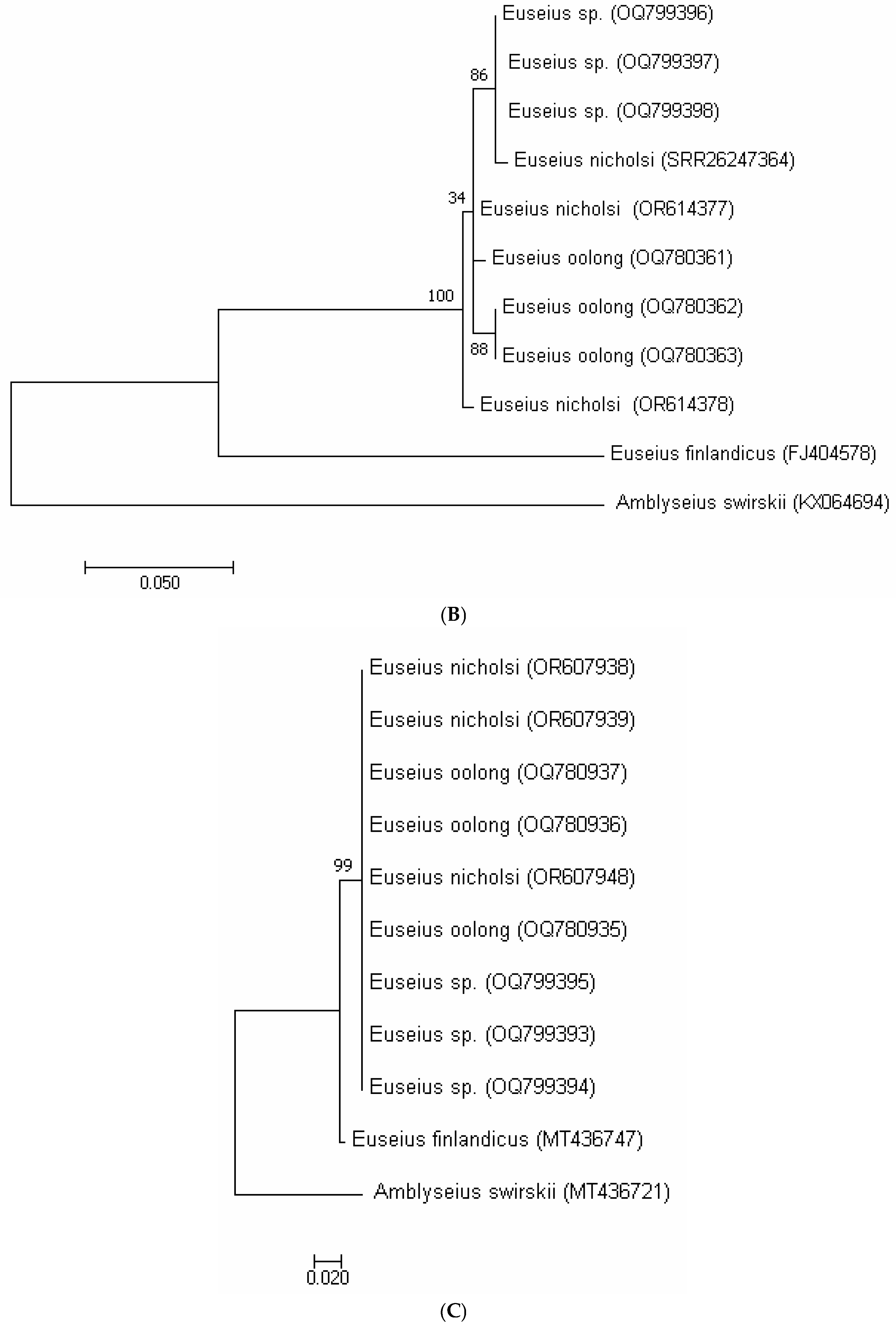
In the phylogenetic tree based on COI sequences (Figure 1A), the putative Euseius sp., E. nicholsi, and E. oolong clustered together within a single clade, supported by bootstrap values of 92–98%, and were clearly separated from the outgroup A. swirskii. A similar pattern was recovered with the 12S rRNA marker (Figure 1B), where all specimens of E. nicholsi, E. oolong, and the putative Euseius sp. formed a single well-supported lineage (bootstrap > 90%). The ITS tree (Figure 1C) showed identical clustering, with no divergence among the three taxa. These consistent topologies indicated that the three groups represent a single evolutionary lineage and provided further support for conspecificity.

3.4. Comparative Synthesis of Morphological and Molecular Results

When the three taxa were compared directly, the specimens from Bajiaozhai (putative Euseius sp.) were generally smaller in body size than E. nicholsi but overlapped partly with E. oolong. Variation was also observed in the number and shape of metapodal platelets: the putative Euseius sp. showed one or two pairs, either hooked or unhooked, whereas E. nicholsi typically has a single hooked pair and E. oolong usually two unhooked pairs. The spermathecal calyx of the Bajiaozhai specimens was tubular and only lightly sclerotized distally, in contrast to the funnel-shaped calyx of E. nicholsi and the cup-shaped calyx of E. oolong. In addition, the dorsal shield bore seven solenostomes, the same as in E. nicholsi but more than the five solenostomes generally described for E. oolong. Measurements of setae (e.g., j3, Sge IV, Sti IV, and St IV) were intermediate between the two named species (Table 6).
The molecular markers confirmed these observations. Genetic distances among the three taxa were minimal (COI: 0–4%; 12S rRNA: 0–2%; and ITS: 0%), far below interspecific levels reported for other phytoseiids. In all phylogenetic reconstructions (Figure 4A–C), the Bajiaozhai specimens, E. nicholsi, and E. oolong formed a single clade, clearly distinct from the reference species E. finlandicus and the outgroup A. swirskii. These results showed that the morphological differences were intraspecific rather than interspecific, and that E. oolong was best treated as a junior synonym of E. nicholsi.
Table 3. Genetic distances between cytochrome oxidase subunit 1 (COI) sequences of putative Euseius sp., E. nicholsi, and E. oolong using the Kimura 2-parameter model.
Putative Euseius sp. (OQ813513)Putative Euseius sp.
(OQ813514)
Putative Euseius sp.
(OQ813515)
E. oolong
(OQ809342)
E. oolong
(OQ809343)
E. oolong
(OQ809344)
E. nicholsi
(OR610851)
E. nicholsi
(OR610849)
E. nicholsi
(OR610850)
E. finlandicus
(OL863178)
Amblyseius
swirskii
(MW074356)
Putative Euseius sp.
(OQ813513)
Putative Euseius sp.
(OQ813514)
0.00
Putative Euseius sp.
(OQ813515)
0.010.01
E. oolong
(OQ809342)
0.020.020.02
E. oolong
(OQ809343)
0.020.020.020.00
E. oolong
(OQ809344)
0.020.020.020.000.00
E. nicholsi
(OR610851)
0.020.020.020.000.000.00
E. nicholsi
(OR610849)
0.010.010.010.020.020.020.02
E. nicholsi
(OR610850)
0.040.040.040.040.040.040.040.04
E. finlandicus
(OL863178)
0.230.230.230.230.230.230.230.240.24
Amblyseius swirskii (MW074356)0.310.310.310.300.300.300.300.310.330.30
Table 4. Genetic distances between 12S rRNA sequences of putative Euseius sp., E. nicholsi, and E. oolong using the Kimura 2-parameter model.
Putative Euseius sp.
(OQ799396)
Putative Euseius sp.
(OQ799397)
Putative Euseius sp.
(OQ799398)
E. oolong
(OQ780361)
E. oolong
(OQ780362)
E. oolong
(OQ780363)
E. nicholsi
(PRJNA1022847)
E. nicholsi
(OR614377)
E. nicholsi
(OR614378)
E. finlandicus
(FJ404578)
Amblyseius
swirskii
(KX064694)
Putative Euseius sp.
(OQ799396)
Putative Euseius sp.
(OQ799397)
0.00
Putative Euseius sp.
(OQ799398)
0.010.01
E. oolong
(OQ780361)
0.020.020.01
E. oolong
(OQ780362)
0.020.020.010.02
E. oolong
(OQ780363)
0.020.020.010.020.00
E. nicholsi
(PRJNA1022847)
0.020.020.020.020.000.00
E. nicholsi
(OR614377)
0.010.010.000.010.010.010.01
E. nicholsi
(OR614378)
0.020.020.010.020.000.000.000.01
E. finlandicus (FJ404578)0.210.210.210.210.220.220.220.210.22
Amblyseius swirskii (KX064694)0.340.340.340.340.350.350.360.340.350.39
Table 5. Genetic distances between ITS sequences of putative Euseius sp., E. nicholsi, and E. oolong using the Kimura 2-parameter model.
Putative Euseius sp.
(OQ799393)
Putative Euseius sp.
(OQ799394)
Putative Euseius sp. (OQ799395)E. oolong
(OQ780935)
E. oolong
(OQ780936)
E. oolong (OQ780937)E. nicholsi
(OR607938)
E. nicholsi
(OR607939)
E. nicholsi
(OR607948)
E. finlandicus
(MT436747)
Amblyseius
swirskii
(MT436721)
Putative Euseius sp.
(OQ799393)
Putative Euseius sp.
(OQ799394)
0.00
Putative Euseius sp. (OQ799395)0.000.00
E. oolong
(OQ780935)
0.000.000.00
E. oolong
(OQ780936)
0.000.000.000.00
E. oolong
(OQ780937)
0.000.000.000.000.00
E. nicholsi
(OR607938)
0.000.000.000.000.000.00
E. nicholsi
(OR607939)
0.000.000.000.000.000.000.00
E. nicholsi
(OR607948)
0.000.000.000.000.000.000.000.00
E. finlandicus
(MT436747)
0.020.020.020.020.020.020.020.020.02
Amblyseius swirskii
( MT436721)
0.180.180.180.180.180.180.180.180.180.18
Table 6. Key diagnostic traits comparing putative Euseius sp., E. nicholsi, and E. oolong.
CharacterPutative Euseius sp.E. nicholsiE. oolong
Female dorsal shield length (µm)299–335352–381323–361
Dorsal solenostomes775 (sometimes 7, weak gd1)
Spermathecal calyxTubular, weakly sclerotizedFunnel-shaped, flaringCup-shaped
Metapodal platelets1–2 pairs, hooked/unhooked1 pair, mostly hooked2 pairs, unhooked
Fixed digit teeth43–45
j3 seta (µm)22–3127–3320–25
St IV (µm)43–6256–7347–61
Molecular divergenceCOI 0–4%; 12S 0–2%; ITS 0%SameSame
Phylogenetic placementSame cladeSame cladeSame clade

4. Discussion

The comparative analysis presented in the Results section showed that morphological variation among the Bajiaozhai specimens, E. nicholsi, and E. oolong was minor and largely overlapped. Features such as differences in spermathecal calyx shape, dorsal solenostomes, metapodal platelets, and setal lengths fell within the range of variation observed for other phytoseiid mites. To determine whether these differences were taxonomically significant, molecular evidence was compared with previous studies of genetic divergence in Phytoseiidae.
For the COI marker, interspecific divergence within phytoseiid genera such as Kampimodromus and Typhlodromus can reach 17–18%, whereas intraspecific variation is typically very low, for example, 0–0.4% in Neoseiulus californicus, 4–6% in K. aberrans, and 6% in K. hmiminai [16,18]. By contrast, the specimens examined here showed only 0–4% divergence, indicating they belonged to the same species. For the 12S rRNA fragment, the maximum divergence between closely related species has been reported at 10% [18], while intraspecific variation generally ranges from 0 to 3% [19,24]. The 0–2% divergence recorded in our study fell within this intraspecific range. Similarly, ITS divergence among congeneric phytoseiids often ranges from 3 to 7% [18], but in our data, the divergence was 0%. These values demonstrated that the putative Euseius sp., E. nicholsi, and E. oolong were conspecific. The congruent clustering of all three taxa in COI, 12S rRNA, and ITS phylogenies (Figure 4A–C), each with high bootstrap support, provided strong molecular evidence that E. nicholsi and E. oolong cannot be separated as distinct species.
Both mitochondrial (COI, 12S) and nuclear (ITS) markers therefore supported synonymy. The phylogenetic trees (Figure 4A–C) clustered all three taxa into a single clade with low genetic distances, clearly distinct from E. finlandicus and A. swirskii. This pattern was consistent with earlier studies, where low bootstrap values and minor genetic variation reflected population-level differentiation rather than species boundaries [16,17,35].
Taken together, the molecular results confirmed that the morphological differences observed among the three taxa represented intraspecific variation. Consequently, E. oolong should be regarded as a junior synonym of E. nicholsi, and the Bajiaozhai specimens can be confidently identified as E. nicholsi. This conclusion contributes to a clearer understanding of phytoseiid systematics and highlights the importance of integrative taxonomy for resolving synonymy issues that complicate biodiversity assessment and biological control programs.

5. Conclusions

According to Tixier et al. [6], the minimal difference of setae length between the means of two specimen lots belonging to two species should be 10.58 μm (for setae < 65 μm) and 33.99 μm (for setae > 65 μm).
Therefore, the two following points can be concluded:
  • The specimens of Euseius sp. from Bajiaozhai National Forest Park were morphologically characterized as E. nicholsi, due to their high similarity. Based on molecular analyses, the specimens identified as E. nicholsi exhibited minimal genetic divergence from Euseius sp., indicating they likely represented the same species. Therefore, the morphological differences between Euseius sp. and E. nicholsi were best interpreted as intraspecific variation.
  • Molecular evidence indicated that E. nicholsi and E. oolong are conspecific, with E. oolong treated as a synonym of E. nicholsi. Variations in body size, metapodal platelets, digit dentition, and setal lengths reflect intraspecific diversity.
According to Ehara and Lee [9], E. nicholsi is characterized by a pair of hook-shaped metapodal platelets, which appear to originate from the joining of two metapodal platelets. However, our molecular analysis revealed highly similar sequences among specimens with both hooked and non-hooked metapodal platelets, as well as among those bearing one or two pairs of platelets (Table 1). Additionally, no molecular divergence was detected among specimens exhibiting slight differences in the number of teeth on the fixed digit. These findings suggest that intraspecific variation is more extensive than previously anticipated.

Author Contributions

Conceptualization, X.F. and J.L.; methodology, X.F., J.L., S.U.M., N.C.F., and J.M.; software, X.F. and S.U.M.; validation, X.F. and J.L.; formal analysis, X.F. and S.U.M.; investigation, X.F., J.L., S.U.M., N.C.F., and J.M.; resources, X.F. and J.L.; data curation, X.F., S.U.M., N.C.F., and J.M.; writing—original draft preparation, X.F., J.L., S.U.M., N.C.F., and J.M.; writing—review and editing, X.F., J.L., S.U.M., N.C.F., and J.M.; visualization, X.F. and J.L.; supervision, X.F. and J.L.; project administration, X.F. and J.L.; funding acquisition, X.F. and J.L. All authors have read and agreed to the published version of the manuscript.

Funding

This study was funded by the National Natural Science Foundation of China (31900352), the GDAS Special Project of Science and Technology Development (2020GDASYL-20200301003), the GDAS Special Project of Science and Technology Development (2022GDASZH-2022010106), and the National Foreign Expert Individual Human Project (Y20240119).

Data Availability Statement

The data sheets are available and can be requested from the corresponding author upon reasonable request.

Acknowledgments

We thank our dear friends Xiaodan Pan and Jianhua Huang for reviewing an earlier draft of this manuscript.

Conflicts of Interest

The authors declare no conflicts of interest. The funders had no role in the design of this study; in the collection, analyses, or interpretation of data; in the writing of the manuscript; or in the decision to publish the results.

References

  1. Wu, W.N.; Ou, J.F.; Huang, J.L. Fauna Sinica, Invertebrata Vol. 47, Arachnida, Acari, Phytoseiidae; Science Press: Beijing, China, 2009; 511p. (In Chinese) [Google Scholar]
  2. Wu, W.N.; Fang, X.D. Phytoseiidae Systematics and Management of Pests; Guangdong Science and Technology Press: Guangzhou, China, 2021; 428p. (In Chinese) [Google Scholar]
  3. McMurtry, J.A.; de Moraes, G.J.; Sourassou, N.F. Revision of the lifestyles of phytoseiid mites (Acari: Phytoseiidae) and implications for biological control strategies. Syst. Appl. Acarol. 2013, 18, 297–320. [Google Scholar] [CrossRef]
  4. Demite, P.R.; De Moraes, G.J.; McMurtry, J.A.; Denmark, H.A.; Castilho, R.C. Phytoseiidae Database. Zootaxa 2014, 3795, 571–577. Available online: www.lea.esalq.usp.br/phytoseiidae/ (accessed on 1 December 2024). [CrossRef] [PubMed]
  5. Tixier, M.S.; Okassa, M.; Kreiter, S. An integrative morphological and molecular diagnostics for Typhlodromus pyri (Acari: Phytoseiidae). Norw. Acad. Sci. Lett. 2012, 41, 68–78. [Google Scholar]
  6. Tixier, M.S. Statistical approaches for morphological continuous characters: A conceptual model applied to Phytoseiidae (Acari: Mesostigmata). Zool. Scr. 2013, 42, 327–334. [Google Scholar]
  7. Dos Santos, V.V.; Tixier, M.S. Molecular markers for analysing phylogenetic relationships within the mite family Phytoseiidae (Acari: Mesostigmata). Cladistics 2017, 28, 1–16. [Google Scholar] [CrossRef]
  8. Döker, I.; Karut, K.; Karaca, M.M.; Cargnus, E.; Kazak, C. Internal Transcribed Spacer (ITS) sequences of some Kampimodromus (Acari: Phytoseiidae) species: Is Kampimodromus ragusai a valid species or a synonym of Kampimodromus aberrans? Syst. Appl. Acarol. 2018, 23, 2237–2243. [Google Scholar] [CrossRef]
  9. Ehara, S.; Lee, L.H.Y. Mites associated with plants in Hong Kong. J. Fac. Educ. Tottori Univ. Nat. Sci. 1971, 22, 61–78. [Google Scholar]
  10. Liao, J.H.; Ho, C.C.; Fang, X.D.; Ko, C.C. Contribution to the knowledge of the genera Euseius wainstein and Gynaseius wainstein (Acari: Mesostigmata: Amblyseiinae) from Taiwan. Exp. Appl. Acarol. 2018, 23, 2192–2213. [Google Scholar] [CrossRef]
  11. Liao, J.R.; Ho, C.C.; Lee, H.C.; Ko, C.C. Phytoseiidae of Taiwan (Acari: Mesostigmata). National Taiwan University Press: Taipei, Taiwan, 2020; 538p. [Google Scholar]
  12. Dos Santos, V.V.; Tixier, M.S. Integrative taxonomy approach for analysing evolutionary history of the tribe Euseiini Chant & McMurtry (Acari: Phytoseiidae). Syst. Biodivers. 2018, 16, 302–319. [Google Scholar] [CrossRef]
  13. Döker, I.; Çelik, S.O.; Karaca, M.M. Review of Euseius wainstein (Parasitiformes: Phytoseiidae) in Türkiye: An integrative taxonomic approach using morphological and molecular data with a new combination Euseius degenerans (Berlese). Exp. Appl. Acarol. 2025, 30, 560–591. [Google Scholar] [CrossRef]
  14. Döker, I.; Khaustov, V.A.; Ben-ami, R.; Tabic, A.; Joharchi, O. Integrative taxonomy reveals a new species of predatory mite in the subgenus Anthoseius De Leon (Acari: Phytoseiidae). Syst. Appl. Acarol. 2025, 30, 286–301. [Google Scholar] [CrossRef]
  15. Kanouh, M.; Tixier, M.S.; Guichou, S.; Cheval, B.; Kreiter, S. Two synonymy cases within the genus Neoseiulella (Acari: Phytoseiidae): Is the molecular evidence so evident? Biol. J. Linn. Soc. 2010, 101, 323–344. [Google Scholar] [CrossRef]
  16. Tixier, M.S.; Kreiter, S.; Ferragut, F.; Cheval, B. The suspected synonymy of Kampimodromus hmiminal and Kampimodromus adrianae (Acari: Phytoseiidae): Morphological and molecular investigations. Can. J. Zool. 2006, 84, 1216–1222. [Google Scholar] [CrossRef]
  17. Tixier, M.S.; Kreiter, S.; Barbar, Z.; Ragusa, S.; Cheval, B. Status of two cryptic species, Typhlodromus exhilaratus Ragusa and Typhlodromus phialatus Athias-Henriot (Acari: Phytoseiidae): Consequences for taxonomy. Zool. Scr. 2006, 35, 115–122. [Google Scholar] [CrossRef]
  18. Tixier, M.S.; Guichou, S.; Kreiter, S. Assessment of the usefulness of eight DNA fragments for phylogenetic studies within the family Phytoseiidae. In Trends in Acarology; Sabelis, M.W., Bruin, J., Eds.; Springer: Dordrecht, The Netherlands, 2010; pp. 41–47. [Google Scholar] [CrossRef]
  19. Tixier, M.S.; Ferrero, M.; Okassa, M.; Guichou, S.; Kreiter, S. On the specific identity of specimens of Phytoseiulus longipes Evans (Mesostigmata: Phytoseiidae) showing different feeding behaviours: Morphological and molecular analysis. Bull. Entomol. Res. 2010, 100, 569–579. [Google Scholar] [CrossRef] [PubMed]
  20. Tixier, M.S.; Otto, J.; Kreiter, S.; dos Santos, V.; Beard, J. Is Neoseiulus wearnei the Neoseiulus californicus of Australia? Exp. Appl. Acarol. 2014, 62, 267–277. [Google Scholar] [CrossRef] [PubMed]
  21. Tixier, M.S.; dos Santos, V.; Douin, M.; Duso, C.; Kreiter, S. Great molecular variation within the species Phytoseius finitimus (Acari: Phytoseiidae): Implications for diagnosis decision within the mite family Phytoseiidae. Acarologia 2017, 57, 493–515. [Google Scholar] [CrossRef]
  22. Tixier, M.S.; Dennj, P.; Douin, M.; Kreiter, S.; Haralabos, T. Mites of the genus Typhlodromus (Acari: Phytoseiidae) from Southern France: Combined morphological and molecular approaches for species identification. Zootaxa 2019, 4604, 242–280. [Google Scholar] [CrossRef]
  23. Tixier M., S.; Auger, P.; Migeon, A.; Douina, M.; Fossoudc, A.; Navajas, M.; Arabuli, T. Integrated taxonomy supports the identification of some species of Phytoseiidae (Acari: Mesostigmata) from Georgia. Acarologia 2021, 61, 824–844. [Google Scholar] [CrossRef]
  24. Okassa, M.; Tixier, M.S.; Cheval, B.; Kreiter, S. Molecular and morphological evidence for a new species status within the genus Euseius (Acari: Phytoseiidae). Can. J. Zool. 2009, 87, 689–698. [Google Scholar] [CrossRef]
  25. Okassa, M.; Tixier, M.S.; Kreiter, S. Morphological and molecular diagnostics of Phytoseiulus persimilis and Phytoseiulus macropilis (Acari: Phytoseiidae). Exp. Appl. Acarol. 2010, 52, 291–303. [Google Scholar] [CrossRef]
  26. Okassa, M.; Kreiter, S.; Tixier, M.S. Obtaining molecular data for all life stages of Typhlodromus (Typhlodromus) Exhilaratus (Mesostigmata: Phytoseiidae): Consequences for species identification. Exp. Appl. Acarol. 2012, 57, 105–116. [Google Scholar] [CrossRef]
  27. Lindquist, E.E.; Evans, G.O. Taxonomic concepts in the Ascidae with a modified setal nomenclature for the idiosoma of the Gamasina (Acarina: Mesostigmata). Mem. Entomol. Soc. Can. 1965, 47, 5–66. [Google Scholar] [CrossRef]
  28. Rowell, H.J.; Chant, D.A.; Hansell, R.I.C. The determination of setal homologies and setal patterns on the dorsal shield in the family Phytoseiidae (Acarina: Mesostigmata). Can. Entomol. 1978, 110, 859–876. [Google Scholar] [CrossRef]
  29. Chant, D.A.; Yoshida-Shaul, E. Adult idiosomal setal patterns in the family Phytoseiidae (Acari: Gamasina). Intern. J. Acarol. 1992, 18, 177–193. [Google Scholar] [CrossRef]
  30. Athias-Henriot, C. Nouvelles notes sur les Amblyseiini. II. Le relevé organotaxique de la face dorsale adulte (Gamasides protoadéniques, Phytoseiidae). Acarologia 1975, 17, 20–29. [Google Scholar]
  31. Folmer, O.; Black, M.; Hoeh, W.; Lutz, R.; Vrijenhoek, R. DNA primers for amplification of mitochondrial cytochrome oxidase subunit I from diverse metazoan invertebrates. Mol. Mar. Biol. Biotechnol. 1994, 3, 294–299. [Google Scholar]
  32. Li, J.B.; Li, Y.X.; Sun, J.T.; Xue, X.F.; Xu, X.N.; Hong, X.Y. COI barcoding as a molecular assay for the identification of phytoseiid mites. Syst. Appl. Acarol. 2012, 17, 397–406. [Google Scholar] [CrossRef]
  33. Jeyaprakash, A.; Hoy, M. Mitochondrial 12S rRNA sequences used to design a molecular ladder assay to identify six commercially available phytoseiids (Acari: Phytoseiidae). Biol. Control 2002, 25, 136–142. [Google Scholar] [CrossRef]
  34. Navajas, M.; Lagnel, J.; Fauvel, G.; de Moraes, G. Sequence variation of ribosomal Internal Transcribed Spacers (ITS) in commercially important Phytoseiidae mites. Exp. Appl. Acarol. 1999, 23, 851–859. [Google Scholar] [CrossRef]
  35. Hosseininia, A.; Khanjani1, M.; Asadi, M.; Soltani1, J. Genetic diversity of predatory mite Typhlodromus bagdasarjani (Acari: Phytoseiidae) populations based on molecular markers. Syst. Appl. Acarol. 2023, 28, 2164–2170. [Google Scholar] [CrossRef]
Figure 1. Euseius sp., female. (A) Dorsal shield; (B) Ventral idiosoma; (C) Chelicera; (D) Spermatheca; (E) Leg IV, genu-basitarsus.
Insects 16 00950 g001
Figure 2. Euseius sp., male. (A) Dorsal idiosoma; (B) Ventral idiosoma; (C) Spermatodactyl.
Insects 16 00950 g002
Figure 3. (From paratypes). E. oolong Female, (A) Dorsal shield; (B) Ventral idiosoma; (C) Chelicera; (D) Spermatheca; (E) Leg IV, genu-basitarsus.
Insects 16 00950 g003
Figure 4. Maximum likelihood phylogenetic trees including putative Euseius sp. from Bajiaozhai National Forest Park, E. oolong from ZIGAS, E. nicholsi from ZIGAS, and also Shaoguan newly collected specimens (Amblyseius swirskii as the outgroup, E. finlandicus as the reference) obtained with (A)—COI mtDNA, (B)—12S rRNA and (C)—ITS markers.
Insects 16 00950 g004aInsects 16 00950 g004b
Table 1. Morphological characteristics and measurements differentiating putative Euseius sp., E. nicholsi, and E. oolong.
Putative Euseius sp. aE. nicholsi bE. nicholsi cE. nicholsi dE. oolong eE. oolong f
Dorsal shield length (µm)316 (299–335)360365–381352 (348–357)341 (324–361)323 (319–328)
Dorsal shield width (µm) (s4 level)239 (223–254)260265–282251 (246–257)246 (236–266)238 (233–243)
Dorsal shieldanterior reticulation and lateral striationmost surface reticulatedmost surface reticulatedmost surface reticulatedlaterally reticulatedlaterally reticulated
Solenostome on dorsal shieldgd1, gd2, gd4, gd5, gd6, gd8, gd9gd1, gd2, gd4, gd5, gd6, gd8, gd9 gd1, gd2, gd4, gd5, gd6, gd8, gd9gd2, gd5, gd6, gd8, gd9gd1, gd2, gd4, gd5, gd6, gd8, gd9
Calyx of spermathecatubular, with distal half only lightly sclerotized; atrium c-shaped incorporated within the calyxtubular, with basal half swollenfunnel-shaped, distal flaring; atrium knobbed, connected directly with calyxfunnel-shaped, distal flaring; atrium knobbed, connected directly with calyxcup-shaped with distal half only lightly sclerotized; atrium c-shaped incorporated within the calyxcup-shaped with distal half only lightly sclerotized; atrium c-shaped incorporated within the calyx
Metapodal plateletone or two pairs, hooked or notone pair, hookedone pair, hooked or notone pair, hooked or nottwo pairs, un-hookedone pair, hooked
Number of teeth on the fixed digit of the chelicerae4 3–4455
Seate j325 (22–31)27 (26–28)3031 (30–33)22 (20–25)22 (20–25)
Macroseta Sge IV41 (33–48)50 (48–53)5248 (45–51)44 (37–50)50 (47–54)
Macroseta Sti IV32 (27–37)38 (38–39)4134 (33–35)34 (27–37)33 (32–34)
Macroseta St IV51 (43–62)68 (60–73)6656 (55–57)58 (50–61)50 (47–51)
a From 12 female specimens in Bajiaozhai National Forest Park; b From Holotype and reference [10]; c From reference [1]; d From newly collected specimens; e From reference [10]; f From two paratypes deposited at IZGAS and also newly collected specimens.
Table 2. Characteristics of E. nicholsi, E. finlandicus, and outgroup species Amblyseius swirskii, and GenBank accession numbers of Cytochrome c Oxidase subunit I (COI), 12S rRNA, and internal transcribed spacer (ITS) markers.
SpeciesCountryLocalityPlant SupportsGenBank Accession Number of COI SequencesGenBank Accession Number of 12S rRNA SequencesGenBank Accession Number of ITS Sequences
Putative Euseius sp.ChinaGuilinCyclobalanopsis glaucaOQ813513
OQ813514
OQ813515
OQ799396
OQ799397
OQ799398
OQ799393
OQ799394
OQ799395
E. oolongChinaGuangzhouBauhinia purpureaOQ809342
OQ809343
OQ809344
OQ780361
OQ780362
OQ780363
OQ780935
OQ780936
OQ780937
E. nicholsiChinaGuangzhou
Shaoguan
Shaoguan
Bauhinia purpurea
Eurya macartneyi
Ficus hispida
OR610851
OR610849
OR610850
SRR26247364
OR614377
OR614378
OR607938
OR607939
OR607948
E. finlandicusRussia
France
Turkey
Sochi
Unknown
Unknown
Ulmus sp.
Prunus cerasus
Unknown
OL863178FJ404578MT436747
Amblyseius swirskiiFrance
The Netherlands
Turkey
Unknown
Unknown
Unknown
Unknown
Rearing population
Unknown
MW074356KX064694MT436721
Disclaimer/Publisher’s Note: The statements, opinions and data contained in all publications are solely those of the individual author(s) and contributor(s) and not of MDPI and/or the editor(s). MDPI and/or the editor(s) disclaim responsibility for any injury to people or property resulting from any ideas, methods, instructions or products referred to in the content.