Article

Tribe Acalyptaini (Hemiptera: Tingidae: Tinginae) Revisited: Can Apomorphies in Secondary and Tertiary Structures of 18S rRNA Length-Variable Regions (LVRs) Support Tribe Validity?

Institute of Biology, University of Opole, 45-052 Opole, Oleska 22, Poland
*
Author to whom correspondence should be addressed.
Insects 2023, 14(7), 600; https://doi.org/10.3390/insects14070600
Submission received: 2 June 2023 / Revised: 25 June 2023 / Accepted: 28 June 2023 / Published: 3 July 2023
(This article belongs to the Section Insect Systematics, Phylogeny and Evolution)

Abstract

:

Simple Summary

The small subunit (SSU) of the nuclear ribosomal DNA that codes 18S rRNA is one of the most frequently sequenced genes in phylogenetic analyses of true bugs (Hemiptera: Heteroptera). However, no studies have been identified that use this method with lace bugs (Tingidae). Furthermore, the secondary and tertiary structures of the 18S rRNA have not been described nor used to interpret relationships among lace bug taxa. The number of nucleotides and shapes of the 18S rRNA length-variable regions (LVRs) have been confirmed to be phylogenetically informative; therefore, we verified their usefulness in resolving the validity issues within the Acalyptaini tribe.

Abstract

The lace bug tribe Acalyptaini (Tingidae: Tinginae) includes five genera, Acalypta, Derephysia, Dictyonota, Kalama, and Recaredus, and it was recently resurrected based on morphological and karyological characters. We aimed to validate the distinctiveness of this tribe using 18S rDNA sequences, which have not been used in previous Tingidae phylogenomic studies. Our results confirmed the monophyly of the tribe. Moreover, the monophyly of the subfamily Cantacaderinae and its basal position within the family Tingidae were indicated, as well as the position of the tribe Litadeini as sister to all other Tinginae. In addition, we attempted to determine the apomorphic morpho-molecular characters in the secondary and tertiary structures of length-variable regions of the 18S rRNA sequences of the analysed species. The results showed that two LVRs (LVR X and LVR L) of the hypervariable region V4 exhibited significant variability in the number of nucleotides and could be considered for apomorphic recognition.

1. Introduction

The family Tingidae (commonly called lace bugs due to their lacelike body appearance) is currently divided into three subfamilies: Vianaidinae, Cantacaderinae, and Tinginae [1]. The Vianaidinae subfamily is a small group, which has not been divided into tribes [1,2], and the Cantacaderinae contain several relatively uncontested tribes [1,3,4]. The internal classification of contemporary Tinginae has not been fully established [1,5,6,7,8]; however, four tribes are usually recognised, Tingini Laporte, 1832, Litadeini Drake and Ruhoff, 1965, Phatnomini Drake and Davis, 1960, and Ypsotingini, Drake and Ruhoff, 1965.
Recently [9], the phylogenetic affinity of the morphological and karyological characters of the genera Acalypta Westwood, 1840, Dictyonota Curtis, 1827, Kalama Puton, 1876, and Derephysia Spinola, 1837 were demonstrated through a comparative analysis, and these genera were then united into a single tribe under the resurrected valid name Acalyptini (Acalyptaini; ICZN Case 3813) [10]. Previously, these genera had been classified into three tribes: Acalyptini Blatchley, 1926, or Tingini Laporte, 1832, for Acalypta and Ypsotingini, Drake and Ruhoff, 1965, for Dictyonota, Derephysia, and Kalama [1,5,7,11].
Moreover, comparative cytogenetic studies of lace bugs have confirmed that the same chromosome numbers and sex chromosome systems exist in these genera, and these differ from those of the species representing the tribe Tingini [12]. Morphological character and genital structure analyses have recently resulted in the genus Recaredus Distant, 1909, which was allocated to the tribe Ypsotingini, to be included in the tribe Acalyptaini [13]. Thus, the tribe Acalyptaini presently contains five genera: Acalypta, Derephysia, Dictyonota, Kalama, and Recaredus [11].
Species of these genera are mainly Holarctic in distribution [1,5,7,9], but some have been identified in the tropical regions of Africa and Asia [5,7,11,13].
The external morphological characters that best identify the tribe [9,11,13] are: (1) a head with two (frontal) or four (frontal and occipital) spines or tubercles (the median spine or tubercle is lacking); (2) buccal laminae, which are not closed anteriorly; (3) a pronotum with slightly oblique paranota, not reflexed and not cyst-forming; (4) a flat posterior process of the pronotum; and (5) a metathoracic scent gland opening, which is absent of a peritreme. Moreover, the structure of the aedeagus in all genera of this tribe (Acalypta, Derephysia, Dictyonota, Kalama, and Recaredus) differs distinctly from that found in other genera of Tinginae tribes and other Tingidae subfamilies [11,13,14]. Two aedeagal structures, the bifurcate ductus seminis and small endosomal diverticula, are considered the most important genital characters for identifying species of this tribe [11,13,14].
Additional essential features suggesting close phylogenetic affinities for the four genera classified within the tribe Acalyptaini (Acalypta, Derephysia, Dictyonota and Kalama) concern the sex chromosome system [9,12]. According to Golub and Golub [9] and Golub et al. [12], the tribe-specific karyotype is 2 n = 12 + X0/XX (male/female), which differs significantly from the karyotype described for species of the tribe Tingini, which is 2 n = 12 + XY/XX (male/female) (these data are summarised in [9] and [12]).
Identification keys have been prepared for these four genera [9] and all five genera within this tribe (Acalypta, Derephysia, Dictyonota, Kalama, and Recaredus) [11]. Photographs of the total habitus of species representing the Acalyptaini genera have been presented by Golub and Golub [9] and Lis et al. [11].
The only previous molecular data analysis of relationships within the entire Tingidae identified [8] utilised sequences of four mitochondrial DNA genes (COI, Leu-tRNA, COII and 16S), with only one of these representing nuclear DNA (28S rDNA). Therefore, we attempted to verify whether an additional nuclear DNA gene (18S rDNA) sequence analysis would confirm the close relationship of the genera currently included in the tribe Acalyptaini.
In addition, based on recent studies on the secondary and tertiary structures of RNA encoded by this gene [15] in Heteroptera, we tested whether morpho-molecular apomorphies in these structures could be included in the analysis of relationships among taxa within the family Tingidae.

2. Materials and Methods

2.1. Selection of Taxa

In this study, 22 terminal taxa were included in the analysis, with 19 forming the ingroup and three as the outgroup (Table S1). The ingroup contained species of the family Tingidae, including three with 18S rDNA complete sequences that were obtained from GenBank (Table S1), and 16 which were newly sequenced in this study (Table S2). Three species of other cimicomorphan families were selected as the outgroup (Table S1). Two of these represented the family Miridae, the sister group of Tingidae within the superfamily Miroidea [1,4,16], and one belonged to the superfamily Naboidea, which is usually considered a sister to Miroidea [4,16].
Taxa names, geographic origins, collector names, University of Opole (Poland) sample numbers (if applicable), and accession numbers for sequences we deposited into GenBank, and those obtained directly from GenBank are provided in Tables S1 and S2.

2.2. DNA Extraction

Ethanol-preserved specimens were used for genomic DNA extraction, except for those of Recaredus rex Distant, 1909, which were obtained from dry museum specimens. The total genomic DNA was extracted from the thorax muscle tissues of each species using the DNeasy Tissue Kit (QIAGEN Inc., Santa Clara, CA, USA), following the manufacturer’s protocol. The remains of the specimens were then inserted into tubes with 96% ethanol and placed in a deep freezer at the Institute of Biology, University of Opole (for the University of Opole sample numbers, see Table S2).

2.3. PCR Amplification, Purification and Sequencing

The PCR amplification for 18S rDNA was performed using a 25 μL reaction volume consisting of 1 μL of DNA template, 1× reaction buffer, 0.5 μL of each primer, 200 μm dNTPs mix, and 0.02 U/μL of HiFiTaq® DNA Polymerase [17]. The length of the 18S rDNA exceeded that required for a single amplification; therefore, we used two sets of primers. The first included three primer pairs: 1F-5R, 3F-18Sbi, and 5F-9R [18,19], and the second consisted of two primer pairs: Ns1-18SP3 and 18SP5-Ns8 [20,21,22,23] (Table S3).
The PCR reactions were conducted using an Eppendorf Master Thermocycler, following the procedure described by Lis et al. [17], with 36 cycles of denaturation at 93 °C for 1 min, annealing at 59 °C for 1 min and extension at 72 °C for 40 s, with an initial denaturation step of 93 °C for 2 min and a final extension step of 72 °C for 5 min. The quality of the final PCR products was evaluated by 1% agarose gel electrophoresis. The successful samples were purified using the Qiaquick PCR Purification Kit (QIAGEN Inc.) and eluted in 30 μL of elution buffer.
All experimental PCR runs were completed concurrent with those of the negative controls (without templating DNA). Purified amplicons were sequenced in the Health Care Center GENOMED (Warsaw, Poland) with appropriate sequencing primers. The obtained sequences were verified using BLAST searches to certify that the results were not those of contaminants. All newly obtained DNA sequences were deposited in GenBank (OR022068–OR022083), and their accession numbers are provided in Tables S1 and S2.

2.4. Phylogenetic Analysis

Sequences were aligned using ClustalW (with default parameters) in the MEGAX software [24] and then truncated at both ends to avoid the influence of missing data from incomplete sequences.
A Maximum Likelihood tree was generated using IQ-TREE [25] on the web server [26] with 10,000 replications of the Ultrafast Bootstrap method [27]. The obtained tree was visualised and edited using the online tool iTOL v5 [28] and prepared for publication with CorelDRAW 21.

2.5. Reconstruction of Secondary Structures

The secondary structure of the 18S rRNA for each species was constructed according to the models provided for Heteroptera [15,22,29]. The three hypervariable regions (V2, V4, V7), which are considered crucial for recovering the phylogenetic relationships among higher-level Heteroptera taxa [15,22,29], were analysed.
Thirteen length-variable regions (LVRs) were identified in the heteropteran 18S rRNA secondary structure models [15,22,29], and those which could potentially serve as morpho-molecular apomorphies (synapomorphies or autapomorphies) were considered for further analysis: three LVRs (E, F, G) in the V2 region, one (L) in the V4 region, and two (S, T) in the V7 region [15,22,29] (Figure 1).
LVR L was the longest and most variable region, and it presented the most appropriate length-variable region for phylogenetic relationship analyses [15,22,29]. Therefore, it was thoroughly examined, and its secondary structures were predicted using the computer program RNAstructure ver. 6.3 [30]. The three-step procedure described by Lis [15] was applied to the comparative sequence analysis. RNAstructure ver. 6.3 suggested a species that exhibited a secondary structure common to two or more sequences, and this was considered the “consensus species” for these sequences [15].
The hypervariable region numbering, the numbering system for the length–variable regions (LVRs), and the nucleotide numbering of the entire gene sequences followed that of Yu et al. [23], Wu et al. [29], and Lis [15]. Subdividing of the secondary structures of LVR L into subregions was performed according to Lis [15].
All secondary structures were visualised using RNAstructure ver. 6.3 [30].

2.6. Prediction of Tertiary Structures

The 18S rRNA gene tertiary structures were predicted with 3dRNA v2.0 Web Server (http://biophy.hust.edu.cn/new/3dRNA, accessed on 8 May 2023) [31]. An optimisation procedure and the ‘ProbKnot’ method were selected for prediction, similar to those used for the 18S rRNA analyses of shield bugs (Pentatomoidea) [15].
LVR L tertiary structures were predicted using RNAComposer (http://rnacomposer.ibch.poznan.pl, accessed on 10 May 2023), which is a fully automated RNA structure modelling server [32,33]. Twenty 3D RNA models were generated for each LVR sequence, and the best model with the lowest free energy was selected for analysis. The tertiary structural images were visualised using PyMol software ver. 2.4.0 [34].

2.7. Morpho-Molecular Structures Potentially Serving as Apomorphies

The concept of morpho-molecular apomorphies (autapomorphies and synapomorphies) of nucleotide sequences in the predicted 18S rRNA secondary structures followed that of Lis [15], Yu et al. [23], Xie et al. [35], and Ouvrard et al. [36].
To recognise the apomorphies in the predicted LVR tertiary structures of the analysed 18S rRNA sequences, a two-step procedure recently proposed by Lis [15] was applied. Only tertiary structures which had their distinctness confirmed at the level of secondary structures were considered apomorphic [15].

3. Results

3.1. Sequence Analysis and Tree Topology

The 18S rDNA genes of 16 of the 17 species analysed were successfully amplified and sequenced; only the extraction and amplification of the dry museum specimen of Recaredus rex Distant, 1909 failed (Table S2).
The final 18S rDNA alignment contained 1931 sites. The number of conserved and variable sites were 1536 and 383, respectively, while 211 sites were parsimony-informative, and 172 were singletons. The alignment file used for the phylogenetic analysis is provided in the Supplementary Materials (File S1).
ModelFinder in the IQ-TREE [24] has tested 88 DNA models for this set of sequences, and the TNe + I + G4 substitution model was chosen as the best fit according to the Bayesian Information Criterion. The IQ-TREE generated 98 initial parsimony trees; the ML consensus tree is shown in Figure 2.
Our analyses showed that the tribe Acalyptaini conceived by Golub et al. [9] was a monophyletic group with a node support of 68% ML bootstrap value (MBLv) (Figure 2). However, the species representing the tribe Tingini formed two independent lineages on the tree, the first including species of the Tingis, Oncochila, and Physatocheila genera, and the second consisted of Stephanitis, Metasalis, Pseudacysta, Corythucha, Copium, Dictyla, and Lasiacantha species.
Nobarnus signatus (Litadeini) was indicated as belonging to the sister group to Acalyptaini + Tingini, and two species of Cantacaderinae were recovered as a strongly supported monophyletic clade with a node support of 99% MBLv. What is essential is that the clade was retrieved as a sister to all species of the subfamily Tinginae. The latter clade (including Acalyptaini, Tingini, and Litadeini) had a ML bootstrap value of 100% (Figure 2).

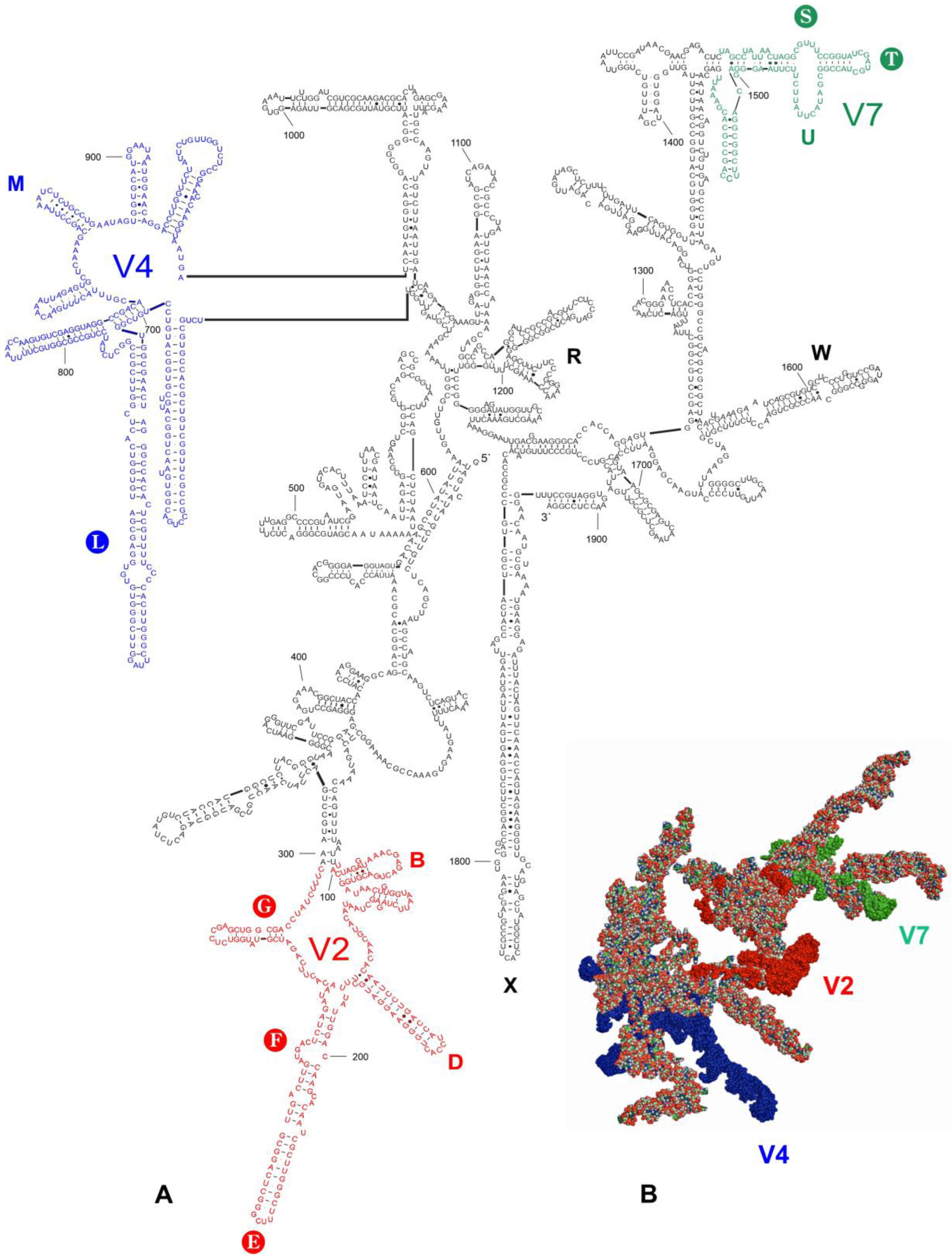
3.2. Secondary Structure Models

The secondary structure models of the entire 18S rRNA gene were predicted for 15 species, including all consensus species (File S2). The prediction included four species of the tribe Acalyptaini, seven of Tingini, one of Litadeini, one of the subfamily Cantacaderinae, and three species of the outgroup. Although the secondary structure models were similar in their general outlines, local differences were found within certain hypervariable regions (V) and length–variable regions (LVRs) (File S2). The secondary structure model of 18S rRNA of Acalypta sauteri Drake, 1942, showing the positions of these regions, is presented in Figure 1.
The nucleotides in the A. sauteri sequence formed 584 pairs (61.2% of all nucleotides in the secondary structure model), with the standard canonical pairs (G–C and A–U) as the most common (450 pairs, 77.1%). The wobble G:U pairs were approximately six times less commonly formed than the standards between the paired nucleotides (83 pairs, 14.2%). The A:G, A:C and other non-canonical pairs were observed rarely (51 pairs, 8.7%). The number of nucleotides that formed particular pair types varied insignificantly among the studied sequences (i.e., ±1.0–1.5% in the standard canonical, ±1.5–3.0% in the wobble G:U, and ±4.5–7.5% in all other non-canonical pairs). This range of variability was consistent with those of previous data for other Heteroptera species [23,29,37].
The alignment results of the entire 18S rRNA sequences analysed (File S1) confirmed the existence of the three hypervariable regions (V2, V4, and V7), which have been described in Heteroptera [15,23,29,37]. The sequence length of the V4 region was highly diverse (299–323 nucleotides), whereas those of the V2 and V7 regions were less variable (198–202 and 90–91 nucleotides, respectively) (File S1, Table 1). However, when considering all analysed species (not only the consensus species), the V4 hypervariable region sequence length (Files S1, Table 1) was stable within the Acalyptaini (at 321 nucleotides) and the Cantacaderinae (at 299 nucleotides). In contrast, this region was highly variable within the Tingini (305–321 nucleotides).
The positions of the LVRs within the gene sequences are shown for Acalypta sauteri (Figure 1) and all analysed consensus species (in File S2). The LVR G, which has been detected in some heteropterans [15,29], was absent in all analysed species. The five LVRs (M, T, U, R, and W) displayed the same number of nucleotides for each region, while six others (B, D, E, F, S, and X) exhibited only insignificant variations in length (one to three nucleotide differences) (Table 2). All 11 of these LVRs were short (three to thirteen nucleotides). In contrast, LVR L was relatively long (from 57 to 81 nucleotides) and showed distinct variations in the sequence lengths (Table 2). Therefore, as suggested in the recent analyses of the 18S rRNA secondary structures in the heteropteran superfamily Pentatomoidea [15], this region was subdivided into subregions to compare the homologous fragments in analysed sequences (Figure 3, Figure S1). The number of nucleotides for each subregion resulting from this comparative analysis is provided in Table 3.
Two subregions, L2 and LA (LA1 + LA2), showed little variability (a single nucleotide). All remaining subregions were variable, with nucleotide numbers ranging from 19–24 in LB, 7–12 in LC, 10–14 in LD, and 9–16 in LE (Table 3).

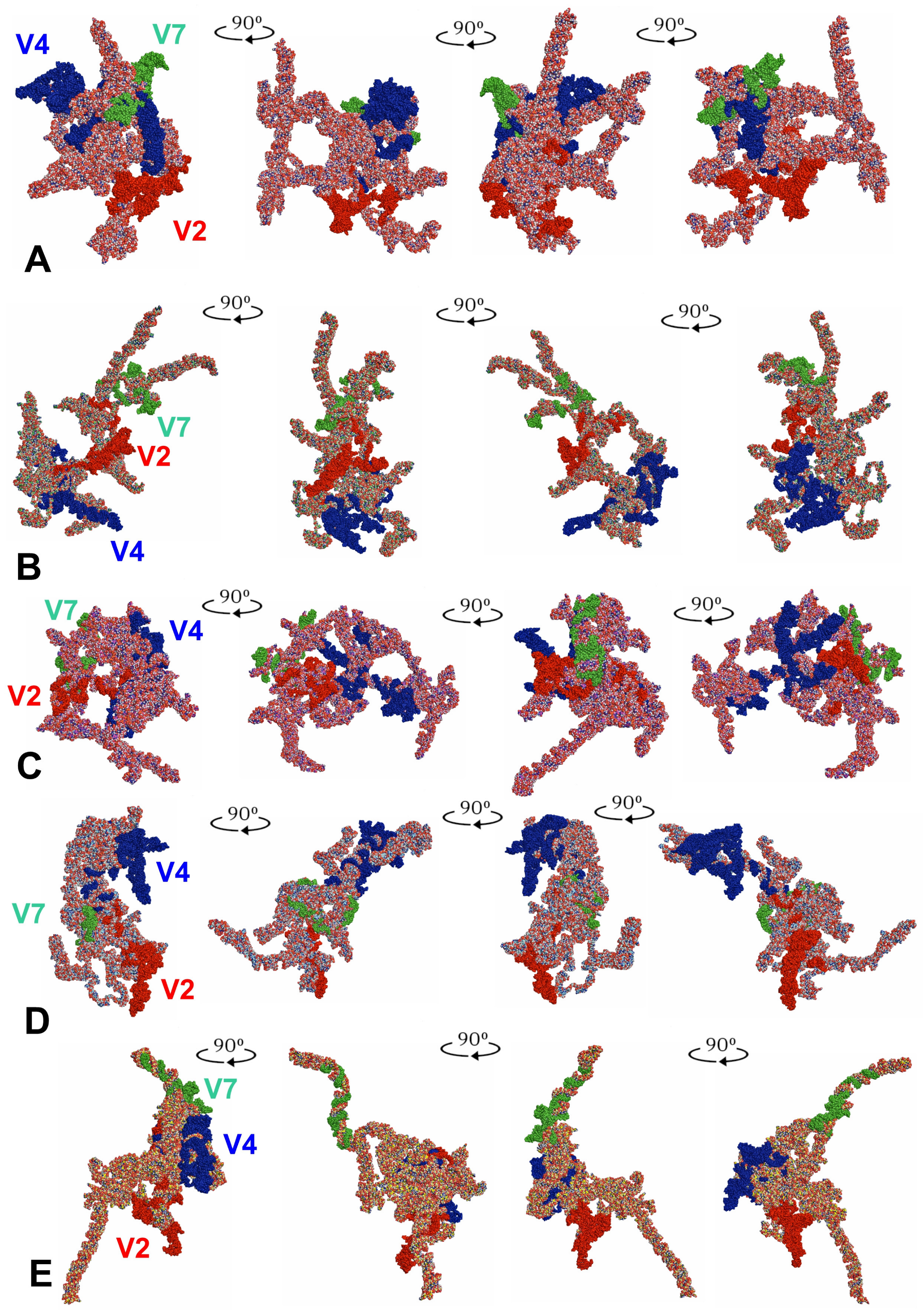
3.3. Tertiary Structure Models

The tertiary structure models of the entire 18S rRNA genes were predicted for all five consensus species (Figure 4).
When the tertiary structures of the consensus species were aligned (Figure 5A), the three species, representing the subfamily Tinginae (A. sauteri, N. signatus, and T. matsumurai) appeared comparable (Figure 5B). This similarity involved the location of the hypervariable regions V2, V4, and V7. However, the general shape of the 18S rRNA tertiary structure for Cantacader lethierryi of the subfamily Cantacaderinae differed significantly, especially when the V7 hypervariable region was considered (Figure 5C).
LVR L, which was the longest segment (57 to 81 nucleotides) of the hypervariable region V4, differed in its level of visibility. This region was indistinct and mainly hidden in the core of the entire tertiary structure in Adelphocoris lineolatus (the outgroup) and Cantacader lethierryi (the subfamily Cantacaderinae of Tingidae) (Figure 6A–B). However, in three consensus species of the subfamily Tinginae (Acalypta sauteri, Nobarnus signatus and Tingis matsumurai), the LVR L was well recognisable (Figure 6C–E).
The predicted tertiary structures of the LVR Ls for all five consensus species are presented in Figure 7 and Figure 8, with all subregions recovered in four of these (A. lineolatus, A. sauteri, N. signatus and T. matsumurai). For C. lethierryi, subregion LB was missing what can be considered its autapomorphy. The other fragments that could serve as morpho-molecular autapomorphies are indicated by the arrows that are colour-coded to the particular LVR L subregion.

4. Discussion

4.1. 18S rRNA Secondary and Tertiary Structures of Tingidae

The 18S rRNA secondary structure models predicted for the four Tingidae consensus species were similar (File S2). Differences were identified within certain hypervariable regions (V2, V4 and V7) (Table 1), especially when modifications in the LVRs were considered (Table 2, Figure 3). Among the LVRs analysed, LVR L appeared the most variable, which corroborates the results of previous studies on 18S rRNA secondary structures in Heteroptera [15,23,29].
Despite similarities among secondary structures of the analysed species, their predicted tertiary structures differed, sometimes substantially (Figure 4 and Figure 5). In particular, the specific tertiary structure of Cantacader lethierryi of the subfamily Cantacaderinae differed significantly from those of all other species representing the subfamily Tinginae (Figure 4, Figure 5 and Figure 6).
Our results support the recent opinion [15] that probable in vivo tertiary configurations of the 18S rRNA are not predictable using existing software (3dRNA v2.0 Web Server). However, small nucleolar RNAs (snoRNAs) activities could have modified the ribosomal RNA tertiary structures [38,39,40,41,42]. The impact of these activities on such structures has not been identified in heteropteran studies; therefore, further research is required in this area [15].

4.2. Potential Apomorphies in Secondary and Tertiary Structures of LVRs

Our analysis demonstrated that the unique nucleotide numbers of two LVRs potentiate these regions as autapomorphies for a particular taxon.
LVR X had a unique number of nucleotides (three to four), which is a potential autapomorphy for the tribe Acalyptaini (Table 2, Figure 1). All other analysed species have five to six nucleotides for this LVR (Table 2, File S2).
However, the most effective method for recovering apomorphic morpho-molecular characters was the comparative analyses of LVR L secondary and tertiary structures (Figure 3, Figure 6, Figure 7 and Figure 8). These results showed the occurrence of several synapomorphies and autapomorphies related to the number of nucleotides in specific subregions (Table 3).
The nineteen nucleotides in the LA subregion, eight in the LE subregion, and ten in the LD subregion can be considered synapomorphies for the Acalyptaini, Litadeini and Tingini of the subfamily Tinginae (Table 3). In addition, an equal number of nucleotides (16) in the LE subregion for Acalyptaini and Litadeini could indicate the synapomorphy of these tribes.
Among the analysed taxa, Cantacaderinae had the highest number of autapomorphies (four) in the specific subregions (except for L2 and LA). Tingini had two autapomorphies (in L2 and LB), while a single autapomorphy was revealed for Acalyptaini and Litadeini (Table 3). The most noteworthy autapomorphy was the absence of the entire subregion LB in the Cantacaderinae; such an extensive deletion in the 18S rDNA sequence has not been recorded in Heteroptera. Strict morpho-molecular autapomorphies in the LVR L tertiary structures were found in four subregions, LB, LC, LD, and L2 (Figure 7 and Figure 8). However, most morpho-molecular autapomorphies involved the LB subregion (Figure 7 and Figure 8) and unique nucleotide numbers for certain tribes (21 for Acalyptaini, 22 for Tingini and 24 for Litadeini). A single morpho-molecular autapomorphy was recovered for the four other subregions (Figure 7 and Figure 8): one in L2 for the Tingini, and one each in LC, LD, and E for Cantacaderinae (Figure 7 and Figure 8). These results indicate that the subfamily Cantacaderinae exhibited the highest number of morpho-molecular autapomorphies in the secondary and tertiary structures of the 18S rRNA. In addition, these results agree with our phylogenomic analysis, which identified the Cantacaderinae as sister to all other Tingidae used by us (Figure 2). Unfortunately, we could not study any representative of the subfamily Vianaidinae, usually considered the sister group to Tingidae (sensu stricto) (Tinginae + Cantacaderinae), based on morphological analyses [2,43,44].
Because the species of Vianaidinae were never included in any known molecular phylogenetic studies, we must bear in mind that the sister relationship between Cantacaderinae and Tinginae presented here cannot be fully supported without the addition of the Vianaidinae into the molecular phylogenetic analysis of the entire Miroidea.

4.3. Identity and Systematic Position of the Tribe Acaltyptaini within the Subfamily Tinginae

Although the tribe Acalyptaini has only recently been validated [9], three of its genera (Acalypta, Dictyonota, and Kalama) have already been included in the first and only phylogenomic analysis of the entire family Tingidae [8]. Five loci were used, including four from mitochondrial genes (COI, Leu-tRNA, COII, and 16S rRNA) and one from a nuclear gene (28S rRNA). That study [8] indicated, among others, that these three genera (Acalypta, Dictyonota, and Kalama) form a monophyletic clade. However, the clade was identified a part of the broadly conceived Tingini [8], which is contrary to our present findings.
Our results confirmed the monophyly of the tribe Acalyptaini, based on an analysis of 18S rDNA sequences, which has not been used in previous phylogenomic studies of Tingidae [8]. We could not include the DNA sequences of Recaredus rex, which was recently included in the tribe Acalyptaini [11], due to the lack of freshly collected specimens. Therefore, placing this genus within the tribe must be based only on morphological characters [11].
In addition, the present study indicated that the tribe Tingini was not monophyletic. The Litadeini, placed within the tribe Tingini by Guilbert et al. [8], was suggested in our study as sister to the clade, which consisted of the Acalyptaini and Tingini. In both molecular analyses [8], in the present study, the tribe Litadeini was represented only by New Caledonian endemic taxa (two genera with three species, and one genus with a single species, respectively). Moreover, the tribe range is considered pantropical [7]. Therefore, all these could be a reason for explaining the different Litadeini position on the phylogenetic tree of Tingidae obtained in both surveys [8] in the present study.

5. Conclusions

  • The results of the present molecular analyses (phylogenetic and structural) validated the recognition of the tribe Acalyptaini within the subfamily Tinginae.
  • The monophyly of the subfamily Cantacaderinae and its basal position within the family Tingidae were indicated, as well as the position of the tribe Litadeini as sister to all other Tinginae.
  • The structural analysis of the predicted tertiary structures of the entire 18S rRNA confirmed the proposed hypothesis that the in vivo configuration of this gene is likely not predictable using only secondary structure models and existing software. However, this may result from small nucleolar RNAs (snoRNAs) activities that can affect changes in the tertiary structures of ribosomal genes.
  • The results showed that two LVRs (LVR X and LVR L) of the hypervariable region V4 exhibited significant variability in the number of nucleotides and could be considered for apomorphic recognition.
  • LVR L appeared to be the most appropriate for phylogenetic relationship analysis within the family Tingidae when considering the secondary and tertiary structure models suitable for identifying morpho-molecular apomorphies.
  • The subfamily Cantacaderinae exhibited the highest number of morpho-molecular autapomorphies in the secondary and tertiary structures of the 18S rRNA. In particular, the absence of the entire subregion LB in this subfamily is the first example of such an extensive deletion in the 18S rDNA sequence in Heteroptera.
  • The tertiary structure of the 18S rRNA exhibited evolutionary properties, which were not detectable in the primary or secondary structures. Therefore, the results of rRNA tertiary structure analyses for phylogenetic considerations are promising. Therefore, including the methods of rRNA tertiary structure analyses in the phylogenetic evaluations in other groups of Heteroptera is strongly suggested.

Supplementary Materials

The following supporting information can be downloaded at: https://www.mdpi.com/article/10.3390/insects14070600/s1, Table S1 [45,46]: List of specimens used in the phylogenetic analysis, their geographic origin (if provided), GenBank accession numbers, and the sources for the sequences downloaded from GenBank. Table S2: List of specimens with 18S used for extraction and amplification during the present study. Their geographic origin, GenBank accession numbers, University of Opole sample numbers, and names of the persons who provided the specimens for analyses are provided. Table S3: Primers used for PCR amplification and sequencing of the nuclear 18S rDNA gene. File S1: The alignment file of the 18S rDNA dataset used for the phylogenetic analysis. File S2: The predicted secondary structure models of the 18S rRNA gene for analysed consensus species. Figure S1: Subdivision of the length-variable regions L (LVRs L) resulting from the alignment of their DNA sequences in five consensus species.

Author Contributions

Conceptualization, B.L. and J.A.L.; Data curation, J.A.L.; Formal analysis, J.A.L. and P.J.D.; Investigations, J.A.L. and P.J.D.; Resources, J.A.L. and P.J.D.; Supervision, J.A.L. and B.L.; Visualization, J.A.L. and P.J.D.; Writing—original draft, J.A.L. and B.L.; Writing—review and editing, J.A.L., B.L. and P.J.D. All authors have read and agreed to the published version of the manuscript.

Funding

This research received no external funding.

Data Availability Statement

The data presented in this study are available in the article and supplementary material.

Acknowledgments

We want to thank S. Kudo (SK, Department of Biology, Naruto University of Education, Tokushima, Japan), and M. Mazur (MM) and A. Wolski (AW) (both of the Institute of Biology, University of Opole, Opole, Poland), for providing us with freshly collected material in ethanol from Japan (SK), Slovakia (MM), Thailand (AW), and Ukraine (MM). Moreover, we want to thank two anonymous reviewers for their comments on the manuscript.

Conflicts of Interest

The authors declare no conflict of interest.

References

  1. Schuh, R.T.; Weirauch, C.H. True Bugs of the World (Hemiptera: Heteroptera): Classification and Natural History, 2nd ed.; Monograph Series; Siri Scientific Press: Rochdale, UK, 2020; Volume 8, 767p. [Google Scholar]
  2. Schuh, R.T.; Cassis, G.; Guilbert, E. Description of the first recent macropterous species of Vianaidinae (Heteroptera: Tingidae) with comments on the phylogenetic relationships of the family within the Cimicomorpha. J. N. Y. Entomol. Soc. 2006, 114, 38–53. [Google Scholar] [CrossRef]
  3. Lis, B. Phylogeny and classification of Cantacaderini [=Cantacaderidae stat. nov.] (Hemiptera: Tingoidea). Ann. Zool. 1999, 49, 157–196. [Google Scholar]
  4. Guilbert, E. Phylogeny of Cantacaderinae (Heteroptera: Tingidae) revisited after the description of a new genus and new species from New Caledonia. Eur. J. Entomol. 2012, 109, 111–116. [Google Scholar] [CrossRef] [Green Version]
  5. Drake, C.J.; Ruhoff, F.A. Lacebugs of the World: A Catalog (Hemiptera: Tingidae). USA Natl. Mus. Bull. 1965, 243, 1–634. [Google Scholar] [CrossRef]
  6. Froeschner, R.C. Zoogeographic and systematic notes on the lace bug tribe Litadeini, with the description of the new genus Stragulotingis (Hemiptera: Tingidae). Great Basin Nat. 1969, 29, 129–132. [Google Scholar]
  7. Froeschner, R.C. Lace Bug Genera of the World, II: Subfamily Tinginae: Tribes Litadeini and Ypsotingini (Heteroptera; Tingidae). Smithson. Contrib. Zool. 2001, 611, 1–28. [Google Scholar] [CrossRef]
  8. Guilbert, E.; Damgaard, J.; D’Haese, C.A. Phylogeny of the lacebugs (Insecta: Heteroptera: Tingidae) using morphological and molecular data. Syst. Entomol. 2014, 39, 431–441. [Google Scholar] [CrossRef]
  9. Golub, V.B.; Golub, N.V. On the status of the genera complex Acalypta, Dictyonota, Kalama and Derephysia (Heteroptera: Tingidae: Tinginae) having common morphological and karyological features. Zoosyst. Ross. 2019, 28, 228–237. [Google Scholar] [CrossRef]
  10. Gapon, D.A.; Golub, V.B.; Knudson, A.H. Case 3813—Acalyptini Thomson, 1859 (Hexapoda, Coleoptera) and Acalyptini Blatchley, 1926 (Hexapoda, Heteroptera): Proposed removal of homonymy by emendation of the latter name to Acalyptaini. Bull. Zool. Nomencl. 2019, 76, 175–178. [Google Scholar] [CrossRef]
  11. Lis, B.; Zielińska, A.; Lis, J.A. The King’s Lace Bug Recaredus rex Distant, 1909 (Hemiptera: Heteroptera: Tingidae): Systematic Position, First Palaearctic and Afrotropical Records, and Ecological Niche Modelling. Insects 2022, 13, 558. [Google Scholar] [CrossRef]
  12. Golub, N.V.; Golub, V.B.; Anokhin, B.A.; Kuznetsova, V.G. Comparative Cytogenetics of Lace Bugs (Tingidae, Heteroptera): New Data and a Brief Overview. Insects 2022, 13, 608. [Google Scholar] [CrossRef] [PubMed]
  13. Lis, B.; Parveen, S.; Ramamurthy, V.V. Redescription of the Oriental lace-bug Recaredus rex Distant, 1909 (Hemiptera: Tingidae: Tinginae), and its new tribal assignment, with a key to Ypsotingini. Zootaxa 2013, 3702, 597–600. [Google Scholar] [CrossRef] [PubMed] [Green Version]
  14. Lis, B. Comparative studies on the ductus seminis of aedeagus in Tingoidea (Hemiptera: Heteroptera). Pol. J. Entomol. 2004, 73, 245–258. [Google Scholar]
  15. Lis, J.A. Molecular Apomorphies in the Secondary and Tertiary Structures of Length-Variable Regions (LVRs) of 18S rRNA Shed Light on the Systematic Position of the Family Thaumastellidae (Hemiptera: Heteroptera: Pentatomoidea). Int. J. Mol. Sci. 2023, 24, 7758. [Google Scholar] [CrossRef] [PubMed]
  16. Weirauch, C.; Schuh, R.T.; Cassis, G.; Wheeler, W.C. Revisiting habitat and lifestyle transitions in Heteroptera (Insecta: Hemiptera): Insights from a combined morphological and molecular phylogeny. Cladistics 2019, 35, 67–105. [Google Scholar] [CrossRef] [Green Version]
  17. Lis, J.A.; Ziaja, D.; Lis, B.; Gradowska, P.A. Non-monophyly of the “cydnoid” complex within Pentatomoidea (Hemiptera: Heteroptera) revealed by Bayesian phylogenetic analysis of nuclear rDNA sequences. Arthropod Syst. Phylogeny 2017, 75, 481–496. [Google Scholar]
  18. Giribet, G.; Carranza, S.; Bagui, J.; Riutort, M.; Ribera, C. First Molecular Evidence for the Existence of a Tardigrada + Arthropoda Clade. Mol. Biol. Evol. 1996, 13, 76–84. [Google Scholar] [CrossRef]
  19. Whiting, M.F.; Carpernter, J.C.; Wheeler, Q.D.; Wheeler, W.C. The Strepsiptera problem: Phylogeny of the holometabolous insect orders inferred from 18S and 28S ribosomal DNA sequences and morphology. Syst. Biol. 1997, 46, 1–68. [Google Scholar] [CrossRef]
  20. Campbell, B.C.; Steffen-Campbell, J.D.; Sorensen, J.T.; Gill, R.J. Paraphyly of Homoptera and Auchenorrhyncha inferred from 18S rDNA nucleotide sequences. Syst. Entomol. 1995, 20, 175–194. [Google Scholar] [CrossRef]
  21. Black, W.C., IV; Klompen, J.S.H.; Keirans, J.E. Phylogenetic relationships among tick subfamilies based on the SSU nuclear rDNA gene. Mol. Phylogenet. Evol. 1997, 7, 129–144. [Google Scholar] [CrossRef]
  22. Barker, S.C.; Whiting, M.; Johnson, K.P.; Murrell, A. Phylogeny of the lice (Insecta, Phthiraptera) inferred from small subunit rRNA. Zool. Scr. 2003, 32, 407–414. [Google Scholar] [CrossRef]
  23. Yu, S.; Wang, Y.; Rédei, D.; Xie, Q.; Bu, W. Secondary structure models of 18S and 28S rRNAs of the true bugs based on complete rDNA sequences of Eurydema maracandica Oshanin, 1871 (Heteroptera, Pentatomidae). ZooKeys 2013, 319, 363–377. [Google Scholar] [CrossRef] [Green Version]
  24. Kumar, S.; Stecher, G.; Li, M.; Knyaz, C.; Tamura, K. MEGA X: Molecular evolutionary genetics analysis across computing platforms. Mol. Biol. Evol. 2018, 35, 1547–1549. [Google Scholar] [CrossRef] [PubMed]
  25. Nguyen, L.-T.; Schmidt, H.A.; von Haeseler, A.; Minh, B.Q. IQ-TREE: A fast and effective stochastic algorithm for estimating maximum likelihood phylogenies. Mol. Biol. Evol. 2015, 32, 268–274. [Google Scholar] [CrossRef] [PubMed]
  26. Trifinopoulos, J.; Nguyen, L.-T.; von Haeseler, A.; Minh, B.Q. W-IQ-TREE: A fast online phylogenetic tool for maximum likelihood analysis. Nucleic Acids Res. 2016, 44, 232–235. [Google Scholar] [CrossRef] [Green Version]
  27. Hoang, D.T.; Chernomor, O.; von Haeseler, A.; Minh, B.Q.; Vinh, L.S. UFBoot2: Improving the Ultrafast Bootstrap Approximation. Mol. Biol. Evol. 2018, 35, 518–522. [Google Scholar] [CrossRef]
  28. Letunic, I.; Bork, P. Interactive Tree of Life (iTOL) v5: An online tool for phylogenetic tree display and annotation. Nucleic Acids Res. 2021, 49, W293–W296. [Google Scholar] [CrossRef]
  29. Wu, Y.Z.; Yu, S.S.; Wang, Y.H.; Wu, Y.H.; Li, X.R.; Men, X.Y.; Zhang, Y.W.; Rédei, D.; Xie, Q.; Bu, W.J. The evolutionary position of Lestoniidae revealed by molecular autapomorphies in the secondary structure of rRNA besides phylogenetic reconstruction (Insecta: Hemiptera: Heteroptera). Zool. J. Linn. Soc. 2016, 177, 750–763. [Google Scholar] [CrossRef]
  30. Reuter, J.S.; Mathews, D.H. RNAstructure: Software for RNA secondary structure prediction and analysis. BMC Bioinform. 2010, 11, 129. [Google Scholar] [CrossRef] [Green Version]
  31. Wang, J.; Homoptera, J.; Huang, Y.; Xiao, Y. 3dRNA v2. 0: An Updated Web Server for RNA 3D Structure Prediction. Int. J. Mol. Sci. 2019, 20, 4116. [Google Scholar] [CrossRef] [Green Version]
  32. Popenda, M.; Szachniuk, M.; Antczak, M.; Purzycka, K.J.; Lukasiak, P.; Bartol, N.; Blazewicz, J.; Adamiak, R.W. Automated 3D structure composition for large RNAs. Nucleic Acids Res. 2012, 40, e112. [Google Scholar] [CrossRef] [PubMed]
  33. Biesiada, M.; Purzycka, K.J.; Szachniuk, M.; Blazewicz, J.; Adamiak, R.W. Automated RNA 3D structure prediction with RNAComposer. In RNA Structure Determination. Methods in Molecular Biology; Turner, D., Mathews, D., Eds.; Humana Press: New York, NY, USA, 2016; Volume 1490, pp. 199–215. [Google Scholar] [CrossRef]
  34. Schrödinger, L.; DeLano, W. PyMOL, Version 2.4.0; Schrödinger, LLC.: New York, NY, USA, 2020; Available online: http://www.pymol.org/pymol (accessed on 11 May 2023).
  35. Xie, Q.; Tian, X.; Qin, Y.; Bu, W. Phylogenetic comparison of local length plasticity of the small subunit of nuclear rDNAs among all Hexapoda orders and the impact of hyper-length-variation on alignment. Mol. Phylogenet. Evol. 2009, 50, 310–316. [Google Scholar] [CrossRef] [PubMed]
  36. Ouvrard, D.; Campbell, B.C.; Bourgoin, T.; Chan, K.L. 18S rRNA Secondary Structure and Phylogenetic Position of Peloridiidae (Insecta, Hemiptera). Mol. Phylogenet. Evol. 2000, 16, 403–417. [Google Scholar] [CrossRef] [Green Version]
  37. Xie, Q.; Tian, Y.; Zheng, L.; Bu, W. 18S rRNA hyper-elongation and the phylogeny of Euhemiptera (Insecta: Hemiptera). Mol. Phylogenet. Evol. 2008, 47, 463–471. [Google Scholar] [CrossRef]
  38. Kiss, T. Small Nucleolar RNAs An Abundant Group of Noncoding RNAs with Diverse Cellular Functions. Cell 2002, 109, 145–148. [Google Scholar] [CrossRef] [Green Version]
  39. Zemann, A.; op de Bekke, A.; Kiefmann, M.; Brosius, J.; Schmitz, J. Evolution of small nucleolar RNAs in nematodes. Nucleic Acids Res. 2006, 34, 2676–2685. [Google Scholar] [CrossRef] [PubMed] [Green Version]
  40. Schmitz, J.; Zemann, A.; Churakov, G.; Kuhl, H.; Grützner, F.; Reinhardt, R.; Brosius, J. Retroposed SNOfall-A mammalian-wide comparison of platypus snoRNAs. Genome Res. 2008, 18, 1005–1010. [Google Scholar] [CrossRef] [Green Version]
  41. Bratkovič, T.; Božič, J.; Rogelj, B. Functional diversity of small nucleolar RNAs. Nucleic Acids Res. 2020, 48, 1627–1651. [Google Scholar] [CrossRef] [PubMed] [Green Version]
  42. Soulé, S.; Mellottee, L.; Arab, A.; Chen, C.; Martin, J.-R. Jouvence, a new small nucleolar RNA required in the gut extends lifespan in Drosophila. Nat. Commun. 2020, 11, 987. [Google Scholar] [CrossRef] [PubMed] [Green Version]
  43. Schuh, R.T.; Štys, P. Phylogenetic analysis of cimicomorphan family relationships (Heteroptera). J. N. Y. Entomol. Soc. 1991, 99, 298–350. [Google Scholar]
  44. Schuh, R.T.; Weirauch, C.; Wheeler, W.C. Phylogenetic relationships within the Cimicomorpha (Hemiptera: Heteroptera): A total-evidence analysis. Syst. Entomol. 2009, 34, 15–48. [Google Scholar] [CrossRef]
  45. Wang, Y.H.; Cui, Y.; Rédei, D.; Baňař, P.; Xie, Q.; Štys, P.; Damgaard, J.; Chen, P.P.; Yi, W.B.; Wang, Y.; et al. Phylogenetic divergences of the true bugs (Insecta: Hemiptera:Heteroptera), with emphasis on the aquatic lineages: The last piece of the aquatic insect jigsaw originated in the Late Permian/EarlyTriassic. Cladistics 2016, 32, 390–405. [Google Scholar] [CrossRef] [PubMed]
  46. Campbell, B.C.; Steffen-Campbell, J.D.; Gill, R.J. Evolutionary origin of whiteflies (Hemiptera: Sternorrhyncha: Aleyrodidae) inferred from 18S rDNA sequences. Insect Mol. Biol. 1994, 3, 73–88. [Google Scholar] [CrossRef] [PubMed]
Figure 1. 18S rRNA of Acalypta sauteri. (A) Secondary structure model. The bases marked in colour represent the hypervariable regions (V2—red, V4—dark blue, V7—green). Thirteen length–variable regions (LVRs) are labelled as capital letters B to W in colours analogous to the base colours representing the hypervariable regions or other sequences’ regions. The capital letters in filled circles indicate six LVRs (E, F, G, L, S, and T) that can serve as molecular synapomorphies or autapomorphies in analyses. Base pairing is shown as follows: standard canonical pairs are lines (G–C, A–U), wobble G:U pairs are dots (G·U), A:G or A:C pairs are open circles (A; G, A; C), and other non-canonical pairs are filled circles (e.g., U and U, A and A). (B) Tertiary structure model. The fragments marked in colour represent the hypervariable regions (V2—red, V4—dark blue, V7—green).
Insects 14 00600 g001
Figure 2. Phylogenetic tree based on a Maximum Likelihood analysis of the 18S rDNA. Node labels—Ultrafast Bootstrap values (see Section 2), and the Bootstrap values over 50% are shown next to the branches, scale bar—number of substitutions per site.
Insects 14 00600 g002
Figure 3. Secondary structure models of the length–variable region L. (A) Adelphocoris lineolatus (outgroup). (B) Acalypta sauteri (Tinginae: Acalyptaini). (C) Nobarnus signatus (Tinginae: Litadeini). (D) Tingis matsumurai (Tinginae: Tingini). (E) Cantacader lethierryi (Cantacaderinae). Specific subregion bases are marked in the same colour.
Insects 14 00600 g003
Figure 4. Tertiary structure models of the 18S rRNA. (A) Adelphocoris lineolatus (outgroup). (B) Acalypta sauteri (Tinginae: Acalyptaini). (C) Nobarnus signatus (Tinginae: Litadeini). (D) Tingis matsumurai (Tinginae: Tingini). (E) Cantacader lethierryi (Cantacaderinae). The hypervariable regions are marked in red (V2), dark blue (V4), and green (V7).
Insects 14 00600 g004
Figure 5. 18S rRNA tertiary structure models. (A) Combined for all five consensus species and aligned to the outgroup (Adelphocoris lineolatus) sequence. (B) Combined for three consensus species of the subfamily Tinginae and aligned to Acalypta sauteri sequence. (C) Combined for four consensus species of Tingidae (Tinginae and Cantacaderinae) and aligned to Cantacader lethierryi sequence. The hypervariable regions are marked in red (V2), dark blue (V4), and green (V7). V7C—hypervariable region V7 in Cantacader lethierryi.
Insects 14 00600 g005
Figure 6. The LVR L position (marked in light blue) within the hypervariable region V4 in the tertiary structure models of the 18S rRNA gene. (A) Adelphocoris lineolatus (outgroup). (B) Cantacader lethierryi (Cantacaderinae). (C) Acalypta sauteri (Tinginae: Acalyptaini). (D) Nobarnus signatus (Tinginae: Lita-deini). (E) Tingis matsumurai (Tinginae: Tingini). The hypervariable regions are marked in red (V2), dark blue (V4), and green (V7). All sequences are aligned to the outgroup (A. lineolatus) sequence.
Insects 14 00600 g006
Figure 7. The predicted tertiary structure models of the LVR L. (A) Adelphocoris lineolatus (outgroup). (B) Acalypta sauteri (Tinginae: Acalyptaini). (C) Nobarnus signatus (Tinginae: Litadeini). (D) Tingis matsumurai (Tinginae: Tingini). (E) Cantacader lethierryi (Cantacaderinae). The LVR subregions are marked in the appropriate colour: L (A1 + A2) in magenta, L (B1 + B2) in red, L (C1 + C2) in blue, L (D1 + D2) in orange, L (E1 + E2) in yellow, and L2 in green. The arrows, corresponding in colour to the particular subregion, indicate the fragments that can serve as potential morpho-molecular derived characters (autapomorphies). All sequences are aligned to the outgroup (A. lineolatus) sequence. The numbers above the arrows show the autapomorphic number of nucleotides in the subregion.
Insects 14 00600 g007
Figure 8. The predicted tertiary structure models of the LVR L. (A) Adelphocoris lineolatus (outgroup). (B) Acalypta sauteri (Tinginae: Acalyptaini). (C) Nobarnus signatus (Tinginae: Litadeini). (D) Tingis matsumurai (Tinginae: Tingini). (E) Cantacader lethierryi (Cantacaderinae). The LVR subregions are marked in the appropriate colour: L (A1 + A2) in magenta, L (B1 + B2) in red, L (C1 + C2) in blue, L (D1 + D2) in orange, L (E1 + E2) in yellow, and L2 in green. The arrows corresponding in colour to the particular subregion indicate the fragments that can serve as potential morpho-molecular derived characters (autapomorphies). All sequences are aligned to A. sauteri sequence. The numbers above the arrows show the autapomorphic number of nucleotides in the subregion.
Insects 14 00600 g008
Table 1. The number of nucleotides of the hypervariable regions V2, V4, and V7 in the 18S rRNA of the analysed taxa. For the ‘consensus species’ definition, see Section 2.
Taxon GroupConsensus SpeciesNumber of Nucleotides
V2V4V7
OutgroupAdelphocoris lineolatus (Goeze, 1778)19831791
Tinginae: AcalyptainiAcalypta sauteri (Drake, 1942)20032190
Tinginae: LitadeiniNobarnus signatus (Distant, 1920)20232390
Tinginae: TinginiTingis matsumurai (Takeya, 1962)20032090
CantacaderinaeCantacader lethierryi (Scott, 1874)19929990
Table 2. The nucleotide numbers of the LVRs in the 18S rRNA of the analysed taxa. The autapomorphy for Acalyptaini is indicated in red.
Taxon GroupNumber of Nucleotides
BDEFGLMSTURWX
outgroup12654060–7846713536
Tinginae: Acalyptaini1154507945713533–4
Tinginae: Litadeini1145508145713536
Tinginae: Tingini1154–55063–7945713535–6
Cantacaderinae1155405745713535
Table 3. The nucleotide numbers of the subregions of the LVR L. The autapomorphies are indicated in red, and the synapomorphies are indicated in green.
Taxon GroupConsensus SpeciesTotal LengthNumber of Nucleotides of the LVR L Subregions
L2LA (A1 + A2)LB (B1 + B2)LC (C1 + C2)LD (D1 + D2)LE (E1 + E2)
outgroupAdelphocoris lineolatus74418 (9 + 9)19 (10 + 9)7 (3 + 4)11 (6 + 5)15 (8 +7)
AcalyptainiAcalypta sauteri79419 (10 + 9)21 (11+ 10)8 (4 + 4)10 (5 + 5)16 (8 + 8)
LitadeiniNobarnus signatus81419 (10 + 9)24 (13 + 11)8 (4 + 4)10 (5 + 5)16 (8 + 8)
TinginiTingis matsumurai78519 (10 + 9)22 (11 + 11)8 (3 + 5)10 (6 + 4)14 (7 + 7)
CantacaderinaeCantacader lethierryi57418 (9 + 9)012 (7 + 5)14 (7 + 7)9 (4 + 5)
Disclaimer/Publisher’s Note: The statements, opinions and data contained in all publications are solely those of the individual author(s) and contributor(s) and not of MDPI and/or the editor(s). MDPI and/or the editor(s) disclaim responsibility for any injury to people or property resulting from any ideas, methods, instructions or products referred to in the content.

