Article

The Key Glutathione S-Transferase Family Genes Involved in the Detoxification of Rice Gramine in Brown Planthopper Nilaparvata lugens

1
College of Plant Protection, China Agricultural University, Beijing 100193, China
2
MOA Key Laboratory of Crop Pest Monitoring and Green Control, College of Plant Protection, China Agricultural University, Beijing 100193, China
3
Department of Entomology, Texas A & M University, College Station, TX 77843, USA
4
College of Plant Sciences, Jilin University, Changchun 130062, China
5
Key Laboratory of Zoonosis Research, Ministry of Education, Jilin University, Changchun 130062, China
*
Author to whom correspondence should be addressed.
These authors equally contributed to this work.
Insects 2021, 12(12), 1055; https://doi.org/10.3390/insects12121055
Submission received: 16 September 2021 / Revised: 14 November 2021 / Accepted: 22 November 2021 / Published: 25 November 2021
(This article belongs to the Section Insect Molecular Biology and Genomics)

Abstract

:

Simple Summary

Plant defensive compounds are the effective measures for host plants to defend against herbivorous insects, whereas in turn insects evolve an array of detoxifying enzymes to overcome the resistance of these compounds. However, the molecular mechanisms underlying the resistance of plant toxins to herbivores are still unclear. This study uses an integrative approach to investigate the molecular basis of how brown planthopper uses glutathione S-transferases to detoxify gramine, an important defensive toxin in rice. We show here that gramine can induce the activity and increase the transcripts of glutathione S-transferase in brown planthopper. Knockdown of seven glutathione S-transferase family genes of brown planthopper increases its sensitivity to gramine. The glutathione S-transferase activity is regulated by the expression of three key genes (NlGST1-1, NlGSTD2, and NlGSTE1), as silencing of these genes significantly inhibits this enzyme’s activity. The current study identifies a few key glutathione S-transferase genes involved in the detoxification of gramine in brown planthopper. Our findings unravel the molecular mechanisms of the detoxification of plant defensive chemicals by glutathione S-transferases, providing a new potential pest control strategy that uses the method of RNA interference to deplete this gene family to increase the efficiency of host resistance to herbivores.

Abstract

Phytochemical toxins are considered a defense measure for herbivore invasion. To adapt this defensive strategy, herbivores use glutathione S-transferases (GSTs) as an important detoxification enzyme to cope with toxic compounds, but the underlying molecular basis for GST genes in this process remains unclear. Here, we investigated the basis of how GST genes in brown planthopper (BPH, Nilaparvata lugens (Stål)) participated in the detoxification of gramine by RNA interference. For BPH, the LC25 and LC50 concentrations of gramine were 7.11 and 14.99 μg/mL at 72 h after feeding, respectively. The transcriptions of seven of eight GST genes in BPH were induced by a low concentration of gramine, and GST activity was activated. Although interferences of seven genes reduced BPH tolerance to gramine, only the expression of NlGST1-1, NlGSTD2, and NlGSTE1 was positively correlated with GST activities, and silencing of these three genes inhibited GST activities in BPH. Our findings reveal that two new key genes, NlGSTD2 and NlGSTE1, play an essential role in the detoxification of gramine such as NlGST1-1 does in BPH, which not only provides the molecular evidence for the coevolution theory, but also provides new insight into the development of an environmentally friendly strategy for herbivore population management.

1. Introduction

Understanding interactions between herbivorous insects and their host plants is challenging when insects colonize. During feeding, herbivorous insects are exposed to an array of plant defensive compounds such as alkaloids and phenolics [1,2,3]. Of them, alkaloids are important nitrogenous compounds in plants, and many alkaloids have been developed as potential pesticides [4,5]. Gramine (indol-3-ylmethyldimethylamine), a simple indole alkaloid, exists widely in wheat Triticum aestivum, rice Oryza sativa, barley Hordeum vulgare, Arundo donax, and other gramineous plants [3,6,7,8,9]. Previous studies have reported that this compound is a deterrent and/or toxicant to some herbivorous insects, such as brown planthopper (BPH, Nilaparvata lugens), aphids Rhopalosiphum padi and Sitobion avenae, bettle Scolytus multistriatus, and cotton bollworm Helicoverpa armigera [3,7,9,10,11,12]. The resistances of gramineous plants to herbivores are highly associated with gramine content in plants. For example, in aphid-resistant wheat varieties and BPH-resistant rice varieties, the contents of gramine are generally higher than that of in the susceptible varieties [3,6,9], leading to the reduction of the survival rate and growth rate of insects after being fed on these resistant varieties. Therefore, herbivores must deal with these defensive compounds in plants properly so as to better adapt to their hosts.
The antagonistic compounds of plants are usually toxic to insects [13], and this toxic effect on insects is closely related to the levels of the secondary metabolites in host plants [9,14]. Although herbivorous insects harbor a large set of these detoxification enzymes, such as glutathione S-transferases (GSTs), cytochrome P450 (CYP450s), and carboxylesterases (CarEs), in most cases only a small number of detoxifying enzymes can metabolize specific plant compounds [15]. Of them, insect GSTs belong to one of the important detoxification-related enzymes that convert toxic phytochemicals into nontoxic products, in order to avoid potential damage to insect cellular processes [14,16]. When herbivorous insects are exposed to relatively low concentrations of xenobiotics, induced GST activity in insects rapidly catabolizes these compounds to avoid toxicity accumulation [9,13,17,18,19,20]. Interestingly, not all members of GST family in insects are involved in the detoxification of identical xenobiotic classes. For example, fruit fly Drosophila melanogaster DmGSTS1-1 and DmGSTD1 are responsible for the detoxification of mustard oils and 4-hydroxynonenal, respectively [21,22]; BPH NlGST1-1 is involved in detoxification of gramine and pyrethroid [9,23]. LsGSTe1 and LsGSTm are required for the metabolism of insecticides in Laodelphax striatellus [24]. Notably, GSTs appear to have substrate specificities since detoxification of some specific xenobiotics largely depends on certain GSTs in the GST family [21,25,26,27]. It is thus foreseeable that identifying these detoxification enzyme genes and blocking their functions would reduce the adaptation of these insects to the toxic phytochemical-containing plants.
BPH is a devastating phloem feeding insect that is widely distributed throughout rice production areas in Asia [28]. Feeding exclusively on rice, BPH directly results in losses of rice yield and straw (bioethanol feedstock) by extracting nutrients [29]. BPH-resistant properties of many rice varieties have been quickly overcome due to BPH biotype development [30,31,32]. It is thus necessary to understand the mechanism and factors that are responsible for manifesting the resistance into the selected cultures with desirable characters so that they can be utilized effectively in the breeding program. Perhaps, BPH that can rapidly adapt to their host plants may be closely related to strong detoxification capability in the insect. GSTs in BPH are important detoxification enzymes that are involved in the degradations of toxic rice compounds [9,19]. Previous evidence indicates that GST genes in BPH are currently divided into six subfamilies: Delta (NlGST1-1, NlGSTD1, and NlGSTD2), Epsilon (NlGSTE1), Sigma (NlGSTS1 and NlGSTS2), Omega (NlGSTO1), Theta (NlGSTT1), Zeta (NlGSTZ1), and a microsomal class [23,27,33]. Of them, eight cytosolic GST genes (NlGST1-1, NlGSTD1, NlGSTD2, NlGSTE1, NlGSTO1, NlGSTS1, NlGSTS2, and NlGSTT1) are highly expressed in midgut and Malpighian tubules, which are the main toxic metabolic organs in BPH [23,27]. Although previous studies have reported that NlGST1-1 is required for detoxifying pyrethroid and gramine, NlGSTD1 and NlGSTE1 are required for detoxifying ferulic acid, and even some GST genes are important in imidacloprid resistance [9,19,23,34], while the molecular basis of how BPH GST family is involved in the detoxification of phytochemicals, especially for gramine of a simple indole alkaloid in rice, remains obscure.
In this work, we re-examined the toxicity of gramine to BPH, and investigated the relationship between rice gramine and BPH GST family genes using the method of RNA interference (RNAi). The GST family genes NlGSTD2 and NlGSTE1 in BPH play an indispensable role in the detoxification of gramine, the same as NlGST1-1. Our research on the coupling of phytochemicals with depletion of BPH detoxification genes provides an efficient management strategy for sap-sucking pests in rice.

2. Materials and Methods

2.1. Plants and Insects

Rice BPH (biotype II) nymphs were obtained from paddy fields in Hubei Province, China. The nymphs were reared on five-leaf stage of susceptible Indica Taichung Native 1 (TN1) rice seedlings in a climate-controlled chamber at 27 ± 2 °C, 90% RH, and 16 h light/8 h dark (unless otherwise indicated). BPH population was reproduced for at least ten generations on TN1 seedlings before they were used in the subsequent experiments.

2.2. Bioassay on Gramine Toxicity to BPH

To understand the toxicity of gramine to BPH in more detail, the lethal concentration 25% (LC25) and lethal concentration 50% (LC50) of gramine were determined following a previously described method [9,35]. BPH artificial diet was prepared according to the previously described methods [35]. Gramine (purity 99%, CAS Number: 87-52-5, Sigma-Aldrich, St. Louis, MO, USA) was distributed into the liquid artificial diet with concentrations of 0.0 (control), 3.0, 6.0, 12.0, and 24.0 μg/mL. The concentration of gramine used in this experiment was approximately equaled to its content in fresh rice leaves, which ranged from 3 to 140 μg/g wet weight [9,36]. The diet packet holding 100 μL of the liquid diet in double layers of stretched Parafilm M (Pechiney Plastic Packaging Company, Chicago, USA) was placed at one end of a transparent cylindrical glass tube (L × D: 10 cm × 2.5 cm, open at both ends) and per diet packet was fed by fifteen BPH nymphs (second or third instar). The other end of the feeding tube was covered with a piece of gauze to allow air to exchange. Each gramine concentration included 3 feeding tubes with 4 replicates. Diets and feeding sachets were changed daily to avoid diet deterioration and microbial contamination. After feeding for 72 h, dead BPH nymphs in different treatments were recorded for LC25 and LC50 calculations. The surviving nymphs were collected for the GST activity assay.
To test the impact of nonlethal doses of gramine on each gene expression in BPH GST family, second- or third-instar BPH nymphs were cultured on artificial diets without or with gramine following described methods above. Referring to nonlethal doses of gramine to BPH, 0.0, 2.0, or 3.0 μg/mL gramine was distributed into the diets as control and treatments with 4 replicates. After feeding for 72 h, surviving nymphs were collected for the expression level analysis by quantitative real-time PCR (qRT-PCR).

2.3. GST Activity Assay

GST activity was evaluated using a previously described method with minor modification [37]. Ten frozen BPH nymphs in 1 mL of 0.1 M phosphate buffer (pH 6.5) at 4 °C were homogenized, centrifuged, and then the supernatants were collected to analyze the GST activity. The reaction mixture consisted of 0.95 mL of 0.1 M phosphate buffer (pH 6.5), 20 μL of 50 mM 1-chloro-2, 4-dinitrobenzene (CDNB), and 20 μL of 50 mM L-glutathione (reduced). Ten microliters of the supernatant were quickly added into the reaction mixture and monitored the change of the absorbance using a spectrophotometer at 340 nm (SP-756P, Shanghai Metash Instruments Co. Ltd., Shanghai, China). The supernatant was replaced with the same volume of phosphate buffer as the control. GST activity was expressed as μmol·min−1·mg protein−1 as calculated by using the extinction coefficient for CDNB (9.6 mM−1·cm−1).
The concentrations of total proteins were determined using a previously described method [38]. The absorbance of the reaction mixture was measured at 595 nm with a spectrophotometer. The standard curve of the protein concentration of BPH samples was established according to the known concentration of bovine serum protein (Sigma-Aldrich, St. Louis, MO, USA).

2.4. qRT-PCR

Total RNA of the BPH nymphs was extracted using RNeasy Mini Kit (QIAGEN, Hilden, Germany), and cDNA was prepared by the FastQuant RT Kit (with gDNase; TIANGEN, Beijing, China) following the manufacturer’s instructions. qRT-PCR was performed on an Applied Biosystems (ABI) 7500 Real-Time PCR System (ABI, Foster City, CA, USA) using SYBR Premix Ex Taq II (Tli RNaseH Plus; TaKaRa, Shiga, Japan). Due to midgut and Malpighian tubules being the main toxic metabolic organs in BPH, we selected eight cytosolic GST genes (NlGST1-1, GenBank No. AF448500.1; NlGSTD1, GenBank No. JQ917469.1; NlGSTD2, GenBank No. JQ917467.1; NlGSTE1, GenBank No. JQ917470.1; NlGSTO1, GenBank No. JQ917471.1; NlGSTS1, GenBank No. JQ917468.1; NlGSTS2, GenBank No. JQ917474.1; NlGSTT1, GenBank No. JQ917472.1) that were highly expressed in these two tissues as target genes [23,27]. The specific primer sequences of BPH GST genes are listed in Table S1. The relative expression levels of target genes were calculated according to the 2–ΔΔCt method [39]. BPH β-actin (GenBank No. EU179846) was used as reference gene.

2.5. Double-Stranded RNA (dsRNA) Synthesis and Feeding Assays

The methods of total RNA extraction and cDNA synthesis of the BPH nymphs are described above. Eight aforementioned BPH GST genes (NlGST1-1, NlGSTD1, NlGSTD2, NlGSTE1, NlGSTO1, NlGSTS1, NlGSTS2, and NlGSTT1) were PCR-amplified, ligated into the pEASY-T1 vector (TransGen Biotech, Beijing, China), and transformed into Trans1-T1 phage resistant chemically competent cell (TransGen Biotech, Beijing, China). After sequencing, the recombinant pEASY-GST plasmids were served as PCR templates to produce the templates for the synthesis of each GST dsRNA with the specific primers containing T7 promoter sequences. The specific primer sequences are listed in Table S1. Each GST dsRNA was generated by using the HiScribe T7 in vitro Transcription Kit (NEB, Ipswich, MA, USA) and purified with the RNA Clean Kit (TIANGEN, Beijing, China) following the manufacturer’s instructions. GFP (green fluorescent protein, GenBank No. AAX31732.1) dsRNA was synthesized as control.
The dsRNAs of eight GST genes were separately dissolved in artificial diets at concentrations of 0.2, 0.4, and 0.8 µg/mL. Among these, NlGST1-1 dsRNA was used as a positive control and GFP dsRNA at 0.4 µg/mL served as a negative control [9]. Feeding tubes containing dsRNAs were assembled, as described in the methods above; each tube was fed with fifteen second- or third-instar BPH nymphs. Each treatment contained 3 sample sizes and 4 replicates. Dead nymphs were recorded for calculating BPH mortality rates. The surviving nymphs were collected to analyze the transcription level of GST genes and GST activity, as described above.

2.6. Cotreatment of dsRNA and Gramine

First, to determine the nonlethal doses of each GST dsRNA to BPH nymphs, each GST dsRNA was dissolved into artificial diets at concentrations of 0.05, 0.1, 0.2, and 0.4 µg/mL. GFP dsRNA at 0.4 µg/mL was used as control. Fifteen second- or third-instar BPH nymphs were fed in the feeding tubes. The tube containing dsRNAs was performed as described above. The experiment was repeated 3 times. Dead nymphs were recorded for calculating nonlethal doses of each GST dsRNA to BPH.
Second, we used the nonlethal doses of each GST dsRNA and gramine to evaluate the effect of each gene interference on BPH sensitivity to gramine. The nymph mortalities among GFP dsRNA (negative control), dsRNA (positive control), gramine (positive control), and cotreatment of dsRNA and gramine were compared to determine BPH sensitivity to gramine. Ninety BPH nymphs for each construct, divided into 6 feeding tubes (15 nymphs/tube), were fed with diets containing the following nonlethal doses of dsRNA. The same concentration of GFP dsRNA as above was used as a control. Three days later, while half of the insects (3 feeding tubes) in each treatment continued to ingest dsRNA-containing diets for additional 3 days, so-called dsRNA control, the other half were provided with gramine-containing diet (3.0 μg/mL) and also fed for 3 more days, denoted as “dsRNA + gramine” (Figure S1). Nymphs that spent the first 3 days on artificial diet only and then transferred to gramine-containing diet for another 3 days functioned as gramine control. In addition, GFP dsRNA was considered as negative control (Figure S1). Each treatment contained 3 sample sizes and 5 replicates. Another 3 days later, dead nymphs of all treatments and controls were used to calculate their mortalities.

2.7. Statistical Analysis

Statistical analyses were performed using IBM SPSS Statistics version 20.0 (SPSS 20.0) (IBM Inc., Chicago, IL, USA). LC25 and LC50 of gramine to BPH were calculated by Probit analysis using SPSS 20.0. The least significant difference (LSD) was used in the analysis of variance (ANOVA) to indicate significant differences between treatments. All counts of nymph mortality were transformed by ln to increase the homogeneity of variances before variance analysis. The correlation of GST activity with the transcriptional level of each gene was assessed by a nonparametric rank correlation test. A probability level of p < 0.05 was considered statistically significant.

3. Results

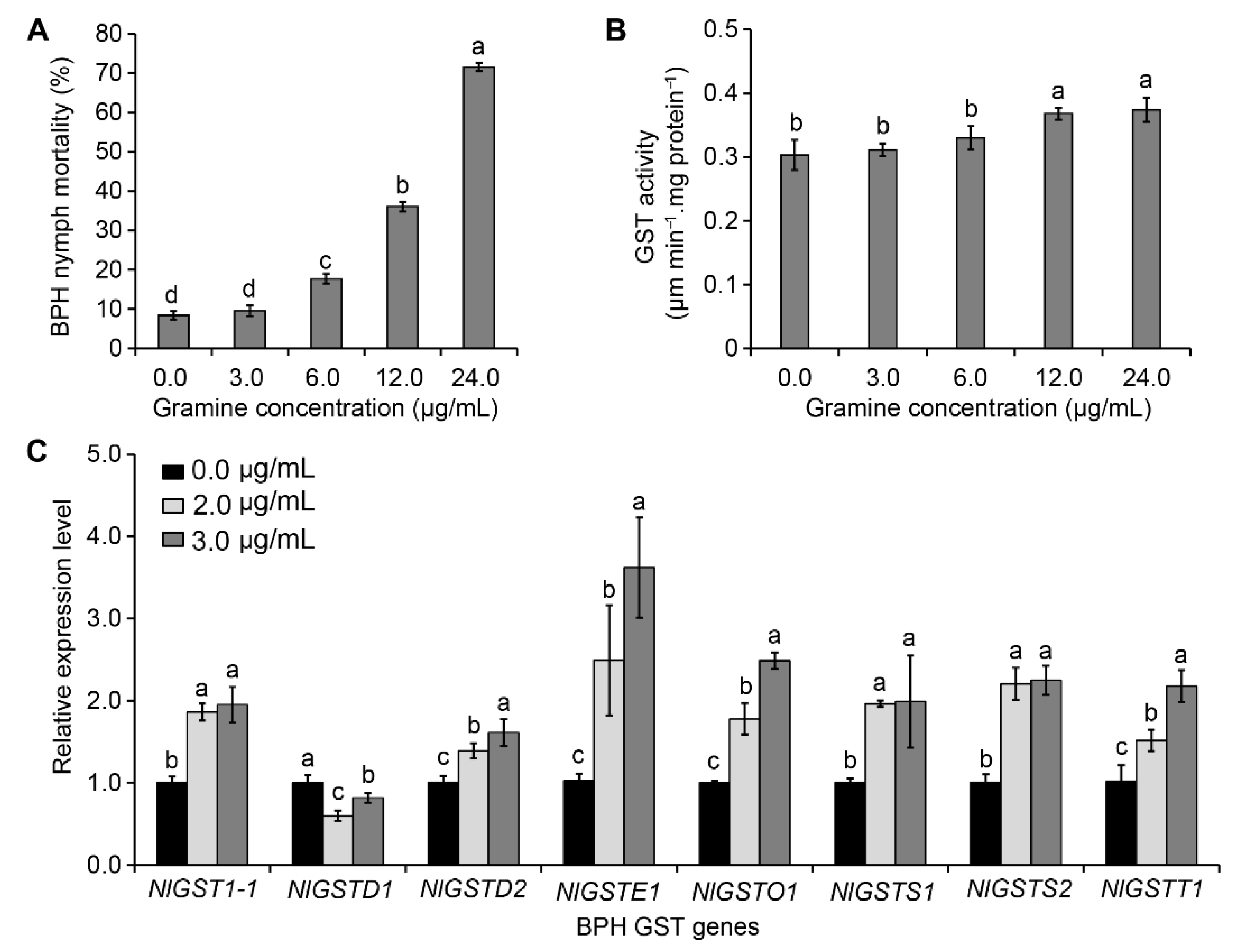
3.1. LC25 and LC50 of Gramine Toxicity to BPH

Bioassay of gramine toxicity to BPH in vitro indicates that increased toxicity to BPH depended on gramine concentrations at 72 h post feeding. The dose-response curves showed that the threshold of gramine observably caused the death of nymphs was 6.0 μg/mL (Figure 1A). Compared with the control, ingestion of 3.0 μg/mL gramine was not sufficient to cause death of BPH. Therefore, this concentration of 3.0 μg/mL gramine was considered as a nonlethal concentration for BPH (Figure 1A). Furthermore, using the Probit scale, we found LC25 and LC50 of gramine to BPH were 7.11 μg/mL (95% confidence interval: 5.07–9.16 μg/mL) and 14.99 μg/mL (95% confidence interval: 11.57–21.88 μg/mL) at 72 h post feeding, respectively (Table 1). When fed with the diets containing gramine, GST activities can be rapidly induced in BPH, despite the induced GST activity of BPH had no significant differences at the low concentration of gramine (3.0 μg/mL) compared with control (Figure 1B). The activation of GST in BPH by gramine was dose-dependent; the threshold dose for significantly activating BPH was 12.0 μg/mL (Figure 1B).

3.2. Effect of Gramine on NlGST Expression in BPH

The nonlethal dosages (≤3.0 μg/mL) of gramine under LC25 were used to verify the impact of gramine on each gene expression in the BPH GST family. Dietary gramine (2.0 and 3.0 μg/mL) significantly induced expression levels of seven GST genes, NlGST1-1, NlGSTD2, NlGSTE1, NlGSTO1, NlGSTS1, NlGSTS2, and NlGSTT1, increasing percentage from 36% to 214% (Figure 1C). Of them, after being fed with 3.0 μg/mL gramine, the expression level of BPH NlGSTE1 was largely increased by 214%, expression levels of others (NlGST1-1, NlGSTO1, NlGSTS1, NlGSTS2, and NlGSTT1) also increased by more than 90% (Figure 1C). However, the transcriptional level of NlGSTD1 was suppressed in the presence of gramine (Figure 1C). The findings indicate that the expression levels of most genes in BPH GST family were significantly raised by gramine, and the sensitivities of these GST genes to gramine showed obvious differences.

3.3. Correlation between Each GST Interference and GST Activity

To determine whether GST transcript levels are correlated with enzymatic activity, we measured the expression level of each GST gene and its GST activity after BPH nymphs ingested dsRNAs of individual genes. As expected, the expression level of each gene was significantly reduced by corresponding gene dsRNA (Figure 2A). When fed the diets containing 0.2, 0.4, and 0.8 μg/mL each GST dsRNA, the transcript levels of NlGST1-1, NlGSTD1, NlGSTD2, NlGSTE1, NlGSTO1, NlGSTT1, NlGSTS1, and NlGSTS2 in BPHs decreased by 2.60–27.60%, 14.60–21.20%, 5.4–21.40%, 27.80–34.20%, −8.40–27.80%, −9.00–28.25%, −9.75–27.50%, and 16.00–23.20%, respectively (Figure 2A). Although dietary dsRNAs caused transcript reduction of all eight GST genes (Figure 2A), only four of them, i.e., NlGST1-1, NlGSTD1, NlGSTD2, and NlGSTE1, significantly suppressed the insect GST activity (Figure 2B). Then, we analyzed the correlation between GST activities and transcript levels of these genes, and found that the expression of three genes (NlGST1-1, NlGSTD2, and NlGSTE1) was positively correlated with their GST activities (Table 2). In addition, each GST dsRNA with higher concentrations (0.4 μg/mL) significantly increased BPH mortality compared to the GFP dsRNA control (Figure 3).

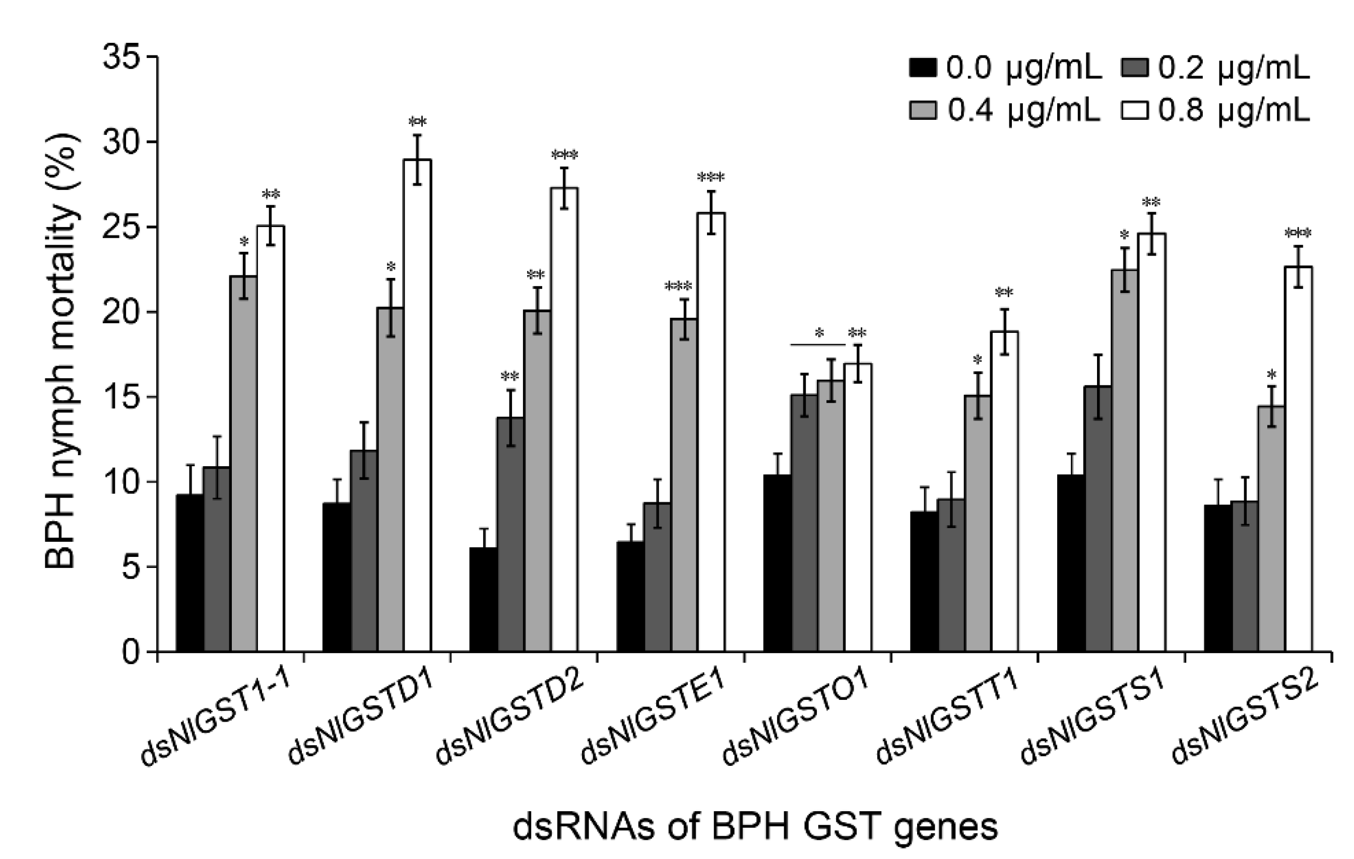
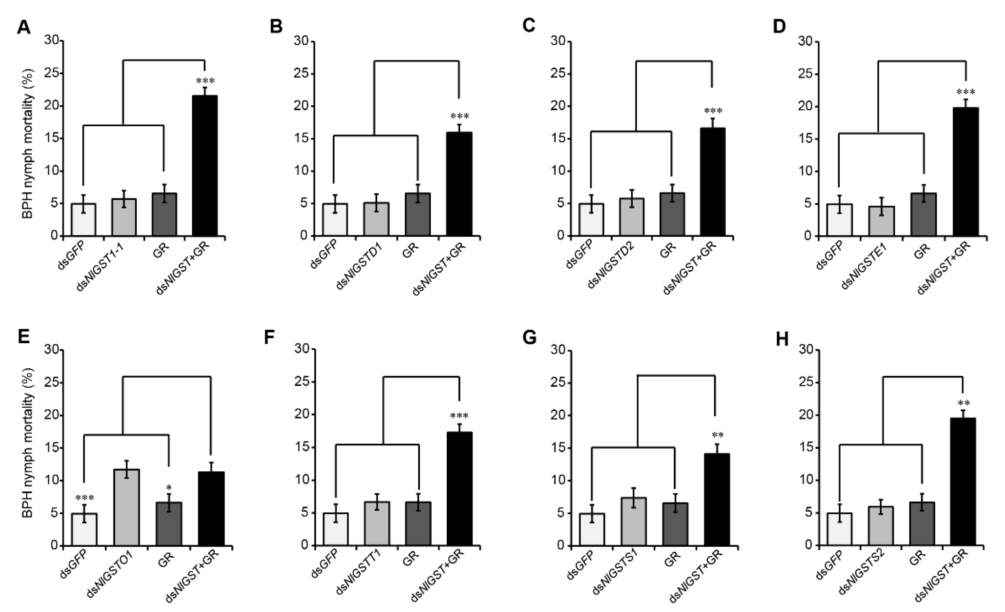
3.4. Suppression of GST Expression Increases BPH Susceptibility to Gramine

First, we also defined the nonlethal doses of GST dsRNAs to BPH, which means the maximum concentration at which GST dsRNAs do not cause significant death for BPH nymphs, compared with the control. There was a difference for the nonlethal doses of each GST dsRNAs to BPH nymphs. Of them, the nonlethal doses of NlGSTD1 and NlGSTS1 dsRNAs were 0.1 μg/mL; NlGST1-1, NlGSTD2, NlGSTS2, NlGSTE1 and NlGSTO1 dsRNAs were 0.2 μg/mL, while only NlGSTT1 dsRNA was 0.4 μg/mL (Figure S2). Next, to assess the potential effect of silencing BPH GST genes on insect detoxification, we recorded mortality of BPH nymphs that fed initially on artificial diets containing the nonlethal doses of each GST dsRNAs for 3 days, and then transferred to the nonlethal doses of gramine-containing diet for an additional 3 days (Figure S1). Similar to the positive control (NlGST1-1), silencing of six other GST genes, namely, NlGSTD1, NlGSTD2, NlGSTE1, NlGSTS1, NlGSTS2, and NlGSTT1, significantly increased the nymph sensitivity to gramine (Figure 4A–D,F–H). The nymph mortality doubled when compared to that of nymphs fed only on gramine (Figure 4A–D,F–H). However, ingestion of NlGSTO1 dsRNA did not increase the sensitivity to gramine for BPH nymphs (Figure 4E), implying that NlGSTO1 may be irrelevant to gramine detoxification in BPH.

4. Discussion

Plant secondary metabolites are important defensive compounds [2,3,7,10]. As a result, herbivorous insects possess a wide range of enzymes by which plant-derived toxicants are often metabolized into nontoxic substances [15]. In this study, we investigated each member in BPH GST family association with GST activity, interacting with phytochemical gramine. Besides NlGST1-1, BPH GST activity was also closely correlated with NlGSTD2 and NlGSTE1 transcript levels; therefore, GST activity in BPH is most likely regulated by a few members in the GST family.
Insects feed on host plants to obtain necessary nutrients, but some toxic compounds from plants can also enter the insect’s body during the feeding process, although their concentrations are low [40,41]. To mitigate the negative impact on their nutritional metabolism, development, and survival, herbivorous insects have evolved counteracting mechanisms such as detoxification, which can be rapidly induced in response to ingestion of toxic compounds, degrading them into nontoxic or less toxic compounds to ensure the normal growth and development of the insects [14,42]. Therefore, blocking detoxification when administrating defensive phytochemicals should be considered as an important strategy for the effective management of key agricultural pests. We have previously demonstrated that rice gramine plays a crucial role in rice resistance to BPH and that the resistance of a rice variety to BPH is highly correlated with its gramine content [9]. We found GST activity of BPH can be induced by the low concentration of gramine in this study, although the previous studies have proved that BPH GST enzyme is an important detoxification enzyme to rice defensive compound—gramine [9,36]. This result is consistent to the previous reports that the GST and carboxylesterase (CarE) activities in S. avenae are induced rapidly by ingestion of gramine and ferulic acid, and that of BPH is induced by ferulic acid [9,11,19]; GST activity of Myzus persicae can be activated by glucosinolates in host plants [42]. The rapid induction of detoxification enzyme activity by toxic compounds is a common strategy of insect adaptation to plant defense. In addition to GSTs, there are other detoxifying enzymes in insects, such as CYP450s and CarEs, which are also dedicated to the detoxification of toxic compounds including plant defensive compounds and pesticides [19,43,44,45,46,47,48,49]; how each enzyme contributes to the detoxification of the same substance still needs further study.
Elevated GST activities by toxic xenobiotics such as phytochemicals and pesticides in herbivores could result from increased transcriptional levels of key genes encoding these proteins [13,17,18]. The insect GST family is divided into six subfamilies: Delta, Epsilon, Sigma, Omega, Theta, and Zeta [50]. In this study, seven GST genes (NlGST1-1, NlGSTD2, NlGSTE1, NlGSTO1, NlGSTS1, NlGSTS2, and NlGSTT1) were induced by low content of gramine. Reduction of NlGST1-1, NlGSTD2, and NlGSTE1 significantly impaired BPH GST activity, implying that the proteins encoded by these genes may be involved in direct detoxification of gramine. Therefore, suppression of the expression of these gramine-inducible GST genes via RNAi approach in BPH increases the sensitivity of the insect to gramine. Interestingly, knockdown of NlGSTS1, NlGSTS2, or NlGSTT1 enhanced BPH sensitivity to gramine, but had no effect on the GST activity. Two reasons may support this result: (1) these particular GSTs may be having activity with other substrates other than the substrate used in this study, because different substrates can cause great differences in the determination of GST activity [42,51]; (2) these genes may be associated with other physiological pathways in BPH. For example, AccGSTS1 in Apis cerana cerana, DmGSTS1-1 in D. melanogaster, and TcGSTS6 in Tribolium castaneum, are involved in the defense of cellular antioxidants [22,52,53]. In Anopheles cracens, AcGSTT1-1 may be a peroxidase that acts as a binding protein for organophosphates to detoxify the insecticides [54]. Of course, we cannot limit our understanding of these GST subfamilies, because it has been reported that the Sigma subfamily of GST is important for insecticide resistance [24,34]. Consistent with the previous findings [27,54], the involvement of NlGSTO1 in BPH metabolism and detoxification of xenobiotics was limited. This is because NlGSTO1 may participate in antioxidant resistance in BPH [55]. Therefore, our results apparently suggest that not all GST genes are involved in the detoxification of gramine in BPH, although GST activity in herbivores is closely related to its detoxification capability against xenobiotics [9,18,20].
With the development of RNAi technology, this method has become one of the effective tools for studying the function of insect genes. However, no matter by feeding or injection, during the RNAi reaction, the dsRNA entered in the insect body is processed to RNA segments 21–23 nucleotides in length to produce an RNAi effect [56,57]. Hence, some sequence’s contiguous match that occurs among different insect species is more common in sibling species and less common in distantly related species [36,58,59]. In the GST family, Delta and Epsilon subfamilies are only found in insects [50]; surprisingly, the GST genes involved in the detoxification of gramine belonged to these two GST subfamilies in BPH. The white-backed planthopper Sogatella furcifera is a sibling species with BPH; the two insects are both important pests of rice, which occur simultaneously in rice, causing serious economic losses. Compared with the sequence similarities of Delta and Epsilon subfamilies of GSTs in BPH and their orthologous genes in S. furcifera, we found they share 89.40% nucleotide sequence similarity between NlGST1-1 and SfGSTD2, 89.55% similarity between NlGSTD2 and SfGSTD2, and 81.28% similarity between NlGSTE1 and SfGSTE1 (Figure S3). The risk assessment of transgenic rice expressing BPH NlGST1-1 dsRNA found that forty-six 21-nt (nucleotide) continuous sequence matches in SfGSTD2 [36]. Thus, we predict that comparable to BPH, the white-backed planthopper SfGSTD2, and even the homologous genes that are highly similar to these two species in other insects, could also involve in detoxicating the phytochemicals to be suitable for host plants. If this speculation is reasonable and feasible, the use of orthologous gene dsRNA in insects at least can be a potential tool for controlling the sibling species of BPH, S. furcifera. Certainly, future studies evaluating the effects of RNAi, especially plant-mediated RNAi on nontarget organisms, are necessary, and this evaluation will give us a holistic view of the impacts of RNAi on these unintended targets [60,61]. Although the current research in this area is very limited, we attempt to provide a new idea for the control of some sibling species of insects. As for the control effect of using orthologous genes in related species, it needs to be further tackled.

5. Conclusions

In summary, we revealed the molecular basis of the BPH GST family genes to the detoxification of gramine, one important rice defensive compound, that NlGSTD2 and NlGSTE1 play an indispensable role in the detoxification of this compound in rice. This research suggests that a few key members of GST family are associated with the tolerance of rice defensive compounds in BPH, providing a new approach for high-efficiency management of the sap-sucking insect population.

Supplementary Materials

The following are available online https://www.mdpi.com/article/10.3390/insects12121055/s1, Figure S1. The process diagram of the effects of silencing NlGSTs on BPH sensitivity to gramine. The process diagram of the effects of silencing NlGSTs on BPH sensitivity to gramine. The second- or third-instar BPH nymphs from rice were introduced into feeding tubes (15 nymphs/tube). Forty-five nymphs distributed into three feeding tubes, were fed with artificial diets (AD) with GST dsRNA (AD + dsRNA), GFP dsRNA (AD + GFP dsRNA), or no dsRNA (AD) for 3 days, and then separately transferred to the new diets with gramine (AD + gramine) and corresponding dsRNA (AD + dsRNA or AD + GFP dsRNA) to continue to feed for additional 3 days as the treatments of “dsRNA + gramine”, “dsRNA positive control”, “gramine control”, and “GFP dsRNA negative control”, respectively. All treatments and controls were replicated 5 times. Final nymph mortality was used for variance analysis to compare significant difference between treatment and control. Figure S2. The mortality of BPH nymphs to GST dsRNAs. Ten second- or third-instar BPH nymphs were fed on artificial diets containing NlGST1-1 (A), NlGSTD1 (B), NlGSTD2 (C), NlGSTE1 (D), NlGSTO1 (E), NlGSTS1 (F), NlGSTS2 (G), and NlGSTT1 (H) dsRNA, respectively, for three days. Concentrations of 0.05, 0.1, 0.2, and 0.4 µg/mL for each GST dsRNA were used in these experiments; 0.4 µg/mL GFP dsRNA was used as control. The mortality of BPH nymphs was recorded and analyzed. Compared with the control, the maximum concentration at which GST dsRNA does not cause significant death for BPH nymphs was considered as a nonlethal dose for BPH GST dsRNA. Data represent the means ± SD from three replicates. Different letters labeled indicate significant differences at p < 0.05. Figure S3. Alignment of Delta and Epsilon subfamilies of GSTs in BPH and their orthologous genes in Sogatella furcifera. NlGST1-1, NlGSTD2, and SfGSTD2 belong to Delta subfamilies of GSTs, and NlGSTE1 and SfGSTE1 are the members of Epsilon subfamilies of GSTs in insects. The homology analysis between NlGST1-1 and SfGSTD2 (A), NlGSTD2 and SfGSTD2 (B), and NlGSTE1 and SfGSTE1 (C) were performed by multiple sequence alignment using the DNAMAN software. Table S1. Primers used in this study.

Author Contributions

Conceptualization, J.Y. and Q.-N.C.; methodology, J.Y., Q.-M.Q., and Q.-N.C.; software, J.Y., X.-D.K., Q.-M.Q., and Q.-N.C.; validation, J.Y., X.-D.K., K.Z.-S., Q.-M.Q., and Q.-N.C.; formal analysis, J.Y., X.-D.K., Q.-M.Q., and Q.-N.C.; investigation, J.Y. and X.-D.K.; resources, J.Y., Q.-M.Q., and Q.-N.C.; data curation, J.Y. and Q.-N.C.; writing—original draft preparation, J.Y. and Q.-N.C.; writing—review and editing, J.Y., X.-D.K., K.Z.-S., Q.-M.Q., and Q.-N.C.; visualization, J.Y., Q.-M.Q., and Q.-N.C.; supervision, Q.-N.C.; project administration, Q.-N.C.; funding acquisition, J.Y. and Q.-N.C. All authors have read and agreed to the published version of the manuscript.

Funding

This research was funded by the Nation Key Research and Development Program of China (2016YFD0300203), and the Chinese Universities Scientific Fund (2015NX001).

Institutional Review Board Statement

Not applicable.

Informed Consent Statement

Not applicable.

Data Availability Statement

All data discussed in the current work are provided in the article and online Supplementary Materials.

Acknowledgments

We are very grateful to Hongxia Hua (Huazhong Agricultural University) for providing original brown planthopper (biotype II).

Conflicts of Interest

The authors declare that they have no competing interest.

References

  1. Chrzanowski, G.; Leszczyński, B.; Czerniewicz, P.; Sytykiewicz, H.; Matok, H.; Krzyżanowski, R.; Sempruch, C. Effect of phenolic acids from black currant, sour cherry and walnut on grain aphid (Sitobion avenae F.) development. Crop Prot. 2012, 35, 71–77. [Google Scholar] [CrossRef]
  2. Corcuera, L.J. Effects of indole alkaloids from gramineae on aphids. Phytochemistry 1984, 23, 539–541. [Google Scholar] [CrossRef]
  3. Zúñiga, G.E.; Salgado, M.S.; Corcuera, L.J. Role of an indole alkaloid in the resistance of barley seedlings to aphids. Phytochemistry 1985, 24, 945–947. [Google Scholar] [CrossRef]
  4. Alvarez, M.A. Plant Secondary Metabolism. In Plant Biotechnology for Health: From Secondary Metabolites to Molecular Farming; Alvarez, M.A., Ed.; Springer: Cham, Switzerland, 2014; pp. 15–31. [Google Scholar]
  5. Jang, J.Y.; Le Dang, Q.; Choi, G.J.; Park, H.W.; Kim, J.C. Control of root-knot nematodes using Waltheria indica producing 4-quinolone alkaloids. Pest Manag. Sci. 2019, 75, 2264–2270. [Google Scholar] [CrossRef]
  6. Cai, Q.N.; Zhang, Q.W.; Zhou, M.Z. Correlation between indole alkaloid content in flag leaves and ears of wheat and its resistance to Sitobion avenae (F.). Plant Prot. 2002, 28, 11–13. [Google Scholar] [CrossRef]
  7. Corcuera, L.J. Biochemical basis for the resistance of barley to aphids. Phytochemistry 1993, 33, 741–747. [Google Scholar] [CrossRef]
  8. Hanson, A.D.; Traynor, P.L.; Ditz, K.M.; Reicosky, D.A. Gramine in barley forage—Effects of genotype and environment. Crop Sci. 1981, 21, 726–730. [Google Scholar] [CrossRef]
  9. Sun, X.-Q.; Zhang, M.-X.; Yu, J.-Y.; Jin, Y.; Ling, B.; Du, J.-P.; Li, G.-H.; Qin, Q.-M.; Cai, Q.-N. Glutathione S-transferase of brown planthoppers (Nilaparvata lugens) is essential for their adaptation to gramine-containing host plants. PLoS ONE 2013, 8, e64026. [Google Scholar] [CrossRef] [PubMed] [Green Version]
  10. Cai, Q.N.; Zhang, Q.W.; Cheo, M. Contribution of indole alkaloids to Sitobion avenae (F.) resistance in wheat. J. Appl. Entomol. 2004, 128, 517–521. [Google Scholar] [CrossRef]
  11. Cai, Q.-N.; Han, Y.; Cao, Y.-Z.; Hu, Y.; Zhao, X.; Bi, J.-L. Detoxification of gramine by the cereal aphid Sitobion avenae. J. Chem. Ecol. 2009, 35, 320–325. [Google Scholar] [CrossRef] [Green Version]
  12. Wang, Y.; Cai, Q.-N.; Zhang, Q.-W.; Han, Y. Effect of the secondary substances from wheat on the growth and digestive physiology of cotton bollworm Helicoverpa armigera (Lepidoptera: Noctuidae). Eur. J. Entomol. 2006, 103, 255–258. [Google Scholar] [CrossRef] [Green Version]
  13. Zhang, M.; Fang, T.; Pu, G.; Sun, X.; Zhou, X.; Cai, Q. Xenobiotic metabolism of plant secondary compounds in the English grain aphid, Sitobion avenae (F.) (Hemiptera: Aphididae). Pestic. Biochem. Physiol. 2013, 107, 44–49. [Google Scholar] [CrossRef]
  14. Zhou, Y.; Han, Z.; Zhang, X.; Pang, C. Influence of rice varieties on the metabolizing enzymes of Nilaparvata lugens Stål. J. Nanjing Agric. Univ. 2003, 26, 24–28. [Google Scholar]
  15. Heidel-Fischer, H.M.; Vogel, H. Molecular mechanisms of insect adaptation to plant secondary compounds. Curr. Opin. Insect Sci. 2015, 8, 8–14. [Google Scholar] [CrossRef]
  16. Zhu-Salzman, K.; Bi, J.L.; Liu, T.X. Molecular strategies of plant defense and insect counter-defense. Insect Sci. 2005, 12, 3–15. [Google Scholar] [CrossRef]
  17. Damásio, J.; Guilhermino, L.; Soares, A.M.; Riva, M.C.; Barata, C. Biochemical mechanisms of resistance in Daphnia magna exposed to the insecticide fenitrothion. Chemosphere 2007, 70, 74–82. [Google Scholar] [CrossRef]
  18. Vontas, J.G.; Small, G.J.; Hemingway, J. Glutathione S-transferases as antioxidant defence agents confer pyrethroid resistance in Nilaparvata lugens. Biochem. J. 2001, 357, 65–72. [Google Scholar] [CrossRef]
  19. Yang, J.; Sun, X.-Q.; Yan, S.-Y.; Pan, W.-J.; Zhang, M.-X.; Cai, Q.-N. Interaction of ferulic acid with glutathione S-transferase and carboxylesterase genes in the brown planthopper, Nilaparvata lugens. J. Chem. Ecol. 2017, 43, 693–702. [Google Scholar] [CrossRef] [PubMed]
  20. Zhang, Y.; Yan, H.; Lu, W.; Li, Y.; Guo, X.; Xu, B. A novel Omega-class glutathione S-transferase gene in Apis cerana cerana: Molecular characterisation of GSTO2 and its protective effects in oxidative stress. Cell Stress Chaperon. 2013, 18, 503–516. [Google Scholar] [CrossRef] [Green Version]
  21. Gloss, A.D.; Vassão, D.G.; Hailey, A.L.; Dittrich, A.C.N.; Schramm, K.; Reichelt, M.; Rast, T.J.; Weichsel, A.; Cravens, M.G.; Gershenzon, J.; et al. Evolution in an ancient detoxification pathway is coupled with a transition to herbivory in the Drosophilidae. Mol. Biol. Evol. 2014, 31, 2441–2456. [Google Scholar] [CrossRef] [Green Version]
  22. Singh, S.P.; Coronella, J.A.; Benes, H.; Cochrane, B.J.; Zimniak, P. Catalytic function of Drosophila melanogaster glutathione S-transferase DmGSTS1-1 (GST-2) in conjugation of lipid peroxidation end products. Eur. J. Biochem. 2001, 268, 2912–2923. [Google Scholar] [CrossRef]
  23. Vontas, J.G.; Small, G.J.; Nikou, D.C.; Ranson, H.; Hemingway, J. Purification, molecular cloning and heterologous expression of a glutathione S-transferase involved in insecticide resistance from the rice brown planthopper, Nilaparvata lugens. Biochem. J. 2002, 362, 329–337. [Google Scholar] [CrossRef] [PubMed]
  24. Zhou, W.-W.; Li, X.-W.; Quan, Y.-H.; Cheng, J.; Zhang, C.-X.; Gurr, G.; Zhu, Z.-R. Identification and expression profiles of nine glutathione S-transferase genes from the important rice phloem sap-sucker and virus vector Laodelphax striatellus (Fallen) (Hemiptera: Delphacidae). Pest Manag. Sci. 2012, 68, 1296–1305. [Google Scholar] [CrossRef] [PubMed]
  25. Lin, Y.H.; Lee, C.M.; Huang, J.H.; Lee, H.J. Circadian regulation of permethrin susceptibility by glutathione S-transferase (BgGSTD1) in the German cockroach (Blattella germanica). J. Insect Physiol. 2014, 65, 45–50. [Google Scholar] [CrossRef]
  26. Mazari, A.M.; Dahlberg, O.; Mannervik, B.; Mannervik, M. Overexpression of glutathione transferase E7 in Drosophila differentially impacts toxicity of organic isothiocyanates in males and females. PLoS ONE 2014, 9, e110103. [Google Scholar] [CrossRef]
  27. Zhou, W.-W.; Liang, Q.-M.; Xu, Y.; Gurr, G.M.; Bao, Y.-Y.; Zhou, X.-P.; Zhang, C.-X.; Cheng, J.; Zhu, Z.-R. Genomic insights into the glutathione S-transferase gene family of two rice planthoppers, Nilaparvata lugens (Stål) and Sogatella furcifera (Horváth) (Hemiptera: Delphacidae). PLoS ONE 2013, 8, e56604. [Google Scholar] [CrossRef] [Green Version]
  28. Ghaffar, M.B.A.; Pritchard, J.; Ford-Lloyd, B. Brown planthopper (Nilaparvata lugens Stal) feeding behavior on rice germplasm as an indicator of resistance. PLoS ONE 2011, 6, e22137. [Google Scholar] [CrossRef] [PubMed]
  29. Singh, A.; Jaswal, A.; Sarkar, S. Incidence of brown planthopper in the rice field with the use of different doses of fertilizers. Agric. Sci. Digest. 2019, 39, 108–113. [Google Scholar] [CrossRef] [Green Version]
  30. Du, B.; Zhang, W.; Liu, B.; Hu, J.; Wei, Z.; Shi, Z.; He, R.; Zhu, L.; Chen, R.; Han, B.; et al. Identification and characterization of Bph14, a gene conferring resistance to brown planthopper in rice. Proc. Natl. Acad. Sci. USA. 2009, 106, 22163–22168. [Google Scholar] [CrossRef] [Green Version]
  31. Jairin, J.; Phengrat, K.; Teangdeerith, S.; Vanavichit, A.; Toojinda, T. Mapping of a broad-spectrum brown planthopper resistance gene, Bph3, on rice chromosome 6. Mol. Breed. 2007, 19, 35–44. [Google Scholar] [CrossRef]
  32. Jena, K.K.; Kim, S.M. Current status of brown planthopper (BPH) resistance and genetics. Rice 2010, 3, 161–171. [Google Scholar] [CrossRef] [Green Version]
  33. Bao, Y.-Y.; Wang, Y.; Wu, W.-J.; Zhao, D.; Xue, J.; Zhang, B.-Q.; Shen, Z.-C.; Zhang, C.-X. De novo intestine-specific transcriptome of the brown planthopper Nilaparvata lugens revealed potential functions in digestion, detoxification and immune response. Genomics 2012, 99, 256–264. [Google Scholar] [CrossRef] [PubMed] [Green Version]
  34. Yang, B.; Lin, X.; Yu, N.; Gao, H.; Zhang, Y.; Liu, W.; Liu, Z. Contribution of glutathione S-transferases to imidacloprid resistance in Nilaparvata lugens. J. Agric. Food Chem. 2020, 68, 15403–15408. [Google Scholar] [CrossRef] [PubMed]
  35. Powell, K.S.; Gatehouse, A.M.R.; Hilder, V.A.; Gatehouse, J.A. Antimetabolic effects of plant lectins and plant and fungal enzymes on the nymphal stages of two important rice pests, Nilaparvata lugens and Nephotettix cinciteps. Entomol. Exp. Appl. 1993, 66, 119–126. [Google Scholar] [CrossRef]
  36. Yang, J.; Sun, X.-Q.; Zhu-Salzman, K.; Qin, Q.-M.; Feng, H.-Q.; Kong, X.-D.; Zhou, X.-G.; Cai, Q.-N. Host-induced gene silencing of brown planthopper glutathione S-transferase gene enhances rice resistance to sap-sucking insect pests. J. Pest Sci. 2021, 94, 769–781. [Google Scholar] [CrossRef]
  37. Franciosa, H.; Berge, J.B. Glutathione S-transferases in housefly (Musca domestica): Location of GST-1 and GST-2 families. Insect Biochem. Mol. Biol. 1995, 25, 311–317. [Google Scholar] [CrossRef]
  38. Bradford, M.M. A rapid and sensitive method for the quantitation of microgram quantities of protein utilizing the principle of protein-dye binding. Anal. Biochem. 1976, 72, 248–254. [Google Scholar] [CrossRef]
  39. Livak, K.J.; Schmittgen, T.D. Analysis of relative gene expression data using real-time quantitative PCR and the 2(-Delta Delta C(T)) Method. Methods 2001, 25, 402–408. [Google Scholar] [CrossRef]
  40. Hayashi, H.; Chino, M. Chemical composition of phloem sap from the uppermost internode of the rice plant. Plant Cell Physiol. 1990, 31, 247–251. [Google Scholar] [CrossRef] [Green Version]
  41. Susumu, K.; Tomoyoshi, F.; Mitsuo, C. Collection of rice phloem sap from stylets of homopterous insects severed by YAG laser. Plant Cell Physiol. 1980, 21, 1319–1327. [Google Scholar] [CrossRef]
  42. Francis, F.; Vanhaelen, N.; Haubruge, E. Glutathione S-transferases in the adaptation to plant secondary metabolites in the Myzus persicae aphid. Arch. Insect Biochem. Physiol. 2005, 58, 166–174. [Google Scholar] [CrossRef]
  43. Bass, C.; Puinean, A.M.; Andrews, M.; Cutler, P.; Daniels, M.; Eliáš, J.; Paul, V.L.; Crossthwaite, A.J.; Denholm, I.; Field, L.M.; et al. Mutation of a nicotinic acetylcholine receptor beta subunit is associated with resistance to neonicotinoid insecticides in the aphid Myzus persicae. BMC Neurosci. 2011, 12, 51. [Google Scholar] [CrossRef] [Green Version]
  44. Ding, Z.; Wen, Y.; Yang, B.; Zhang, Y.; Liu, S.; Liu, Z.; Han, Z. Biochemical mechanisms of imidacloprid resistance in Nilaparvata lugens: Over-expression of cytochrome P450 CYP6AY1. Insect Biochem. Mol. Biol. 2013, 43, 1021–1027. [Google Scholar] [CrossRef]
  45. Gong, Y.; Shi, X.; Desneux, N.; Gao, X. Effects of spirotetramat treatments on fecundity and carboxylesterase expression of Aphis gossypii Glover. Ecotoxicology 2016, 25, 655–663. [Google Scholar] [CrossRef]
  46. Pang, R.; Li, Y.; Dong, Y.; Liang, Z.; Zhang, Y.; Zhang, W. Identification of promoter polymorphisms in the cytochrome P450 CYP6AY1 linked with insecticide resistance in the brown planthopper, Nilaparvata lugens. Insect Mol. Biol. 2014, 23, 768–778. [Google Scholar] [CrossRef] [PubMed]
  47. Yang, Z.; Zhang, Y.; Liu, X.; Wang, X. Two novel cytochrome P450 genes CYP6CS1 and CYP6CW1 from Nilaparvata lugens (Hemiptera: Delphacidae): cDNA cloning and induction by host resistant rice. Bull. Entomol. Res. 2011, 101, 73–80. [Google Scholar] [CrossRef]
  48. Zhang, J.; Ge, P.; Li, D.; Guo, Y.; Zhu, K.Y.; Ma, E.; Zhang, J. Two homologous carboxylesterase genes from Locusta migratoria with different tissue expression patterns and roles in insecticide detoxification. J. Insect Physiol. 2015, 77, 1–8. [Google Scholar] [CrossRef] [PubMed]
  49. Yang, Y.X.; Yu, N.; Zhang, J.H.; Zhang, Y.X.; Liu, Z.W. Induction of P450 genes in Nilaparvata lugens and Sogatella furcifera by two neonicotinoid insecticides. Insect Sci. 2018, 25, 401–408. [Google Scholar] [CrossRef]
  50. Friedman, R. Genomic organization of the glutathione S-transferase family in insects. Mol. Phylogenet. Evol. 2011, 61, 924–932. [Google Scholar] [CrossRef] [PubMed]
  51. Pavlidi, N.; Khalighi, M.; Myridakis, A.; Dermauw, W.; Wybouw, N.; Tsakireli, D.; Stephanou, E.G.; Labrou, N.E.; Vontas, J.; Van Leeuwen, T. A glutathione-S-transferase (TuGSTd05) associated with acaricide resistance in Tetranychus urticae directly metabolizes the complex II inhibitor cyflumetofen. Insect Biochem. Mol. Biol. 2017, 80, 101–115. [Google Scholar] [CrossRef]
  52. Song, X.-W.; Zhong, Q.-S.; Ji, Y.-H.; Zhang, Y.-M.; Tang, J.; Feng, F.; Bi, J.-X.; Xie, J.; Li, B. Characterization of a sigma class GST (GSTS6) required for cellular detoxification and embryogenesis in Tribolium castaneum. Insect Sci. 2021, 1–15. [Google Scholar] [CrossRef] [PubMed]
  53. Yan, H.; Jia, H.; Gao, H.; Guo, X.; Xu, B. Identification, genomic organization, and oxidative stress response of a sigma class glutathione S-transferase gene (AccGSTS1) in the honey bee, Apis cerana cerana. Cell Stress Chaperon. 2013, 18, 415–426. [Google Scholar] [CrossRef] [PubMed] [Green Version]
  54. Wongtrakul, J.; Pongjaroenkit, S.; Leelapat, P.; Nachaiwieng, W.; Prapanthadara, L.A.; Ketterman, A.J. Expression and characterization of three new glutathione transferases, an Epsilon (AcGSTE2-2), Omega (AcGSTO1-1), and Theta (AcGSTT1-1) from Anopheles cracens (Diptera: Culicidae), a major Thai Malaria vector. J. Med. Entomol. 2010, 47, 162–171. [Google Scholar] [CrossRef] [PubMed]
  55. Saruta, F.; Yamada, N.; Yamamoto, K. An omega-class glutathione S-transferase in the brown planthopper Nilaparvata lugens exhibits glutathione transferase and dehydroascorbate reductase activities. Arch. Insect Biochem. Physiol. 2019, 102, e21599. [Google Scholar] [CrossRef]
  56. Preall, J.B.; He, Z.; Gorra, J.M.; Sontheimer, E.J. Short interfering RNA strand selection is independent of dsRNA processing polarity during RNAi in Drosophila. Cur. Biol. 2006, 16, 530–535. [Google Scholar] [CrossRef] [Green Version]
  57. Zamore, P.D.; Tuschl, T.; Sharp, P.A.; Bartel, D.P. RNAi: Double-stranded RNA directs the ATP-dependent cleavage of mRNA at 21 to 23 nucleotide intervals. Cell 2000, 101, 25–33. [Google Scholar] [CrossRef] [Green Version]
  58. Pan, H.; Xu, L.; Noland, J.E.; Li, H.; Siegfried, B.D.; Zhou, X. Assessment of potential risks of dietary rnai to a soil micro-arthropod, Sinella curviseta Brook (Collembola: Entomobryidae). Front. Plant Sci. 2016, 7, 1028. [Google Scholar] [CrossRef] [Green Version]
  59. Pan, H.; Yang, X.; Bidne, K.; Hellmich, R.L.; Siegfried, B.D.; Zhou, X. Dietary risk assessment of v-ATPase A dsRNAs on monarch butterfly larvae. Front. Plant Sci. 2017, 8, 242. [Google Scholar] [CrossRef] [Green Version]
  60. Casacuberta, J.M.; Devos, Y.; du Jardin, P.; Ramon, M.; Vaucheret, H.; Nogué, F. Biotechnological uses of RNAi in plants: Risk assessment considerations. Trends Biotechnol. 2015, 33, 145–147. [Google Scholar] [CrossRef] [Green Version]
  61. Devos, Y.; Álvarez-Alfageme, F.; Gennaro, A.; Mestdagh, S. Assessment of unanticipated unintended effects of genetically modified plants on non-target organisms: A controversy worthy of pursuit? J. Appl. Entomol. 2016, 140, 1–10. [Google Scholar] [CrossRef]
Figure 1. Gramine toxicity to BPH and effect of gramine on BPH nymph GST gene expression levels and GST activity. (A) Nymph mortality of BPH to gramine. (B) GST activity of BPH nymphs fed with different concentrations of gramine. (C) The expression level of GST gene in BPH nymphs after feeding on diets containing a nonlethal dose of gramine. Data are presented as mean ± standard error of mean (SEM) from 4 replicates. Different letters labeled indicate significant differences at p < 0.05.
Insects 12 01055 g001
Figure 2. Effect of GST dsRNAs on each GST gene transcriptional level and GST activity. The second- or third-instar BPH nymphs were fed with artificial diets supplemented with 0 (containing 0.4 μg/mL GFP dsRNA), 0.2, 0.4, and 0.8 μg/mL of the indicated dsRNA for 3 days. The transcriptional level of target gene (A) and GST activity (B) in the nymphs were analyzed. The label “ds + gene” represents the dsRNA of the corresponding GST gene. Data represent the means ± SEM from 4 replicates. Different letters labeled indicate significant differences at p < 0.05.
Insects 12 01055 g002
Figure 3. Effect of GST dsRNAs on BPH nymph mortality. The second- or third-instar BPH nymphs were fed with artificial diets containing 0.2, 0.4, or 0.8 ng/µL of each GST dsRNA or 0.4 ng/µL GFP dsRNA (control, without 0.0 ng/µL target gene dsRNA). The nymph mortalities in all the treatments and control were statistically analyzed. The label “ds + gene” represents dsRNA of the corresponding gene. Data represent the means ± SEM from 4 replicates; *, **, *** indicate significance at p < 0.05, 0.01, and 0.001, respectively.
Insects 12 01055 g003
Figure 4. Sensitivity of dsRNA prefed BPH nymphs to gramine-containing diets. BPH nymphs were first fed with the dsRNA-containing diets for 3 days, and then, transferred to new diets with gramine (nonlethal dose) for an additional 3 days, the mortality of the nymphs was used to evaluate the combinative effects of gramine and the dsRNA of NlGST1-1 (a positive control) (A), NlGSTD1 (B), NlGSTD2 (C), NlGSTE1 (D), NlGSTO1 (E), NlGSTT1 (F), NlGSTS1 (G), or NlGSTS2 (H). The label “ds + gene” represents the corresponding dsRNA of each gene; dsGFP: GFP dsRNA was used as a control. GR: gramine. Data represent the means ± SEM from 5 replicates. *, **, *** indicate significance at p < 0.05, 0.01, and 0.001, respectively.
Insects 12 01055 g004
Table 1. LC25 and LC50 of gramine against BPH.
nLC25 (μg/mL) aLC50 (μg/mL) aSlope ± SE b2) cp
1867.11 (5.07–9.16)14.99 (11.57–21.88)2.084 ± 0.372.710.987
a lethal concentration causing 25% and 50% mortality after 3 d with 95% confidence limits; b slope ± standard error of the concentration–mortality regression line; c chi square.
Table 2. Correlation of GST gene transcription level with GST activity in BPH.
GST Transcript-GST ActivitynEquationrpSignificance
NlGST1-1-GSTs 4y = 1.6649x − 0.78650.9719 0.0141 *
NlGSTD1-GSTs4y = 0.3453x + 0.53470.7612 0.1194 ns
NlGSTD2-GSTs4y = 1.1624x − 0.17540.9839 0.0080 **
NlGSTE1-GSTs4y = 0.6451x + 0.22130.9541 0.0230 *
NlGSTO1-GSTs4y = 0.2159x + 0.92050.5605 0.2197 ns
NlGSTS1-GSTs4y = 0.1235x + 0.99280.4532 0.2734 ns
NlGSTS2-GSTs4y = 0.2489x + 0.8809 0.8772 0.0614 ns
NlGSTT1-GSTs4y = 0.3803x + 0.7793 0.8408 0.0796 ns
*, **: significance at p < 0.05 and 0.01, respectively; ns: p > 0.05. A bivariate Spearman’s nonparametric rank correlation test was used in the correlation analysis.
Publisher’s Note: MDPI stays neutral with regard to jurisdictional claims in published maps and institutional affiliations.

