Article

Resolving the Taxonomic Status of Potential Biocontrol Agents Belonging to the Neglected Genus Lipolexis Förster (Hymenoptera, Braconidae, Aphidiinae) with Descriptions of Six New Species †

by
Korana Kocić
1,
Andjeljko Petrović
1,
Jelisaveta Čkrkić
1,
Nickolas G. Kavallieratos
2,*,
Ehsan Rakhshani
3,
Judit Arnó
4,
Yahana Aparicio
4,
Paul D. N. Hebert
5 and
Željko Tomanović
1
1
Institute of Zoology, Faculty of Biology, University of Belgrade, Studentski trg 16, 11000 Belgrade, Serbia
2
Laboratory of Agricultural Zoology and Entomology, Department of Crop Science, Agricultural University of Athens, 75 Iera Odos str., 11885 Athens, Greece
3
Department of Plant Protection, College of Agriculture, University of Zabol, Zabol 98615-538, Iran
4
IRTA Cabrils, 08348 Cabrils, Spain
5
Centre for Biodiversity Genomics, Biodiversity Institute of Ontario, University of Guelph, 50 Stone Road, East Guelph, ON N1G 2W1, Canada
*
Author to whom correspondence should be addressed.
http://zoobank.org/53AED1C9-A652-414A-9F09-B333FD6F6567.
Insects 2020, 11(10), 667; https://doi.org/10.3390/insects11100667
Submission received: 3 September 2020 / Revised: 23 September 2020 / Accepted: 23 September 2020 / Published: 29 September 2020
(This article belongs to the Collection Biology and Management of Sap-Sucking Pests)

Abstract

:

Simple Summary

Lipolexis is small but widely distributed genus across Europe and Asia. Nevertheless, its taxonomic distinctiveness was subsequently questioned by some authors who considered it as a synonym of the genus Diaeretus. Although Lipolexis is widely distributed and one species (Lipolexis oregmae Gahan) is an important biological control agent, the last taxonomic study on it was conducted more than 50 years ago. Our study employs an integrative approach (morphology and molecular analysis (COI barcode region)), to examine Lipolexis specimens that were sampled worldwide, including specimens from BOLD database. It led to the description of six new species. Each of the new species possesses clear morphological characters that distinguishes it from its congeners. Our findings suggest that two groups can be differentiated within the genus—oregmae and gracilis. Furthermore, we present a key for the identification to all known Lipolexis species of the world.

Abstract

Lipolexis is a small genus in the subfamily Aphidiinae represented by one species in Europe (Lipolexis gracilis Förster) and by four in Asia (Lipolexis wuyiensis Chen, L. oregmae Gahan, L. myzakkaiae Pramanik and Raychaudhuri and L. pseudoscutellaris Pramanik and Raychaudhuri). Although L. oregmae is employed in biological control programs against pest aphids, the last morphological study on the genus was completed over 50 years ago. This study employs an integrative approach (morphology and molecular analysis (COI barcode region)), to examine Lipolexis specimens that were sampled worldwide, including specimens from BOLD database. These results establish that two currently recognized species of Lipolexis (L. gracilis, L. oregmae) are actually a species complex and also reveal phylogenetic relationships within the genus. Six new species are described and a global key for the identification of Lipolexis species is provided.

1. Introduction

Lipolexis, a small genus in the subfamily Aphidiinae, is widely distributed across Europe and Asia. Like all members of this subfamily, species of Lipolexis are solitary koinobiont endoparasitoids of aphids (Aphididae). Förster (1862) [1] described it as monotypic with Lipolexis gracilis as the type species. However, its taxonomic distinctiveness was subsequently questioned by some authors who considered it as a synonym of the genus Diaeretus [2,3]. Starý [3] re-established Lipolexis on the basis of type material and specimens collected in former Czechoslovakia. A second member of this genus, Lipolexis oregmae Gahan, was first described as Diaeretus oregmae by Gahan [4]. However, in his revision of generic boundaries in the family Aphidiidae (now subfamily Aphidiinae), Starý [5] stated that, aside from its type species, the genus Diaeretus was monotypic and other taxa should be transferred to other genera. Consequently, he reassigned D. oregmae to Lipolexis [6]. Two years later, Mackauer [7] described the third species, Lipolexis scutellaris Mackauer, from the Oriental region. However, after re-examining L. oregmae and L. scutellaris, Starý concluded that L. scutellaris was a junior synonym of L. oregmae [8]. Since this time, additional species of Lipolexis have been reported. Lipolexis chinensis Chen was described by Chen [9], but three years later Starý and Ghosh [10] placed it as a junior synonym of L. gracilis. However, three more species were subsequently described from the Oriental region: Lipolexis wuyiensis Chen [11] from China, and both Lipolexis myzakkaiae Pramanik and Raychaudhuri and Lipolexis pseudoscutellaris Pramanik and Raychaudhuri [12] from India. Although these three species are still regarded as valid, their descriptions leave doubt. Moreover, it seems that neither species has been mentioned in the scientific literature since their description. Lipolexis gracilis has a Palaearctic distribution and it is the only Lipolexis currently thought to be widely distributed in Europe [13,14,15]. By contrast, L. oregmae, an Oriental species, has been introduced to USA (Florida) [16], Jamaica [17], Dominica [18] and Costa Rica [19]. While the potential value of L. gracilis in biocontrol of black bean aphid, Aphis fabae Scopoli [20], and soybean aphid, Aphis glycines Matsumura [21] has been considered, L. oregmae has proven effective against the brown citrus aphid, Toxoptera citricida (Kirkaldy). The latter aphid causes serious damage to citrus plantations both by transmitting the tristeza closterovirus [22] and by direct damage to the plant. Lipolexis oregmae has been successfully introduced to Florida from Guam in a biological control program directed against this aphid pest [16]. Hoy et al. [17] reported the unintentional establishment of L. oregmae in Jamaica, where the invasion of T. citricida has led to serious losses in citrus production. Furthermore, Cocco et al. [18] and Zamora et al. [19] reported the same situation for Dominica and Costa Rica, respectively.
As Mackauer [23] considered Lipolexis as phylogenetically close to the genera Trioxys and Binodoxys, he placed it within the subtribe Trioxina. This subtribe is classified by different authors within tribe Trioxini [24] or tribe Aphidiini [23]. However, morphological examination of its immature stages led Finlayson [25] to recognize that larvae of Lipolexis possess an epistoma, a unique character for Aphidiinae genera. Based on this observation, she suggested that Lipolexis should be placed in a separate subtribe despite the morphological similarity of the adult to those of Trioxina species.
Although the genus Lipolexis is widely distributed and one species is an important biological control agent, the last taxonomic study on it was conducted more than 50 years ago by Starý [3]. Several molecular studies on Aphidiinae included representatives of this genus, but conclusions concerning the position of Lipolexis were ambiguous. Based on phylogenetic analysis of the mitochondrial NADH 1 dehydrogenase gene using neighbor joining analysis, Smith et al. [26] positioned L. gracilis as a sister group to the rest of Aphidiina and Trioxina species. In the same study, both weighted and unweighted parsimony analysis positioned L. gracilis within the tribe Aphidiini. Furthermore, in the weighted parsimony analysis of the 16S RNA sequence conducted by Kambhampati et al. [27], L. gracilis was placed as a sister group to the genera Binodoxys and Trioxys with high bootstrap value supporting placement of Lipolexis within the subtribe Trioxina. However, the same study placed L. gracilis with the subtribe Aphidiina with low bootstrap support in the unweighted parsimony analysis. The results of phylogenetic analysis of 18S RNA region [28] positioned L. gracilis as a sister group to the genus Trioxys, supporting Mackauers’ [23] classification. The study of Shi and Chen [29] based on three genes (16S RNA, 18S RNA, ATPase 6) also supports the traditional classification of Lipolexis within the subtribe Trioxina. Furthermore, analysis of the barcode region of cytochrome c oxidase subunit I (COI) yielded a phylogenetic tree with L. gracilis grouped as a sister taxon to Falciconus pseudoplatani Marshall within species in the subtribes Trioxina and Monoctonina [30]. Based on these results, it is evident that the position of Lipolexis within Aphidiinae remains uncertain and merits further research.
Our exhaustive studies on populations of L. gracilis across Europe and L. oregmae in the far East and USA (Florida) revealed considerable variation in taxonomically important morphological characters, such as the number of maxillary and labial palpomeres, as well as in the shape of flagellomere 1 (F1) and petiole. Motivated by these observations, both morphology and molecular analysis (COI barcode region) were used to re-examine specimens of L. gracilis and L. oregmae from various sites across their distributions. Our research sought to ascertain if L. gracilis and L. oregmae include cryptic species and to reconstruct phylogenetic relationships within the genus. It led to recognition of six new species and a key for the identification to all known Lipolexis species.

2. Materials and Methods

2.1. Sample Collection

Specimens were collected over 12 years at sites throughout Europe (Serbia, Montenegro, Croatia, Belgium, Slovenia, Bosnia and Herzegovina, Greece, Spain and Czech Republic), Middle East (Turkey, Iran, Pakistan), in the Far East (Bangladesh, China) and in USA (Florida) (Supplementary Materials Table S1). Parasitoids were collected by net sweeping and Malaise traps by the Barcode of Life initiative or by sampling parts of the plant infested with aphid colonies. In the latter case, plants with aphid colonies were placed in plastic containers covered by nylon tulles to allow ventilation, and transported to the laboratory where they were held under controlled conditions (22.5 °C, 65% relative humidity, 16 h light/8 h dark). Upon emergence, parasitoids were preserved in 96% ethanol. To establish tri-trophic associations, aphid and plant samples were identified to a species or genus level. Parasitoid specimens were examined under the ZEISS Discovery V8 (Carl Zeiss MicroImaging GmbH, Göttingen, Germany) or Olympus SZX9 (Olympus, Tokio, Japan) stereomicroscopes before being dissected and slide mounted. Slides were photographed with a Leica DM LS phase contrast microscope (Leica Microsystems GmbH, Wetzlar, Germany), while relevant measurements were made using the ImageJ [31] software. Terminology of morphological characters follows Sharkey and Wharton [32]. The parasitoids examined in this study are deposited in the collection at the Institute of Zoology, Faculty of Biology, University of Belgrade (FBUB), in the Croatian Natural History Museum, Zagreb, Croatia, and in the Canadian National Collection of Insects, Arachnids, and Nematodes, Ottawa (CNC). Specimen collector abbreviations are following: ŽT (Željko Tomanović), KK (Korana Kocić), KKos (Katarina Kos), NK (Nickolas Kavallieratos), YA (Yahana Aparicio), VŽ (Vladimir Žikić), AP (Andjeljko Petrović), Jelisaveta Čkrkić (JČ), Petr Starý (PS) and MK (Marina Kaiser).

2.2. Molecular Analysis

Genomic DNA was extracted nondestructively with the QIAGEN Dneasy® Blood & Tissue Kit (Qiagen Inc., Valencia, CA, USA) from 23 specimens, following the protocol provided by the manufacturer. Universal primers LCO1490 and HCO2891 were used for amplification of the barcode COI region [33]. The final volume of the amplification mixture was 20 µL, which contained 11.8 µL of nuclease-free water, 0.2 µL of Taq Polymerase, 1 µL of primers (each), 1.2 µL of nucleotides, 1.8 µL of MgCl2, 2 µL of buffer and finally 1 µL of the extracted DNA. The following PCR temperature and cycle program was used: 5 min of initial denaturation, 35 cycles of 60 s denaturation (94 °C), 60 s annealing (54 °C) and 90 s extension (72 °C) and 7 min of final extension (72 °C). Amplification products were purified with QIAquick PCR purification Kit (Qiagen Inc., Valencia, CA, USA) and sequenced by Macrogen Inc. (Seoul, Korea). After DNA extraction, specimens were examined morphologically.

2.3. Phylogenetic Analysis

Electropherograms were visualized in Finch TV Geospiza Inc. (Seattle, WA, USA) and manually edited and aligned using BioEdit [34] software. The analysis of evolutionary divergence was conducted in MEGA 6 [35] software using the Kimura 2-parameter distance model. The Tamura 3-parameter model [36] with gamma distribution and invariate sites (T92+G+I) was indicated by MEGA 6 as the best fitting model test. Bayesian evolutionary analysis was performed with BEAST 2.5 [37] software employing the initial data set constructed in BEAUti v1.10.4 [37] with designated strict clock type and Yule process of speciation. The analysis ran for 10 million generations, the sampling was conducted every 1000 generations, while the first million trees were discarded as a burn in. The effective sample size (ESS) of the parameters of the Markov chain Monte Carlo was estimated by Tracer v1.7.1 [38]. The saturation level for the third codon position was inspected in DAMBE software [39] using Xia model test [40]. The phylogenetic tree was visualized using FigTree 1.4.3 software [41]. Maximum likelihood (ML) and Neighbor Joining (NJ) phylogenetic trees were constructed in MEGA 6 (Tamura 3-parameter model, 2000 replicates, total of 683 positions in the final dataset). In all analyses, the outgroup species was Venturia canescens (Gravenhorst). Haplotype diversity was estimated by the software DNAsp [42] and haplotype networks were constructed by Network (version 5.0.0.1). The final dataset contained 64 COI sequences (one outgroup sequence, additional 40 sequences acquired from BOLD systems database (25) and GenBank (15), and 23 newly recovered sequences).

3. Results

3.1. Molecular Analysis

COI sequences were acquired from 63 specimens of Lipolexis, five assigned to L. oregmae and 58 to L. gracilis (Table S1). All three phylogenetic reconstruction methods (Bayesian, ML, NJ) yielded a tree with similar topology (Figure 1). The sequences fell into two main lineages (Table 1) showing deep sequence divergences (22.7–23.2%); the first group included all 58 individuals identified as L. gracilis (“gracilis” group) while the second included the five specimens of L. oregmae (“oregmae” group) (Figure 1).
The five sequences initially assigned to L. oregmae were separated into two distinct clades with an average genetic distance of 19.9%. A haplotype network supported the presence of two species (L. oregmae, Lipolexis bengalensis sp. n) with high sequence divergence (77 mutational steps) at COI (Figure 2).
The 58 sequences recovered from members of L. gracilis group included 26 haplotypes that fell into two main groups (“group 1” and “group 2”) (Figure 1) with the average sequence divergence of 11.4%. The first group included European specimens which were separated into two clusters with an average COI distance of 7.3% (L. gracilis s.s., Lipolexis labialis sp. n.). The second group included three clades: the first represented by a single specimen from China (Lipolexis takadai sp. n.) with an average genetic distance of 4.5% and 7.2%, from the other two clades. The second clade (Lipolexis pelopsi sp. n.) included six individuals from the Mediterranean, while the third included specimens from the Middle East, the Orient, and Spain. The average evolutionary divergence between the Mediterranean and the third clade was 8%. The Middle East and Oriental region clade, which also contained several specimens from Spain, was further separated into two clades (Lipolexis pakistanicus sp. n. and Lipolexis peregrinus sp. n.) with mean distance of 2.2%. The 26 haplotypes of L. gracilis s.l. (Figure 3) were separated into six distinct groups that corresponded to the named clades shown in the phylogenetic tree.
Morphological analysis of the Lipolexis specimens in each clade revealed diagnostic differences supporting the recognition of six new species which is in concordance with the phylogenetic results. The balance of the Results section describes the new taxa and redescribes L. gracilis and L. oregmae.

3.2. Description of Six New Species

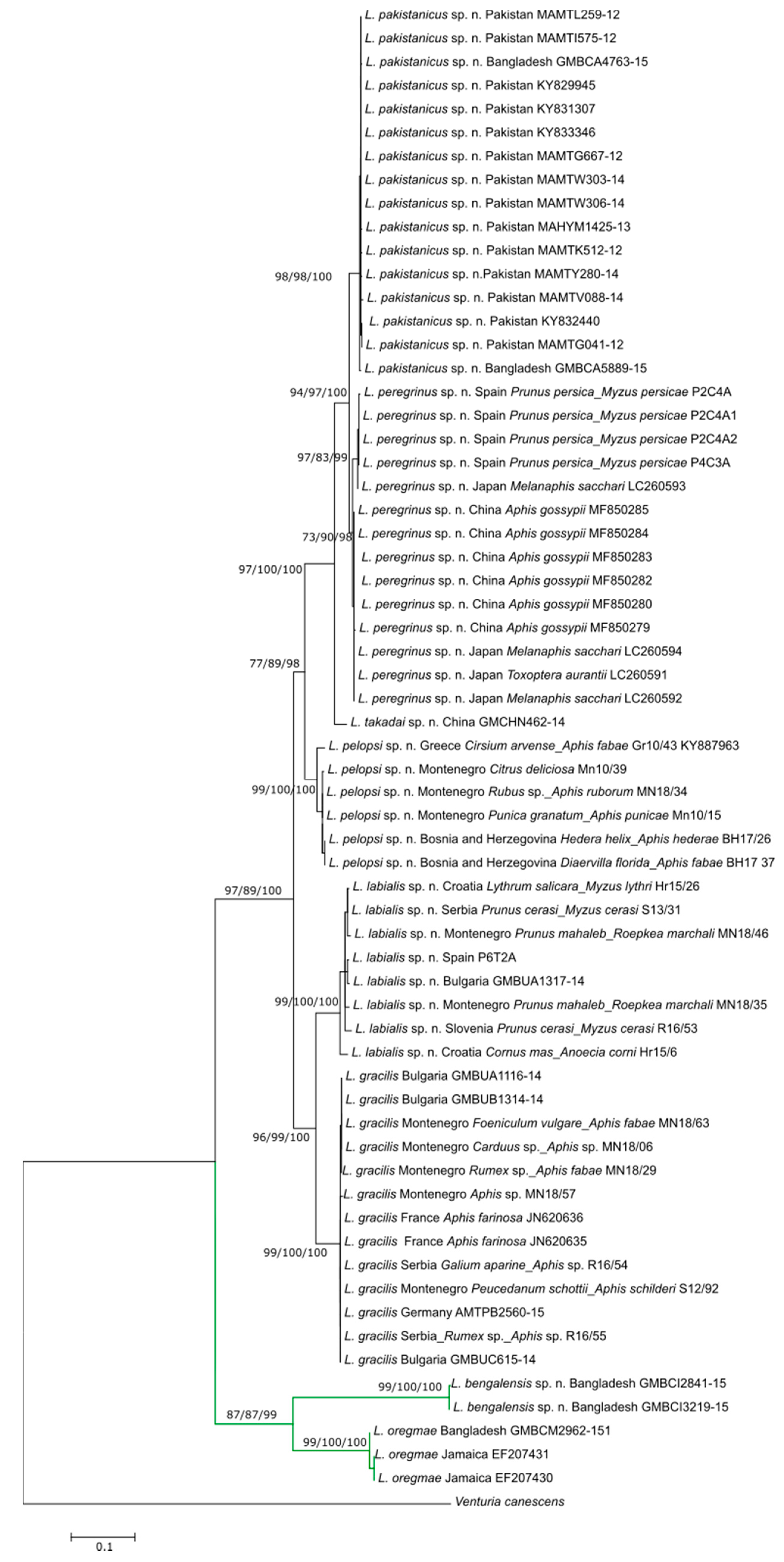
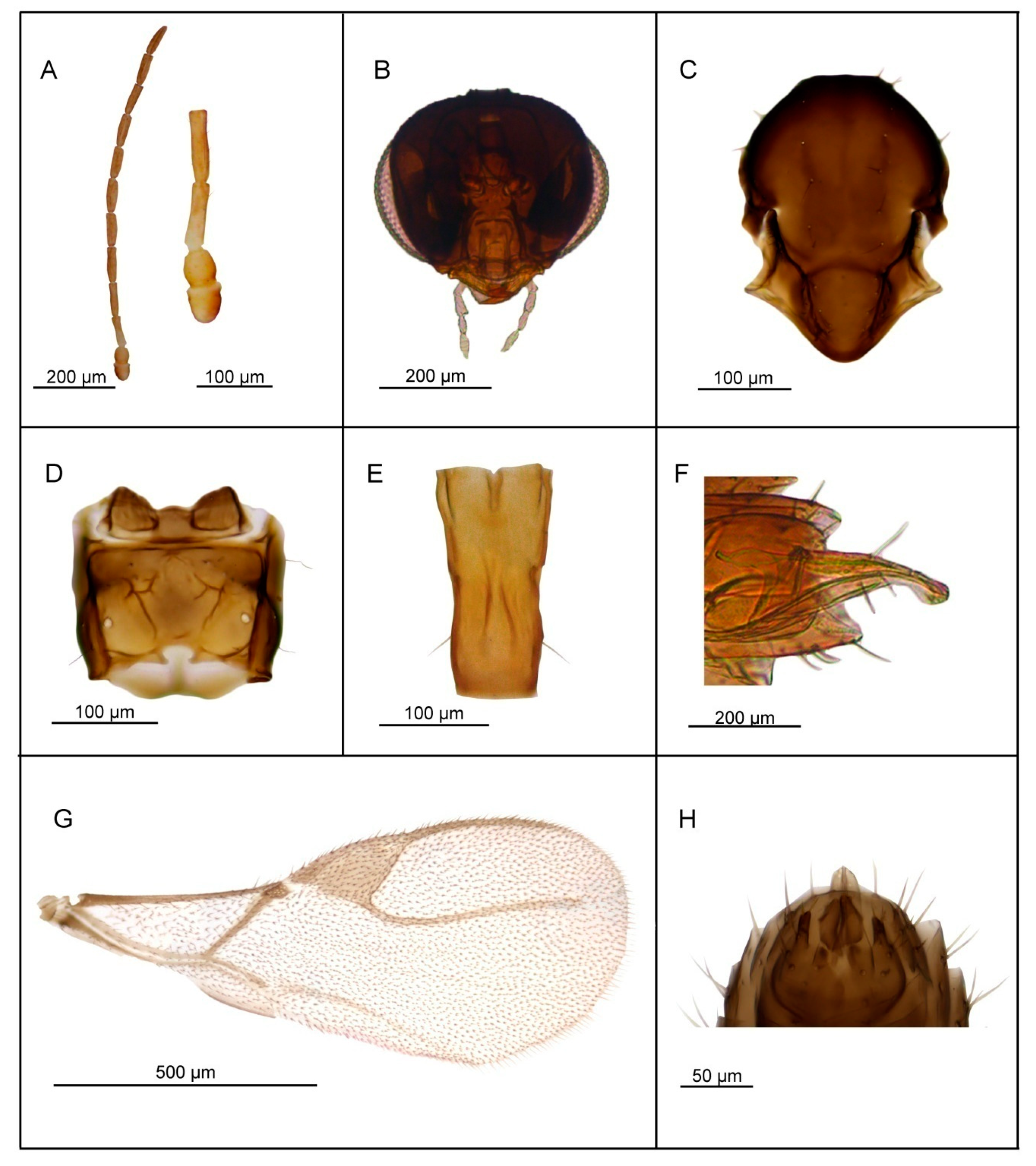
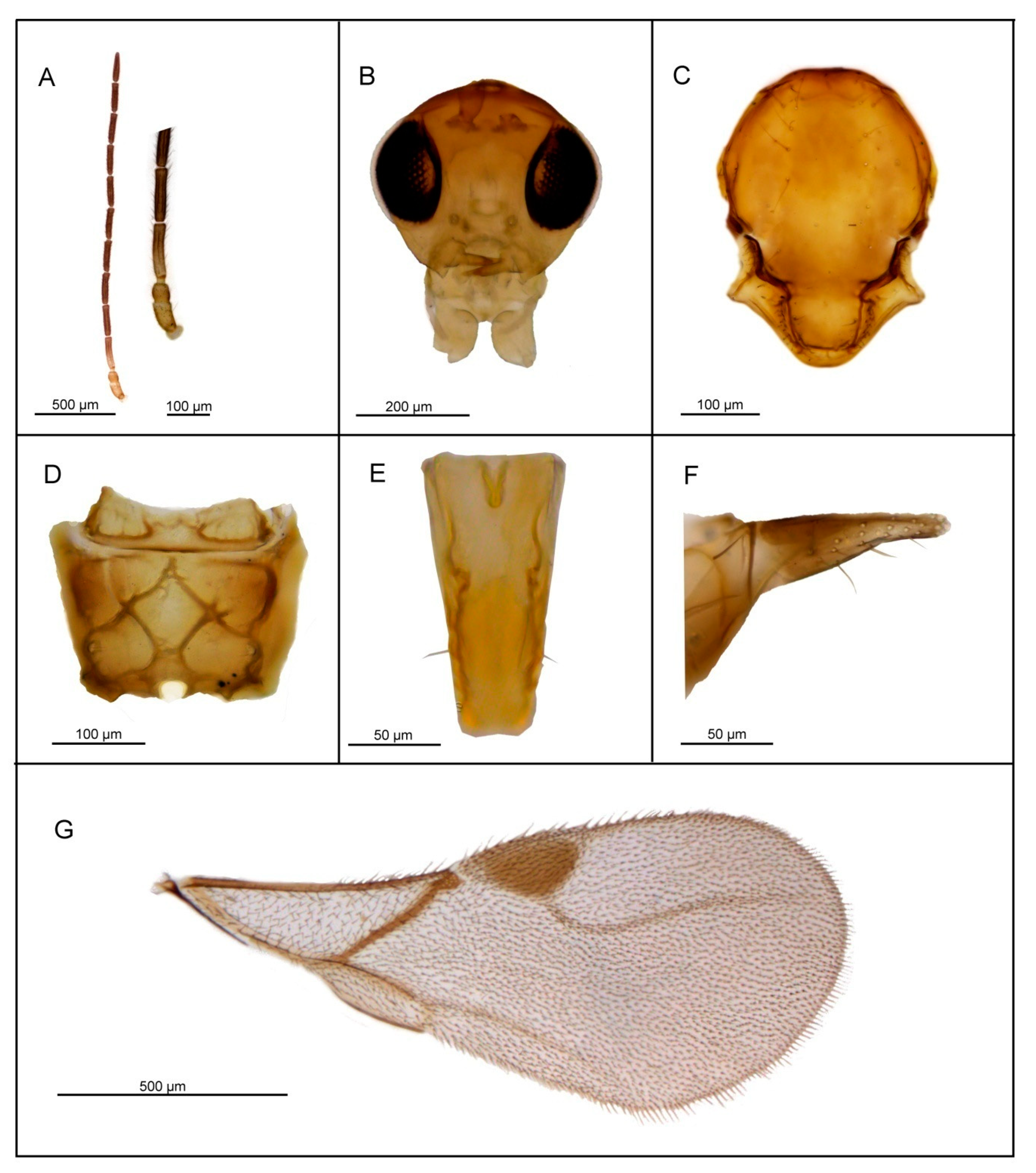
3.2.1. Lipolexis bengalensis sp. n. Tomanović and Kocić

Diagnosis. Lipolexis bengalensis sp. n. possesses crenulated longitudinal carinae along the sides of petiole and belongs to the oregmae group. However, it differs from the nominative species, L. oregmae, because its maxillary palps have two palpomeres (L. oregmae has three maxillary palpomeres). Additionally, L. bengalensis sp. n. has a shorter F1 (proportions between length and maximum width at the middle of F1 are 4.0 in L. bengalensis sp. n. vs. 4.45 in L. oregmae) and a stouter petiole (proportions between length and width of petiole at spiracles 2.6–2.9 in L. bengalensis sp. n. and 3.3 in L. oregmae) (Figure 4).

Female

Head (Figure 4B). Head wider than mesosoma (proportion between head and mesoscutum width 1.40–1.50), with sparse long setae. Eyes large, oval, laterally prominent. Face uniformly pubescent with moderately long setae. Clypeus protrudent with 6–7 long setae. Tentorial index 0.20. Malar space 0.15× as long as longitudinal eye diameter. Mandible bidentate, with 8–10 long setae on outer surface. Maxillary palps with two palpomeres. Labial palps with one palpomere. Antenna damaged (Figure 4A) (antenna broken, missing F8–F10), filiform; antennal segments long and cylindrical with semi-erected long sparse setae that are longer than half of flagellomeres diameter. F1 (Figure 4A) equal to F2; F1 and F2 bearing 1–2 and 2–3 longitudinal placodes, respectively. Proportion between length and maximum width at middle of F1 and F2, 4.0.
Mesosoma. Mesoscutum (Figure 4C) smooth, covering pronotum above; notaulices distinct in very short ascedent portion of anterolateral margin, effaced dorsally, with a series of 6–7 long setae along the latero-dorsal part of mesoscutum, almost reaching to scutellum. Scutellum nearly triangular, bearing 2 long setae on lateral margins. Propodeum (Figure 4D) clearly areolated, with a wide central areola, with pronounced oblique antero-central carinae extending to the spriracles. External and dentiparal areolae of propodeum with 5–6 and 1–2 long setae in each side, respectively. Forewing (Figure 4G) densely pubescent, marginal setae longer than the surface setae; venation hyaline; pterostigma triangular, 2.3× as long as wide and equal to R1 vein; vein r and RS long, reaching near the apex of the wing.
Metasoma. Petiole (Figure 4E) long and slender, slightly widened at apex and narrow posteriorly; its length 2.6–2.9× as long as wide at spiracles; petiole is dorsally smooth, but it bears crenulated longitudinal carinae along the sides, a feature typical for the oregmae group, and two long setae near the base of petiole at each side. Ovipositor sheath long (Figure 4F), wide at base and curved downwards, bearing scattered very long setae in the middle portion; distally dilated slightly. Length of ovipositor sheath 2.5–2.8× as long as maximum width at base and 7.3–7.6× as long as minimum width at tip. Second valvulae with a convex dorsal outline, second valvifer narrow, widened at the joint in point with ovipositor sheath.
Body length. 1.5–1.8 mm.
Coloration. Head dark brown, face brown. Mouthparts yellow to light brown. Scape and pedicel light brown. F1 segment brown with very narrow yellow or light brown ring at the base, remaining antennal flagellomere brown. Mesosoma brown with light brown legs. Metasoma dark brown. Ovipositor sheath light brown.

Male

Unknown.
Distribution: Bangladesh.
Etymology: L. bengalensis sp. n. takes its name after Bengal, the old name of Bangladesh, reflecting the current known distribution of this species.
Material. Holotype: 1♀, 12. IV 2014, Chittagong, Bangladesh (BOLD ID: GMBCJ2865-15). Paratypes: 1♀, 12. IV 2014, Chittagong, Bangladesh (BOLD ID: GMBCJ1757-15); 2♀, 11.VI 2014, Chittagong, Bangladesh (BOLD ID: GMBCI2841-15, GMBCI3219-15). Holotype is slide mounted and deposited in the Canadian National Collection of Insects, Arachnids and Nematodes, Ottawa (CNC). Paratype specimens are slide mounted and deposited in the collection of Institute of Zoology, Faculty of Biology, University of Belgrade (FBUB).

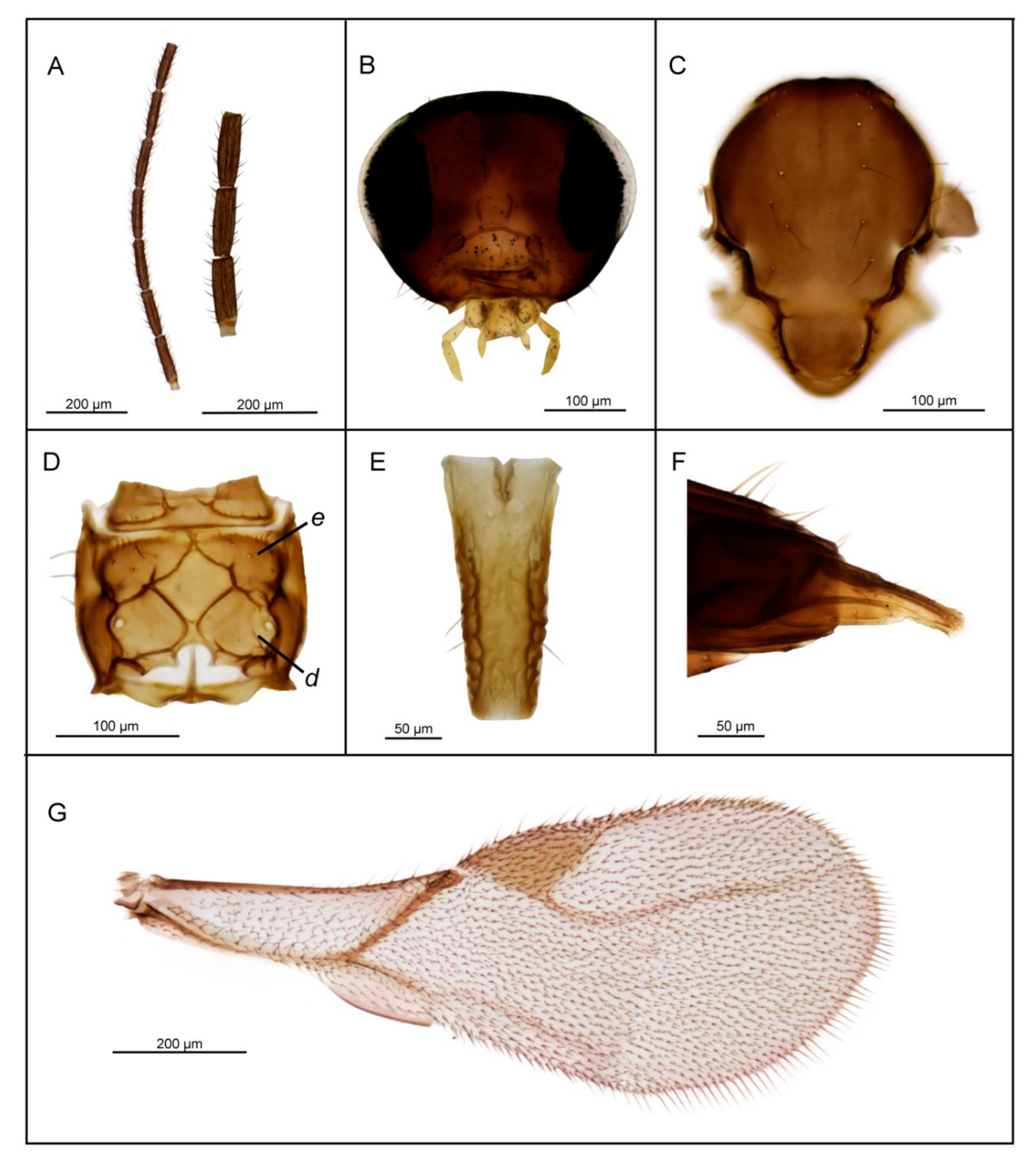
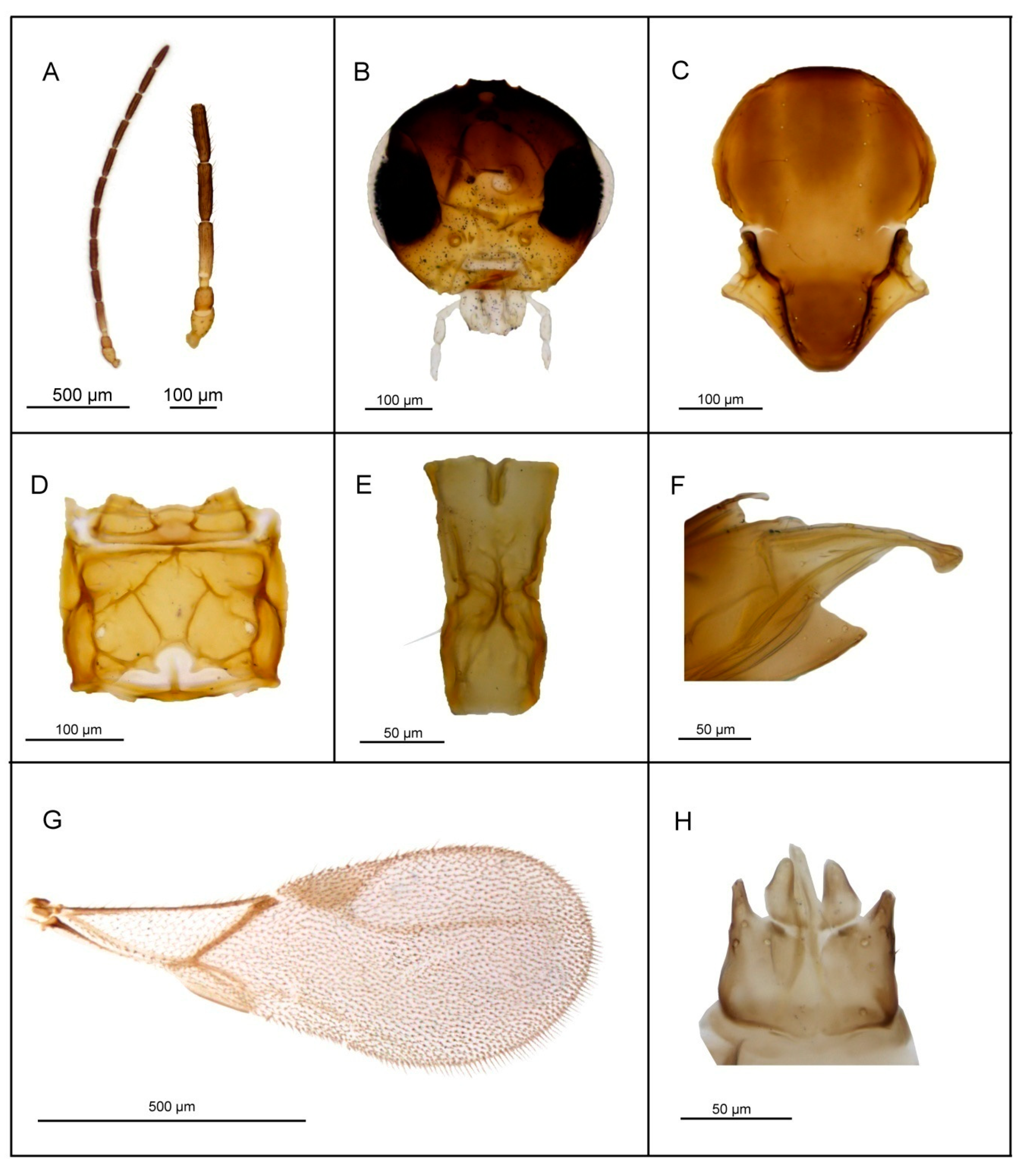
3.2.2. Lipolexis labialis sp. n. Tomanović and Kocić

Diagnosis. L. labialis sp. n. possesses maxillary palps with four palpomeres, a feature shared with two other members of the gracilis group—L. gracilis and L. pelopsi sp. n. However, it is the only known species of Lipolexis with labial palps with two palpomeres. Additionally, it differs from L. gracilis and L. pelopsi sp. n. by having a more elongated F1 (proportions between length and maximum width at middle of F1, 3.7–4.0 in L. labialis sp. n. vs. 3.0–3.6 in L. gracilis and 3.4–3.6 in L. pelopsi sp. n.) and generally smaller number of longitudinal placodes on F1 and F2 than L. gracilis and L. pelopsi sp. n. (L. labialis sp. n. has 1–2 on F1 and 2–4 longitudinal placodes on F2, while L. gracilis has 2–4 on F1 and 4–6 on F2 and L. pelopsi sp. n. has 3–4 on F1 and 4–5 on F2) (Figure 5).

Female

Head (Figure 5B) transverse, wider than mesosoma at tegulae (proportion between wide of head and mesoscutum 1.37–1.45), with sparse long setae. Eyes large, oval, laterally prominent. Clypeus with 6–7 long setae. Tentorial index 0.28–0.31. Malar space equal to 0.18–0.20× as long as longitudinal eye diameter. Mandible bidentate, with 7–10 long setae on outer surface. Maxillary palps with four palpomeres. Labial palps with two palpomeres. Antenna 12—segmented (Figure 5A), filiform; antennal segments long and cylindrical with semi-erected long sparse setae equal to about half of flagellomeres diameter or little longer. F1 (Figure 5A) equal or slightly longer than F2; F1 and F2 bearing 1–2 and 2–4 longitudinal placodes, respectively. Proportions between length and maximum width at middle of F1 and F2, 3.7–4.0 and 3.3–4.0, respectively.
Mesosoma. Mesoscutum (Figure 5C) smooth, covering pronotum above; notaulices distinct in very short ascendant portion of anterolateral margin, effaced dorsally, with a series of 8–10 long setae along the latero-dorsal part of mesoscutum. Scutellum nearly triangular, bearing 2–3 long setae on lateral margins. Propodeum (Figure 5D) areolated, with a wide central areola, sometimes with no clear anterior carinae. External and dentiparal areolae of propodeum with 2–3 and 1 long setae in each side, respectively. Central areola with rugosities. Forewing (Figure 5G) densely pubescent, marginal setae longer than the surface setae; pterostigma triangular, 2.5–2.9× as long as wide and subequal to R1 vein (proportion between pterostigma length and R1 vein length 0.9–1.1); vein r and RS long, reaching near the apex of the wing.
Metasoma. Petiole (Figure 5E) long and slender, slightly widened at apex; its length 2.7–3.2× as long as wide at the weak prominent spiracles; a pair of central carinae, distinctly prominent along dorsal surface of the petiole, diverging toward base. Ovipositor sheath strong, long and slightly curved (Figure 5F), dilated at the apex. Length of ovipositor sheath 2.5× as long as wide at the base, and approx. 10× as long as tip.
Body length: 1.6–1.8 mm.
Coloration: Head brown, face brown. Mouthparts yellow. Scape, pedicel and F1 yellow till light brown, remaining flagellomeres brown. Mesosoma brown. Metasoma brown with light brown petiole and ovipositor sheath.

Male

Antennae 13—segmented. F1 and F2 shorter than in females, 2.8–3.1× as long as wide and 3.0–3.1× as long as wide, respectively. Pterostigma 2.44–2.75× as long as wide. Aedeagus funnel shaped, apodemae long, volsellae prominent and pointed; paramerae short, triangular, bearing 4–5 long setae (Figure 5H). Color same as in female.
Body length: 1.4–1.6 mm.
Hosts: Anoecia corni (F.), Dysaphis sp. Börner, Myzus cerasi (F.), Myzus lythri (Schrank), Roepkea marchali (Börner).
Distribution. European distribution. Recorded from Serbia, Croatia, Spain, Montenegro, Slovenia, and Bulgaria.
Etymology. Lipolexis labialis sp. n. takes its name from its possession of the highest number of labial palpomeres (2) within Lipolexis.
Material: Holotype: 1♀ reared from Dysaphis sp. Börner on Malus sp., 12. VI 2013, Umčari, Kotlova, Serbia; Paratypes: 1♀1♂, Dysaphis sp. Börner on Malus sp., 12. VI 2013, Umčari, Kotlova, Serbia (MK); 3♀5♂, M. cerasi on Prunus cerasus, 16. VI 2013, Umčari, Parloge, Serbia (MK); 1♀1♂, M. lythri on Lythrum salicaria, 21. VI 2015, Plitvička jezera, Kozjak, Croatia (ŽT); 6♀5♂, A. corni on Cornus mas, 21.VI 2015, Plitvička jezera, Kozjak, Croatia (ŽT); 1♀, 2016, Segria, Spain (YA); 6♀6♂, R. marchali (Börner) on Prunus mahaleb, 18. VI 2018, Lovćen, Njeguši, Montenegro (KK); 1♀2♂, M. cerasi (F.) on Prunus cerasi, 30.V 2016, Škocjanske jame, Slovenia (KK).
Holotype is slide mounted and deposited in the collection of Institute of Zoology, Faculty of Biology, University of Belgrade (FBUB). Four female and one male paratypes are slide mounted and deposited in the collection of Institute of Zoology, Faculty of Biology, University of Belgrade (FBUB). Paratypes collected in National Park Plitvice, Croatia are deposited in the Croatian Natural History Museum, Zagreb, Croatia. Two female and two male paratypes (Umčari, Parloge, Serbia) are deposited in the Canadian National Collection of Insects, Arachnids and Nematodes, Ottawa (CNC).

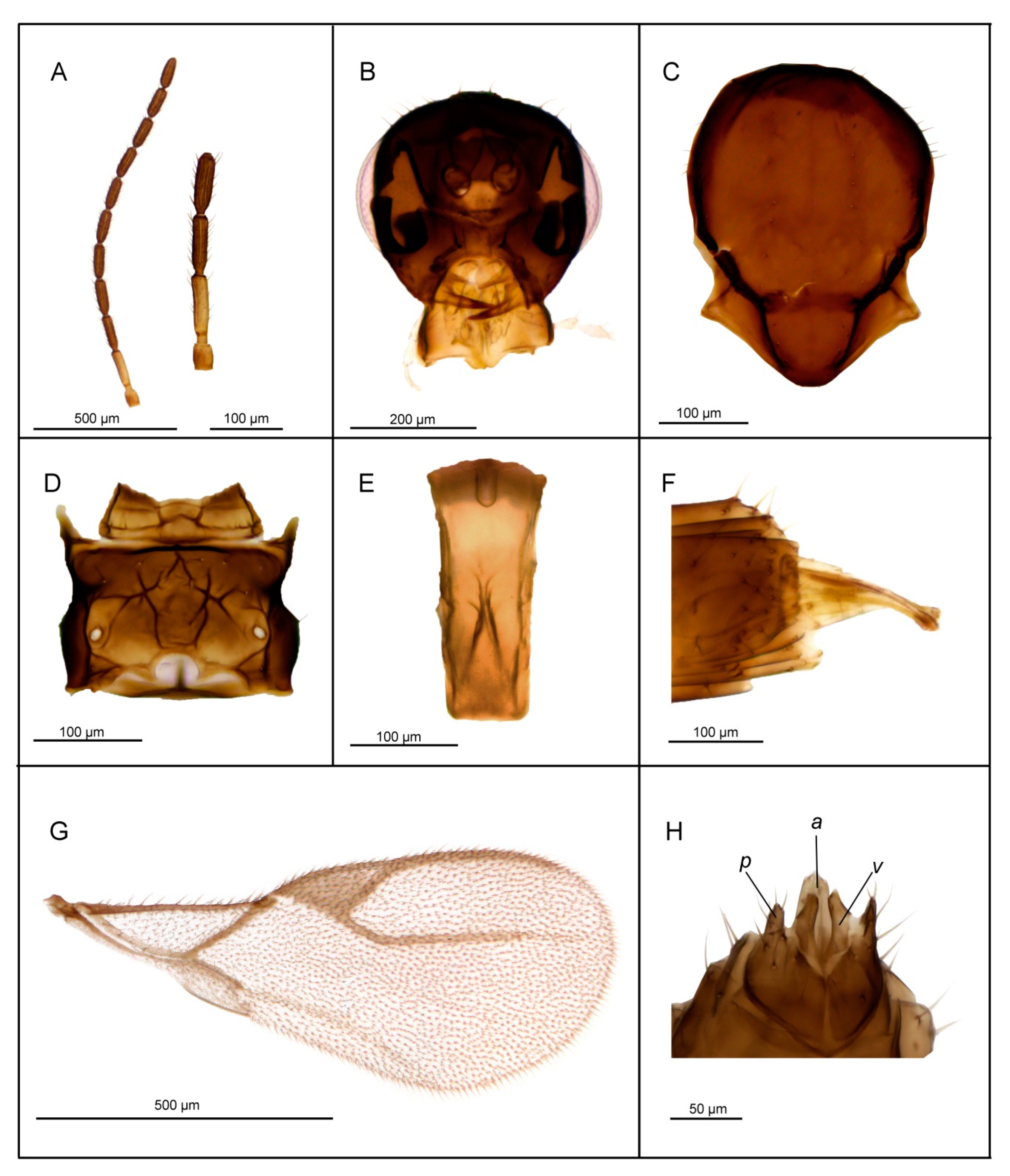
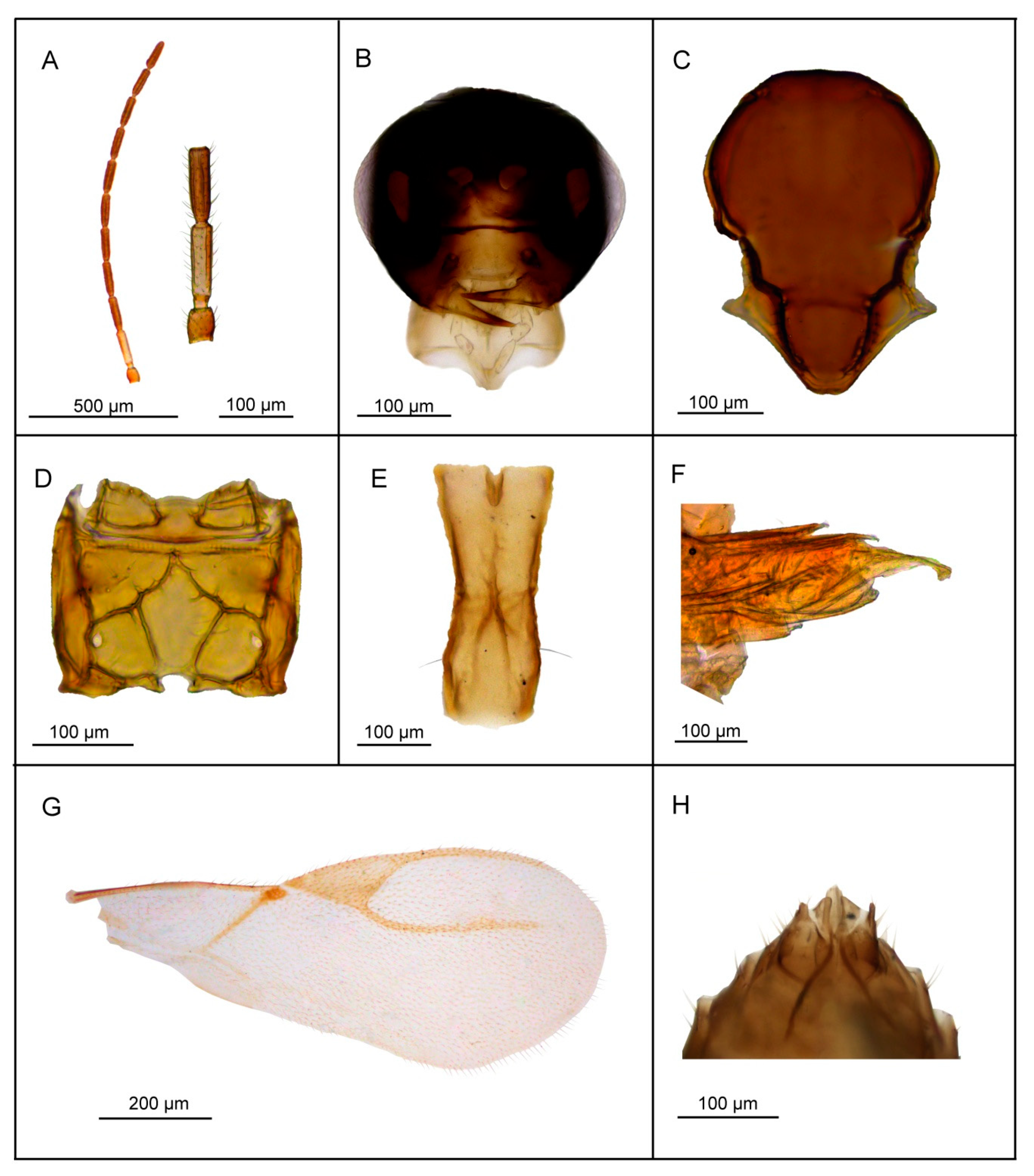
3.2.3. Lipolexis takadai sp. n. Tomanović and Kocić

Diagnosis. Lipolexis takadai sp. n. differs from all known species of Lipolexis by the following combination of morphological characters: maxillary palps with three palpomeres and labial palps with one palpomere, and very long F1 and F2 (proportions between length and maximum width at middle of F1 and F2, 4.75 and 4.80–5.0, respectively). All known Lipolexis species possess proportions between length and maximum width at middle of F1 and F2, 3.0–4.45 and 2.70–4.25, respectively (Figure 6).

Female

Head (Figure 6B) is wider than mesosoma at tegulae (proportion between head and mesoscutum width 1.30), with sparse long setae. Eyes large, oval, laterally prominent. Face uniformly pubescent with moderately long setae. Clypeus protrudent with 7–8 long setae. Tentorial index 0.30–0.35. Malar space equal to 0.15–0.20× as long as longitudinal eye diameter. Mandible bidentate, with 8–10 long setae on outer surface. Maxillary palps with three palpomeres. Third palpomere long but undivided. Labial palps with one palpomere. Antenna 12—segmented, filiform (Figure 6A); antennal segments long and cylindrical with semi-erected long sparse setae length about half of flagellomeres diameter. F1 (Figure 6A) longer than F2; F1 and F2 bearing 2 and 3 longitudinal placodes, respectively. Proportions between length and maximum width at the middle of F1 and F2, 4.75 and 4.8–5.0, respectively.
Mesosoma. Mesoscutum (Figure 6C) smooth, covering pronotum above; notaulices distinct in very short ascedent portion of anterolateral margin, effaced dorsally, with a series of 9–10 long setae along the latero-dorsal part of mesoscutum, reaching to scutellum. Scutellum nearly triangular, noticeably crenulated along lateral margins, bearing 2–3 long setae. Propodeum (Figure 6D) areolated, with a wide central areola, oblique antero-central carinae extending to the spriracles. External and dentiparal areolae of propodeum with 3–4 and without long setae in each side, respectively. Forewing (Figure 6G) densely pubescent, marginal setae longer than the surface setae; venation hyaline; pterostigma triangular, 2.6–2.7× as long as wide and subequal to R1 vein; vein r and RS long, reaching near to the apex of the wing.
Metasoma. Petiole (Figure 6E) long and slender, slightly widened at apex; its length 2.8× as long as wide at spiracles, with prominent central carina. Ovipositor sheath (Figure 6G) long and slender, strongly curved downwards, wide at base, narrowing in the last third and slightly wider at the tip. Length to width ratio of the ovipositor sheath is 2.7 at the widest site, and 10 at the tip.
Body length: 1.5–1.8 mm.
Coloration: Head brown. Mouthparts yellow. Scape and pedicel yellow, F1 light brown, remaining antennal segments brown. Mesosoma brown. Metasoma brown with light brown petiole. Ovipositor sheath light brown.

Male—Unknown

Distribution. Japan and China.
Host. Aphis gossypii Glover.
Etymology. Lipolexis takadai sp. n. is named in honor of the famous Japanese entomologist and taxonomist, Prof. Hajimu Takada, for his exceptional contribution to the knowledge of the parasitoid fauna of aphids in Japan, Far East and worldwide.
Material. Holotype: 1♀, 27. VII 2012, Shaanxi, Yinggezhen, China (BOLD ID: GMHCN 462-14); Paratypes: 1♀, reared from Aphis gossypii Glover on Solanum melongena, 25. VII 1962, “Moraica”, Japan; 1♀, reared from A. gossypii Glover, Kyoto, Japan, leg. Hajimu Takada. Holotype is slide mounted and deposited in the Canadian National Collection of Insects, Arachnids and Nematodes, Ottawa (CNC). Paratypes are slide mounted and deposited in the collection of Institute of Zoology, Faculty of Biology, University of Belgrade (FBUB).

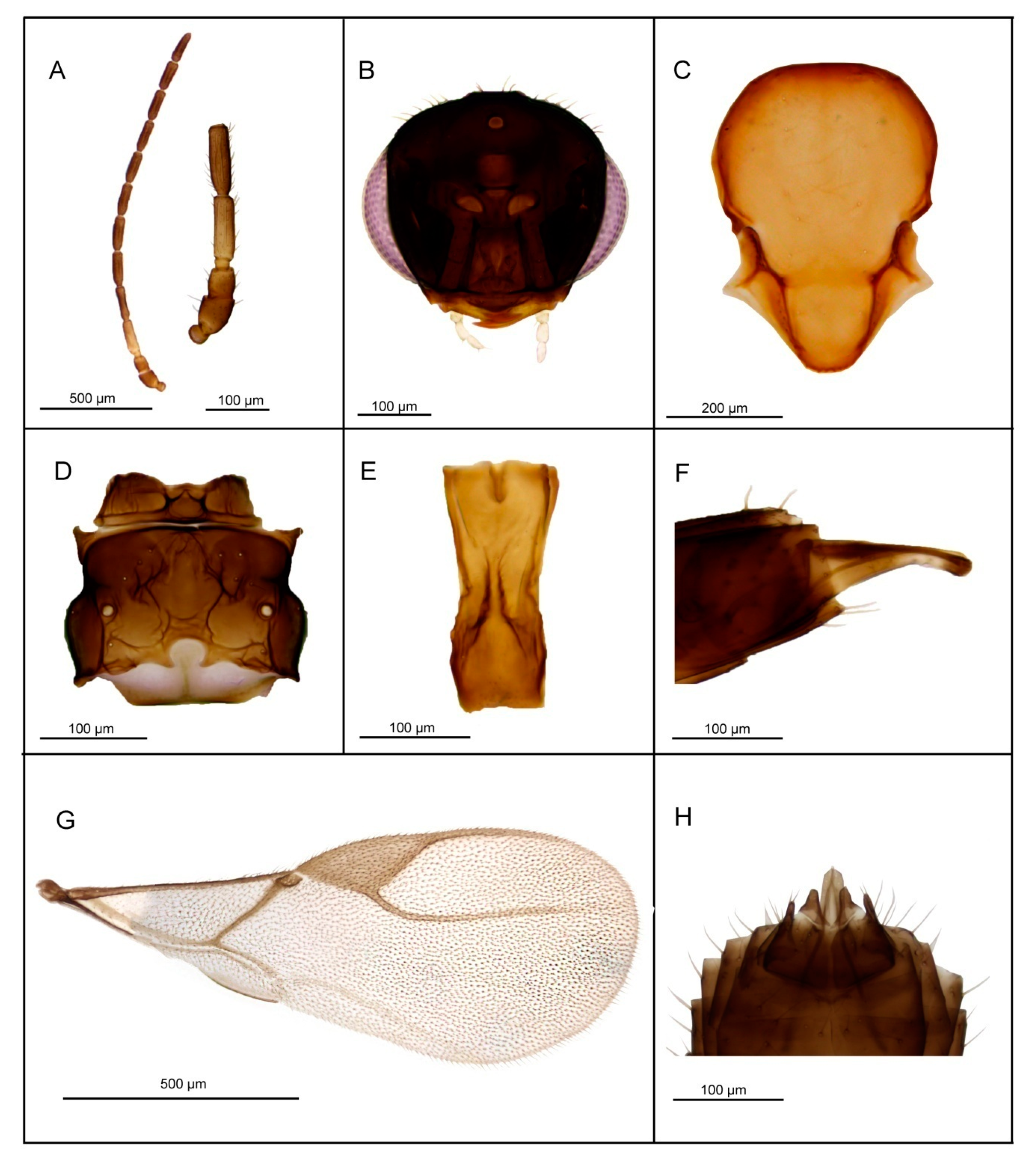
3.2.4. Lipolexis pelopsi sp. n. Tomanović and Kavallieratos

Diagnosis. Lipolexis pelopsi sp. n. differs from all known Lipolexis species by its combination of the following morphological characters: maxillary palps with four palpomeres, labial palps with one palpomere (the same combination as L. gracilis), pubescent body and stout petiole (proportions between length and maximum width of petiole at spiracles, 2.4–2.6 in L. pelopsi sp. n., vs. 3.0–3.6 in L. gracilis) (Figure 7).

Female

Head (Figure 7B) transverse, wider than mesosoma at tegulae (proportion between wide of head and mesoscutum, 1.2–1.5), with sparse long setae. Eyes large, oval, laterally prominent. Face densely pubescent with moderately long setae. Clypeus protrudent with 9–12 long setae. Tentorial index 0.25–0.35. Malar space equal to 0.12–0.18× as long as longitudinal eye diameter. Maxillary palps with four palpomeres. Labial palps with one palpomere. Antenna 12—segmented (Figure 7A), filiform; antennal segments long and cylindrical with semi-erected long sparse setae shorter than half of flagellomeres diameter. F1 (Figure 7A) subequal to F2; F1 and F2 bearing 3–4 and 4–5 longitudinal placodes, respectively. Proportions between length and maximum width at middle of F1 and F2, 3.4–3.6 and 3.6, respectively.
Mesosoma. Mesoscutum (Figure 7C) smooth, covering pronotum above; notaulices distinct in very short ascedent portion of anterolateral margin, effaced dorsally, with a series of 6 to 10 long setae along the latero-dorsal parts of mesoscutum, reaching to scutellum. Specimens collected in Greece possess densely pubescent mesoscutum with 18 to 20 setae. Scutellum nearly triangular, bearing 5–6 setae on lateral margins. Propodeum (Figure 7D) areolated, with a wide central areola, sometimes with undefined upper carinae. External and dentiparal areolae of propodeum with 5–6 and 0–1 long setae in each side, respectively. Forewing (Figure 7G) densely pubescent, marginal setae long and longer than the surface setae; venation reduced; pterostigma triangular, 2.3–2.6× as long as wide and subequal to R1 vein; vein r and RS long, reaching near to the apex of the wing.
Metasoma. Petiole (Figure 7E) long and slender, slightly widened at apex; its length 2.4–2.6× as long as wide at spiracles, slightly prominent in lateral margin; a pair of central carinae short, prominent along dorsal surface of the petiole; Ovipositor sheath narrow and curved downwards (Figure 7F), dilated at the apex, bearing 3 large long setae in the middle portion. Ovipositor 3.1× as long as wide at the widest part and 9.1× as long as wide at the tip.
Body length: 1.5–2.0 mm.
Coloration: Head brown, eyes black; mouthparts yellow. Scape, pedicel and F1 light brown, remainder of antennae brown. Mesosoma brown. Legs light brown. Petiole yellow. Metasoma dorsally dark brown.

Male

Antennae 13–segmented, F1 and F2 bearing 3 and 4 longitudinal placodes, respectively. Pterostigma 2.70× as long as wide and clearly shorter than R1 vein. Aedeagus long, funnel shaped; volsellae strong, apodemae long (Figure 7H). Paramerae short, pointed apically, bearing 3–4 long setae.
Body length: 1.4–1.6 mm.
Distribution. Mediterranean distribution, recorded from Bosnia and Herzegovina, Montenegro and Greece.
Host. Species in the genus Aphis L.
Etymology. Lipolexis pelopsi sp. n. takes its name after Pelops, mythological king of Peloponnese region, which is the area of the first collected specimens.
Material: Holotype: 1♀ reared from Aphis punicae Passerini on Punica granatum, 19. V 2010, Tivat, Montenegro (VŽ); Paratypes: 4♀7♂, reared from A. fabae Scopoli on Diervilla florida, 23. V 2017, Blagaj, Bosnia and Herzegovina (ŽT); 1♂, reared from Aphis hederae Kaltenbach on Hedera helix, 23. V 2017, Buna, Bosnia and Herzegovina (ŽT); 1♀ reared from Aphis sp. on Citrus deliciosa, 18. V 2010, Petrovac, Montenegro (VŽ); 4♀4♂, reared from Aphis ruborum (Börner and Schilder) on Rubus sp., 15. VI 2018, Rijeka Crnojevića, Montenegro; 8♀4♂, reared from Aphis fabae cirsiiacanthoides Scopoli on Cirsium arvense, 01. V 2010, Kyparissia, Greece (ŽT, NK). Holotype and 5♀1♂ paratypes slide mounted and deposited in the collection of Institute of Zoology, Faculty of Biology, University of Belgrade (FBUB) (the rest of specimens are kept in 96% ethanol). Two female and two male paratypes (Blagaj, Bosnia and Herzegovina) are deposited in the Canadian National Collection of Insects, Arachnids and Nematodes, Ottawa (CNC).

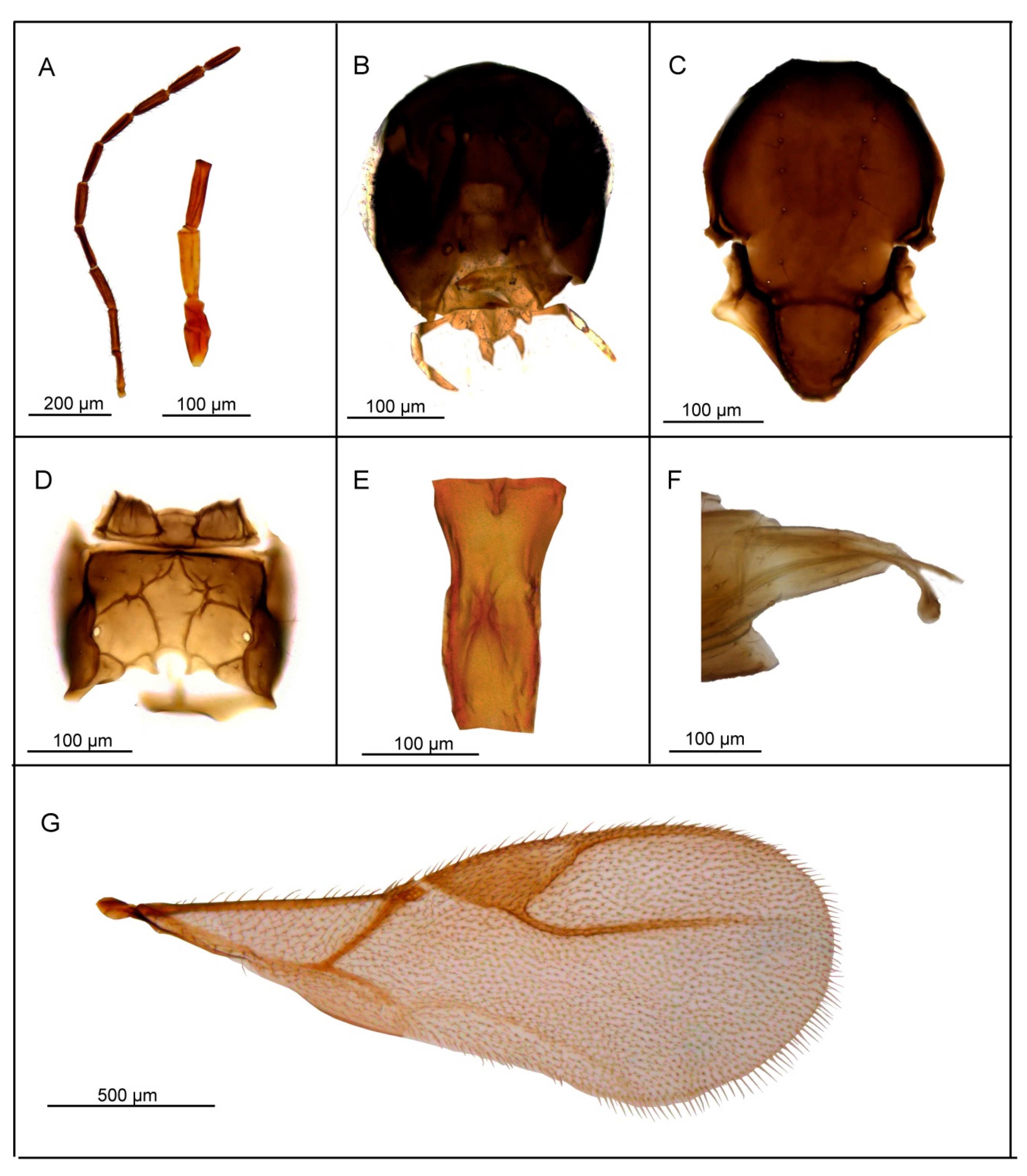
3.2.5. Lipolexis pakistanicus sp. n. Tomanović and Kocić

Diagnosis. Lipolexis pakistanicus sp. n. belongs to “gracilis” group and shares the same number of maxillary (three) and labial palpomeres (one) as L. peregrinus sp. n. and L. takadai sp. n. However, it has a more elongate pterostigma than these species (2.9 length to width ratio in L. pakistanicus sp. n., vs. 2.6–2.7 and 2.4–2.7 in L. takadai sp.n. and L. peregrinus sp. n., respectively) (Figure 8).

Female

Head (Figure 8B) wider than mesosoma (proportion between head width and mesoscutum width 1.5), with sparse long setae. Eyes large, oval, laterally prominent. Face uniformly pubescent with moderately long setae. Clypeus protrudent with 6–7 long setae. Tentorial index 0.30. Malar space equal to 0.20× as long as longitudinal eye diameter. Mandible bidentate, maxillary palps with three palpomeres and labial palps with one palpomere. Antenna 12—segmented (Figure 8A), filiform; antennal segments long and cylindrical with semi-erected long sparse setae length about half of flagellomeres diameter. F1 (Figure 8A) equal or subequal to F2; F1 and F2 bearing 3–4 and 4–5 longitudinal placodes, respectively. Proportion between length and maximum width at middle of F1 and F2, 4.25.
Mesosoma. Mesoscutum (Figure 8C) smooth, covering pronotum above; notaulices distinct in very short ascedent portion of anterolateral margin, effaced dorsally, with a series of 6–7 long setae along the latero-dorsal part of mesoscutum, reaching to scutellum. Scutellum nearly triangular, slightly crenulated along lateral margins, bearing 2–3 long setae on lateral margins. Propodeum (Figure 8D) clearly areolated, with a wide central areola, oblique antero-central carinae extending to the spriracles. External and dentiparal areolae of propodeum with 3–4 and without long setae in each side, respectively. Forewing (Figure 8G) densely pubescent, marginal setae longer than the surface setae; venation reduced; pterostigma triangular, 2.9× as long as wide and little shorter than R1 vein (proportion between pterostigma length and R1 vein length 0.90); vein r and RS long, reaching near to the apex of the wing.
Metasoma. Petiole (Figure 8E) slightly widened at apex; its length 2.7× as long as wide at spiracles, prominent in lateral margin; dorsally smooth but it bears short and strong mediodorsal carina with two short longitudinal carinae diverging along the sides to the posterior part of petiole. Petiole bears two very long setae along both sides near base. Ovipositor sheath (Figure 8F) long, wide at base, curved downwards, with no visible setae; distally more dilated than in other species, upper part of the ovipositor sheath more sclerotized along the whole length. Length of ovipositor sheath 2.56× as long as wide at base and 6.5× as long as minimum width at tip.
Body length: 1.5–1.8 mm.
Coloration: Head dark brown, face brown. Mouthparts yellow to light brown. Scape, pedicel and base of F1 yellow to light brown, remaining parts of antenna brown. Mesosoma brown with light brown legs. Metasoma brown or light brown with yellow petiole.

Male

F1 subequal to F2. Mesoscutum smooth, with a series of 7–8 long setae. Propodeum with clear central areola. Petiole shorter and broader than in female. Aedeagus funnel shaped, pointed apically, volsellae strong and triangular (Figure 8H). Parameare short, bearing 3–4 setae. Head testaceus, antennae, mesoscutum, propodeum and abdomen brown, legs yellow.
Host. Aphis gossypii Glover.
Distribution. Pakistan, Bangladesh and Moldova.
Etymology. Lipolexis pakistanicus sp. n. takes its name from the country where the majority of specimens were collected.
Material: Holotype: 1♀, 28. III 2012, Islamabad, Pakistan (BOLD ID: MAMTG667-12); Paratypes: 1♀, 11. IV 2012, Islamabad, Pakistan (BOLD ID: MAMTI575-12); 1♀1♂, 15. XI 2012, Islamabad, Pakistan (BOLD IDs: MAMTW303-14, MAMTW306-14); 1♀, 12. IV 2014, Chittagong, Bangladesh (BOLD ID: GMBCA4763-15), 1♀, reared from A. gossypii on Cucumis sp., 26. VII 1972, Pakistan; 2♀, Moldova, date and location record of these samples is unknown.
The holotype is slide mounted and deposited in the Canadian National Collection of Insects, Arachnids and Nematodes, Ottawa (CNC). Paratypes are slide mounted and deposited in the collection of Institute of Zoology, Faculty of Biology, University of Belgrade (FBUB).

3.2.6. Lipolexis peregrinus sp. n. Tomanović and Kocić

Diagnosis. Lipolexis peregrinus sp. n. belongs to “gracilis” group and possesses three maxillar palpomeres and one labial palpomere as L. takadai sp. n. and L. pakistanicus sp. n. However, it clearly differs from both species by having shorter F1 (proportions between length and maximum width at middle of F1 in L. peregrinus sp. n., 3.4–3.8 vs. 4.75 in L. takadai sp. n. and 4.25 in L. pakistanicus sp. n., respectively) and more elongated petiole (proportion between length and width of petiole at spiracles in L. peregrinus sp. n., 3.1–3.3 vs. 2.8 in L. takadai sp. n. and 2.7 in L. pakistanicus sp. n., respectively (Figure 9).

Female

Head (Figure 9B) transverse, wider than mesosoma at tegulae (proportion between width of head and mesoscutum 1.40–1.50), with sparse long setae. Eyes large, oval, laterally prominent. Face with moderately long setae. Clypeus with 7–8 long setae. Tentorial index 0.23–0.30. Malar space equal to 0.13–0.18× as long as longitudinal eye diameter. Mandible bidentate, with 11–12 long setae on outer surface. Maxillary palps with three palpomeres. Labial palps with one palpomere. Antenna 12—segmented (Figure 9A), filiform; antennal segments long and cylindrical with semi-erected long setae length about half of flagellomeres diameter. F1 (Figure 9A) equal to F2 or somewhat longer; F1 and F2 bearing 2–3 and 4–5 longitudinal placodes, respectively. Proportions between length and maximum width at middle of F1 and F2, 3.4–3.8 and 3.2–3.6, respectively.
Mesosoma. Mesoscutum (Figure 9C) smooth, covering pronotum above; notaulices distinct in very short ascedent portion of anterolateral margin, effaced dorsally. Mesoscutum sparsely setous with a 5–7 long setae along the latero-dorsal parts. Scutellum nearly triangular, bearing 1–3 long setae on lateral margins. Propodeum (Figure 9D) areolated, with a wide central areola, oblique antero-central carinae clearly extending to the spriracles. External and dentiparal areolae of propodeum with 3–5 and 0–1 long setae on each side, respectively. Forewing (Figure 9G) densely pubescent with setae longer than the ones on surface; pterostigma triangular, 2.4–2.7× as long as wide and shorter than R1 vein (proportion between pterostigma length and R1 vein length 0.90); vein r and RS long, reaching near the apex of the wing.
Metasoma. Petiole (Figure 9E) long and slender, slightly widened at apex; length 3.1–3.3× as long as wide at spiracles, slightly prominent at lateral margin; a pair of central carinae, distinctly prominent along dorsal surface of the petiole, merged or separate in anterior part, but diverging toward base; one long seta at the base of each lateral side; spiracular tubercules smooth, positioned beyond the first half of the segment. Ovipositor sheath (Figure 9F) long, wide at base, curved downwards; distally dilated slightly, upper part of the ovipositor sheath more sclerotized along the whole length. Length of ovipositor sheath 2.8–2.9× as long as maximum width at base and 8.0–9.0× as long as minimum width at tip.
Body length: 1.5–2.0 mm.
Coloration: Head light brown. Mouthparts yellow. Scape, pedicel and F1 yellow to light brown, remaining antennal segments brown. Mesosoma and metasoma light brown to brown. Petiole light brown.

Male

Antennae 13—segmented. Flagellomeres somewhat shorter than in females. F1 and F2, 3.30 and 3.60× as long as wide, respectively. Pterostigma more triangular than in female (2.20–2.30× as long as wide). Aedeagus funnel shaped, apodemae long; volsellae wide, triangular. Paramerae short, bearing 3 setae (Figure 9H). Body darker than in females with yellow legs and mouthparts.
Body length: 1.4–1.6 mm.
Hosts. Myzus persicae (Sulzer), Aphis sp., A. gossypii Glover, Toxoptera aurantii (Boyer de Fonscolomber), Melanaphis sacchari (Zehntner).
Distribution. Europe (Spain and Slovenia) and Oriental region (China and Japan—mined from GenBank, see Table S1 for sample information).
Etymology. Lipolexis peregrinus sp. n. takes its name in regard to its unknown place of origin (latin peregrinus = foreign, from abroad).
Material. Holotype: 1♀ reared from M. persicae on P. persica, 04. VI 2015, Alfarras, Spain. Paratypes: 1♀1♂, reared from M. persicae on P. persica, 04. VI 2015, Alfarras, Spain; 1♀ reared from M. persicae on P. persica, 2015, Alfarras, Lleida, Spain; 2♀ reared from Aphis sp. on Solanum tuberosum, 27. VI 2009, Zaleb, Slovenia (KKos). Holotype and paratype specimens are dissected and slide mounted, and kept in the collection of Institute of Zoology, Faculty of Biology, University of Belgrade (FBUB) Two female paratypes (Alfarras, Lleida, Spain) are deposited in the Canadian National Collection of Insects, Arachnids and Nematodes, Ottawa (CNC).

3.3. Re-Description of L. gracilis and L. oregmae

3.3.1. Lipolexis gracilis Förster, 1862, re-description

Lipolexis gracilis Förster, 1862, Verh. Nat. Ver. Preuss.Rheinl.19:249.
Gynocryptus pieltaini Quilis, 1930, Eos, Madrid 7: 27–29.
Aphidius palpator Gautier and Bonnamour, 1931, Bull. Soc. Ent. Fr., 166–167.
Lipolexis chinensis Chen, 1980, Entomotaxonomia 2: 169–172.

Female

Diagnosis. Lipolexis gracilis has a petiole with central bifurcating carinae, a feature that positions it within the gracilis species group. It possesses maxillary palps with four palpomeres, labial palps with one palpomere (the same combination as L. pelopsi). However, it differs from the latter by a less pubescent body and elongated petiole (proportions between length and maximum width of petiole at spiracles, 3.0–3.6 in L. gracilis vs. 2.4–2.6 in L. pelopsi sp. n.).
Head (Figure 10B) transverse, wider than mesosoma at tegulae, with sparse long setae. Eyes large, oval, laterally prominent. Face uniformly pubescent with moderately long setae. Clypeus protrudent with 7–9 long setae. Tentorial index 0.25–0.40. Malar space equal to 0.10–0.15× as long as longitudinal eye diameter. Mandible bidentate, with 6–9 long setae on outer surface. Maxillary palps with four palpomeres. Labial palps with one palpomere. Antenna 12—segmented (Figure 10A), filiform; antennal segments long and cylindrical with semi-erected long sparse setae of length about half of flagellomeres diameter. F1 (Figure 10A) equal or slightly longer than F2; F1 and F2 bearing 2–4 and 4–6 longitudinal placodes, respectively. Proportions between length and maximum width at middle of F1 and F2, 3.0–3.6 and 2.7–3.2, respectively.
Mesosoma. Mesoscutum (Figure 10C) smooth, covering pronotum above; notaulices distinct in very short ascedent portion of anterolateral margin, effaced dorsally, with a series of 6–8 long setae along the latero-dorsal part of mesoscutum, reaching to scutellum. Scutellum nearly triangular, bearing 2–3 long setae on lateral margins. Propodeum (Figure 10D) areolated, with a wide central areola, oblique antero-central carinae extending to the spriracles. External and dentiparal areolae of propodeum with 3–4 and 0–1 long setae in each side, respectively. Forewing (Figure 10G) densely pubescent, the upper marginal setae slightly longer than the surface setae; venation reduced; pterostigma triangular, 2.3–2.5× as long as wide and equal or subequal to R1 vein (proportion between pterostigma length and R1 vein length 1.0–1.1); vein r and RS long, reaching near to the apex of the wing.
Metasoma. Petiole (Figure 10E) long and slender, slightly widened at apex; length 2.7–3.2× as long as wide at spiracles, slightly prominent in lateral margin; a pair of central carinae, distinctly prominent along dorsal surface of petiole, merged or separate in anterior part, but diverging toward base; spiracular tubercules smooth, positioned beyond the first half of the segment. Ovipositor sheath (Figure 10F) long, wide at base, distally dilated slightly and slightly curved downwards. As in all Lipolexis species, the upper part of the ovipositor sheath more sclerotized along the whole length. Length of ovipositor sheath 2.9× as long as maximum width at base and 9.6× as long as minimum width at tip. Body length: 1.5–1.8 mm.
Coloration: Head dark brown, face brown. Mouthparts except tips of mandibles yellow. Scape, pedicel and F1 brown, other segments darker. Pronotum brown; mesoscutum and mesopleuron dark brown. Propodeum brown. Legs light brown, hind femur and tibia slightly darker at the middle. Wings slightly infumated, venation yellowish brown. Petiole brown. Metasoma dorsally dark brown. Ovipositor sheath yellow, dorsally darker.

Male

Antennae 13—segmented. Aedeagus funnel-shaped, with sub-parallel lateral margin in upper part and sharply widening toward the base (Figure 10H). Apodemae distinctly longer than trunk of the aedeagus. Volsellae wide, triangular; parameare bearing two long setae at the tip. Tentorial index 0.40, malar space equal to quarter to eye length.
Body length: 1.4–1.6 mm.
Hosts. Acyrthosiphon Mordvilko, Aphis L., Brachycaudus van der Goot, Capitophorus van der Goot, Liosomaphis Walker, Lipaphis Mordvilko, Melanaphis van der Goot, Metopeurum Mordvilko, Myzocallis Passerini, Myzus, Rhopalosiphum, Semiaphis van der Goot, Therioaphis Walker and Toxoptera [43].
Material: 41♀, 12♂ reared from Aphis sp. on Rumex sp.: 13. V 2016, Novi Beograd, Serbia (KK); 22♀18♂ reared from Aphis sp. on Galium aparine: 25. V 2016, Surčinsko jezero, Serbia (KK); 1♀ reared from Aphis schilderi (Börner) on Peucedanum schottii: 27. VII 2012, Durmitor, Montenegro (AP); 2♀2♂ reared from Aphis sp. on Carduus sp.: 15. VI 2018, Cetinje, Lipska pećina, Montenegro (AP); 1♀ reared from A. fabae on Rumex sp.: 17. VI 2018, Lovćen, Montenegro (JČ); 1♀ reared from Aphis sp. on Rubus sp.: 17. VI 2018, Rijeka Crnojevića, Montenegro (JČ); 1♀ reared from A. fabae on Foeniculum vulgare: 17. VI 2018, Lovćen, Montenegro (KK); 2♀ reared from Aphis sambuci on Sambucus nigra: VI 2013, Češke Budejovice, Czech Republic (PS).

3.3.2. Lipolexis oregmae (Gahan 1932)

Diaeretus oregmae (Gahan, 1932), Ann Entomol Soc Am, 25, 736–757.
Lipolexis scutellaris Mackauer, 1962, Entomophaga, 7, 1, 37–45.
Remarks about L. wuyiensis, L. pseudosctullearis and L. myzakkaiae. On the basis of its description, L. wuyiensis is very similar to L. oregmae and it parasitizes Ceratovacuna lanigera Zehntner (=Oregma lanigera), the same aphid host as L. oregmae in the Philippines. Furthermore, apart from the drawing of the petiole of L. myzakkaiae, which does not follow the original description (i.e., two lateral longitudinal wavy branched carinae), both L. myzakkaiae and L. pseudoscutellaris are morphologically similar to L. oregmae. Moreover, it seems that neither species has been mentioned in the scientific literature since their description. In our opinion, all three species are likely to be conspecific with L. oregmae. However, due the absence of type material, the authors refrained from official synonymization.

Female

Diagnosis. Lipolexis oregmae possesses a petiole that bears lateral longitudinal carinae, which is a morphological character of the oregmae group of species. It differs from the other member of the group, L. bengalensis sp. n. by the number of maxillary palpomeres (three maxillar palpomeres in L. oregmae vs, two in L. bengalensis sp. n.).
Head (Figure 11B) wider than mesosoma at tegulae (proportion between head and mesoscutum width 1.45), with sparse long setae. Eyes large, oval, laterally prominent. Face uniformly pubescent with moderately long setae. Clypeus protrudent with 6–7 long setae. Tentorial index 0.25. Malar space 0.15x as long as longitudinal eye diameter. Maxillary palps with three palpomeres and labial palps with one palpomere. Antenna 12—segmented (Figure 11A), filiform; antennal segments long and cylindrical with semi-erected long sparse setae with length of about half of flagellomeres diameter. F1 (Figure 11A) longer than F2; F1 and F2 bearing 2–3 and 3–4 longitudinal placodes, respectively. Proportion between length and width maximum width at middle of F1 and F2, 4.45×.
Mesosoma. Mesoscutum (Figure 11C) smooth, covering pronotum above; notaulices distinct in very short ascedent portion of anterolateral margin, effaced dorsally, with a series of 9–10 long setae along the latero-dorsal part of mesoscutum, reaching to scutellum. Scutellum nearly triangular, bearing 2 long setae on lateral margins. Propodeum (Figure 11D) clearly areolated, with a wide central areola, oblique antero-central carinae extending to the spiracles. External and dentiparal areolae of propodeum with 6–7 and with one long setae on each side, respectively. Forewing (Figure 11G) densely pubescent, marginal setae longer than the surface setae; venation reduced; pterostigma triangular, 2.2× as long as wide and equal to R1 vein; vein r and RS long, reaching near the apex of the wing.
Metasoma. Petiole (Figure 11E) long and slender, wide at base, narrowing towards the apex and then slightly widened; length 3.3× as long as wide at spiracles, dorsally smooth, bearing lateral longitudinal carinae. Ovipositor sheath (Figure 11F) long, wide at base, curved downwards, bearing 3 scattered long setae in the middle portion; distally dilated slightly, upper part of the ovipositor sheath more sclerotized along the whole length. Length of ovipositor sheath 2.8× as long as maximum width at base and 10× as long as minimum width at tip. Body length: 1.5–1.8 mm.
Coloration: Head dark brown, mouthparts yellow. Scape, pedicel and F1 yellow, remainder antenna brown. Mesosoma brown with light brown legs. Metasoma brown.

Male

Antennae 13—segmented. Aedeagus funnel-shaped, with sub-parallel lateral margin in upper part and sharply widening toward the base. Apodemae distinctly longer than trunk of the aedeagus. Tentorial index 0.40, malar space equal to quarter to eye length.
Body length: 1.4–1.6 mm.
Hosts. Aphis, Cavariella del Guercio, Ceratovacuna Zehtner, Greenidea Schouteden, Liosomaphis, Myzus, Pentalonia Coquerel, Rhopalosiphum, Semiaphis, Sitobion Mordvilko, Toxoptera, Tuberolachnus Mordvilko [43].
Material: 1♀, 26. II 2015, Chittagong, Bangladesh (BOLD ID: GMBCM 2962-15), 61♀13♂, reared from T. citricida (Kirkaldy) on Citrus sp., Florida, USA; 1♀1♂, India (date and location record of these samples is unknown).

3.4. Identification Key to the World Species of Genus Lipolexis

1.Petiole dorsally smooth, bearing crenulated lateral longitudinal carinae (Figure 4E and Figure 11E). ………2
oregmae group
-Petiole with prominent bifurcating central carina dorsally, without crenulated lateral longitudinal carinae (Figure 5E, Figure 6E, Figure 7E, Figure 8E, Figure 9E, and Figure 10E).…......3
gracilis group
2.Maxillary palps with 3 palpomeres, labial palps with 1 palpomere. F1 4.45× as long as wide, number of longitudinal placodes on F1 and F2, 2–3 and 4–5, respectively (Figure 11A).Lipolexis oregmae
-Maxillary palps with 2 palpomeres, labial palps with 1 palpomere. F1 4× as long as wide, F2 with 2–3 number of longitudinal placodes on F1 and F2, 1–2 and 2–3, respectively (Figure 4A).Lipolexis bengalensis sp. n.
3.Maxillary palps with 4 palpomeres.………4
-Maxillary palps with 3 palpomeres.………6
4.Labial palps with 2 palpomeres, F1 3.7–4.0× as long as wide, number of longitudinal placodes on F1 and F2, 1–2 and 2–4, respectively (Figure 5A).Lipolexis labialis sp. n.
-Labial palps with one palpomere, F1 3.0–3.6× as long as wide, number of longitudinal placodes on F1 and F2, 2–4 and 4–6, respectively (Figure 7A and Figure 10A).………5
5.Petiole 2.4–2.6× as long as wide (Figure 7E). R1 slightly longer than pterostigma (Figure 7G).Lipolexis pelopsi sp. n.
-Petiole 2.7–3.2× as long as wide (Figure 10E). R1 equal or slightly shorter than pterostigma (Figure 10G).Lipolexis gracilis
6.F1 4.2–4.7× as long as wide (Figure 6A and Figure 8A), petiole 2.7–2.8× as long as wide (Figure 6E and Figure 8E).………..7
-F1 3.4–3.8× as long as wide (Figure 9A), petiole 3.1–3.3× as long as wide (Figure 9E)Lipolexis peregrinus sp. n.
7.F1 and F2 4.7 and 4.8–5.0× as long as wide, respectively (Figure 6A), scutellum laterally crenulated along sides (Figure 6C), number of longitudinal placodes on F1 and F2 2 and 3, respectively (Figure 6A)Lipolexis takadai sp. n.
-Both F1 and F2 4.2× as long as wide (Figure 8A), scutellum laterally not bearing crenulations (Figure 8C), F1 and F2 with 2–3 and 4–5 longitudinal placodes, respectivelyLipolexis pakistanicus sp. n.

4. Discussion

Until recently, taxonomists relied on morphological and ecological studies as a basis for describing new species or identifying newly collected specimens. However, easy access to DNA sequence information has now enabled integrative taxonomy which combines morphological/ecological and molecular data to clarify species boundaries. The barcode region of COI is by far the most widely utilized molecular marker used for molecular studies on the subfamily Aphidiinae [30,44,45]. Aside from aiding the identification of the newly collected specimens, this gene region aids the delineation of species boundaries, an approach of particular value for aphidiine genera that are suspected to include cryptic species complexes [46,47,48,49]. Our study reinforced the value of COI as it separated Lipolexis species with high resolution and bootstrap support. The molecular results were concordant with those from morphological study leading to the description of six new species of Lipolexis. Each of these species possessed diagnostic morphological characters which separates it from its congeners which makes the fact that they went unnoticed surprising.
Based on morphological and molecular differences, Lipolexis can be separated into two main groups, i.e., gracilis and oregmae. The average genetic distance between species in these two groups is high, ranging from 21.7% to 23.2%. Furthermore, gracilis and oregmae groups are morphologically distinguished by the differing shape of the petiole: members of the L. gracilis group have a petiole with prominent central carinae, while those in the L. oregmae group have a petiole that is smooth dorsally, but with noticeably crenulated lateral longitudinal carinae. Additionally, members of the gracilis group have a slightly shorter metacarpal vein (R1) than members of the oregmae group (proportion between length of pterostigma and metacarpal vein is 0.90–1.1 in gracilis group versus 0.75–0.90 in oregmae group).
Five new species belonging to the L. gracilis group were discovered in this study: Lipolexis labialis sp. n., L. pelopsi sp. n., L. takadai sp. n., L. pakistanicus sp. n. and L. peregrinus sp. n. Furthermore, within the gracilis group, two additional clusters are formed, one constituted of L. gracilis s. str. and L. labialis sp. n., and the second of the remaining species (L. pelopsi sp. n., L. takadai sp. n., L. pakistanicus sp. n. and L. peregrinus sp. n.) with the average between group distance of 11.4%. Although L. labialis sp. n. is most closely related to L. gracilis (average genetic distance of 7.3%), the two species are readily distinguished by their differing number of labial palpomeres. L. gracilis possesses one labial palpomere while L. labialis sp. n. has two. These two species have overlapping distributions in Europe, but may use different hosts. All reared specimens of L. labialis employed Macrosiphini aphid hosts (Myzus Passerini, Roepkea Hille Ris Lambers) excepting one reared from Anoecia corni (F.). Although L. gracilis has also been reported from Myzus [43,50], in the light of the new species discoveries, it is not clear whether the specimens reared from Myzus are indeed L. gracilis or might be some other Lipolexis species, such as L. labialis sp. n. or L. peregrinus sp. n.
In this phylogenetic analysis, within the second cluster of the gracilis group, L. takadai sp. n. wasrepresented by just a single specimen from China. However, morphological examination of other Lipolexis revealed two specimens from Japan which fully correspond to it. Although molecular data indicate that L. takadai sp. n. is most closely related to L. peregrinus sp. n., L. pakistanicus sp. n. and L. pelopsi sp. n. (3.9%, 4.5%, 6.8% average COI divergence respectively), it possesses several morphological differences from other species of Lipolexis. Although it is only known from China and Japan, its actual distribution may be much broader. Lipolexis takadai sp. n. was reared from Aphis gossypii Glover, a common host for other Lipolexis.
A second species from this cluster, Lipolexis pelopsi sp. n., is most closely related to L. peregrinus sp. n., L. pakistanicus sp. n. and L. takadai sp. n., with 6.9%, 8.6%, 6.8% average COI distance, respectively. It has a Mediterranean distribution as it was reared from aphids collected in Greece, Bosnia and Herzegovina, Croatia, and Montenegro. It is a parasitoid of aphids in the genus Aphis attacking various plant species. Lipolexis pelopsi sp. n. coccurs in this region with L. gracilis and L. labialis sp. n., and also shares an aphid host (Aphis sp.) with L. gracilis. It is characterised by a pubescent body (specimens from Greece possess heavily setose mesoscutum), a feature that distinguishes it from all other species.
Lipolexis peregrinus sp. n., another species in the same cluster, is most closely related to L. pakistanicus sp. n., L. takadai sp. n. and L. pelopsi sp. n. (2.2%, 3.9%, 6.9% average COI distance, respectively). Its specimens were collected in both Asia (Japan, China) and Europe (Spain, Slovenia). Further investigations are needed to ascertain if L. peregrinus sp. n., is present across the Palaearctic or if it was introduced from Asia to Europe or vice versa. Given its phylogenetic position within the gracilis group and its number of palpomeres, it probably originates from the Oriental region. Lipolexis peregrinus sp. n. is phylogenetically closest to Asian species and its maxillary palps have 3 palpomeres, a trait that characterizes Asian species, while the European species (i.e., L. gracilis, L. pelopsi sp. n. and L. labialis sp. n.) possess maxillary palps with 4 palpomeres. In Asia, L. peregrinus sp. n. was found to parasitize aphid genera Aphis L., Melanaphis and Toxoptera Koch [51,52] (in both studies identified as L. gracilis), while in Europe it was reared from M. persicae and Aphis sp.
Lipolexis pakistanicus sp. n. is phylogenetically most closely related to L. peregrinus sp. n., L. pelopsi sp. n. and L. takadai sp. n. (2.2%, 8.6%, 4.5% average COI distances, respectively). With the exception of two specimens from Bangladesh and two from Moldova, all material was collected in Pakistan. As most specimens were collected in Malaise traps, the aphid host spectrum is unknown, but one specimen from Pakistan was reared from A. gossypii. This species is likely distributed across the Oriental region. In Bangladesh, it co-occurs with L. oregmae and L. bengalensis sp. n., but more information is needed on host use to ascertain the extent of overlap.
Although L. bengalensis sp. n. is a member of the oregmae group, based on both sequence results and morphology, it is genetically distant from L. oregmae (19.9% average), implying the species have long been separate. At the moment, L. bengalensis sp. n. is only known from Bangladesh where it was collected in a vegetable crop field where L. oregmae was also collected, indicating their co-occurence. Its aphid host is unknown because it was collected by Malaise trap.
Starý [53] reported that L. gracilis is common in steppe habitat, orchards, and wood edges. Our material of L. gracilis originates from high montane habitat, mixed forests, steppes, orchards, and urban area. Although L. gracilis mostly parasitizes hosts from the tribes Aphidini (genus Aphis Linnaeus) and Macrosiphini (genera Brachycaudus and somewhat less Myzus Passerini), it has been reared from other unrelated aphid genera, such as Anoecia Koch, Myzocallis and Therioaphis [43]. By comparison, the Oriental L. oregmae is mostly associated with aphids from the tribes Aphidini (genera Aphis, Rhopalosiphum Koch and Toxoptera Koch) and Macrosiphini (genera Cavariella, Liosomaphis, Myzus, Pentalonia, Semiaphis and Sitobion), but it also attacks representatives of some tribes ignored by L. gracilis such as Cerataphidini (Ceratovacuna lanigera Zehntner), Greenideini (Greenidea psidii van der Goot (=formosana (Maki)) and Tuberolachnini (Tuberolachnus salignus (J. F. Gmelin)) [43]. However, in light of our discovery of six new species, host use might not be that broad as previously thought so trophic associations for these two species require careful validation.
With the exception of L. labialis sp. n., all other species of Lipolexis parasitize Aphis despite their sympatry in the same habitat. For instance, in Europe, both L. gracilis and L. labialis sp. n. parasitize Aphis spp. Furthermore, three species were found in Montenegro, i.e., L. labialis sp. n., L. pelopsi sp. n. and L. gracilis, while, L. labialis sp. n., L. gracilis and L. peregrinus sp. n. were recorded from Slovenia. As a result, it seems clear that speciation in the genus Lipolexis has not been driven by host specialization.
Several studies have reported unusually high COI distances between species within the subfamily Aphidiinae [47,49]. For example Kocić et al. [47] found COI average distances of up to 20.7% among species in the phylogenetically old aphidiine genus Ephedrus. Similarly, some species of Lipolexis showed even higher average distances (i.e., 23.2% between L. bengalensis sp. n. and L. peregrinus sp. n.). While adults of Ephedrus possess several plesiomorphic taxonomic characters, this is not the case for Lipolexis. In fact, Lipolexis is characterized by several apomorphic taxonomic characters, such as reduced wing venation, elongated petiole, and shape of the ovipositor sheath. Its apomorphies and phylogenetic studies at the subfamily level (where it is clustered with evolutionary younger genera Binodoxys and Trioxys) suggest this genus evolved quite recently. However, the average genetic distances of COI, which are even higher than those in Ephedrus, might indicate that Lipolexis diversification was not a recent event. Furthermore, Finlayson [25] stated that the larval morphology of Lipolexis exhibits plesiomorphic morphological characters. One possibility might be that Lipolexis separated early in the evolution of Aphidiinae and acquired adult apomorphic traits independently. Further research is needed in order to try to resolve the complex and poorly investigated position of Lipolexis and its relationships with the other aphidiine members.

5. Conclusions

Our study revealed six new species of Lipolexis (L. peregrinus sp. n., L. pelopsi sp. n., L. labialis sp. n., L. takadai sp. n., L. pakistanicus sp. n. and L. bengalensis sp. n.). Each of these species possesses clear morphological characters that distinguishes it from its congeners. Moreover, two groups can be differentiated within the genus—oregmae and gracilis. The genetic distances between them are even higher than intrageneric distances within the potentially genus Ephedrus. The oregmae group consists out of L. oregmae and L. bengalensis sp. n. of Oriental origin. The gracilis group of species can be separated into two clades, one consisting of L. gracilis and L. labialis sp. n., and the other of L. peregrinus sp. n., L. takadai sp. n., L. pelopsi sp. n. and L. pakistanicus sp. n. Furthermore, the consistency of number of palpomeres is in concordance with the geographical distribution: the European species (L. gracilis, L. labialis sp. n. and L. pelopsi sp. n.) possess four maxillar palpomeres, while the Oriental ones (L. oregmae, L. takadai, L. bengalensis, L. peregrinus and L. pakistanicus) have three. We provide a key to the world Lipolexis species.

Supplementary Materials

The following files are available online at https://www.mdpi.com/2075-4450/11/10/667/s1, Table S1: List of samples used in the analyses.

Author Contributions

K.K. and Ž.T. conceived and designed the experiments. Ž.T., A.P., J.Č., K.K., E.R., N.G.K., Y.A., J.A., and P.D.N.H. provided specimens that were used for morphological or molecular analyses. K.K., J.Č., and A.P. carried out laboratory work and data analysis, Ž.T., K.K., and N.G.K. (L. pelopsi sp. n.) provided species descriptions. K.K. and Ž.T. wrote the original draft of the paper. All authors have read and agreed to the published version of the manuscript.

Funding

This research was funded by the Ministry of Education, Science and Technological Development of the Republic of Serbia (project III43001), by a grant from the Canada First Research Excellence Fund to PDNH through the Food FROM Thought research program at the University of Guelph. Judit Arno and Yahana Aparicio were funded by the Spanish Ministry of Economy and Competitiveness (project AGL2016-77373-C2-1-R), CERCA Programme/Generalitat de Catalunya and Yahana Aparicio was supported by a grant provided by CONACyT (Mexico).

Acknowledgments

We thank Plitvice (Croatia) and Durmitor (Montenegro) national parks for permits and assistance with specimen collections. We thank Petr Starý, Marjorie Hoy (Department of Entomology and Nematology, University of Florida, Gainsville, USA), Hajimu Takada (Kyoto Prefectural University, Japan) and Katarina Kos (Department of Agronomy, University of Ljubljana, Slovenia) for the loan of Lipolexis specimens. We also thank Erna Karalija and Adi Vesnić from the Faculty of Science, Department of Biology (University of Sarajevo, Bosnia and Herzegovina) for help with field collections in Bosnia and Herzegovina, and Katica Poljak for her remarkable help in collecting material in NP Plitvice.

Conflicts of Interest

The authors declare no conflict of interest. The funders had no role in the design of the study, in the collection, analysis or interpretation of data, in the writing of the manuscript or in the decision to publish the results.

References

  1. Forster, A. Synopsis der familien und gattungen der Braconiden. Verhandlungen des Naturhistorischen Vereins der Preussischen Rheinlande und Westfalens 1862, 19, 225–288. [Google Scholar]
  2. Smith, C.F. The Aphidiinae of North America (Hymenoptera: Braconidae); The Ohio State University: Columbus, OH, USA, 1944; Volume 6, pp. 1–154. [Google Scholar]
  3. Starý, P. Redescription of the aphidiine genus Lipolexis Förster, 1862 (Hym., Braconidae). Acta Soc. Entomol. Čechoslov. 1959, 59, 93. [Google Scholar]
  4. Gahan, A.B. Miscellaneous descriptions and notes on parasitic Hymenoptera. Ann. Entomol. Soc. Am. 1932, 25, 736–757. [Google Scholar] [CrossRef]
  5. Starý, P. The generic classification of the family Aphidiidae (Hymenoptera). Acta Soc. Entomol. Čechoslov. 1960, 57, 238–252. [Google Scholar]
  6. Starý, P. The Systematic Position of “Diaeretus oregmae Gahan” (Hymenoptera: Aphidiidae). Insecta Matsumurana 1960, 23, 109–111. [Google Scholar]
  7. Mackauer, M. Drei neue blattlaus-parasiten aus Hongkong. Entomophaga 1962, 34, 87–103. [Google Scholar] [CrossRef]
  8. Miller, R.H.; Pike, K.S.; Starý, P. Aphid Parasitoids (Hymenoptera: Aphidiidae) on Guam. Micronesica 2002, 34, 87–103. [Google Scholar]
  9. Chen, J. Description of a new species of the genus Lipolexis Forster (Hymenoptera: Aphidiidae). Entomotaxonomia 1980, 2, 169–172. [Google Scholar]
  10. Starý, P.; Ghosh, A.K. Aphid Parasitoids of India and Adjacent Countries (Hymenoptera: Aphidiidae); India, G., Ed.; Zoological Survey of India: Calcuta, India, 1983.
  11. Chen, J. Description of a new species on aphid parasites of Oregma lanigera (Zehntner) (Hymenoptera, Aphidiidae). J. Fuj. Agri. Coll. 1981, 6, 33–38. [Google Scholar]
  12. Pramanik, D.R.; Raychaudhuri, D. New aphid-parasitoids (Hymenoptera: Aphidiidae) from Nagaland, Northeast India. Akitu 1984, 61, 1–4. [Google Scholar]
  13. Kavallieratos, N.G.; Tomanović, Ž.; Starý, P.; Athanassiou, C.G.; Sarlis, G.P.; Petrović, O.; Niketić, M.; Veroniki, M.A. A survey of aphid parasitoids (Hymenoptera: Braconidae: Aphidiinae) of Southeastern Europe and their aphid-plant associations. Appl. Entomol. Zool. 2004, 39, 527–563. [Google Scholar] [CrossRef] [Green Version]
  14. Mifsud, D.; Starý, P. Lipolexis gracilis Forster, 1862—New record of an aphid parasitoid from Malta (Hymenoptera, Braconidae, Aphidiinae). Bull. Entomol. Soc. Malta 2012, 5, 175–177. [Google Scholar]
  15. Kaliuzhna, M.O. A review of the genus Lipolexis Förster, 1862 (Hymenoptera, Braconidae: Aphidiinae) in the Fauna of Ukraine. Ukr. Entomol. J. 2018, 15, 22–27. [Google Scholar] [CrossRef]
  16. Persad, A.B.; Hoy, M.A.; Nguyen, R. Establishment of Lipolexis oregmae (Hymenoptera: Aphidiidae) in a lassical biological control program directed against the brown citrus aphid (Homoptera: Aphididae) in Florida. Fla. Entomol. 2007, 90, 204–213. [Google Scholar] [CrossRef]
  17. Hoy, M.A.; Jeyaprakash, A.; Clarke-Harris, D.; Rhodes, L. Molecular and field analyses of the fortuitous establishment of Lipolexis oregmae (Hymenoptera: Aphidiidae) in Jamaica as a natural enemy of the brown citrus aphid. Biocontrol Sci. Technol. 2007, 17, 473–482. [Google Scholar] [CrossRef]
  18. Cocco, A.; Jeyaprakash, A.; Hoy, M.A. Parasitism of the brown citrus aphid in Dominica by Lysiphlebus testaceipes and Lipolexis oregmae (Hymenoptera: Aphidiinae). Fla. Entomol. 2009, 92, 497. [Google Scholar] [CrossRef]
  19. Zamora Mejías, D.; Hanson, P.E.; Starý, P.; Rakhshani, E. Parasitoid (Hym., Braconidae, Aphidiinae) complex of the black citrus aphid, Toxoptera citricidus (Kirkaldy) (Hem., Aphididae) in Costa Rica and its relationships to nearby areas. J. Entomol. Res. Soc. 2011, 13, 107–115. [Google Scholar]
  20. Stanković, S.; Ilić Milošević, M.; Žikić, V. Potential candidates for biological control of the black bean aphid Aphis fabae in Serbia. Biol. Nyssana 2015, 6, 49–54. [Google Scholar]
  21. Heimpel, G.E.; Ragsdale, D.W.; Venette, R.; Hopper, K.R.; O’Neil, R.J.; Rutledge, C.E.; Wu, Z. Prospects for importation biological control of the soybean aphid: Anticipating potential costs and benefits. Ann. Entomol. Soc. Am. 2004, 97, 249–258. [Google Scholar] [CrossRef]
  22. Knorr, L.C.; DuCharme, E.P. The relationship between Argentina’s lepra explosiva and Florida’s scaly bark, with implications for the Florida citrus grower. Plant Dis. Rpt. 1951, 35, 70–75. [Google Scholar]
  23. Mackauer, M. Hymenopterorum Catalogus. Pars 3 Aphidiidae; Dr. W. Junk: The Hague, The Netherland, 1968. [Google Scholar]
  24. Belshaw, R.; Quicke, D.L.J. A molecular phylogeny of the Aphidiinae (Hymenoptera: Braconidae). Mol. Phylogenet. Evol. 1997, 7, 281–293. [Google Scholar] [CrossRef] [PubMed]
  25. Finlayson, T. The systematics and taxonomy of final-instar larvae of the family Aphidiidae (Hymenoptera). Mem. Entomol. Soc. Can. 1990, 122, 3–74. [Google Scholar] [CrossRef]
  26. Smith, P.T.; Kambhampati, S.; Volkl, W.; Mackauer, M. A phylogeny of aphid parasitoids (Hymenoptera: Braconidae: Aphidiinae) inferred from mitochondrial NADH 1 dehydrogenase gene sequence. Mol. Phylogenet. Evol. 1999, 11, 236–245. [Google Scholar] [CrossRef] [PubMed] [Green Version]
  27. Kambhampati, S.; Volkl, W.; Mackauer, M. Phylogenetic relationship among genera of Aphidiinae (Hymenoptera: Braconidae) based on DNA sequence of the mitochrondrial 16S rDNA gene. Syst. Entomol. 2000, 25, 437–445. [Google Scholar] [CrossRef]
  28. Sanchis, A.; Latorre, A.; Gonzales-Candelas, F.; Michelena, J.M. An 18S rDNA-based molecular phylogeny of Aphidiinae (Hymenoptera: Braconidae). Mol. Phylogenet. Evol. 2000, 14, 180–194. [Google Scholar] [CrossRef] [Green Version]
  29. Shi, M.; Chen, X.X. Molecular phylogeny of the Aphidiinae (Hymenoptera: Braconidae) based on DNA sequences of 16s rRNA, 18S rDNA and ATPase 6 genes. Eur. J. Entomol. 2005, 102, 133–138. [Google Scholar] [CrossRef] [Green Version]
  30. Derocles, S.A.P.; Le Ralec, A.; Plantegenest, M.; Chaubet, B.; Cruaud, C.; Cruaud, A.; Rasplus, J.Y. Identification of molecular markers for DNA barcoding in the Aphidiinae (Hym. Braconidae). Mol. Ecol. Resour. 2012, 12, 197–208. [Google Scholar] [CrossRef]
  31. Schneider, C.; Rasband, W.; Eliceiri, K. NIH Image to ImageJ: 25 years of image analysis. Nat. Methods 2012, 9, 671–675. [Google Scholar] [CrossRef]
  32. Sharkey, M.J.; Wharton, R.A. Morphology and terminology. In Manual of the New World Genera of the Family Braconidae; Wharton, R.A., Marsh, P.M., Sharkey, M.J., Eds.; International Society of Hymenopterists: Washington, DC, USA, 1997. [Google Scholar]
  33. Folmer, O.; Black, M.; Hoeh, W.; Lutz, R.; Vrijenhoek, R. DNA primers for amplification of mitochondrial cytochrome c oxidase subunit I from diverse metazoan invertebrates. Mol. Mar. Biol. Biotechnol. 1994, 3, 294–299. [Google Scholar]
  34. Hall, T.A. BioEdit: A user-friendly biological sequence alignment editor and analysis program for Windows 95/98/NT. Nucl. Acids. Symp. 1999, 95–98. [Google Scholar] [CrossRef]
  35. Tamura, K.; Stecher, G.; Peterson, D.; Filipski, A. MEGA 6: Molecular evolutionary genetics analysis version 6.0. Mol. Biol. Evol. 2013, 30, 2725–2729. [Google Scholar] [CrossRef] [Green Version]
  36. Tamura, K. Estimation of the number of nucleotide substitutions when there are strong transition-transversion and G+C-content biases. Mol. Biol. Evol. 1992, 9, 678–687. [Google Scholar] [PubMed] [Green Version]
  37. Drummond, A.J.; Suchard, M.A.; Xie, D.; Rambaut, A. Bayesian phylogenetics with BEAUti and the BEAST 1.7. Mol. Biol. Evol. 2012, 29, 1969–1973. [Google Scholar] [CrossRef] [Green Version]
  38. Rambaut, A.; Drummond, A.; Xie, D.; Baele, G.; Suchard, M. Posterior summarisation in Bayesian phylogenetics using Tracer 1.7. Syst. Biol. 2018, 67, 901–904. [Google Scholar] [CrossRef] [PubMed] [Green Version]
  39. Xia, X. DAMBE7: New and improved tools for data analysis in molecular biology and evolution. Mol. Biol. Evol. 2018, 35, 1550–1552. [Google Scholar] [CrossRef] [PubMed] [Green Version]
  40. Xia, X.; Xie, Z.; Salemi, M.; Chen, L.; Wang, Y. An index of substitution saturation and its application. Mol. Phylogenet. Evol. 2003, 26, 1–7. [Google Scholar] [CrossRef]
  41. Rambaut, A. FigTree Version 1.4.4. 2009. Available online: http://tree.bio.ed.ac.uk/software/figtree/ (accessed on 25 September 2020).
  42. Rozas, J.; Ferrer-Mata, A.; Sanchez-DelBarrio, J.C.; Guirao-Rico, S.; Librado, P.; Ramos-Onsis, S.E.; Sanchez-Gracia, A. DnaSP v6: DNA sequence polymorphism analysis of large datasets. Mol. Biol. Evol. 2017, 34, 3299–3302. [Google Scholar] [CrossRef]
  43. Yu, D.S.; van Achterberg, C.; Horstmann, K. Taxapad 2012—World Ichneumonoidea 2011. Taxonomy, Biology, Morphology and Distribution; Taxapad: Vancouver, BC, Canada. Available online: www.taxapad.com (accessed on 29 March 2019).
  44. Tomanović, Ž.; Mitrović, M.; Petrović, A.; Kavallieratos, N.G.; Žikić, V.; Ivanović, A.; Rakhshani, E.; Starý, P.; Vorburger, C. Revision of the European Lysiphlebus species (Hymenoptera: Braconidae: Aphidiinae) on the basis of COI and 28SD2 molecular markers and morphology. Arthropod Syst. Phylogeny 2018, 76, 179–213. [Google Scholar]
  45. Ye, Z.; Vollhardt, I.M.G.; Tomanovic, Z.; Traugott, M. Evaluation of three molecular markers for identification of European primary parasitoids of cereal aphids and their hyperparasitoids. PLoS ONE 2017, 12, e0177376. [Google Scholar] [CrossRef]
  46. Derocles, S.A.P.; Plantegenest, M.; Rasplus, J.Y.; Marie, A.; Evans, D.M.; Lunt, D.H.; Le Ralec, A. Are generalist Aphidiinae (Hym. Braconidae) mostly cryptic species complexes? Syst. Entomol. 2016, 41, 379–391. [Google Scholar] [CrossRef]
  47. Kocić, K.; Petrović, A.; Čkrkic, J.; Mitrović, M.; Tomanović, Ž. Phylogenetic relationships and subgeneric classification of European Ephedrus species (Hymenoptera, Braconidae, Aphidiinae). Zookeys 2019, 878, 1–22. [Google Scholar] [CrossRef] [PubMed]
  48. Mitrovski-Bogdanović, A.; Petrović, A.; Mitrović, M.; Ivanović, A.; Žikić, V.; Starý, P.; Vorburger, C.; Tomanović, Ž. Identification of two cryptic species within the Praon abjectum group (Hymenoptera: Braconidae: Aphidiinae) using molecular markers and geometric morphometrics. Ann. Entomol. Soc. Am. 2013, 106, 170–180. [Google Scholar] [CrossRef]
  49. Čkrkic, J.; Petrović, A.; Kocić, K.; Mitrović, M.; Kavallieratos, N.G.; van Achterberg, C.; Hebert, P.D.N.; Tomanović, Ž. Phylogeny of the subtribe Monoctonina (Hymenoptera, Braconidae, Aphidiinae). Insects 2020, 11, 160. [Google Scholar] [CrossRef] [PubMed] [Green Version]
  50. Starý, P.; Schlinger, E.I. A Revision of the Far East Asian Aphidiidae (Hymenoptera); Dr W. Junk: The Hague, The Netherlands, 1967. [Google Scholar]
  51. Uesugi, R.; Nagasaka, K. Mitochondrial COI Sequences to identify species of primary and secondary parasitoid wasps of aphids in the agricultural environment in Japan. Annu. Rep. Kanto-Tosan Plant Prot. Soc. 2017, 2017, 143–145. [Google Scholar] [CrossRef]
  52. Dong, Z.; Liu, S.; Zhang, Z. Efficacy of using DNA barcoding to identify parasitoid wasps of the melon-cotton aphid (Aphis gossypii) in watermelon cropping system. BioControl 2018, 63, 677–685. [Google Scholar] [CrossRef]
  53. Starý, P. Biology of Aphid Parasites (Hymenoptera: Aphidiidae) with Respect to Integrated Control; Dr W. Junk: The Hague, The Netherlands, 1970. [Google Scholar]
Figure 1. Phylogenetic tree based on 63 cytochrome oxidase c subunit I (COI) sequences of Lipolexis. obtained using Neighbor Joining analysis. Bootstrap values/Bayesian posterior probabilities above 50% are shown on the branches in ML/NJ/Bayesian analysis order. Specimen data are presented in following array: parasitoid/country of origin/aphid and plant host (if available)/BOLD, GenBank or private code. Green branches represent L. oregmae lineage, black L. gracilis.
Insects 11 00667 g001
Figure 2. Haplotype network of COI sequences obtained from specimens in the L. oregmae group. The circle size indicates the number of specimens with a particular haplotype; each black dot represents a nucleotide substitution.
Insects 11 00667 g002
Figure 3. Haplotype network for COI sequences from 58 specimens belonging to the L. gracilis group. The circle size indicates the number of specimens with a haplotype; each black dot represents a nucleotide substitution, red median vectors.
Insects 11 00667 g003
Figure 4. Lipolexis bengalensis sp. n., female: (A) antenna and F1–F3; (B) head; (C) mesoscutum; (D) propodeum; (E) petiole, dorsal view; (F) ovipositor sheath; (G) forewing; e external areola, d dentiparal areola.
Insects 11 00667 g004
Figure 5. Lipolexis labialis sp. n.: female (A) antenna and F1–F3; (B) head; (C) mesoscutum; (D) propodeum; (E) petiole, dorsal view; (F) ovipositor sheath; (G) forewing; male (H) aedeagus; p paramere, a aedeagus, v volsella.
Insects 11 00667 g005
Figure 6. Lipolexis takadai sp. n. female: (A) antenna and F1 and F2; (B) head; (C) mesoscutum; (D) propodeum; (E) petiole, dorsal view; (F) ovipositor sheath; (G) forewing.
Insects 11 00667 g006
Figure 7. Lipolexis pelopsi sp. n.: female (A) antenna and F1 and F2; (B) head; (C) mesoscutum; (D) propodeum; (E) petiole, dorsal view; (F) ovipositor sheath; (G) forewing; male (H) aedeagus.
Insects 11 00667 g007
Figure 8. Lipolexis pakistanicus sp. n.: female (A)antenna and F1–F3; (B) head; (C) mesoscutum; (D) propodeum; (E) petiole, dorsal view; (F) ovipositor sheath; (G)forewing; male (H) aedeagus.
Insects 11 00667 g008
Figure 9. Lipolexis peregrinus sp. n.: female (A) antenna and F1 and F2; (B)head; (C) mesoscutum; (D) propodeum; (E) petiole, dorsal view; (F) ovipositor sheath; (G) forewing; male (H) aedeagus.
Insects 11 00667 g009
Figure 10. Lipolexis gracilis: female (A) antenna and F1 and F2; (B) head; (C) mesoscutum; (D) propodeum; (E) petiole, dorsal view; (F) ovipositor sheath; (G) forewing; male (H) aedeagus.
Insects 11 00667 g010
Figure 11. Lipolexis oregmae: (A) antenna and F1–F3; (B) head; (C) mesoscutum; (D) propodeum; (E) petiole, dorsal view; (F) ovipositor sheath; (G) forewing.
Insects 11 00667 g011
Table 1. Average genetic distances for the COI gene between eight Lipolexis species.
SpeciesL. peregrinus sp. n.L. gracilisL. pelopsi sp. n.L. labialis sp. n.L. pakistanicus sp. n.L. takadai sp. n.L. bengalensis sp. n.
L. peregrinus sp. n.
L. gracilis0.111
L. pelopsi sp. n.0.0690.086
L. labialis sp. n.0.1230.0730.097
L. pakistanicus sp. n.0.0220.1100.0860.132
L. takadai sp. n.0.0390.0970.0680.1060.045
L. bengalensis sp. n.0.2320.2170.2270.2280.2310.229
L. oregmae0.2060.2150.2180.2320.2130.2000.199

