Article

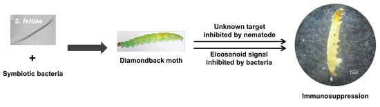
Host Immunosuppression Induced by Steinernema feltiae, an Entomopathogenic Nematode, through Inhibition of Eicosanoid Biosynthesis

1
Department of Plant Medicals, Andong National University, Andong 36729, Korea
2
School of Environmental Ecology and Tourism, Kyungpook National University, Sangju 37224, Korea
*
Author to whom correspondence should be addressed.
Insects 2020, 11(1), 33; https://doi.org/10.3390/insects11010033
Submission received: 10 December 2019 / Revised: 27 December 2019 / Accepted: 28 December 2019 / Published: 31 December 2019
(This article belongs to the Special Issue Insects, Nematodes and Their Symbiotic Bacteria)

Abstract

:
Steinernema feltiae K1 (Filipjev) (Nematode: Steinernematidae), an entomopathogenic nematode, was isolated and identified based on its morphological and molecular diagnostic characteristics. Its infective juveniles (IJs) were highly pathogenic to three lepidopteran (LC50 = 23.7–25.0 IJs/larva) and one coleopteran (LC50 = 39.3 IJs/larva) insect species. Infected larvae of the diamondback moth, Plutella xylostella (L.) (Insecta: Lepidoptera), exhibited significant reduction in phospholipase A2 (PLA2) activity in their plasma. The decrease of PLA2 activity was followed by significant septicemia of the larvae infected with S. feltiae. Insecticidal activity induced by S. feltiae was explained by significant immunosuppression in cellular immune responses measured by hemocyte nodule formation and total hemocyte count (THC). Although S. feltiae infection suppressed nodule formation and THC in the larvae, an addition of arachidonic acid (AA, a catalytic product of PLA2) rescued these larvae from fatal immunosuppression. In contrast, an addition of dexamethasone (a specific PLA2 inhibitor) enhanced the nematode’s pathogenicity in a dose-dependent manner. To discriminate the immunosuppressive activity of a symbiotic bacterium (Xenorhabdus bovienii (Proteobacteria: Enterobacterales)) from the nematode, kanamycin was applied to after nematode infection. It significantly inhibited the bacterial growth in the hemolymph. Compared to nematode treatment alone, the addition of antibiotics to nematode infection partially rescued the immunosuppression measured by phenol oxidase activity. Consequently, treatment with antibiotics significantly rescued the larvae from the insecticidal activity of S. feltiae. These results suggest that immunosuppression induced by infection of S. feltiae depends on its symbiotic bacteria by inhibiting eicosanoid biosynthesis, resulting in significant insect mortality. However, the addition of antibiotics or AA could not completely rescue the virulence of the nematode, suggesting that the nematode itself also plays a role in its insecticidal activity.

Graphical Abstract

1. Introduction

Insects exhibit cellular and humoral immune responses against various pathogens including microorganisms and multicellular parasites through their robust innate immune system [1]. Cellular immune responses use a hemocyte-spreading behavior to exhibit phagocytosis, nodule formation, and encapsulation [2]. In contrast, humoral immune responses represent chemical defenses using antimicrobial peptides (AMPs) and phenol oxidase (PO) [3,4]. PO catalyzes the melanin coat around encapsulated pathogens and produces chemically reactive quinones that are toxic to microbial pathogens [5]. PO activity is required for both cellular and humoral immune responses. Insects exert these immune functions soon after recognizing pathogen-associated molecular patterns by their pattern recognition receptors [6].
Entomopathogenic nematodes (EPNs) are especially promising because they possess various attributes that can allow them to be used as biological control agents for insect pests [7,8]. EPNs are classified into two genera: Steinernema and Heterorhabditis. Steinernema consists of at least 90 species including S. affine, S. beddingi, S. carpocapsae, S. feltiae, S. glaseri, S. longicaudum, and S. monticolum. Heterorhabditis consists of at least 20 species including H. bacteriophora, H. indica, and H. megidis [9]. Steinernema and Heterorhabditis EPNs possess symbiotic bacteria Xenorhabdus and Photorhabdus, respectively [10,11]. Generally, single nematode species can host one symbiotic bacterial species whereas each bacterial species can be hosted by more than one symbiotic nematode species [12]. Both EPN genera exhibit a converged evolutionary trait in their life cycle [13]. Infective juveniles (IJs) of EPNs live in the soil of diverse ecological systems and look for potential target insect hosts [14]. Once IJs infect a host through natural openings such as the mouth, anus, and spiracles, they can release symbiotic bacteria from nematode’s gut to insect hemolymph [15]. Bacteria can then suppress immune attacks of insect hosts to protect themselves and their symbiotic nematodes [16]. Under immunosuppressive conditions, these bacteria can multiply in the hemocoel and kill insects by septicemia or toxemia. Subsequently, nematodes can develop and reproduce in the insect cadaver [17].
To induce immunosuppression, symbiotic bacteria of EPNs can inhibit phospholipase A2 (PLA2) to shutdown eicosanoid biosynthesis of target insects [18]. Eicosanoids are oxygenated C20 polyunsaturated fatty acid (PUFA). They can be classified into three major groups: prostaglandins (PGs), leukotrienes (LTs), and epoxyeicosatrienoic acids (EETs) [19]. In vertebrates, arachidonic acid (AA) released from phospholipids (PLs) by PLA2 catalytic activity is usually a precursor for eicosanoid biosynthesis [20]. AA is then oxygenated into PGs by cyclooxygenase, LTs by lipoxygenase, and EETs by epoxidase activities of monooxygenases [21]. However, most terrestrial insects lack C20 PUFA presumably due to oxidative stress [22]. Instead, linoleic acid, which is rich in insect PLs, is released by PLA2 and changed into AA by desaturases and long chin fatty acid elongase [23]. Resulting PGs and LTs can mediate cellular immune responses by activating a hemocyte-spreading behavior [24] and humoral immune responses through activation of PO [25,26] and induction of AMP gene expression [27]. In mosquitoes, EETs are likely to mediate the humoral immunity in the midgut by elevating expression levels of some AMP genes [28]. The shutdown of eicosanoid biosynthesis by inhibiting PLA2 would prevent the synthesis of three different eicosanoids and lead to significant immunosuppression. This is one of pathogenicity strategies used by Xenorhabdus and Photorhabdus [16]. Thus, an addition of PLA2-catalytic product such as AA can significantly rescue bacterial pathogenicity [23,29]. However, such a rescue effect was little understood in insects infected with EPNs. If nematode contributes to the suppression of insect immune responses by targeting non-PLA2, AA addition would then incompletely rescue the immunosuppression induced by EPNs.
This study identified a new strain of S. feltiae and analyzed its pathogenicity by assessing insecticidal activity and immunosuppression via measuring PLA2 activity, counting total hemocyte count (THC) and phenoloxidase (PO) activity. It also tested a hypothesis that AA addition could rescue the immunosuppression induced by S. feltiae infection. Finally, it assessed the role of the nematode in suppressing immunosuppression by attenuating symbiotic bacteria by antibiotics.

2. Materials and Methods

2.1. Insect Rearing

Three different lepidopteran (Spodoptera exigua (Hűbner), Maruca vitrata (Fabricius), and Plutella xylostella (L.)) and one coleopteran (Tenebrio molitor L.) insects were used in this study. Larvae of S. exigua were collected from Welsh onion (Allium fistulsum L.) and reared on an artificial diet [30]. Larvae of P. xylostella were collected from cabbage (Brassica rapa L.) and reared on cabbage. Both S. exigua and P. xylostella were collected from Andong, Korea. Larvae of M. vitrata were collected from adzuki bean (Vigna angularis (Willd.) (Plantae: Fabales)) in Suwon, Korea and reared on an artificial diet [31]. Larvae of T. molitor were purchased from Bio Utility Institute, Inc. (Andong, Korea). Rearing conditions were: temperature of 25 ± 2 °C, photoperiod of 16:8 h (L:D), and relative humidity of 60 ± 5%.

2.2. Chemicals

Arachidonic acid (AA: 5,8,11,14-eicosatetraenoic acid) and dexamethasone (DEX: (11β, 16α)-9-fluoro-11,17,21-trihydroxy-16-methylpregna-1,4-diene-3) were purchased from Sigma-Aldrich Korea (Seoul, Korea) and dissolved in dimethyl sulfoxide (DMSO). Anticoagulant buffer (ACB, pH 4.5) consisting of 186 mM NaCl, 17 mM Na2EDTA, and 41 mM citric acid was prepared.

2.3. EPN Source and Culturing

An EPN isolate was obtained from a laboratory strain maintained in Nambo, Inc. (Jinju, Korea). The IJs were multiplied using fifth instar (L5) larvae of S. exigua. Briefly, approximately 200 IJs in 500 µL distilled water were applied to Petri dish (9 cm diameter, 3 cm height) containing an individual of L5 larva. Treated larvae were reared with an artificial diet for 3–5 days at rearing conditions. Brown-colored dead larvae were used to collect IJs using a White trap [32]. These collected IJs were then kept at 10 °C.

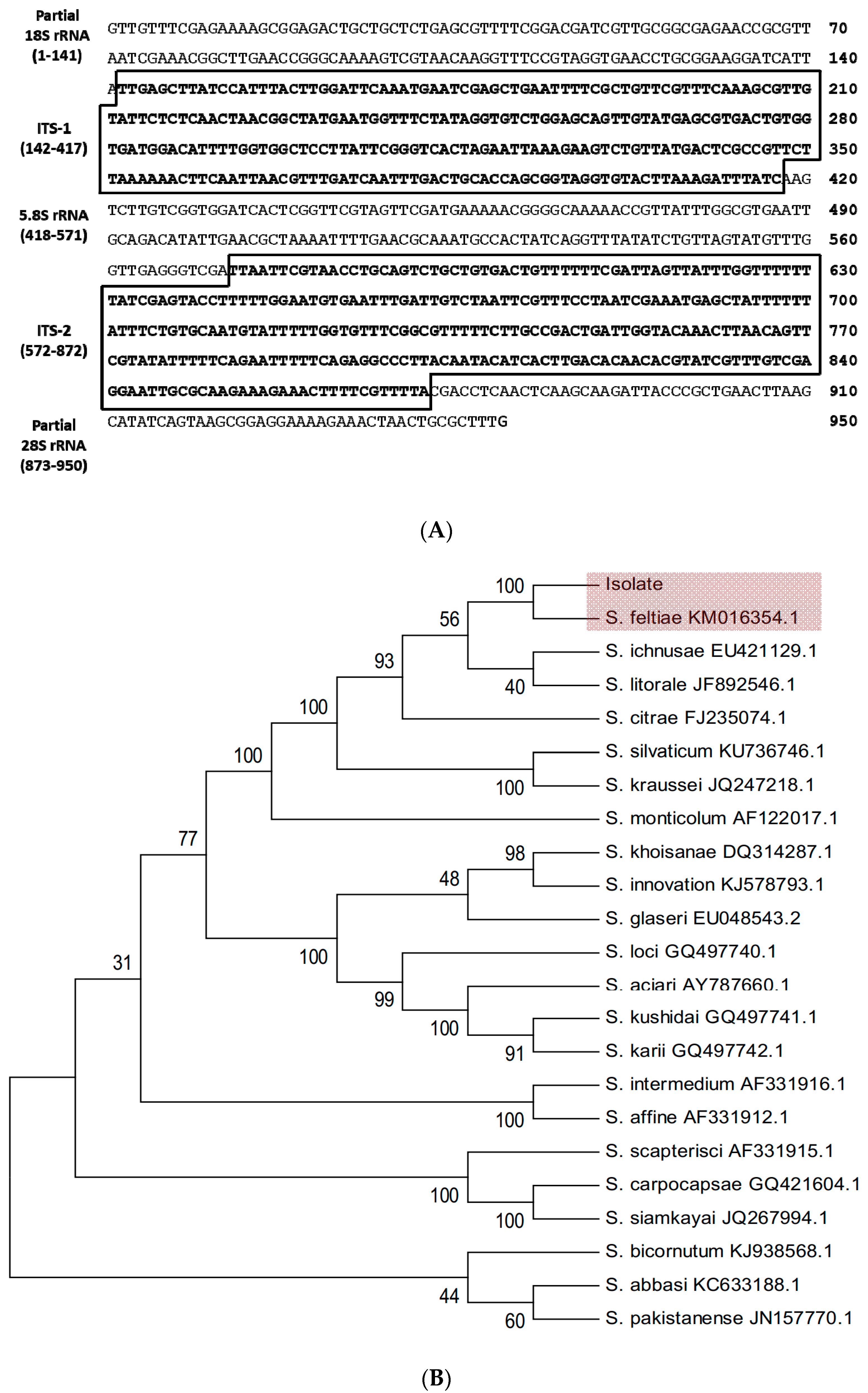
2.4. Molecular Identification of EPN Isolate

IJs (approximately 0.5 g) were used to extract genomic DNA (gDNA) using a method described by Kang et al. [33]. The internal transcribed spacer (ITS) region was amplified using primers GTTTTCCCAGTCACGACTTGATTACGTCCCTGCCCTTT and CAGGAAACAGCTATGACTTTCACTCGCCGTTACTAAGG reported by Vrain et al. [34]. Underlined sequences represent M13 forward and reverse sequences, respectively. For PCR amplification of the ITS region, the extracted gDNA (100 ng per 25 μL reaction volume) was used as a template. PCR consisted of 35 cycles of 1 min at 94 °C for denaturation, 1 min at 46 °C for annealing, and 1 min at 72 °C for extension. The PCR product was used for direct sequencing with M13 universal primers by Macrogen sequencing company (Seoul, Korea). Obtained nucleotide sequences were analyzed using BlastN program of the National Center for Biotechnology Information (www.ncbi.nlm.nih.gov). Phylogenetic tree analysis was performed using a Neighbor-Joining method with MEGA6 [35]. Bootstrap values on branches were estimated with 1000 repetitions.

2.5. Virulence Assay of EPN

Insecticidal activity of the EPN isolate was determined by applying different concentrations (0–320 IJs/larva) to filter paper in Petri dish containing 10 larvae. The experimental unit (=Petri dish) was replicated three times per nematode concentration. Third instar larvae of three lepidopteran species (S. exigua, M. vitrata, and P. xylostella) were used. Larvae of a coleopteran insect (T. molitor) with body length of approximately 2 cm were also used in the assay. Treated larvae were provided with diets and kept in rearing conditions. Median lethal concentration (LC50) was estimated from the mortality data at 72 h after treatment. Median lethal time (LT50) was estimated after treatment with an IJ concentration (80 IJs/larva). To estimate LT50, mortality was assessed every 8 h for 3 days after nematode infection. LC50 and LT50 values were estimated using EPA Probit program, version 1.5 (U.S. Environmental Protection Agency, Cincinnati, OH, USA).

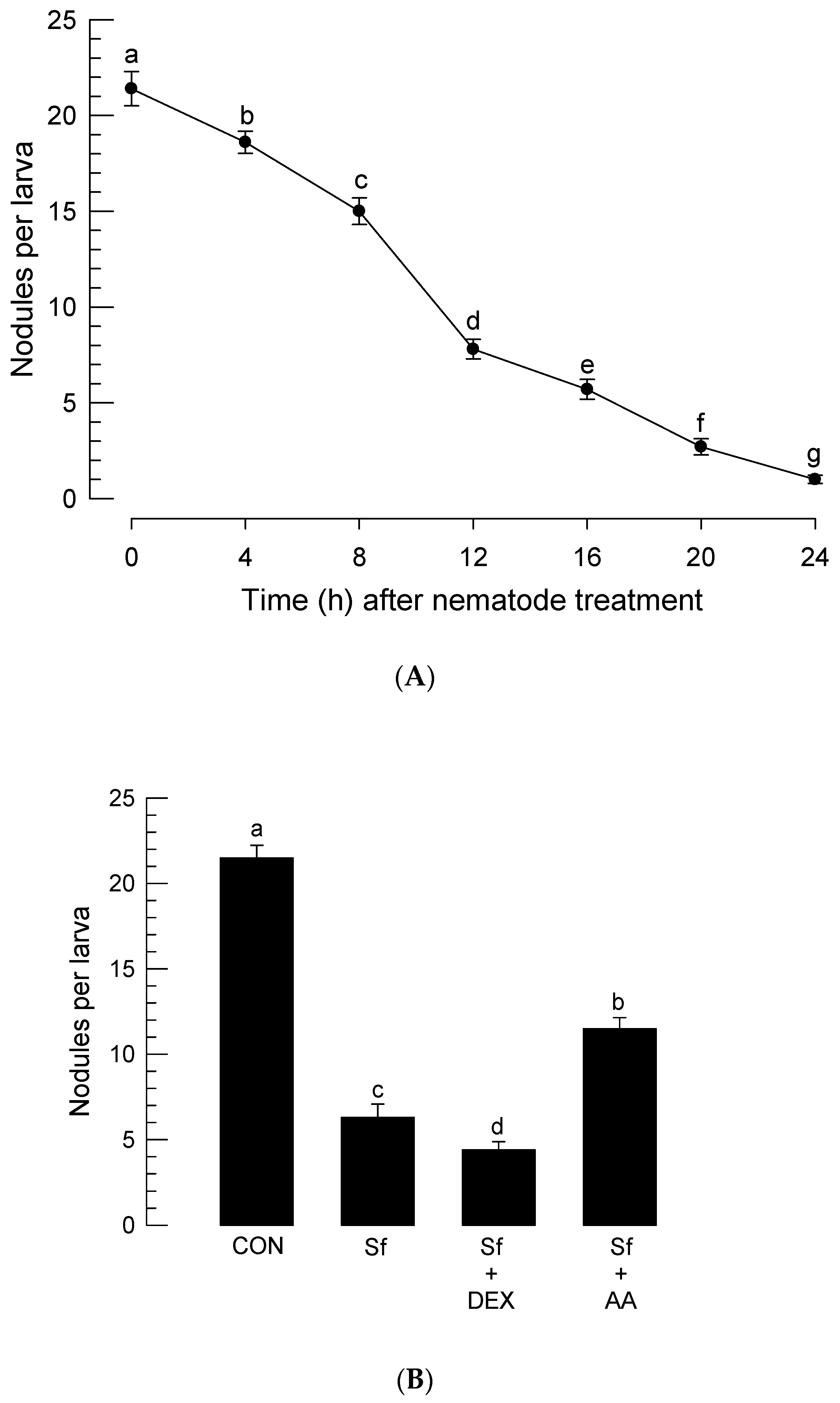
2.6. Assessment of Nodule Formation after Nematode Infection

For nodulation assay, Escherichia coli Top10 cells were used. These bacteria were heat-killed at 95 °C for 10 min. Fourth instar larvae of P. xylostella were infected with 80 IJs/larva. After nematode infection, larvae were randomly selected every 4 h and injected with 1.95 × 10⁴ cells/larva using a Sutter CO2 picopump injector (PV830, World Precision Instrument, Sarasota, FL, USA). Control larvae without nematode infection were also injected with bacteria at the same time periods for 24 h. At 8 h after bacterial injection, nodules were counted by dissecting larvae under a stereomicroscope (Stemi SV 11, Zeiss, Jena, Germany) at 50× magnification. For each time point, 10 larvae were used in each of control and nematode treatments.
To assess effects of AA or DEX on nodule formation in larvae infected with S. feltiae, at 24 h after treatment with nematode (80 IJs/larva), larvae were injected with 10 µg of AA or DEX along with bacterial injection. At 8 h after bacterial injection, the number of nodules formed was counted as described above.

2.7. Bioassay of P. xylostella Larvae Infected with S. feltiae with Addition of DEX or AA

For nematode treatment, 80 IJs/larva were used in the Petri dish assay (10 larvae of P. xylostella per dish). Just before nematode treatment, test larvae were injected with DEX or AA at different doses [0 (DMSO only), 0.1, 1, 10, and 100 µg]. Mortality was assessed at 72 h after nematode infection. Each treatment consisted of three replicates.

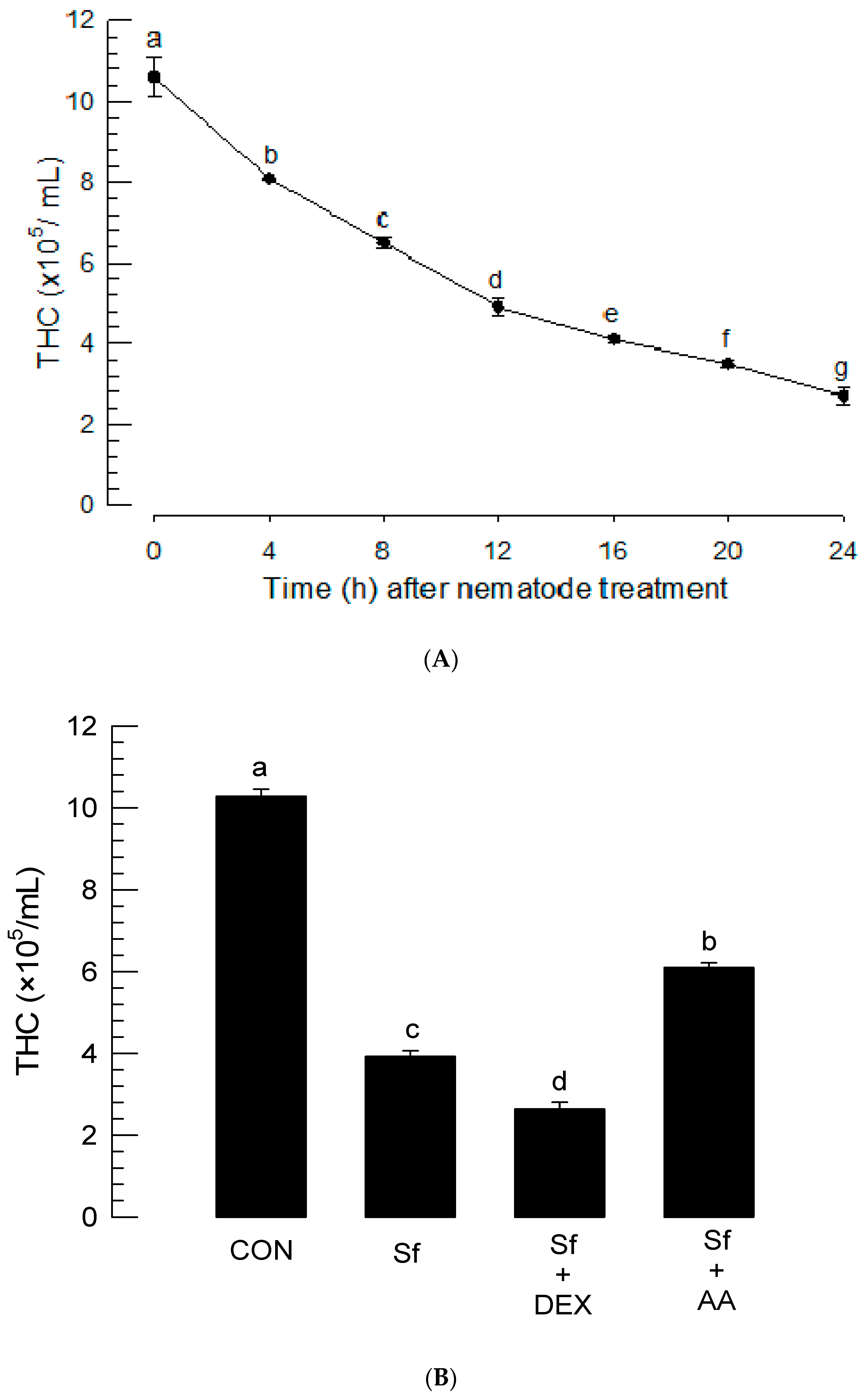
2.8. Total Hemocyte Count (THC) Analysis after Nematode Treatment

Fourth instar larvae of P. xylostella were treated with the nematode isolate (80 IJs/larva). Hemolymph was then collected into ACB by cutting a proleg on the abdomen and aspirating the exuded hemolymph with glass capillaries (TW100-4, World Precision Instrument, Sarasota, FL, USA) at 4 h intervals for 24 h. Hemocytes were counted with a hemocytometer (Neubauer improved bright line, Superior Marienfield, Lauda-Königshofen, Germany) under a phase contrast microscope (BX41, Olympus, Tokyo, Japan). Each treatment was independently replicated three times.
To evaluate effect of AA on THC, test larvae were injected with AA or DEX at a dose of 10 µg of just before nematode treatment (80 IJs/larva). Hemolymph was collected at 16 h after nematode treatment and assessed for THC as described above.

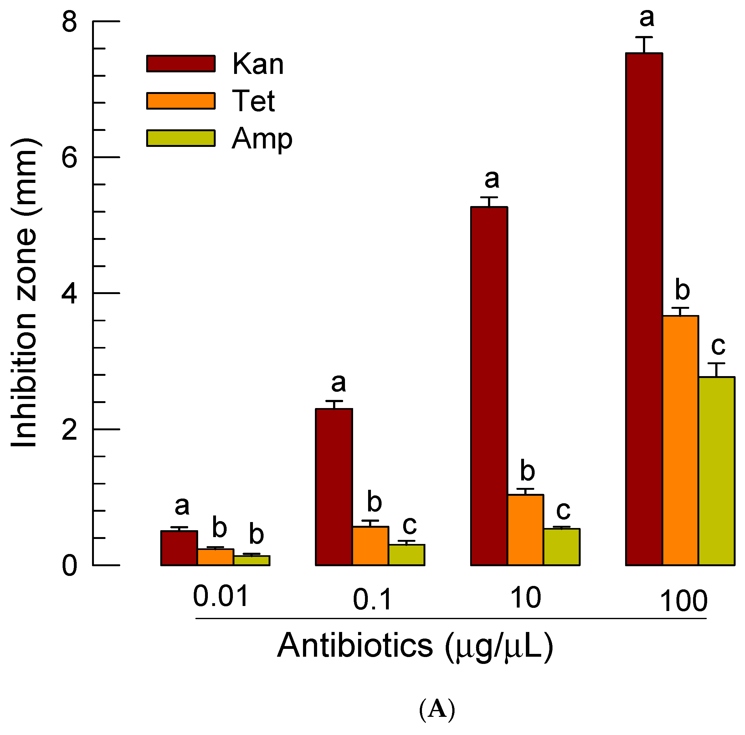
2.9. Antibiotic Screening against X. bovienii

Three different antibiotics (ampicillin, tetracycline, and kanamycin from Sigma-Aldrich Korea) were prepared at 0, 0.01, 0.1, 10, and 100 μg/µL. A symbiotic bacterium, X. bovienii SS 2004 (Proteobacteria: Enterobacterales), was kindly provided by Dr. Sophie Gaudriault (INRA, Monfelliae, France). The bacterium was cultured in Luria-Bertani (LB) medium at 28 °C. Thirty µL of an overnight grown bacterial suspension was plated onto LB agar medium and left for 5 min under a clean bench. Filter paper discs (10 mm diameter) impregnated with different antibiotics were placed on the agar surface. After 16 h of culture at 28 °C, the clear zone around the disc was measured in the distance from the edge of the disc. Each dose treatment was replicated three times.

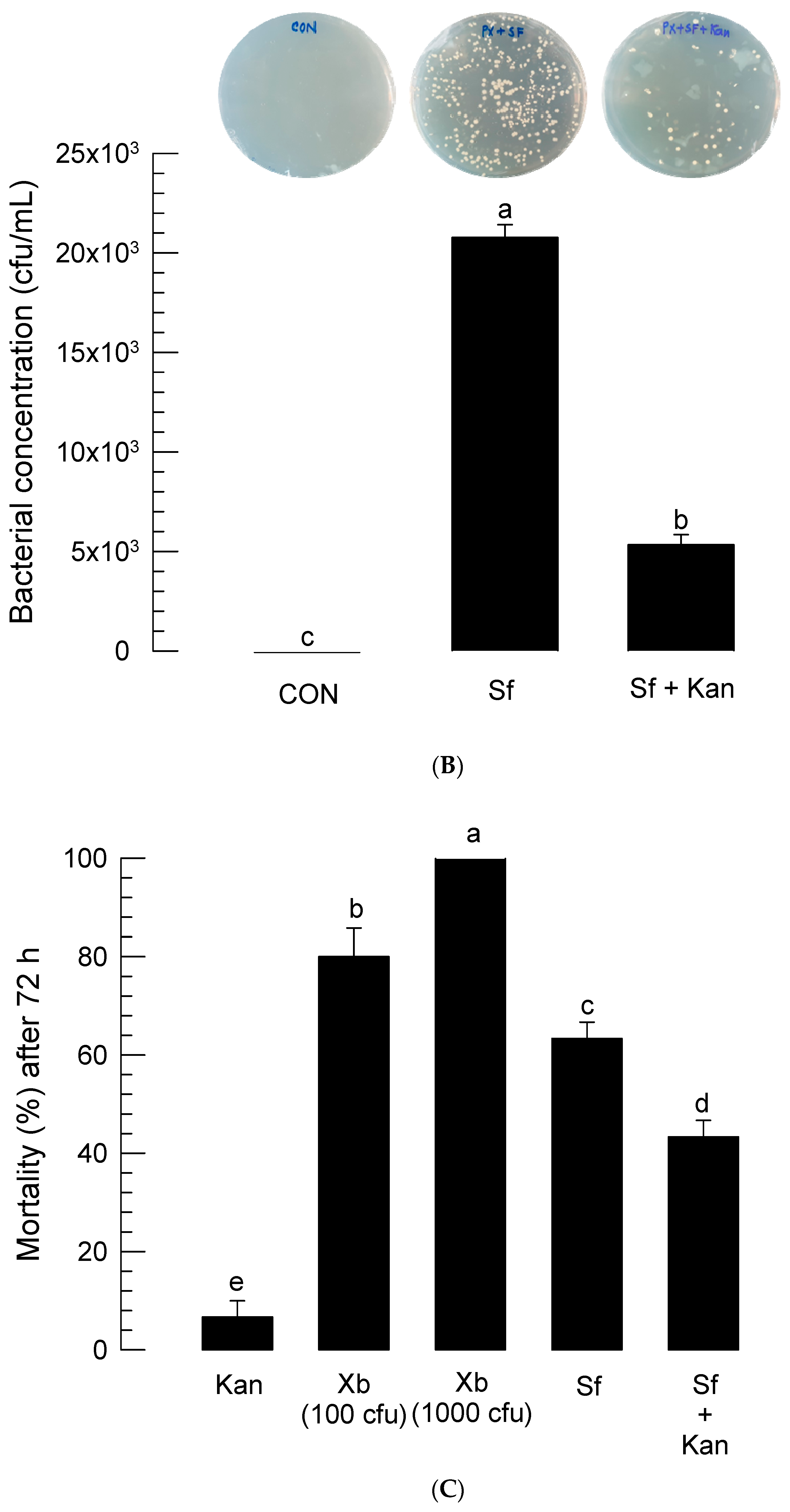
2.10. Effect of Kanamycin in Suppressing Bacterial Growth in P. xylostella Larvae Infected with S. feltiae

To determine the optimal concentration of kanamycin for injection, different kanamycin concentrations were injected to fourth instar larvae of P. xylostella. Mortality was measured at 72 h after treatment. Each treatment used 10 larvae and replicated three times.
Larvae were then injected with 0.1 µg kanamycin and treated with nematode S. feltiae at 80 IJs/larvae. At 8 h after the nematode infection, hemolymph was collected and plated onto LB plate to count bacterial colonies after culturing at 28 °C for 16 h. Each treatment was replicated three times. For each replication, 10 larvae were used.

2.11. Phenol Oxidase (PO) Enzyme Activity

PO activity was determined using 3,4-dihydroxy-L-phenylalanine (DOPA, Sigma-Aldrich Korea) as a substrate. DOPA was dissolved in 100 mM phosphate-buffered saline (PBS, pH 7.4). Fourth instar larvae of P. xylostella larvae were infected with nematodes at 80 IJs/larva. PO activity was measured at 8 h after the nematode infection. Hemolymph samples were collected from ~30 treated larvae as described above and centrifuged at 4 °C for 5 min at 800× g to obtain supernatant plasma samples. Each reaction volume (200 µL) for PO activity measurement consisted of 180 µL of 10 mM DOPA and 20 µL of plasma sample. Absorbance was read at 495 nm using a VICTOR multi label Plate reader (PerkinElmer, Waltham, MA, USA). PO activity was expressed as ΔABS/min/µL plasma. Each treatment consisted of three biologically independent replicates.
To determine the effect of kanamycin on PO activity of nematode-infected larvae, test larvae were injected with 0.1 µg of kanamycin before nematode infection. Untreated naive larvae were also analyzed. PO activity was analyzed at the same time interval as described above.

2.12. PLA2 Activity Measurement in Plasma after S. feltiae Infection

Fourth instar larvae of P. xylostella were used in PLA2 enzyme assay. For nematode treatment 80 IJs per larva were used by spraying on filter paper in a Petri dish. Control used water spray. At different time points, the hemolymph was collected as described above. These collected hemolymph samples were then centrifuged at 800× g for 3 min. The supernatant was used as a plasma sample. Each plasma sample was obtained from 10 larvae. Each treatment was replicated three times. To measure PLA2 activity of the collected plasma sample, a commercial assay kit (sPLA2 Assay Kit, Cayman Chemical, Ann Arbor, MI, USA) was used with diheptanoyl thiophosphatidylcholine as a substrate for the enzyme. Protein concentration was determined by Bradford [36] assay using bovine serum albumin as standard. Catalyzed product was then allowed to react with Ellman’s reagent (5,5′-dithio-bis-(2-nitrobenzoic acid), DTNB) to create 5-thio-2-nitrobenzoic acid, a colored product. DTNB was prepared in 10 mM in 0.4 M Tris buffer (pH 8.0). Assay buffer was 25 mM Tris (pH 7.5) containing 10 mM CaCl2, 100 mM KCl, and 0.3 mM Triton X-100. A reaction volume of 175 μL contained 10 μL plasma sample, 10 μL DTNB, 5 μL assay buffer, and 150 μL of 1.66 mM substrate. As a negative control, the same volume of reaction mixture consisted of 10 μL DTNB, 15 μL assay buffer, and 150 μL substrate. Absorbance changes at 405 nm were measured and plotted to obtain the slope of a linear portion of the curve. Absorbance for non-enzymatic blank control was calculated and subtracted from sample wells. The actual extinction coefficient for DTNB was 10.66 mM−1cm−1.

2.13. Statistical Analysis

Mortality data were transformed by arsine for analysis of variance (ANOVA). Means and variances of treatments were analyzed by one-way ANOVA using PROC GLM of SAS program [37]. All means were compared by least squared difference (LSD) test at Type I error = 0.05.

3. Results

3.1. Identification of a Nematode Isolate

IJs of the nematode isolates were infective to larvae of P. xylostella. Larvae were killed by the nematode, becoming brown-colored cadavers (Figure 1A). The nematode infection reproduced several hundreds of subsequent IJs (Figure 1B). In cadavers, different developmental stages of nematodes were observed (Figure 1C–G).
To identify the nematode isolate, ITS regions were amplified and sequenced (Figure 2A). A total of 950 bp was sequenced for the isolate and compared with a known sequence of Caenorhabditis elegans (Nematoda: Rhabditida) [38] to confirm that these sequenced regions included ITS. The determined sequence contained 5.8S rRNA, ITS-1, and ITS-2 regions as well as partial sequences of 18S and 28S rRNAs. Its sequence was deposited at GenBank with an accession number of MN093395.1. The sequence showed high similarities (>99%) with known ITS sequences of several S. feltiae strains (Table 1). To evaluate the relationship between the isolate and other known Steinernema spp., a phylogenetic tree was constructed using ITS sequences (Figure 2B). The isolate was significantly (bootstrap value = 100) clustered with a local isolate of S. feltiae from Czech Republic [39] among 25 different species of Steinernema.
Molecular identification was further supported by morphometric characteristics of the isolate. All morphological characteristics were obtained from IJ stage, including total body length and width (Figure 1C), esophagus length (Figure 1D), and tail length (Figure 1E). Sizes or ratios of these morphological characteristics were within variations of those in known S. feltiae species (Table 2). The isolate was identified as S. feltiae K1.

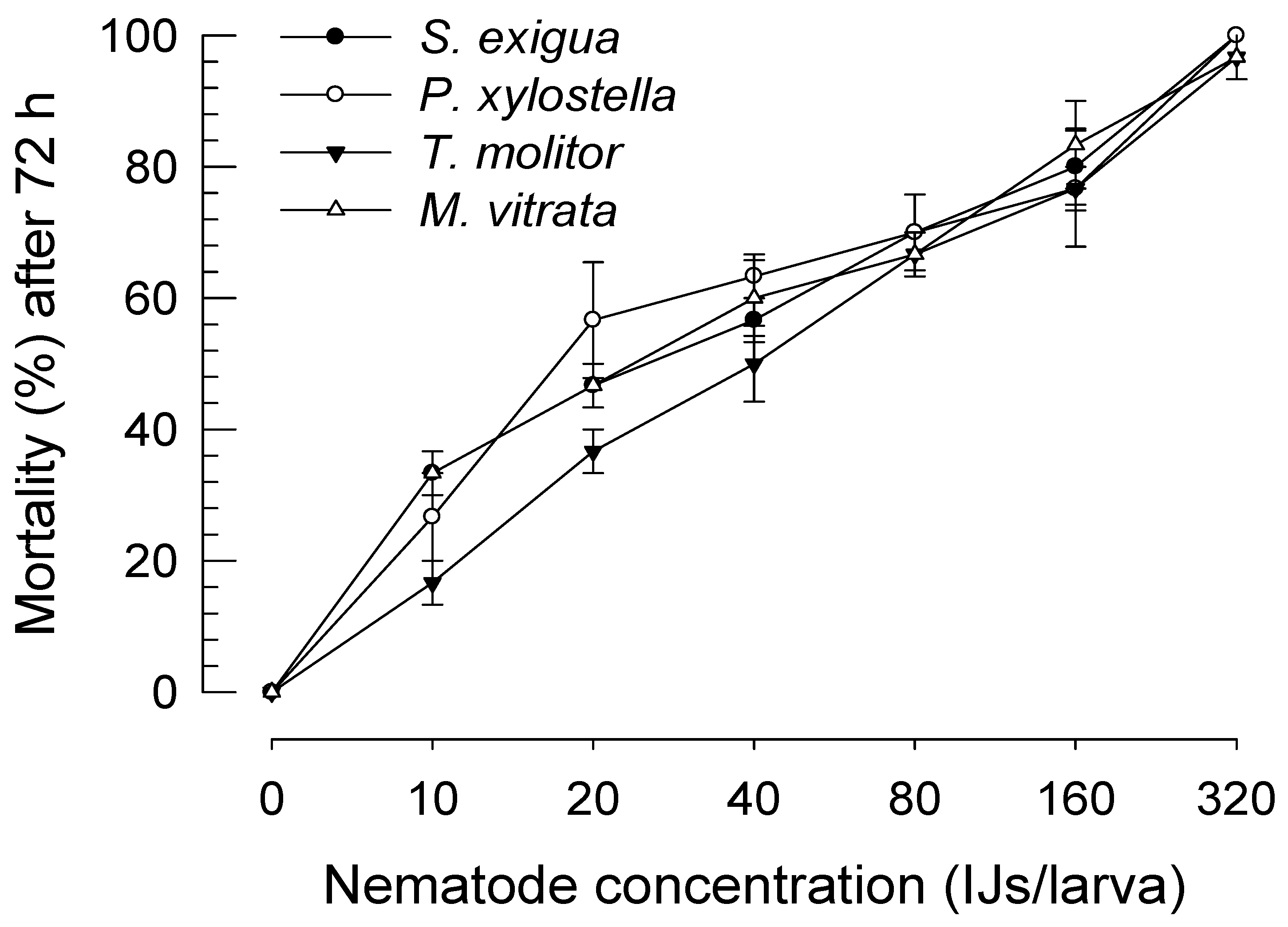
3.2. Insecticidal Activity of S. feltiae K1

S. feltiae K1 was virulent against larvae of three lepidopteran (P. xylostella, M. vitrata, and S. exigua) species and one coleopteran (T. molitor) species (Figure 3). Different concentrations of IJ treatment resulted in significant differences in mortality of these four species of insects (F = 246.74; df = 6, 56; p < 0.0001). Susceptibilities of these insect species to the nematode were slightly different, with T. molitor being the least susceptible to infection (F = 3.49; df = 3, 56; p = 0.0215). Also, estimates of LC50 indicated that S. exigua, P. xylostella, and M. vitrata larvae were much more susceptible to the nematode than T. molitor larvae (Table 3). However, there was little significant difference in the speed-to-kill of S. feltiae K1 against these four insect species based on similar LT50s.

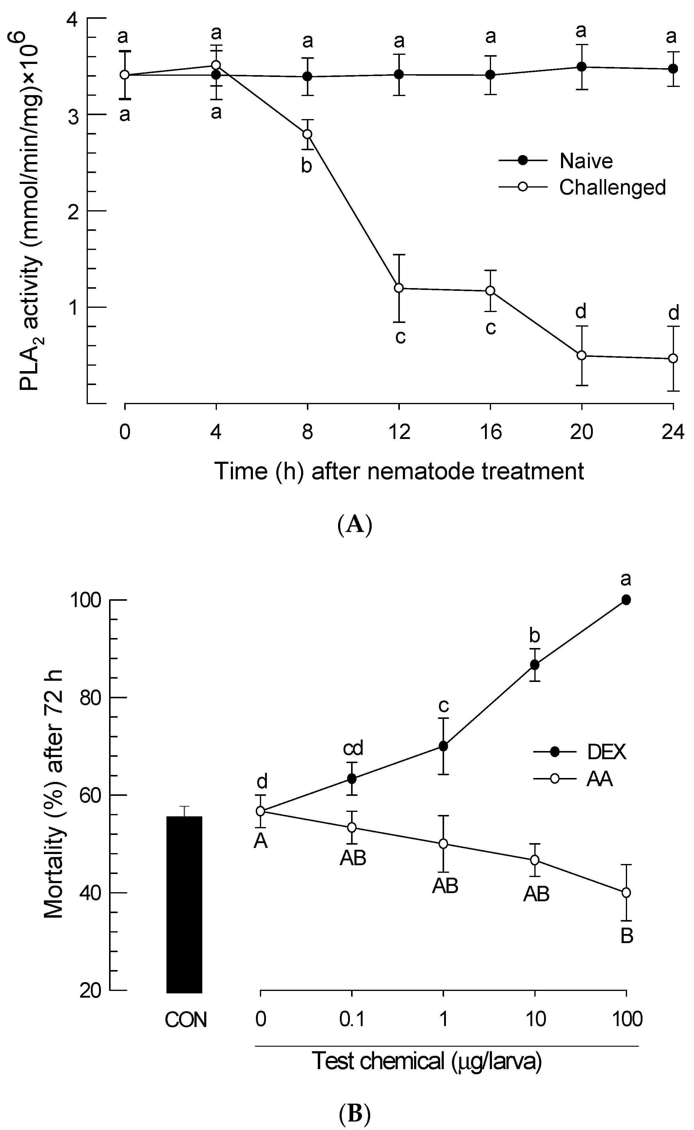
3.3. Insecticidal Virulence of S. feltiae K1 by Down-Regulating PLA2 Activity

After P. xylostella larvae were infected with S. feltiae K1, PLA2 activity changes in the plasma of infected larvae were monitored for 24 h (Figure 4A). The nematode infection significantly (p < 0.05) inhibited plasma PLA2 activity. The inhibition began at 8 h after treatment with the nematode. At 24 h after the treatment, some infected larvae began to die. In contrast, untreated (naive) larvae did not show any significant change in PLA2 activity during the test period.
To see the effect of PLA2 inhibition on insecticidal activity of the nematode, dexamethasone (‘DEX’, a PLA2 inhibitor) was used for treatment. It further significantly (p < 0.05) increased the virulence of the nematode against P. xylostella larvae (Figure 4B). In contrast, addition of arachidonic acid (‘AA’, a catalytic product of PLA2) to larvae infected with S. feltiae K1 at dose of 100 μg/larva significantly (p < 0.05) rescued these larvae from the lethality.

3.4. Infection of S. feltiae K1 Resulted in Immunosuppression and Cytotoxicity

The inhibition of PLA2 by the infection of S. feltiae K1 suggested that there might be an immunosuppression due to the lack of eicosanoid immune mediators [16]. To test this hypothesis, a cellular immune response measured by nodule formation was monitored for 24 h after infection with the nematode (Figure 5A). The infection with the nematode significantly (p < 0.05) suppressed nodule formation. The suppression began at 4 h after infection with the nematode. At 24 h post-infection, larvae infected with S. feltiae K1 almost lost their nodule formation ability. To test a hypothesis that the immunosuppression might be induced by downregulated PLA2 activity inhibited by the nematode, DEX or AA was added to nematode-infected larvae (Figure 5B). DEX treatment further significantly (p < 0.05) suppressed nodule formation while AA addition significantly (p < 0.05) rescued such immunosuppression.
To explain the insecticidal activity of S. feltiae K1, cytotoxicity of the nematode infection was assessed in hemocytes (Figure 6A). P. xylostella larvae had over 106 hemocytes per mL of hemolymph. With increase of infection period, S. feltiae K1 significantly (p < 0.05) induced cytotoxicity against hemocytes of P. xylostella. Significant cytotoxicity began at 4 h post-infection. At 24 h, infected larvae lost more than 70% hemocytes and had only 3 × 10⁵ hemocytes per mL of hemolymph. To explain the cytotoxicity caused by the inhibitory activity of S. feltiae K1 against PLA2, DEX, or AA was added to nematode-infected larvae (Figure 6B). DEX treatment significantly (p < 0.05) potentiated the cytotoxicity of nematode while AA addition significantly (p < 0.05) rescued hemocytes.

3.5. Role of Symbiotic Bacteria in Insecticidal Activity of S. feltiae K1

S. feltiae possesses species-specific symbiotic bacterium X. bovienii in the intestine [41]. This suggests that the insecticidal activity of S. feltiae K1 might be induced by both symbiotic bacteria and their host, the nematode. To discriminate bacterial contribution from the nematode, treatment with antibiotics was performed after nematode infection. First, we selected an effective antibiotic against X. bovienii based on an inhibition zone assay (Figure 7A). Among three antibiotics used, kanamycin was the most effective one in suppressing the bacterial growth at all test concentrations. We then treated P. xylostella larvae with different concentrations of kanamycin (Figure S1). At a high concentration (100 μg/larva), kanamycin was harmful for the survival of P. xylostella larvae. When a relatively low concentration (0.1 μg/larva) of kanamycin was injected to larvae already treated with S. feltiae K1, bacterial growth was suppressed by more than 75% (Figure 7B). Under these antibacterial treatment conditions, contributions of bacteria and nematode to insecticidal activity against P. xylostella were separately assessed (Figure 7C). Kanamycin treatment along with nematode infection significantly (p < 0.05) decreased the insecticidal activity induced by infection of S. feltiae K1 alone. However, it did not completely rescue nematode-infected larvae.
PO activity of P. xylostella, which would be required for both cellular and humoral immune responses, was significantly (p < 0.05) suppressed by the injection with live X. bovienii bacteria while such activity was significantly enhanced by the injection with heat-killed X. bovienii bacteria (Figure 7D). The nematode infection also significantly suppressed PO activity. However, an addition of kanamycin partially rescued the PO activity suppressed by the nematode infection.

4. Discussion

EPN infection can lead to septicemia of target insects. Such septicemia is induced by immunosuppression [29]. Eicosanoids play a crucial role in mediating cellular and humoral immune responses in insects [16]. Both symbiotic bacteria (Xenorhabdus and Photorhabdus) of EPNs can inhibit PLA2 to shut down eicosanoid biosynthesis of target insects [18]. Indeed, Hasan et al. [23] have demonstrated a positive correlation between the degree of PLA2 inhibition by EPNs and their insecticidal activities. However, the role of EPN without symbiotic bacteria in killing target insects by immunosuppression was unclear. This study analyzed independent roles of bacteria and nematodes in inducing immunosuppression and insecticidal activity ultimately.
A new isolate of S. feltiae K1 was identified based on molecular and morphological characteristics. ITS sequence and IJ morphological characteristics indicated that the nematode isolate was a strain of S. feltiae. ITS sequences are powerful for identifying EPNs without morphological characteristics. For example, in an ecological analysis of EPNs and their symbiotic bacteria in Thailand, ITS sequences alone have been used to identify EPNs collected from soil samples [42]. This current study supports the usefulness of ITS sequence for EPN identification because morphological characteristics clearly support the molecular identification.
Infection by S. feltiae K1 had significant lethal effects on four different insect species (three lepidopteran species and one coleopteran species). There was a variation in insecticidal activities among insect species. Especially, the coleopteran species, T. molitor, was less susceptible to the nematode infection than the three lepidopteran species. Compared to other similar nematode species [23], S. feltiae K1 was more potent (more than two times based on LC50 values) than S. carpocapsae against the same host under the same assay conditions. However, LT50s (= speed-to-killing insects) of S. feltiae showed some variations against different insect species. This indicates that the insecticidal activity of S. feltiae K1 is not determined by IJ density only if we knew the number of the infected IJs within the target insects. Symbiotic bacteria might also play a role in the insecticidal activity of the nematode. Jung and Kim [43] have measured the speed-to-kill of X. nematophila (Proteobacteria: Enterobacterales) is 16 h against P. xylostella larvae. Our current LT50s ranged from 46 to 50 h post-infection. Although bacterial species are different, at least 30 h might be necessary to reach the lethal level of bacterial population required to cause lethal septicemia. Thus, the initial 30 h post-infection would be a struggling period between host (insect) defense and virulence of the nematode/bacterial complex. The pathogen–insect interaction involved in the immunosuppression induced by the nematode/bacterial complex was further analyzed in the following section.
Cellular immune response of P. xylostella was severely impaired by the infection of S. feltiae K1. More than 20 nodules were formed in each larva of P. xylostella in response to nonpathogenic bacterial infection. However, the nematode infection progressively suppressed the cellular immune response and led to one or two nodules per larva in response to the same number of bacteria used for challenge at 24 h post-infection. With temporal decrease of cellular immune response, there was a similar decrease of PLA2 activity in the plasma of P. xylostella after the nematode infection. PLA2 activity in the plasma is due to a secretory type of PLA2 (sPLA2) in Spodoptera exigua [44]. Our current PLA2 activity was measured using sPLA2 substrate. Thus, the decrease of PLA2 activity in the plasma of P. xylostella can be explained by the inhibition of sPLA2 of S. feltiae K1. The sPLA2 of S. exigua plays a crucial role in eicosanoid biosynthesis. It also exhibits a direct antibacterial activity like other antimicrobial peptides [44]. Thus, inhibition of plasma PLA2 activity should be required for the pathogenicity of S. feltiae K1 to protect the symbiotic bacteria. In addition, eicosanoid biosynthesis can be inhibited by suppressing PLA2 activity. Eicosanoids are known to mediate nodule formation of hemocytes by activating the hemocyte-spreading behavior via stimulation of actin polymerization factors [2]. After infection with S. feltiae K1, PLA2 activity and nodulation were also downregulated. Hemolysis accompanying decrease of THC was induced by the infection of S. feltiae K1. Interestingly, the cytotoxicity of S. feltiae K1 could be manipulated by the addition of DEX or AA. This suggests that the cytotoxicity of S. feltiae K1 is influenced by eicosanoids. Cho and Kim [45] have reported that Xenorhabdus and Photorhabdus are cytotoxic to insect hemocytes via apoptosis. Hemocytes of the larvae infected with these bacteria exhibited membrane blebbing, nuclear chromatin condensation, and DNA fragmentation [45]. This suggests that S. feltiae K1 can release its symbiotic bacteria that can then synthesize and release apoptosis-inducing factor(s) to kill hemocytes. Indeed, X. bovienii, a symbiotic bacterium of S. feltiae, can synthesize cytotoxic xenocycloin compounds [46]. Thus, the regulation of cytotoxicity by DEX or AA can be explained by the control of immune responses of P. xylostella which manipulates selective pressure on bacterial survival and subsequent xenocycloin production level.
Effective treatment with antibiotics against X. bovienii rescued the larvae infected with S. feltiae K1. Kanamycin was the most effective one in preventing the growth of X. bovienii. Indeed, treatment with antibiotics suppressed the bacterial population in the hemolymph of P. xylostella larvae after infection with S. feltiae K1. Under this condition, S. feltiae K1 partially lost its insecticidal activity. This clearly exhibits the insecticidal role of symbiotic bacteria. There is a variation in insecticidal activity among X. bovienii strains, in which the composition of functional genome associated with virulence is different [47]. Interestingly, treatment with antibiotics to kill symbiotic bacteria did not completely rescue the larvae infected by S. feltiae K1. This suggests that the nematode itself possesses insecticidal activity without the presence of symbiotic bacteria. Furthermore, PO analysis indicated that treating larvae infected by the nematode with antibiotics could still significantly suppress the induction of the enzyme activity. These results suggest that the contribution of nematode to immunosuppression is distinct from that of the symbiotic bacteria. CS03 strain of X. bovienii is a non-virulent strain. However, it exhibits low virulence when it is associated with its Steinernema symbiont [48]. This supports the role of the nematode in insecticidal activity without the presence of symbiotic bacteria. Metabolite analysis of S. feltiae IJs using mass spectrometry identified 266 proteins, among which 52 proteins were conserved among nematode metabolites known to be associated with tissue-damaging and immune-modulating proteins [49]. In addition, the nematode cuticle plays a crucial role in avoiding insect immune surveillance by disguising nematode surface [50]. Thus, several chemical and physical factors derived from S. feltiae K1 might be responsible for its immunosuppression and insecticidal activity. However, we cannot avoid the pathogenic contribution of the gut microbiota in the host insects, which might infect the insect hemocoel during penetration of IJs from gut lumen. To clarify the role of nematode on the immunosuppression, use of axenic IJs might be optimal in a future study rather than use of antibiotics to kill the symbiotic bacteria.

5. Conclusions

This study identified a nematode isolate to be S. feltiae K1 based on its morphological and molecular characteristics. S. feltiae K1 was highly pathogenic to lepidopteran and coleopteran insect pests. Its pathogenicity was dependent on the inhibition of target insect PLA2 activity because addition of AA, an enzyme catalytic product, significantly rescued the virulence of S. feltiae K1. Treatment with an antibiotic to inhibit the growth of symbiotic bacteria significantly rescued nematode-infected insects. However, this study indicated a pathogenic role of the nematode in suppressing insect immune response. It might be independent of the contribution of its symbiotic bacteria.

Supplementary Materials

The following are available online at https://www.mdpi.com/2075-4450/11/1/33/s1, Figure S1: Screening to find the optimum concentration of kanamycin against fourth instar larva of P. xylostella by mortality assay. Kanamycin at indicated concentration was injected into the hemocoel. Each concentration was replicated three times. For each replicate, 10 larvae of target insects were used. Different letters above standard error bars indicate significant differences among means at Type I error = 0.05 (LSD test).

Author Contributions

Conceptualization, Y.K.; Methodology, D.L. and M.C.R.; Formal Analysis, M.C.R.; Writing—Original Draft Preparation, M.C.R.; Writing—Review and Editing, Y.K.; Funding Acquisition, Y.K. All authors have read and agreed to the published version of the manuscript.

Funding

This study was supported by a grant (no. 2017R1A2133009815) of the National Research Foundation (NRF) funded by the Ministry of Science, ICT and Future Planning, Republic of Korea.

Acknowledgments

We appreciate Youngim Song (Andong National University, Andong, Korea) for supplying materials and purchasing chemicals. We also thank Abdullah Al Baki and Mohammad Vatanparast (Andong National University) for their assistance in insect collections.

Conflicts of Interest

The authors declare no conflict of interest.

References

  1. Lemaitre, B.; Hoffmann, J. The host defense of Drosophila melanogaster. Annu. Rev. Immunol. 2007, 25, 697–743. [Google Scholar] [CrossRef] [Green Version]
  2. Ahmed, S.; Kim, Y. PGE2 mediates cytoskeletal rearrangement of hemocytes via Cdc42, a small G protein, to activate actin-remodeling factors in Spodoptera exigua (Lepidoptera: Noctuidae). Arch. Insect Biochem. Physiol. 2019, 102, e21607. [Google Scholar] [CrossRef]
  3. Imler, J.L.; Bulet, P. Antimicrobial peptides in Drosophila: Structures, activities and gene regulation. Chem. Immunol. Allergy. 2005, 86, 1–21. [Google Scholar]
  4. Söderhäll, K.; Cerenius, L. Role of the prophenoloxidase-activating system in invertebrate immunity. Curr. Opin. Immunol. 1998, 10, 23–28. [Google Scholar] [CrossRef]
  5. Castillo, J.C.; Renolds, S.E.; Eleftherianos, I. Insect immune responses to nematode parasites. Trends Parasitol. 2011, 27, 537–547. [Google Scholar] [CrossRef] [PubMed]
  6. Lu, Y.; Su, F.; Li, Q.; Zhang, J.; Li, Y.; Tang, T.; Hu, Q.; Yu, X.Q. Pattern recognition receptors in Drosophila immune responses. Dev. Comp. Immunol. 2020, 102, 103468. [Google Scholar] [CrossRef] [PubMed]
  7. Georgis, R.; Manweiler, S.A. Entomopathogenic nematodes: A developing biological control technology. Agric. Zool. Rev. 1994, 6, 63–94. [Google Scholar]
  8. Poinar, G. Nematode biopesticides. Fundam. Appl. Nematol. 1998, 21, 733–737. [Google Scholar]
  9. Shapiro-Ilan, D.I.; Hazir, S.; Glazer, I. Basic and applied research: Entomopathogenic nematodes. In Microbial Agents for Control of Insect Pests: From Discovery to Commercial Development and Use; Lacey, L.A., Ed.; Academic Press: Amsterdam, The Netherlands, 2017; pp. 91–105. [Google Scholar]
  10. Akhurst, R.J. Taxonomic study of Xenorhabdus, a genus of bacteria symbiotically associated with insect pathogenic nematodes. Int. J. Syst. Bacteriol. 1983, 33, 38–45. [Google Scholar] [CrossRef] [Green Version]
  11. Boemare, N.E. Biology, taxonomy and systematics of Photorhabdus and Xenorhabdus. In Entomopathogenic Nematology; Gaugler, R., Ed.; CABI: New York, NY, USA, 2002; pp. 35–56. [Google Scholar]
  12. Goodrich-Blair, H.; Clarke, D.J. Mutualism and pathogenesis in Xenorhabdus and Photorhabdus: Two roads to the same destination. Mol. Microbiol. 2007, 64, 260–268. [Google Scholar] [CrossRef]
  13. Kaya, H.K.; Aguillera, M.M.; Alumai, A.; Choo, H.Y.; De la Torre, M.; Fodor, A.; Ganguly, S.; Hazir, S.; Lakatos, S.; Pye, A.; et al. Status of entomopathogenic nematodes and their symbiotic bacteria from selected countries or regions of the world. Biol. Control 2006, 38, 134–155. [Google Scholar] [CrossRef]
  14. Dillman, A.R.; Chaston, J.M.; Adams, B.J.; Ciche, T.A.; Goodrich-Blair, H.; Stock, S.P.; Sternberg, P.W. An entomopathogenic nematode by any other name. PLoS Pathog. 2012, 8, e1002527. [Google Scholar] [CrossRef] [PubMed] [Green Version]
  15. Dowds, B.C.A.; Peters, A. Virulence mechanisms. In Entomopathogenic Nematology; Gaugler, R., Ed.; CABI: New York, NY, USA, 2002; pp. 79–98. [Google Scholar]
  16. Kim, Y.; Ahmed, S.; Stanley, D.; An, C. Eicosanoid-mediated immunity in insects. Dev. Comp. Immunol. 2018, 83, 130–143. [Google Scholar] [CrossRef] [PubMed]
  17. Herbert, E.E.; Goodrich-Blair, H. Friend and foe: The two faces of Xenorhabdus nematophila. Nature Rev. Microbiol. 2007, 5, 634–646. [Google Scholar] [CrossRef] [PubMed]
  18. Kim, Y.; Ji, D.; Cho, S.; Park, Y. Two groups of entomopathogenic bacteria, Photorhabdus and Xenorhabdus, share an inhibitory action against phospholipase A2 to induce host immunodepression. J. Invertebr. Pathol. 2005, 89, 258–264. [Google Scholar] [CrossRef] [PubMed]
  19. Stanley, D.W. Eicosanoids in Invertebrate Signal Transduction Systems; Princeton University Press: Princeton, NJ, USA, 2000. [Google Scholar]
  20. Burke, J.E.; Dennis, E.A. Phospholipase A2 structure/function, mechanism, and signaling. J. Lipid Res. 2009, 50, S237–S242. [Google Scholar] [CrossRef] [Green Version]
  21. Stanley, D.; Kim, Y. Prostaglandins and other eicosanoids in insects: Biosynthesis and biological actions. Front. Physiol. 2019, 9, 1927. [Google Scholar] [CrossRef] [Green Version]
  22. Stanley, D.; Kim, Y. Why most insects have very low proportions of C20 polyunsaturated fatty acids: The oxidative stress hypothesis. Arch. Insect Biochem. Physiol. 2020, 103, e21622. [Google Scholar] [CrossRef]
  23. Hasan, M.A.; Ahmed, S.; Mollah, M.M.I.; Lee, D.; Kim, Y. Variation in pathogenicity of different strains of Xenorhabdus nematophila; Differential immunosuppressive activities and secondary metabolite production. J. Invertebr. Pathol. 2019, 166, 107221. [Google Scholar] [CrossRef]
  24. Shrestha, S.; Kim, Y. Biochemical characteristics of immune-associated phospholipase A2 and its inhibition by an entomopathogenic bacterium, Xenorhabdus nematophila. J. Microbiol. 2009, 47, 774–782. [Google Scholar] [CrossRef]
  25. Shrestha, S.; Kim, Y. Eicosanoids mediate prophenoloxidase release from oenocytoids in the beet armyworm, Spodoptera exigua. Insect Biochem. Mol. Biol. 2008, 38, 99–112. [Google Scholar] [CrossRef] [PubMed]
  26. Shrestha, S.; Kim, Y.; Stanley, D. PGE2 induces oenocytoid cell lysis via a G protein-coupled receptor in the beet armyworm, Spodoptera exigua. J. Insect Physiol. 2011, 57, 1568–1576. [Google Scholar] [CrossRef]
  27. Hwang, J.; Park, Y.; Kim, Y.; Hwang, J.; Lee, D. An entomopathogenic bacterium, Xenorhabdus nematophila, suppresses expression of antimicrobial peptides controlled by Toll and Imd pathways by blocking eicosanoid biosynthesis. Arch. Insect Biochem. Physiol. 2013, 83, 151–169. [Google Scholar] [CrossRef] [PubMed]
  28. Xu, J.; Morisseau, C.; Yang, J.; Lee, K.S.; Kamita, S.G.; Hammock, B.D. Ingestion of the epoxide hydrolase inhibitor AUDA modulates immune responses of the mosquito, Culex quinquefasciatus during blood feeding. Insect Biochem. Mol. Biol. 2016, 76, 62–69. [Google Scholar] [CrossRef] [PubMed] [Green Version]
  29. Park, Y.; Kim, Y. Eicosanoids rescue Spodoptera exigua infected with Xenorhabdus nematophilus, the symbiotic bacteria to the entomopathogenic nematode Steinernema carpocapsae. J. Insect Physiol. 2000, 46, 1469–1476. [Google Scholar] [CrossRef]
  30. Goh, H.G.; Lee, S.G.; Lee, B.P.; Choi, K.M.; Kim, J.H. Simple mass-rearing of beet armyworm, Spodoptera exigua (Hübner) (Lepidoptera: Noctuidae), on an artificial diet. Korean J. Appl. Entomol. 1990, 29, 180–183. [Google Scholar]
  31. Jung, J.K.; Seo, B.Y.; Cho, C.R.; Kwon, Y.H.; Kim, G.H. Occurrence of lepidopteran insect pests and injury aspects in Adzuki bean fields. Korean J. Appl. Entomol. 2009, 48, 29–35. [Google Scholar] [CrossRef]
  32. Lee, S.; Kim, Y.; Han, S. An improved collecting method of the infective juveniles of the entomopathogenic nematode, Steinernema carpocapsae Weiser. Korean J. Soil Zool. 2000, 5, 97–100. [Google Scholar]
  33. Kang, S.; Han, S.; Kim, Y. Identification and pathogenic characteristics of two Korean isolates of Heterorhabditis megidis. J. Asia Pac. Entomol. 2005, 8, 411–418. [Google Scholar] [CrossRef]
  34. Vrain, T.C.; Wakarchuk, D.A.; Levesque, A.C.; Hamilton, R.I. Intraspecific rDNA restriction fragment length polymorphism in the Xiphinema americanum group. Fund. Appl. Nematol. 1992, 15, 563–574. [Google Scholar]
  35. Tamura, K.; Stecher, G.; Peterson, D.; Filipski, A.; Kumar, S. MEGA6: Molecular evolutionary genetics analysis version 6.0. Mol. Biol. Evol. 2013, 30, 2725–2729. [Google Scholar] [CrossRef] [PubMed] [Green Version]
  36. Bradford, M.M. A rapid and sensitive method for the quantitation of microgram quantities of protein utilizing the principle of protein-dye finding. Anal. Biochem. 1972, 72, 248–254. [Google Scholar] [CrossRef]
  37. SAS Institute, Inc. SAS/STAT User’s Guide; SAS Institute: Cary, NC, USA, 1989. [Google Scholar]
  38. Ellis, H. Genetic control of programmed cell death in the nematode C. Elegans. Cell 1986, 44, 817–829. [Google Scholar] [CrossRef]
  39. Puza, V.; Chundelova, D.; Nermut, J.; Zurovcova, M.; Mracek, Z. Intra-individual variability of ITS region in entomopathogenic nematodes (Steinernematidae: Nematoda): Implications for their taxonomy. Biocontrol 2015, 60, 547–554. [Google Scholar] [CrossRef]
  40. Nguyen, K.B.; Smart, G.C., Jr. Identification of entomopathogenic nematodes in the Steinernematidae and Heterorhabditidae (Nemata: Rhabditida). J. Nematol. 1996, 28, 286–300. [Google Scholar] [PubMed]
  41. Murfin, K.E.; Whooley, A.C.; Klassen, J.L.; Goodrich-Blair, H. Comparison of Xenorhabdus bovienii bacterial strain genomes reveals diversity in symbiotic functions. BMC Genomics 2015, 16, 889. [Google Scholar] [CrossRef] [PubMed] [Green Version]
  42. Yooyangket, T.; Muangpat, P.; Polseela, R.; Tandhavanant, S.; Thanwisai, A.; Vitta, A. Identification of entomopathogenic nematodes and symbiotic bacteria from Nam Nao National Park in Thailand and larvicidal activity of symbiotic bacteria against Aedes aegypti and Aedes albopictus. PLoS ONE 2018, 13, e0195681. [Google Scholar] [CrossRef]
  43. Jung, S.C.; Kim, Y. Potentiating effect of Bacillus thuringiensis subsp. kurstaki on pathogenicity of entomopathogenic bacterium Xenorhabdus nematophila K1 against diamondback moth (Lepidoptera: Plutellidae). J. Econ. Entomol. 2007, 100, 246–250. [Google Scholar]
  44. Vatanparast, M.; Ahmed, S.; Herrero, S.; Kim, Y. A non-venomous sPLA2 of a lepidopteran insect: Its physiological functions in development and immunity. Dev. Comp. Immunol. 2018, 89, 83–92. [Google Scholar] [CrossRef]
  45. Cho, S.; Kim, Y. Hemocyte Apoptosis Induced by Entomopathogenic Bacteria, Xenorhabdus and Photorhabdus, in Bombyx mori. J. Asia Pac. Entomol. 2004, 72, 195–200. [Google Scholar] [CrossRef]
  46. Proschak, A.; Zhou, Q.; Schöner, T.; Thanwisai, A.; Kresovic, D.; Dowling, A.; ffrench-Constant, R.; Proschak, E.; Bode, H.B. Biosynthesis of the insecticidal xenocyloins in Xenorhabdus bovienii. Chembiochem 2014, 15, 369–372. [Google Scholar] [CrossRef] [PubMed]
  47. Bisch, G.; Ogier, J.C.; Médigue, C.; Rouy, Z.; Vincent, S.; Tailliez, P.; Givaudan, A.; Gaudriault, S. Comparative genomics between two Xenorhabdus bovienii strains highlights differential evolutionary scenarios within an entomopathogenic bacterial species. Genome Biol. Evol. 2016, 8, 148–160. [Google Scholar] [CrossRef] [PubMed] [Green Version]
  48. Bisch, G.; Pagès, S.; McMullen, J.G.; Stock, S.P.; Duvic, B.; Givaudan, A.; Gaudriault, S. Xenorhabdus bovienii CS03, the bacterial symbiont of the entomopathogenic nematode Steinernema weiseri, is a non-virulent strain against lepidopteran insects. J. Invertebr. Pathol. 2015, 124, 15–22. [Google Scholar] [CrossRef] [PubMed]
  49. Chang, D.Z.; Serra, L.; Lu, D.; Mortazavi, A.; Dillman, A.R. A core set of venom proteins is released by entomopathogenic nematodes in the genus Steinernema. PLoS Pathog. 2019, 15, e1007626. [Google Scholar] [CrossRef] [PubMed] [Green Version]
  50. Brivio, M.F.; Mastore, M. Nematobacterial complexes and insect hosts: Different weapons for the same war. Insects 2018, 9, 117. [Google Scholar] [CrossRef] [PubMed] [Green Version]
Figure 1. Morphological structures of S. feltiae isolate at different developmental stages. (A) Dead symptom of P. xylostella larva infected with the nematode isolate. (B) Multiplied nematodes released from the insect cadaver. (C) Whole body shape of the isolate infective juvenile (IJ). (D) IJ esophagus and excretory pore in circle. (E) IJ tail showing anus in circle. (F) Whole body of an adult male. (G) Whole body of an adult female.
Insects 11 00033 g001
Figure 2. Molecular identification of S. feltiae isolate. (A) Sequence analysis of ITS/rRNA region of the nematode isolate. Two ITS regions are marked with boxes. The DNA sequence was deposited to GenBank with an accession number of MN093395.1. (B) Phylogenetic relationship of the nematode isolate with other Steinernema species based on ITS and rRNA sequences. The tree was constructed with the Neighbor-joining method using MEGA6.0. Bootstrapping values on branches were obtained with 1000 repetitions. GenBank accession numbers of ITS/rRNA sequence are followed by species names.
Insects 11 00033 g002
Figure 3. Virulence of S. feltiae K1 to larvae of four target insects: S. exigua, T. molitor, Maruca vitrata, and P. xylostella. Infective juveniles (IJs) at different concentrations were applied to a filter paper in a Petri dish (9 cm in diameter, 3 cm in height) containing 10 larvae of target insects. Each treatment was replicated three times. Nematode dose was calculated by dividing the applied IJ number by 10 larvae to get IJs per larva. Mortality was recorded at 72 h after IJ treatment. Dead insects were dissected to determine whether the lethality was due to the nematode infection.
Insects 11 00033 g003
Figure 4. Effect of PLA2 inhibition induced by S. feltiae K1 infection on insecticidal activity against P. xylostella larvae. (A) Inhibition of PLA2 activity in the plasma of P. xylostella larvae by the nematode infection (‘Challenged’) of S. feltiae K1. Hemolymph was collected every 4 h after the nematode infection to prepare plasma samples. Each treatment was replicated three times. (B) Influence of arachidonic acid (‘AA’, 10 μg/larva) or dexamethasone (‘DEX’, 10 μg/larva) addition on mortality of P. xylostella larvae infected with S. feltiae K1. Indicated concentrations of test chemicals were injected into hemocoel of test insects followed by nematode treatment (80 IJs/larva). Subsequent mortality was measured at 72 h after nematode treatment. Each treatment was replicated three times. For each replication, 10 larvae were used. Different letters above standard error bars indicate significant difference among means at Type I error = 0.05 (LSD test).
Insects 11 00033 g004
Figure 5. Suppression of hemocyte nodule formation in P. xylostella larvae infected with S. feltiae K1. (A) Temporal change in immune response after nematode infection. Fourth instar larvae of P. xylostella were treated with 80 IJs per larva. At different time points, these treated larvae were injected with E. coli (1.86 × 10⁴ cells/larva). Nodules were counted at 8 h after the bacterial treatment. (B) Effect of arachidonic acid (‘AA’, 10 μg/larva) or dexamethasone (‘DEX’, 10 μg/larva) addition on nodulation of P. xylostella larvae infected with S. feltiae K1 (‘Sf’). Each treatment was replicated three times and each replication used 10 individual larvae per treatment. Different letters above standard error bars indicate significant difference among means at Type I error = 0.05 (LSD test).
Insects 11 00033 g005
Figure 6. Suppression of total hemocyte counts (THC) of P. xylostella larvae infected with S. feltiae K1. (A) Temporal change of THC after the nematode infection. Fourth instar larvae of P. xylostella were treated with 80 IJs per larva. (B) Rescue effect of arachidonic acid (‘AA’, 10 μg/larva) or dexamethasone (‘DEX’, 10 μg/larva) addition on THC. Test chemicals were injected just before nematode (‘Sf’) treatment (80 IJs/larva). THC was assessed at 16 h after the nematode treatment. Each treatment was replicated three times. Different letters above standard error bars indicate significant difference among means at Type I error = 0.05 (LSD test).
Insects 11 00033 g006
Figure 7. Independent roles of nematode and its symbiotic bacteria of S. feltiae K1 (‘Sf’) in their pathogenicity against P. xylostella. (A) Screening effective antibiotics against X. bovienii SS2004 growth using inhibition zone assay. Kanamycin (‘Kan’), tetracyclin (‘Tet’), and ampicillin (‘Amp’) were screened. (B) In vivo effect of Kan (0.1 μg/larva) on bacterial growth in P. xylostella larvae treated with 80 IJs of S. feltiae K1 per larva. Antibiotics treatment was performed just before the nematode treatment. Bacterial colonies were counted at 8 h after nematode treatment and expressed as colony forming units (cfu) per mL of hemolymph. Photos above the graph demonstrate bacterial colonies in each treatment. (C) Rescue effect of Kan (0.1 μg/larva) on larvae treated with 80 IJs of S. feltiae K1 per larva. Two different concentrations of X. bovienii (‘Xb’) were injected to hemocoel of P. xylostella larvae. Each treatment was replicated three times. Each replication used 10 larvae. (D) Rescue effect of Kan (0.1 μg/larva) on phenoloxidase (PO) activity of the larvae treated with 80 IJs of S. feltiae K1 per larva. Heat-killed X. bovienii (‘HK-Xb’) was injected to larvae at a concentration of 1000 cells per larva. PO activity was measured at 8 h post infection. Each treatment was replicated three times. Different letters above standard error bars indicate significant difference among means at Type I error = 0.05 (LSD test).
Insects 11 00033 g007aInsects 11 00033 g007bInsects 11 00033 g007c
Table 1. Identification of an EPN isolate using ITS sequence. The sequence region (950 nucleotide (nt)) contains partial 18S rRNA (141 nt), ITS1 (276 nt), 5.8S rRNA (154 nt), ITS2 (301 nt), and partial 28S rRNA (78 nt).
Species Blasted in NCBI-GenBankGenBank Accession NumberTotal ScoreE ValueIdentity (%)
S. feltiae strain 626KM016348.117240.099.68
S. feltiae isolate WG-01MK294325.117180.099.58
S. feltiae strain SNAF121050.217180.099.58
S. feltiae strain T 92AY230185.117140.099.47
S. feltiae strain 626 KM016351.117140.099.47
Table 2. Identification of S. feltiae isolate based on morphological characteristics of infective juveniles.
Morphometric CharactersS. feltae Isolate
Mean ± SE (n = 20)
S. feltae1
TBL (total body length), µm805 ± 10.6849 (736–950)
MBW (maximum body width), µm26.8 ± 0.626 (22–29)
EP (excretory pore), µm62.0 ± 1.562 (53–67)
ES (esophagus length), µm138 ± 2.4136 (115–150)
TL (tail length), µm83.9 ± 1.781 (70–92)
TBL/MBW30.3 ± 0.831 (29–33)
TBL/ES5.9 ± 0.16.0 (5.3–6.4)
TBL/TL9.6 ± 0.210.4 (9.2–12.6)
EP/ES × 10045.1 ± 1.345 (42–51)
EP/TL × 10074.2 ± 2.278 (69–86)
1 Nguyen and Smart [40].
Table 3. Insecticidal activities of S. feltiae K1 against four different insect species. Third instar larvae of three lepidopteran species were used in bioassays. Larvae of a coleopteran species, T. molitor, with body length of approximately 2 cm were used in bioassays. To estimate the median lethal concentrations (LC50s), test nematode concentrations were 0, 10, 20, 40, 80, 160, and 320 IJs per larva. An experimental unit (=each Petri dish) contained 10 larvae. It was replicated three times per nematode concentration. Median lethal times (LT50s) were estimated as nematode concentration of 80 IJs per larva.
Target InsectsNLC50 (IJs/Larva)Slope ± SELT50 (h)Slope ± SE
S. exigua3024.85 (11.9 ± 51.6)1.08 ± 0.1646.36 (39.0 ± 55.1)4.15 ± 0.04
T. molitor3039.33 (23.7 ± 64.3)1.67 ± 0.1146.99 (38.3 ± 57.7)3.36 ± 0.05
P. xylostella3023.67 (10.9 ± 51.2)1.02 ± 0.1749.04 (40.7 ± 58.1)3.82 ± 0.04
M. vitrata3025.04 (13.9 ± 45.3)1.39 ± 0.1350.80 (41.5 ± 62.2)3.43 ± 0.05

