Article

Reproduction of Distinct Varroa destructor Genotypes on Honey Bee Worker Brood

1
Guangdong Key Laboratory of Animal Conservation and Resource Utilization, Guangdong Public Laboratory of Wild Animal Conservation and Utilization, Guangdong Institute of Applied Biological Resources, Guangzhou 510260, China
2
Department of Entomology, Michigan State University, East Lansing, MI 48824, USA
3
USDA-ARS Bee Research Laboratory, Bldg. 306, BARC-East, Beltsville, MD 20705, USA
*
Author to whom correspondence should be addressed.
Insects 2019, 10(11), 372; https://doi.org/10.3390/insects10110372
Submission received: 29 August 2019 / Revised: 21 October 2019 / Accepted: 22 October 2019 / Published: 25 October 2019
(This article belongs to the Collection Biology of Social Insect Diseases)

Abstract

:
Honey bees play important roles in pollination for many crops and wild plants, but have been facing great threats posed by various pathogens and parasites. Among them, Varroa destructor, an obligate ectoparasite of honey bees, is considered the most damaging. Within the last century, V. destructor shifted from the original host, the Asian honey bee Apis cerana to the new host, the European honey bee A. mellifera. However, the reproduction of Varroa mites, especially of different haplotypes in the two hosts, is still largely unknown. In this study, we first investigated the existing Varroa haplotypes in local colonies in southern China, and then compared the reproduction of different haplotypes on the worker brood of both the original and new hosts by artificial inoculation. We confirmed that there are two haplotypes of V. destructor in southern China, one is the Korea haplotype and the other is the China haplotype, and the two types parasitized different honey bee species. Although Varroa females from A. mellifera (Korea haplotype) are able to reproduce on the worker brood of both honey bee species, they showed better reproductive performance in the new host A. mellifera with significantly higher fecundity (number of offspring per mother mite) and reproductive rate (number of adult daughters per mother mite), suggesting that this parasite gains higher fitness after host shift. The data further showed that a short stay of Varroa females inside the A. cerana worker cells decreased their fecundity and especially the reproductive rate in a time-dependent manner, suggesting that the A. cerana worker cells may inhibit Varroa reproduction. In contrast, Varroa mites derived from A. cerana colonies (China haplotype) were entirely sterile in A. mellifera worker cells during two sequential inoculations, while the control mites from A. mellifera colonies (Korea haplotype) reproduced normally. In addition, all the infertile mites were found to defecate on the abdomen of bee pupae. We have revealed that two haplotypes of V. destructor exhibit differential reproduction on the worker brood of the original and new host honey bees, providing novel insights into the diversity and complexity of the reproduction of V. destructor.

1. Introduction

The honey bee is one of the most important economic insects. By providing the essential pollination services for crops and wild plants, honey bees play a vital role in the sustainable development of modern agricultural industries and the global ecosystem, and produce a wide range of benefits for human well-being [1,2]. There is now mounting evidence indicating that the honey bee colony numbers in Western Europe and the US are in decline, which results in a severe pollination crisis and threatens crop production [3,4,5]. Multiple factors have been proposed driving force behind the global honey bee colony losses, where colony-associated parasites and pathogens are specifically emphasized [2,4,6,7]. Among them, it is believed that Varroa destructor, an obligate ectoparasite of honey bees, is the most threatening to bee health [8,9].
V. destructor had been misclassified to be another honey bee Varroa parasite, V. jacobsoni Oudemans for decades until 2000 [10]. V. destructor originally parasitizes the Asian honey bees, Apis cerana, but successfully switched its host to the European honey bees, A. mellifera likely in the Far East in the first half of the last century [11]. The infestation of A. mellifera colonies by V. destructor was initially found in East and South Asia in the 1950s, and then spread to the rest of the world shortly after, excluding Australia [12]. Unlike in its original host, V. destructor has posed a deadly threat to the survival of the new host A. mellifera, which lacks the resistant mechanisms as seen in A. cerana, due to long-term coevolution [13,14]. There are at least seven haplotypes of V. destructor identified based on a 458 bp fragment of the mitochondrial CO-I gene, including Korea, Japan/Thailand, Nepal, Vietnam, China, China 2, and Sri Lanka haplotypes [10,15], and much greater genetic variation among V. destructor (18 haplotypes) has been revealed using the concatenated mtDNA sequences (2700 bp) [16]. However, only two of them, the Korea haplotype and the Japan/Thailand haplotype, have been found to infest the new host A. mellifera. Mites of Korea haplotype have globally spread on A. mellifera, while the Japan/Thailand haplotype has a more restricted distribution and only been reported in Japan, Thailand, and North and South America [10].
The life cycle of V. destructor can be divided into two phases, the phoretic phase and the reproductive phase. In the phoretic phase, Varroa females parasitize honey bee adults, and are previously thought to feed on the host hemolymph, but recently found to consume the fat body [17]. Reproduction of Varroa mites only happens inside the sealed honey bee brood cells during the reproductive phase [12]. Drone brood cells are more attractive to mites compared to worker brood, but much less available in natural colonies, as they are reared only during swarming season. The ability to reproduce on worker brood emerges as a critical adaption to the new host, remarkably facilitating the development of Varroa population and causing greater damage to A. mellifera colonies [18]. In fact, Varroa mites that infest A. mellifera colonies are able to reproduce as effectively on worker brood as on drone brood [12].
In contrast, V. destructor has been found to reproduce exclusively on the drone brood and remain sterile on the worker brood of the original host A. cerana [12,14,19,20,21,22,23,24,25,26]. This phenomenon is considered crucial for the tolerance of A. cerana towards V. destructor, and the balance of the host-parasite relationship [14,20]. The high susceptibility of immature workers to Varroa infestation has been proposed as another resistant mechanism [27]. However, more and more studies have raised different arguments on this issue. By both direct observations of natural infestation and artificial inoculation, many studies showed that V. destructor are capable of reproducing on the worker brood of A. cerana [18,20,25,28,29,30]. It should be pointed out that the haplotypes of Varroa mites used in the above studies were different. Interestingly, the mites that reproduced on A. cerana worker brood were either the Korea haplotype or the Japan/Thailand haplotype, which are found to infest A. mellifera; while those that were considered sterile on the A. cerana worker brood were neither Korea haplotype nor Japan/Thailand haplotype. On the other hand, Varroa mites derived from A. cerana colonies in southwestern China and northern Vietnam, which were both identified to be Vietnam haplotype [10,15], were almost infertile in the worker cells of local A. mellifera colonies after artificial inoculation [20,25]. These findings suggest that the genetic variation among Varroa mites themselves might determine their reproductive capability on worker brood hosts, and mites of different haplotypes show varying reproductive capability on the worker brood of the same honey bee species, but further studies are needed to confirm this.
Anderson and Trueman [10] first reported a new Varroa haplotype, namely, China type in A. cerana colonies in southern China (Guangzhou, Guangdong Province). This haplotype was later identified in the same area, but in different cities (Zhongshan and Zhuhai, Guangdong Province) [15]. Additionally, Varroa mites derived from A. mellifera colonies in the same location were exclusively characterized to be Korea haplotype [15]. The availability of various Varroa haplotypes makes it possible to simultaneously investigate their reproductive capability on honey bee worker brood. In the current study, we first confirmed the genetic identity of V. destructor from local A. cerana and A. mellifera colonies in Guangzhou, China. We then artificially inoculated the A. cerana and A. mellifera worker brood with A. mellifera derived Varroa mites (Korea haplotype), and determined their reproductive outputs. We also sampled Varroa mites from A. cerana colonies (China haplotype) and inoculated A. mellifera worker cells. The reproduction of these mites was compared with controls (using A. mellifera derived mites). We found that A. mellifera derived Varroa mites (Korea haplotype) reproduced on both A. cerana and A. mellifera worker brood, but had better fecundity on the latter, while Varroa mites from A. cerana colonies (China haplotype) were completely sterile on A. mellifera worker brood in two sequential inoculation experiments. Our findings provide novel information on the reproduction of V. destructor in its original and new host. The fact that distinct Varroa haplotypes have different reproductive capability and fitness not only improves our understanding of the complexity of Varroa reproduction, but also reminds us to monitor the emergence of new Varroa haplotypes more closely in both A. cerana and A. mellifera colonies, since the new mite haplotypes may have better reproductive performance and result in a more severe damage to the honey bee hosts.

2. Methods and Materials

2.1. Honey Bees and Varroa Mites

A. mellifera ligustica (Aml) and A. cerana cerana (Acc) colonies were maintained at the experimental apiary of Guangdong Institute of Applied Biological Resources, Guangzhou, China (23.13° N, 113.29° E), where V. destructor were collected. The queen-right colonies were fed with fresh syrup and pollen patty accordingly.
For Varroa genetic identification, female adults of V. destructor were harvested directly from the sealed worker/drone brood of randomly selected colonies. In brief, brood frames were retrieved from the Aml and Acc colonies. Then the caps of sealed brood cells were opened individually, and female adult mites were collected using a soft paintbrush. The collected mites were frozen immediately at −80 °C.
For Varroa artificial inoculation, six Aml colonies that were regularly treated with fluvalinate strips to clear Varroa mites and free of obvious clinical symptoms of diseases were selected and marked as healthy colonies, and six healthy Acc colonies were picked out and labeled as well. Five extra Aml colonies that were kept purposely without Varroa treatment for at least three months were used as V. destructor donor colonies. High level of infestation with V. destructor is rarely seen in Acc colonies. Only one Acc colony was found to have a low level of Varroa infestation, and was used as a V. destructor donor colony.

2.2. Genetic Identification of Varroa Mites

Total DNA was isolated from individual mites using OMEGA reagent (Omega bio-tec, Guangzhou, China). Mitochondrial CO-I gene was amplified from the total DNA by polymerase chain reaction (PCR) assay with the oligo primers: 5′-GG(A/G)GG(A/T)GA(C/T)CC(A/T)ATT(C/T)T(A/T)TATCAAC-3′ (Forward) and 5′-CCTGT(A/T)A(A/T)AATAGCAAATAC-3′ (Reverse) [31]. The PCR amplification was performed in the following conditions—5 min at 94 °C followed by 30 cycles of 1 min at 94 °C, 30 s at 50 °C and 90 s at 72 °C, then a final extension at 72 °C for 5 min. Three to five mites from each colony were used for genetic identification.
The amplified mtDNA CO-I fragment was treated with Xho I and Sac I restriction enzymes respectively, according to the manufacturer’s instructions (Thermo Fisher Scientific, Rockford, IL, USA). The resulting bands were visualized on agarose gel. The PCR product was cut from the agarose gel, purified, and cloned into a pEASY-T vector (TransGen Biotech, Beijing, China), then sequenced using the M13 forward primer (5′-GTAAAACGACGGCCAGT-3′) and reverse primer (5′-AACAGCTATGACCATG-3′) at the Sangon Biotech (Guangzhou, China). Multiple alignments of Varroa CO-I gene sequences was performed by using DNAstar software (v 7.1). A phylogenetic tree of 10 CO-I gene nucleotide sequences was constructed with Geneious Software (v 11.1.3; Biomatters, Auckland, New Zealand). The tree-building method was UPGMA (unweighted paire group method with arithmetic mean), and the genetic distance model used was Tamura-Nei. This phylogenetic tree was tested with 100 bootstrap replicates.

2.3. Preparation of Honey Bee Larvae

The fifth instar worker larvae were identified and prepared according to the method described by previous studies [32,33]. Briefly, for both Aml and Acc colonies, the individual brood frames with uncapped brood were removed out from the healthy colonies and placed with a transparent sheet on the top of the frames. The position and capping status of individual comb cells of a brood frame was marked on the transparent sheet. The brood frames were placed back to the colonies. The next morning, the brood frames were taken out from the colonies and mapped to the transparent sheets. The brood frames with newly-capped brood cells over the past 12 h were identified to contain the fifth instar larvae, and were temporarily kept in an incubator (34 °C, 70% RH).

2.4. Collection and Inoculation of Varroa Mites

Female adults of V. destructor were collected from newly-emerged bees (1 to 2 day old). In brief, frames with sealed brood from Varroa donor colonies were removed and placed into mesh-walled cages individually, and then maintained in an insect growth chamber at 34 ± 1 °C, 55 ± 5% RH for 48 h. Varroa mites were collected from bee body using a fine needle and a soft paintbrush, and stored in a glass petri dish with wet filter paper on the bottom. Three to five mites from the sample pools for each inoculation were randomly selected and individually sequenced to confirm their genetic identity, and the results were consistent with the former characterization: Aml colony derived mites were Korea haplotype, while Acc colony derived ones were China haplotype.
All obtained Varroa mites were used within 1 h to avoid the loss of activity. They were manually introduced into the fifth instar Aml and Acc worker larvae that were prepared as above. Briefly, the wax cap of a selected worker cell was cut along the inner edge of the cell wall and lifted up to make a small gap using a scalpel. Then one single mite was introduced into the cell through the gap using a soft paintbrush. Lastly, the cap was immediately pushed down and re-sealed using a hot steel needle, and the cell was labeled with a different color. The brood frames were put into plastic frame holders until the whole inoculation was accomplished, and then incubated at 34 °C, 70% RH.
To compare the differences of the reproductive outputs between Varroa mites derived from distinct honey bee host species, two successive experiments were conducted.

2.5. Experiment I. Reproduction of Varroa Mites Derived from A. mellifera Colonies

For Experiment I, the V. destructor mites were collected from A. mellifera colonies, and hence, named as ‘Varroa-Aml’, which were identified as Korea haplotype. The experiment consisted of four treatment groups: (1) Aml_always. The Varroa females were exposed to the fifth instar Aml worker larvae and stayed inside of the Aml brood cells for the whole testing period (0 h of stay inside the Acc worker cells). (2) Transferred_8 h. The Varroa females were first introduced to the fifth instar Acc worker larvae and stayed for 8 h (8 h of stay inside the Acc worker cells), and then transferred to the fifth instar Aml worker larvae. (3) Transferred_24 h. The Varroa females were first introduced to the fifth instar Acc worker larvae and stayed for 24 h (24 h of stay inside the Acc worker cells), and then transferred to the fifth instar Aml worker larvae. (4) Acc_Always. The Varroa mites were introduced to the fifth instar Acc worker larvae and kept inside of the Acc brood cells all the time (maximum duration of stay inside the Acc worker cells). The preparation of the fifth instar honey bee larvae and mite collection and inoculation were performed as described above. All the inoculated frames were kept in an incubator (34 °C, 70% RH), and the newly emerged bees were removed every 12 h to avoid hygienic cleaning of mites.
Every infested worker cell was opened and examined at one day before the expected adult eclosion (eleven and ten days post-inoculation for A. mellifera and A. cerana, respectively). The reproduction of every mother mite was recorded using the following parameters: Number of offspring per mother mite, number of adult daughters per mother mite, and a fertility rate of mother mites (percentage of the mother mites that produced offspring). We also measured the survival rate of mother mites (percentage of living mother mites). Inoculated cells with the mother mites missing were not included in the data analyses.
There were three replicates for each treatment in the whole study except Transferred_8 h (two replicates), performed in spring and early summer 2018, and spring 2019, respectively. In total the sample size for each treatment was as follows: n = 80 for Aml_always, n = 52 for transferred_8 h, n = 88 for Transferred_24 h, and n = 97 for Acc_always.

2.6. Experiment II. Reproduction of Varroa Mites Derived from A. cerana Colonies

For Experiment II, the Varroa mites were sampled from A. cerana colonies and named as ‘Varroa-Acc’, which were characterized as China haplotype. Due to the limited availability of Varroa mites from Acc colonies, our test only focused on the observation to determine if the mites from Acc colonies were able to reproduce on Aml worker brood as Varroa-Aml did. The experiment consisted of two treatments: The first was to inoculate fifth instar Aml worker larvae with Varroa-Acc (n = 13), and the second was to inoculate fifth instar Aml worker larvae with Varroa-Aml (Control, n = 19). The inoculation was done as described above. All artificially infested brood frames were incubated at 34 °C, 70% RH, and newly emerged bees were removed every 12 h. Similarly, all treated cells were checked at one day before the expected adult eclosion, and the host appearance was recorded by photos. The same parameters used in Experiment I were employed to report the reproductive outputs of mother mites, and the survival rate was recorded as well. After all these operations, namely, the first infestation, were accomplished, the mother mites were retrieved and transferred immediately to new fifth instar Aml worker larvae, that was the second infestation (Varroa-Acc: n = 11; Varroa-Aml: n = 17). The entire experiment was conducted in spring 2019.

2.7. Statistical Analyses

For the data from Experiment I, normality of distribution and homogeneity of variance was checked using Shapiro-Wilk test and Levene’s test, respectively, to see if the assumptions of analysis of variance (ANOVA) were met. The number of offspring and adult daughters per mother mite did not meet the assumptions, and were analyzed with nonparametric Kruskal-Wallis test followed by Wilcoxon rank sum test with BH adjustment for post hoc comparison. Data of fertility and survival rate of mother mites met with the ANOVA requirements, and then were tested with one-way ANOVA followed by Tukey HSD test for post hoc comparisons. For the data from Experiment II, a significant difference was determined by unpaired two-sample Wilcoxon test regarding the data on the number of offspring and adult daughters per mother mite, and Pearson’s Chi-squared test for data of fertility and mortality of mother mites. In all cases, a p value of <0.05 was taken to be significant. All statistical analyses were performed in software R v 3.6.0 [34].

3. Results

3.1. Genetic Difference between Varroa Destructor from Aml and Acc Colonies

Restriction enzyme analyses showed that the CO-I gene of Aml-derived mites was only cut by Xho I, while the CO-I gene of Acc-derived mites was digested by both of Xho I and Sac I (Figure S1A), which indicated that the Varroa mites from A. mellifera colonies were Korea haplotype, and those from A. cerana colonies were either China or China 2 haplotype.
Sequence analyses of CO-I genes further demonstrated the genetic differences between Aml and Acc derived Varroa mites (Figure 1 and Figure S1B). The results showed that the CO-I gene sequence of Acc-derived Varroa mites was identical to that of China haplotype, which is consistent with earlier studies [10,15]. The CO-I gene sequence of Aml-derived Varroa mites was the same as that of Korea haplotype. Therefore, there were two distinct V. destructor haplotypes identified in the local colonies, one was China haplotype, and the other was Korea haplotype.
The sequences of CO-I genes were submitted to GenBank and received an accession number MN551178 for Aml-derived Varroa mites and MN551179 for Acc-derived Varroa mites.

3.2. Reproduction of Aml-Derived Varroa Mites on both Acc and Aml Worker Brood

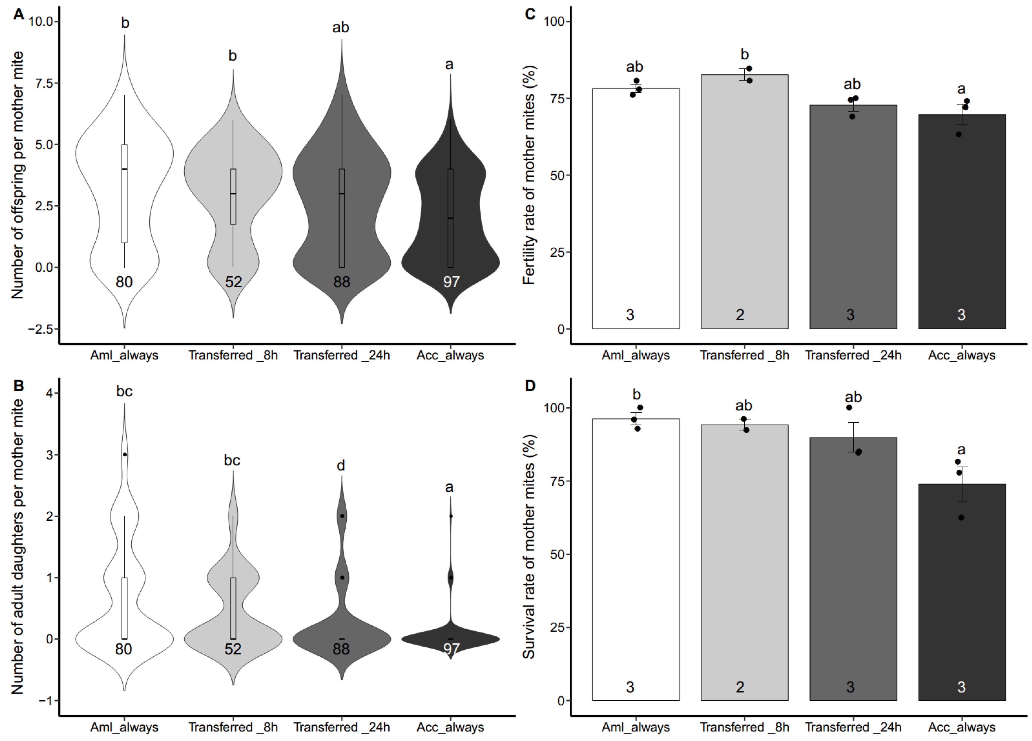
We investigated the reproductive performance of V. destructor from Aml colonies (Korea haplotype) on the worker brood of both host species. As shown in Figure 2, the Varroa mites reproduced differentially on Acc and Aml worker brood, and the reproduction was also affected by the duration of stay inside the Acc worker cells. The fecundity of mother mites among four groups was significantly different (Figure 2A. Kruskal-Wallis test, χ2 = 18.125, df = 3, p < 0.001). The duration of stay inside the Acc worker cells decreased the number of offspring per mother mite. The Varroa females that always stayed in the Acc worker cells reproduced the lowest number of offspring, while those living in the Aml worker cells all the time had the highest number of offspring. Pairwise comparison further revealed the significant differences between groups (Figure 2A. Wilcoxon rank sum test, Acc_always vs. Aml_always: p < 0.001; Acc_always vs. Transferred_8 h: p = 0.0028).
Similarly, the number of adult daughters differed significantly among the four treatments and decreased with the increase of the duration of stay inside the Acc worker cells (Figure 2B. Kruskal-Wallis test, χ2 = 40.684, df = 3, p < 0.001). The mother mites that always stayed in the Acc worker cells had the lowest number of mature daughters, and reproduced significantly less adult daughters compared with those from any of the other treatments (Figure 2B. Wilcoxon rank sum test, Acc_always vs. Aml_always: p < 0.001; Acc_always vs. Transferred_8 h: p < 0.001; Acc_always vs. Transferred_24 h: p = 0.0090). Additionally, the number of adult daughters per mother mite of Transferred_24 h was much smaller than that of Aml_always or Transferred_8 h (Figure 2B. Wilcoxon rank sum test, Transferred_24 h vs. Aml_always: p = 0.0011; Transferred_24 h vs. Transferred_8 h: p = 0.034).
The fertility rate of mother mites presented a small, but significant difference among the four treatments (Figure 2C. one-way ANOVA, F (3, 7) = 5.360, p = 0.031). Only mites of the Transferred_8 h group had a significantly higher fertility rate than those of Acc_always (Figure 2C. Tukey HSD test, p = 0.033). The survival rates of mother mites showed a significant difference among the four treatments (Figure 2D. one-way ANOVA, F (3, 7) = 5.289, p = 0.032). post hoc comparison showed that only mites of Aml_always had a significantly higher survival rate than those of Acc_always (Figure 2D. Tukey HSD test, p = 0.032).

3.3. Reproduction of Acc-Derived Varroa Mites on Aml Worker Brood

We further examined the reproductive performance of Varroa mites from A. cerana colonies (China haplotype). Only the reproduction on the Aml worker brood was included because of the limited number of mites obtained. Two sequential infestations were performed and monitored using the same mother mites. In both infestations, all the Acc-derived mother mites survived and had an equal survival rate to the controls (Aml-derived mother mites), but none reproduced, while the control mites reproduced normally (Table 1). More precisely, for the first infestation, reproductive outputs between Varroa-Acc and Varroa-Aml are statistically different (Table 1. Number of offspring, p < 0.001; number of adult daughters, p = 0.030; fertility rate, p = 0.001). Similarly, for the second infestation reproductive outputs between Varroa-Acc and Varroa-Aml were also statistically different (Table 1. Number of offspring, p = 0.0054; fertility rate, p = 0.012), despite of the number of adult daughters showing no difference (p = 0.056, Table 1).
We also examined the defecating behavior of both Acc and Aml derived mites on Aml worker cells. All Acc-derived Varroa females, which did not reproduce, were found to defecate on the bee abdomen. Most mites from Aml colonies were fertile and found to defecate on the cell walls, while few were sterile, and excreted on pupae body (Figure S2).

4. Discussion

In this study, we identified two types (Korea and China haplotype) of V. destructor from honey bee colonies located in southern China. We found that these two types of Varroa mites were from distinct species of honey bee colonies: The Korea haplotypes only detected in A. mellifera colonies, while China haplotype only from A. cerana colonies. These results are consistent with the findings reported in the same area two decades ago [15]. Further studies have also described the phenomenon of Varroa haplotypes being associated with specific honey bee species (without coexistence) in the same area, such as in southwestern China (China 2 or Vietnam haplotype with A. cerana vs. Korea haplotype with A. mellifera) and in northern Vietnam (Vietnam haplotype with A. cerana vs. Korea haplotype with A. mellifera) [15,16].
It seems that different types of Varroa mites are naturally separated, even though their living space overlaps, even when, as in this case, they are in the same apiary [35]. The inter-colony mite transmission, however, is feasible and mostly mediated by honey bee drifting and robbing behaviors [36]. Honey bee colonies collapsing from high infestations of Varroa mites can act as lures for robber bees, resulting in the elevation of Varroa population in the neighboring colonies [36]. Robbing between A. cerana and A. mellifera colonies is common, especially when the bees are suffering from food shortage. The lack of detection of different haplotypes coexisting in the same host species may be due to some other unknown reasons. We notice that in the areas, such as southern and southwestern China and northern Vietnam only Korea haplotype colonizes A. mellifera colonies, while the other types are confined to the original hosts, but it does not mean that these haplotypes are not able, or do not have the opportunities to shift to A. mellifera. The details of Varroa host shifts are still mysterious, but the sympatric distribution of both honey bee species provides essential opportunities for the parasite to infest the new host [11,12,37]. Monitoring the emergence of new types of Varroa in A. mellifera colonies seems to be important as they pose a potential threat to the new hosts.
Our results show that Varroa mites derived from A. cerana and A. mellifera colonies are not only distinct haplotypes, but also reproduce differentially on worker brood hosts. By artificial inoculation, we first examined the reproduction of A. mellifera derived mites (Korea haplotype) on both A. cerana and A. mellifera worker brood. The results confirm that in addition to the new host, the mother mites also have the capability to reproduce on the worker brood of the original host A. cerana. This has also been demonstrated in previous studies, which were conducted in different parts of China, South Korea, and northern Vietnam, but all involved as Korea haplotype Varroa mites [18,20,25,28,29]. Our data revealed that Varroa mites produced less offspring and adult daughters in the original host than the new host. It is known that immature workers of A. cerana are more susceptible to Varroa infestations than those of A. mellifera, evidenced by strikingly delayed development and high mortality [27,29], and low tolerance to Varroa-derived toxic protein [38]. We suspect that the abnormal development and death of the A. cerana worker brood resulted in a shortage of nutrient and energy supplies for the parasite development, contributing to the lower fecundity of mother mites. Obviously, Varroa mites fare better following host switch. The better reproductive performance of V. destructor on worker brood also accelerates the growth of Varroa population, but exaggerates the damage to the new host.
Interestingly, we also found that a short stay of mother mites on the A. cerana worker brood decreased their fecundity, especially the number of adult daughters, in a time-dependent manner. This suggests that the A. cerana worker cells are able to suppress Varroa reproduction. After entering a brood cell, including a fifth instar larva, the mother mite starts to feed on the host fat body [17], certain proteins of which are then transported and stored directly in the ovary of the mite [39]. It is likely that after long-term interaction, the original host has evolved an effective resistant strategy whereby producing toxic and/or nutrient-deficient substances in the hemolymph to suppress reproduction of Varroa parasites. On the other hand mite, reproduction can be either triggered or inhibited by the stimuli of a brood cell [12,40]. The oogenesis of V. destructor can be induced by the stimuli extracted from the bee larva [41,42,43]. Among them, fatty acid ethyl and methyl esters are most likely to initiate the oogenesis [41]. Meanwhile, inhibitors of Varroa reproduction also exist in the host cells. A particular isomer, (Z)-8-heptadecene from infested cells has been shown to decrease the fecundity of Varroa females under both laboratory and field conditions [44,45]. We suspect that the original host may suppress the reproduction of Varroa mites by regulating the expression of stimuli, and this inhibitive effect can last until reproduction is re-triggered in the new host.
Additionally, the initiation of Varroa reproduction was focused during this experimental design, because it is the key step of the whole reproductive process of Varroa mites [12,40]. Previous studies have shown that stimuli that promote egg maturation and trigger Varroa reproduction are present in capped cells containing larvae and active only within 24 h after cells are sealed [46]. Recently, a study by Frey et al. [41] revealed that the signal that induced Varroa oogenesis was produced within 12 h after capping of worker larvae and within 36 h after capping of drone larvae. Once the period had elapsed, another signal began to act in the opposite way, which interrupted the reproduction of Varroa mites [41]. This suggests that Varroa reproduction is initiated within a short period after the worker cells are capped. For this reason, we made the Transferred_8 h and Transferred_24 h groups to test if a short stay of Varroa mites on the original host worker brood inhibits their reproduction.
Likewise, we tested the reproduction of Varroa mites from A. cerana (China haplotype), but only on the worker brood of new hosts, because of the limited number of mites we could obtain. Since Varroa females can reproduce continuously without a phoretic phase [47], we designed an experiment with two sequential inoculations using the same mother mites. Surprisingly, no reproduction was seen in any Varroa female in either the first or second infestation, while the control mites from A. mellifera colonies (Korea haplotype) still reproduced normally. No matter reproduced or not, both types of Varroa mites survived well on the new host, suggesting other living activities, such as feeding and moving were not disturbed. Similar findings were described in previous studies using Varroa mites from A. cerana (Vietnam haplotype) [20,25]. In addition, Zhou [25] further demonstrated that the mites only reproduced on the drone brood of the original host, suggesting more strict confinement to the reproduction of Varroa mites. Despite being infertile on the new host, all the mites from A. cerana are found to defecate on the host body. This phenomenon is tightly associated with reproduction of Varroa mites, making it a sign of Varroa infertility: If the mites defecate on the bee’s body, they will be found to be sterile [48,49]. Normally, the mite families in a cell defecate in the same location on the cell wall, called fecal accumulation site, which acts as a rendezvous platform for mating [50]. The reason why the infertile mites choose to defecate on the bee’s body is still not clear.
Reproduction is the crucial step for the successful parasitism of V. destructor in honey bees, and also a main target to suppress the growth of Varroa population [12,40]. Our findings further reveal the complexity of Varroa reproduction: Different Varroa haplotypes reproduce differentially in the original and new host. Considering the economic importance of honey bees and the urgency for the control of Varroa mites, our results may help the honey bee conservation from the following aspects in future: (1) To enhance the monitoring of the new Varroa haplotypes in both honey bee species; (2) to set up the natural separation between the honey bee populations with different Varroa haplotypes to avoid the spillover and/or spillback of the parasites.

5. Conclusions

We have confirmed that there are two haplotypes of V. destructor in southern China, one is Korea haplotype, and the other is China haplotype. Although their ecological distribution overlaps, the two Varroa haplotypes are naturally isolated as they strictly parasitize different honey bee species. Moreover, Varroa females from A. mellifera colonies have the capability to reproduce on the worker brood of both the original and new hosts; however, better reproductive outputs are obtained on the latter. We further show that the A. cerana worker cells might inhibit the reproduction of Varroa mites. In contrast, A. cerana derived Varroa females are entirely sterile in A. mellifera worker cells and display abnormal defecation habits. Generally, it is known that infertility on the worker brood is due to a trait of V. destructor [20,25], however, despite the fact that they belong to different haplotypes, what exact genetic variations that underpin the differences in reproduction is largely unknown. The genome sequence of V. destructor (Korea haplotype) has been already available [51,52], which together with the rapid development of functional genomics, makes the prospect of answering the questions in future.

Supplementary Materials

The following are available online at https://www.mdpi.com/2075-4450/10/11/372/s1, Figure S1: Genetic identification of the Varroa mites derived from either Apis mellifera colonies or A. cerana colonies, Figure S2: Defecation of Varroa mites on honey bee abdomen.

Author Contributions

Conceptualization, W.L. and R.H.; Data curation, W.L.; Formal analysis, W.L. and C.W.; Funding acquisition, W.L. and R.H.; Investigation, W.L. and C.W.; Project administration, W.L. and R.H.; Resources, W.L. and R.H.; Supervision, R.H.; Validation, W.L. and C.W.; Visualization, W.L. and C.W.; Writing—original draft, W.L. and C.W.; Writing—review and editing, W.L., C.W., Z.Y.H., Y.C. and R.H.

Funding

This work was funded by the GDAS Special Project of Science and Technology Development (grant number: 2018GDASCX-0931; 2018GDASCX-0107; 2019GDASYL-0104019), the National Natural Science Foundation of China (grant number: 31741117), the Guangzhou Science and Technology Plan Project (grant number: 201904010115), and the Talent Project of Guangdong Institute of Applied Biological Resources (grant number: GIABR-gjrc201702). The funders had no role in study design, data collection and analysis, decision to publish, or preparation of the manuscript.

Acknowledgments

We thank Yi Zhang, Wenjing Wu and Peng Zhang for the assistance of sample collection and beekeeping. We also thank the two reviewers for their valuable comments on the manuscript.

Conflicts of Interest

The authors declare no conflict of interest.

References

  1. Klein, A.M.; Vaissière, B.E.; Cane, J.H.; Steffan-Dewenter, I.; Cunningham, S.A.; Kremen, C.; Tscharntke, T. Importance of pollinators in changing landscapes for world crops. Proc. R. Soc. B 2007, 274, 303. [Google Scholar] [CrossRef] [PubMed]
  2. Potts, S.G.; Imperatriz-Fonseca, V.; Ngo, H.T.; Aizen, M.A.; Biesmeijer, J.C.; Breeze, T.D.; Dicks, L.V.; Garibaldi, L.A.; Hill, R.; Settele, J.; et al. Safeguarding pollinators and their values to human well-being. Nature 2016, 540, 220. [Google Scholar] [CrossRef] [PubMed]
  3. Neumann, P.; Carreck, N.L. Honey bee colony losses. J. Apic. Res. 2010, 49, 1–6. [Google Scholar] [CrossRef] [Green Version]
  4. Potts, S.G.; Biesmeijer, J.C.; Kremen, C.; Neumann, P.; Schweiger, O.; Kunin, W.E. Global pollinator declines: Trends, impacts and drivers. Trends Ecol. Evol. 2010, 25, 345–353. [Google Scholar] [CrossRef] [PubMed]
  5. VanEngelsdorp, D.; Meixner, M.D. A historical review of managed honey bee populations in europe and the united states and the factors that may affect them. J. Invertebr. Pathol. 2010, 103, S80–S95. [Google Scholar] [CrossRef] [PubMed]
  6. Cox-Foster, D.L.; Conlan, S.; Holmes, E.C.; Palacios, G.; Evans, J.D.; Moran, N.A.; Quan, P.-L.; Briese, T.; Hornig, M.; Geiser, D.M.; et al. A metagenomic survey of microbes in honey bee colony collapse disorder. Science 2007, 318, 283–287. [Google Scholar] [CrossRef] [PubMed]
  7. Ratnieks, F.L.W.; Carreck, N.L. Clarity on honey bee collapse? Science 2010, 327, 152–153. [Google Scholar] [CrossRef] [PubMed]
  8. Dietemann, V.; Pflugfelder, J.; Anderson, D.; Charrière, J.-D.; Chejanovsky, N.; Dainat, B.; de Miranda, J.; Delaplane, K.; Dillier, F.-X.; Fuch, S.; et al. Varroa destructor: Research avenues towards sustainable control. J. Apic. Res. 2012, 51, 125–132. [Google Scholar] [CrossRef]
  9. Genersch, E. Honey bee pathology: Current threats to honey bees and beekeeping. Appl. Microbiol. Biotechnol. 2010, 87, 87–97. [Google Scholar] [CrossRef]
  10. Anderson, D.L.; Trueman, J.W.H. Varroa jacobsoni (acari: Varroidae) is more than one species. Exp. Appl. Acarol. 2000, 24, 165–189. [Google Scholar] [CrossRef]
  11. Oldroyd, B.P. Coevolution while you wait: Varroa jacobsoni, a new parasite of western honeybees. Trends Ecol. Evol. 1999, 14, 312–315. [Google Scholar] [CrossRef]
  12. Rosenkranz, P.; Aumeier, P.; Ziegelmann, B. Biology and control of Varroa destructor. J. Invertebr. Pathol. 2010, 103, S96–S119. [Google Scholar] [CrossRef] [PubMed]
  13. Peng, Y.-S.; Fang, Y.; Xu, S.; Ge, L. The resistance mechanism of the asian honey bee, Apis cerana fabr., to an ectoparasitic mite, Varroa jacobsoni oudemans. J. Invertebr. Pathol. 1987, 49, 54–60. [Google Scholar] [CrossRef]
  14. Rath, W. Co-adaptation of Apis cerana Fabr. and Varroa jacobsoni oud. Apidologie 1999, 30, 97–110. [Google Scholar] [CrossRef]
  15. Zhou, T.; Anderson, D.L.; Huang, Z.Y.; Huang, S.; Yao, J.; Ken, T.; Zhang, Q. Identification of Varroa mites (acari: Varroidae) infesting Apis cerana and Apis mellifera in China. Apidologie 2004, 35, 645–654. [Google Scholar] [CrossRef]
  16. Navajas, M.; Anderson, D.L.; de Guzman, L.I.; Huang, Z.Y.; Clement, J.; Zhou, T.; Le Conte, Y. New Asian types of Varroa destructor: A potential new threat for world apiculture. Apidologie 2010, 41, 181–193. [Google Scholar] [CrossRef]
  17. Ramsey, S.D.; Ochoa, R.; Bauchan, G.; Gulbronson, C.; Mowery, J.D.; Cohen, A.; Lim, D.; Joklik, J.; Cicero, J.M.; Ellis, J.D.; et al. Varroa destructor feeds primarily on honey bee fat body tissue and not hemolymph. Proc. Natl. Acad. Sci. USA 2019, 116, 1792–1801. [Google Scholar] [CrossRef]
  18. De Jong, D. Varroa jacobsoni does reproduce in worker cells of Apis cerana in South Korea. Apidologie 1988, 19, 241–244. [Google Scholar] [CrossRef]
  19. Boot, W.J.; Tan, N.Q.; Dien, P.C.; Huan, L.V.; Dung, N.V.; Long, L.T.; Beetsma, J. Reproductive success of Varroa jacobsoni in brood of its original host, Apis cerana, in comparison to that of its new host, A. mellifera (hymenoptera: Apidae). Bull. Entomol. Res. 1997, 87, 119–126. [Google Scholar] [CrossRef]
  20. Boot, W.J.; Calis, J.N.M.; Beetsma, J.; Hai, D.M.; Lan, N.K.; Toan, T.V.; Trung, L.Q.; Minh, N.H. Natural selection of Varroa jacobsoni explains the different reproductive strategies in colonies of Apis cerana and Apis mellifera. Exp. Appl. Acarol. 1999, 23, 133–144. [Google Scholar] [CrossRef]
  21. Koeniger, N.; Koeniger, G.; Delfinado-Baker, M. Observations on mites of the Asian honeybee species (Apis cerana, Apis dorsata, Apis florea). Apidologie 1983, 14, 197–204. [Google Scholar] [CrossRef]
  22. Koeniger, N.; Koeniger, G.; Wijayagunasekara, N.H. Boebachtungen uber die anpassung von Varroa jacobsoni an ihren naturlichen wirt Apis cerana in Sri Lanka. Apidologie 1981, 12, 37–40. [Google Scholar] [CrossRef]
  23. Rosenkranz, P.; Tewarson, N.C.; Rachinsky, A.; Strambi, A.; Strambi, C.; Engels, W. Juvenile hormone titer and reproduction of Varroa jacobsoni in capped brood stages of Apis cerana indica in comparison to Apis mellifera ligustica. Apidologie 1993, 24, 375–382. [Google Scholar] [CrossRef]
  24. Tewarson, N.C.; Singh, A.; Engels, W. Reproduction of Varroa jacobsoni in colonies of Apis cerana indica under natural and experimental conditions. Apidologie 1992, 23, 161–171. [Google Scholar] [CrossRef]
  25. Zhou, T. The Biological Characteristics and the Natural Distribution of Varroa Destructor in China. Ph.D. Thesis, China Agricultural University, Beijing, China, 2005. [Google Scholar]
  26. Anderson, D.L. Variation in the parasitic bee mite Varroa jacobsoni oud. Apidologie 2000, 31, 281–292. [Google Scholar] [CrossRef]
  27. Page, P.; Lin, Z.; Buawangpong, N.; Zheng, H.; Hu, F.; Neumann, P.; Chantawannakul, P.; Dietemann, V. Social apoptosis in honey bee superorganisms. Sci. Rep. 2016, 6, 27210. [Google Scholar] [CrossRef] [Green Version]
  28. Le Conte, Y.; Huang, Z.Y.; Roux, M.; Zeng, Z.J.; Christidès, J.P.; Bagnères, A.G. Varroa destructor changes its cuticular hydrocarbons to mimic new hosts. Biol. Lett. 2015, 11, 20150233. [Google Scholar] [CrossRef]
  29. Lin, Z.; Qin, Y.; Page, P.; Wang, S.; Li, L.; Wen, Z.; Hu, F.; Neumann, P.; Zheng, H.; Dietemann, V. Reproduction of parasitic mites Varroa destructor in original and new honeybee hosts. Ecol. Evol. 2018, 8, 2135–2145. [Google Scholar] [CrossRef]
  30. Sasaki, M. The reason why Apis cerana japonica is resistant to Varroa mite. Honeybee Sci. 1989, 10, 28–36. [Google Scholar]
  31. Anderson, D.L.; Fuchs, S. Two genetically distinct populations of Varroa jacobsoni with contrasting reproductive abilities on Apis mellifera. J. Apic. Res. 1998, 37, 69–78. [Google Scholar] [CrossRef]
  32. Nazzi, F.; Milani, N. A technique for reproduction of Varroa jacobsoni Oud under laboratory conditions. Apidologie 1994, 25, 579–584. [Google Scholar] [CrossRef]
  33. Di Prisco, G.; Annoscia, D.; Margiotta, M.; Ferrara, R.; Varricchio, P.; Zanni, V.; Caprio, E.; Nazzi, F.; Pennacchio, F. A mutualistic symbiosis between a parasitic mite and a pathogenic virus undermines honey bee immunity and health. Proc. Natl. Acad. Sci. USA 2016, 113, 3203–3208. [Google Scholar] [CrossRef] [Green Version]
  34. R Core Team. R: A Language and Environment for Statistical Computing; R Foundation for Statistical Computing: Vienna, Austria, 2019; Available online: https://www.R-project.org/ (accessed on 24 May 2019).
  35. Beaurepaire, A.L.; Truong, T.A.; Fajardo, A.C.; Dinh, T.Q.; Cervancia, C.; Moritz, R.F.A. Host specificity in the honeybee parasitic mite, Varroa spp. in Apis mellifera and Apis cerana. PLoS ONE 2015, 10, e0135103. [Google Scholar] [CrossRef] [PubMed]
  36. Peck, D.T.; Seeley, T.D. Mite bombs or robber lures? The roles of drifting and robbing in Varroa destructor transmission from collapsing honey bee colonies to their neighbors. PLoS ONE 2019, 14, e0218392. [Google Scholar] [CrossRef] [PubMed]
  37. Dietemann, V.; Beaurepaire, A.; Page, P.; Yañez, O.; Buawangpong, N.; Chantawannakul, P.; Neumann, P. Population genetics of ectoparasitic mites Varroa spp. in Eastern and Western honey bees. Parasitology 2019, 146, 1429–1439. [Google Scholar] [CrossRef] [PubMed]
  38. Zhang, Y.; Han, R. A saliva protein of Varroa mites contributes to the toxicity toward Apis cerana and the DWV elevation in A. mellifera. Sci. Rep. 2018, 8, 3387. [Google Scholar] [CrossRef]
  39. Steiner, J.; Dittmann, F.; Rosenkranz, P.; Engels, W. The first gonocycle of the parasitic mite (Varroa juobsoni) in relation to preimaginal development of its host, the honey bee (Apis mellifera carnicar). Invertebr. Reprod. Dev. 1994, 25, 175–183. [Google Scholar] [CrossRef]
  40. Nazzi, F.; Le Conte, Y. Ecology of Varroa destructor, the major ectoparasite of the western honey bee, Apis mellifera. Annu. Rev. Entomol. 2016, 61, 417–432. [Google Scholar] [CrossRef]
  41. Frey, E.; Odemer, R.; Blum, T.; Rosenkranz, P. Activation and interruption of the reproduction of Varroa destructor is triggered by host signals (Apis mellifera). J. Invertebr. Pathol. 2013, 113, 56–62. [Google Scholar] [CrossRef]
  42. Garrido, C.; Rosenkranz, P. Volatiles of the honey bee larva initiate oogenesisin the parasitic mite Varroa destructor. Chemoecology 2004, 14, 193–197. [Google Scholar]
  43. Trouiller, J.; Milani, N. Stimulation of Varroa jacobsoni oud. oviposition with semiochemicals from honeybee brood. Apidologie 1999, 30, 3–12. [Google Scholar] [CrossRef]
  44. Milani, N.; Vedova, G.D.; Nazzi, F. (Z)-8-heptadecene reduces the reproduction of Varroa destructor in brood cells. Apidologie 2004, 35, 265–273. [Google Scholar] [CrossRef]
  45. Nazzi, F.; Milani, N.; Vedova, G.D. (Z)-8-heptadecene from infested cells reduces the reproduction of Varroa destructor under laboratory conditions. J. Chem. Ecol. 2002, 28, 2181–2190. [Google Scholar] [CrossRef] [PubMed]
  46. Milani, N.; Chiesa, F. Some factors affecting the reproduction of Varroa jacobsoni Oud. under laboratory conditions. Apicoltura 1990, 6, 33–42. [Google Scholar]
  47. De Ruijter, A. Reproduction of Varroa jacobsoni during successive brood cycles of the honeybee. Apidologie 1987, 18, 321–326. [Google Scholar] [CrossRef]
  48. Huang, Z. Varroa mite reproductive biology. Am. Bee J. 2012, 152, 981. [Google Scholar]
  49. Posada-Florez, F.; Sonenshine, D.E.; Egekwu, N.I.; Rice, C.; Lupitskyy, R.; Cook, S.C. Insights into the metabolism and behaviour of Varroa destructor mites from analysis of their waste excretions. Parasitology 2018, 146, 527–532. [Google Scholar] [CrossRef]
  50. Donzé, G.; Guerin, P.M. Behavioral attributes and parental care of Varroa mites parasitizing honeybee brood. Behav. Ecol. Sociobiol. 1994, 34, 305–319. [Google Scholar] [CrossRef]
  51. Cornman, R.S.; Schatz, M.C.; Johnston, J.S.; Chen, Y.-P.; Pettis, J.; Hunt, G.; Bourgeois, L.; Elsik, C.; Anderson, D.; Grozinger, C.M.; et al. Genomic survey of the ectoparasitic mite Varroa destructor, a major pest of the honey bee Apis mellifera. BMC Genom. 2010, 11, 1–15. [Google Scholar] [CrossRef]
  52. Techer, M.A.; Rane, R.V.; Grau, M.L.; Roberts, J.M.K.; Sullivan, S.T.; Liachko, I.; Childers, A.K.; Evans, J.D.; Mikheyev, A.S. Divergent evolutionary trajectories following speciation in two ectoparasitic honey bee mites. Commun. Biol. 2019, 2, 357. [Google Scholar] [CrossRef]
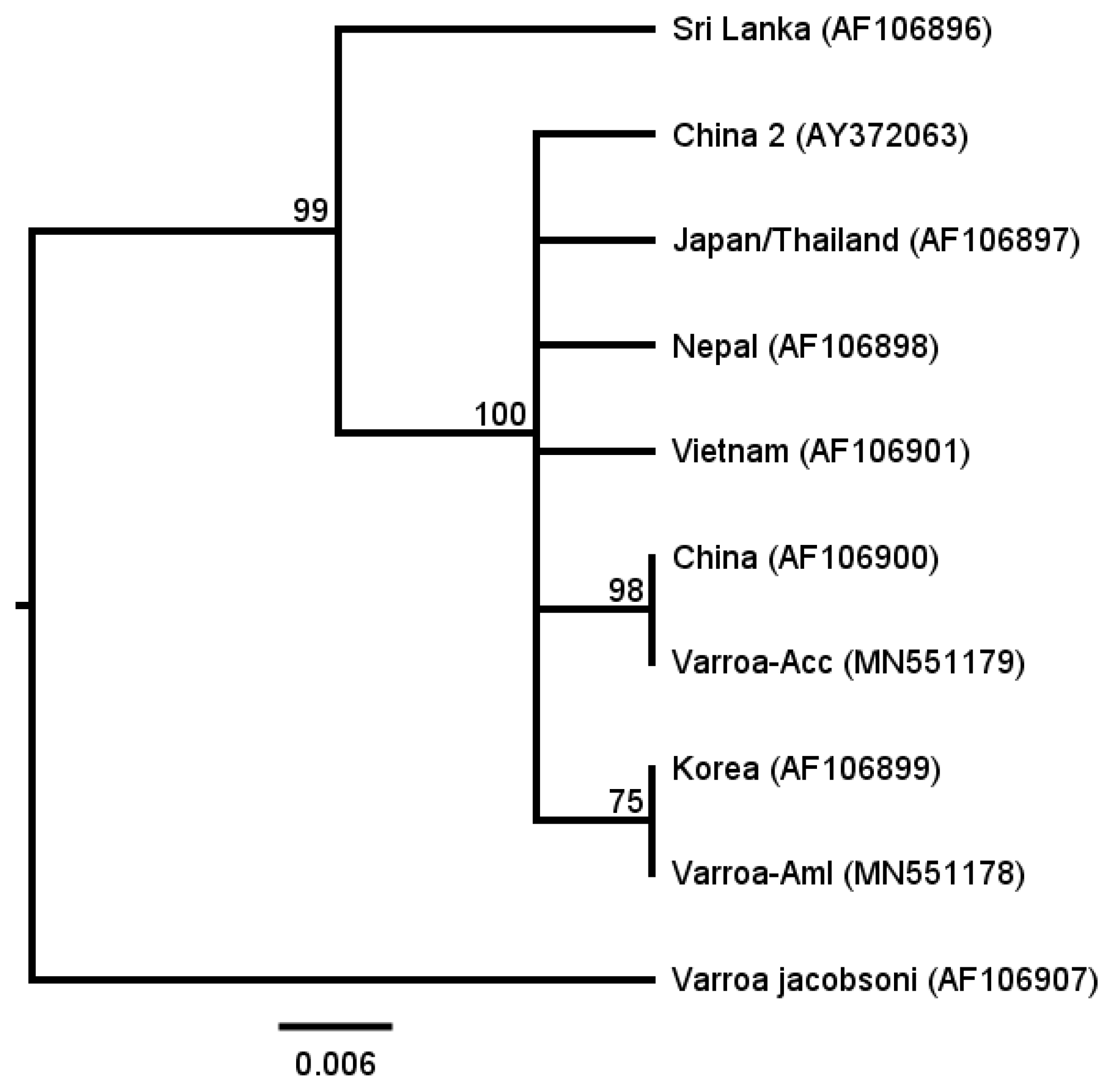
Figure 1. Phylogenetic analysis of CO-I gene nucleotide sequences from different Varroa destructor haplotypes. Varroa-Acc and Varroa-Aml stand for the CO-I gene sequences of Acc and Aml derived Varroa mites, respectively, and CO-I gene sequences of seven known V. destructor haplotypes and one V. jacobsoni haplotype are included (the GenBank accession number are associated). Among them, V. destructor mites of Sri Lanka, China, China 2, Nepal, and Vietnam haplotypes were sampled from A. cerana, while V. destructor mites of Korea and Japan/Thailand haplotypes were collected from both A. cerana and A. mellifera [10,15]. Bootstrap values (100 replicates) are displayed by the nodes. The genetic distance is drawn to scale. The phylogenetic analysis suggests that the Varroa mites collected in the current are either China haplotype (Acc derived) or Korea haplotype (Aml derived).
Insects 10 00372 g001
Figure 2. Reproductive outputs of the Apis mellifera derived Varroa mites on both A. mellifera and A. cerana worker brood. The mother mites of V. destructor were harvested from A. mellifera donor colonies, then four different treatments were applied: (1) Mites were inoculated to A. mellifera ligustica (Aml) worker brood and then stayed inside all the way (Aml_always); (2) mites were inoculated to Acc worker brood for 8 h, then transferred to Aml worker cells, and kept for the rest of time (Transferred_8 h); (3) mites were inoculated to Acc worker brood for 24 h, then transferred to Aml worker brood, and stayed inside for the rest of time (Transferred_24 h); (4) mites were inoculated to A. cerana cerana (Acc) worker brood and then kept inside all the way (Acc_always). (A) Number of offspring per mother mite. (B) Number of adult daughters per mother mite. Data represent the sum of animals in multiple experiments. (C) Fertility rate of mother mites. (D) Survival rate of mother mites. Each dot represents data of one independent experiment. For (A,B), data are presented as median and interquartile range. For (C,D), data are presented as mean ± SEM, and the numbers of tested mother mites or assays are shown in the bottom of each column. For (A,B), the statistical significance was analyzed by Kruskal-Wallis test followed by Wilcoxon rank sum test with BH adjustment for post hoc analysis. For (C,D), the significant difference was determined by one-way ANOVA followed by Tukey HSD test for post hoc analysis. Lowercase letters above bars denote post hoc significance (p < 0.05).
Insects 10 00372 g002
Table 1. Comparison of the reproductive outputs between Apis mellifera and A. cerana derived Varroa mites on A. mellifera worker brood.
First InfestationSecond Infestation
Varroa-Acc a (n = 13)Varroa-Aml b (n = 19)Significance dVarroa-Acc (n = 11)Varroa-Aml (n = 17)Significance d
Number of offspring per mother mite02.47 ± 0.52 cp < 0.00101.65 ± 0.44 cp = 0.005
Number of adult daughters per mother mite00.63 ± 0.24 cp = 0.03000.29 ± 0.11 cp = 0.056
Fertility rate of mother mites (%)063.16p = 0.001052.94p = 0.012
Survival rate of mother mites (%)10089.47p = 0.640100100p = 1
a.Varroa-Acc: Varroa mites derived from A. cerana cerana colonies. b. Varroa-Aml: Varroa mites derived from A. mellifera ligustica colonies. c. Data are presented as mean ± SEM. d. Significant difference was determined by unpaired two-sample Wilcoxon test for data of number of offspring and adult daughters per mother mite, and Pearson’s Chi-squared test for data of fertility and mortality of mother mites.