Toxicity Analysis of Nano-Minimum Quantity Lubrication Machining—A Review


Abstract
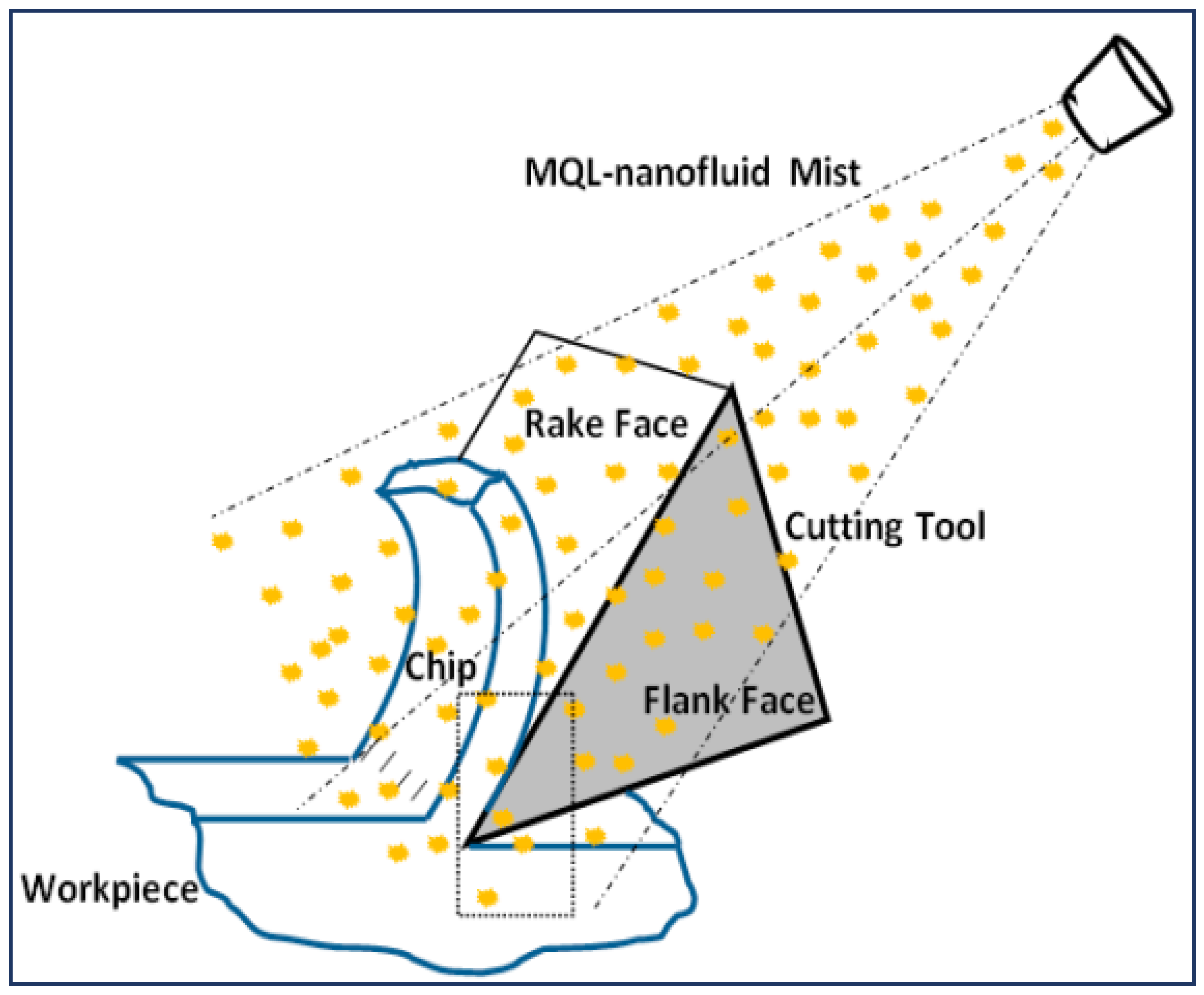
:1. Introduction
2. Research Motivation and Methodology
- Review development phase—The review was developed in two stages. In stage one, the effectiveness of NMQL cooling strategy in machining was established from studies available in literature, and six nanoparticles (some nanoparticle families) were chosen for toxicity analysis in stage two of the review. Due to the presence of detailed review papers on NMQL machining’s performance, this section of the review was kept brief, and it highlights only some studies in each of the machining categories. Further, in this section, reference is made to available review papers in current literature on NMQL machining’s performance. Stage two focused on building the toxicity review for each of the nanoparticles selected in stage one, and it is the main contribution of this review paper. The following methodology was used for stage two:
- In vivo and in vitro studies on both human cells and other living organisms were showcased for all nanoparticles. Studies on aquatic life and bacteria helped with estimating the impacts of the nanoparticles on the environment and may help create a safe disposal procedure. Further, in vitro studies on human cells provided information on the possible impacts of working with nanoparticles during the machining process.
- The following nanoparticle and toxicity test characteristics were made available from each study: nanoparticle size, nanoparticle concentration in the test medium and duration of exposure to the nanoparticle.
- For the development of the toxicity chart, only studies measuring cell viability for seven different human cell lines (human lung epithelial cells (A549), human bronchial epithelial cells (Nl-20), AGS human gastric cells, human epidermal cell (HEK), human liver-derived cells (HepG2), human endothelial cells and human peripheral cells) were considered.
- Results communication and dissemination phase—The following considerations were made in presenting the results in this review:
- The results from the toxicity studies were presented in table format for each of the selected nanoparticles and in understandable terms.
- All studies on each investigated nanoparticle were presented in a chronological order. This was to account for the developing technologies as well as to provide a better insight into some contradictory toxicity results available in the literature.
- For the development of the toxicity chart, only studies measuring cell viability for seven different human cell lines (human lung epithelial cells (A549), human bronchial epithelial cells (Nl-20), AGS human gastric cells, human epidermal cell (HEK), human liver-derived cells (HepG2), human endothelial cells and human peripheral cells) were considered.
3. Toxicity Studies of Nanoparticles
- In vitro studies—Toxicity studies conducted outside a living organism. Usually, a cell culture is developed and the nanoparticles are then added to the cell culture for certain durations of time to examine their effects.
- In vivo studies—Toxicity studies conducted by injecting a living organism/animal with a certain dose of nanoparticles. The impacts on the organs/functions of the animal are then studied.
- Cell viability—Cell viability is defined as the number of healthy cells in a sample and can be expressed in percentage. Many studies focus on evaluating concentrations of nanoparticles that reduce the cell viability of a given sample to 50%.
- Cell morphology—Describes the shape, structure, form and size of cells. Changes in cells’ morphology might indicate negative impacts on cell function.
3.1. Molybdenum Disulfide (MoS2)
- The in vitro studies on human cell lines generally provide a very low toxicity when MoS2 nanoparticles are added to cell cultures.
- The method of nanoparticle exfoliation is highlighted as critical in determining the toxicity levels of MoS2 nanoparticles.
- A study on Escherichia coli to study the effects of MoS2 in natural water provided a high mortality rate. Thus, it indicates a need to be careful in the disposal of the nanoparticles. Further, a lack of in vivo studies on MoS2 has been noted. This is attributed to the relative newness of MoS2 nanoparticles when compared to CNT and metal oxides.
3.2. Tungsten Disulfide (WS2)
- Similar to MoS2 nanoparticles, in vitro studies on human cell lines generally provide a very low toxicity for WS2 nanoparticles.
- A study on Escherichia coli to study the effect of WS2 in natural water provided a high mortality rate. Additionality, a study on the effects of WS2 nanoparticles on a fungus also resulted in high levels of toxicity. Therefore, it is essential to develop a safe disposal mechanism to protect the environment from exposure to these nanoparticles.
- Further, both WS2 and MoS2 have limited number of in-vivo investigation of their toxicity. This is attributed to the relative newness of TMDs.
3.3. Hexagonal Boron Nitride (hBN)
- The early literature on hBN provided contradictory results on toxicity. However, further research has highlighted the cause for the discrepancy in results. The lengths of nanoparticles are crucial in determining the particles’ toxicity levels.
- In vivo studies on mice showcased a dose-dependent increase in toxicity. Therefore, it is very important to understand the toxicity of hBN nanoparticles relative to their concentration levels.
- Soil worms (C. elegans) were impacted by the presence of nanoparticles in their systems. This highlights the need to be cautious in the disposal of the nanoparticles in the environment.
- Study by Xin et al. [51] estimated 40 µg as equal to almost approximately 2–3 decades of work exposure to humans, and 4 µg was estimated to be about 2–7 years of work exposure. Such estimates are important in understanding the safety criteria for the use and implementation of these nanoparticles.

| Type of Study | Concentration | Diameter (nm) | Time of Exposure | Cell Line/Organism | Major Outcomes |
|---|---|---|---|---|---|
| In vitro | 5 µg/mL of PEI-BNNT (1:10) | 72 h | Human neuroblastoma cell line (SH-SY5Y) | No adverse effects on metabolism, viability or cellular replication were reported. A good cell viability was maintained throughout the test period [49]. | |
| In vitro | 2 µg/mL | <80 | 48 h | Human lung epithelial cells (A549) alveolar macrophages (RAW 264.7) fibroblast cells (3T3-L1) Human embryonic kidney cells (HEK293) | Shape and geometry are crucial parameters that dictate the toxicity of nanomaterials. BNNT was found to exhbit toxicity to cell lines at low concentrations [47]. |
| In vitro | 0–100 µg/mL | 75–220 | 72 h | Human vein endothelial cells (HUVECs) | BNNT had a non-significant effect on the cells. It was reported that a modest reduction in cell viability occurred at only the highest concentrations (100 µg mL−1) [48]. |
| In vitro | 0–100 µg/mL | 10–80 | 24–48–72 h | Human neuroblastoma SH-SY5Y cells Human umbilical vein endothelial cells (HUVECs) | Both cell lines exhibited a high viability even at high concentrations of 20 µg/mL. A shorter BNNT was observed to have a low cytotoxicity when compared to longer nanotubes. The same BNNTs with longer lengths (10 nm) were found to be toxic at concentrations as low as 2 µm [52]. |
| In vitro | 25 µg/mL | 24 h | Human cells | Cell stiffness was calculated using atomic forced microscopy. It was seen that there was no significant change in the cell stiffness before and after hBN uptakes. Therefore, the authors posed it as safe for biomedical use. Further in vivo studies are encouraged [53]. | |
| In vitro/in vivo | 0–100 µg/mL and 40 µg | 49 | 24 h | NLRP3-deficient human monocytic cells C57BL/6 J male mice | Both in vitro and in vivo studies resulted in acute inflammation and toxicity due to BNNT contaminations [54]. |
| In vitro | 0–20 µg/mL | <50 | Human hepatoma HepG2 | At 30 μg/mL, MoS2 and BN nanoparticles reduced cell viability [38]. | |
| In vivo | 1–500 µg/mL | 150 | 0–30 days | Caenorhabditis elegans (C. elegans) | It was seen that up to a concentration of 100 µg mL−1, BNNTs did not cause any significant alteration to the growth, locomotion, lifespan or progeny of the C. elegans nematodes. However, at concentrations over 100 µg mL−1, BNNTs significantly reduced growth and locomotion and affected other characteristics [55]. |
| In vitro | 0.025–0.4 mg/mL | 50–190 | 24 and 48 h | Human normal skin fibroblast (CCD-1094Sk and ATCC® CRL 2120™) Madin–Darby canine kidney (MDCK) cells | At a low concentration of 0.025–0.1 mg/mL, no cytotoxcity was observed. However, at concentrations over 0.2 mg/mL, a mild cytotoxicity was noted on CRL-2120 cells. The authors concluded that at concentrations below 0.1 mg/mL, hBN can be a safe oral care product [56]. |
| In vivo | 4 and 40 µg | 13–23 | 4 h 1–7 days 1–2 months | Male C57BL/6 J mice | A concentration of 40 µg caused the greatest amount of damage to the lungs. 40 µg was estimated as equal to almost approximately 2–3 decades of work exposure to humans. 4µg was estimated to be about 2–7 years of work exposure, but resulted in no toxicity [51]. |
| In vivo | 50–3200 µg/kg | 50–200 | 24 h | Wistar albino rats | At concentrations below 1600 µg/kg, no toxicity was observed. Concentrations of 1600 µg/kg and 3200 µg/kg caused significant damage to the liver [57]. |
3.4. Aluminum Oxide (AL2O3)
- AL2O3 nanoparticles are relatively less toxic when compared to other metal oxides, such ZnO and SiO2.
- Dose-dependent increases in toxicity were observed. Low concentrations of AL2O3 nanoparticles of up to 100 µg/mL−1 resulted in low toxicity levels in human cell lines. However, in fish cells, higher toxicity levels were observed for the same levels of nanoparticle concentration.
- In vivo studies on mice also showcased inflammation and damage to the liver.
3.5. Zinc Oxide (ZnO)
- High levels of toxicity were observed at even low concentrations within in vitro cytotoxicity studies on human cell lines.
- In vivo studies also showcased high levels of toxicity and damage to the liver.
3.6. Carbon Nanotubes (CNT, SWCNT and MWCNT)
- High levels of toxicity were observed at even low concentrations within in vitro cytotoxicity studies on human cell lines. Some contradictory results are also available in the literature.
- In vivo studies highlighted short-term impairments of fear, memory and morphological changes and an increased heartbeat.
4. Discussion
5. Conclusions
- Transition metal dichalcogenides (MoS2 and WS2) exhibit a very low toxicity when compared to other nanoparticles and provide a very good machining performance with a good surface finish and lower cutting forces. Among the MoS2 and WS2 nanoparticles, MoS2 provides a better surface finish and exhibits a lower toxicity. However, a lack of in vivo studies and the relative infancy of the toxicity research on these nanoparticles must be considered.
- The toxicity of hBN nanoparticles varies depending on the length. A high toxicity was observed at larger lengths, while very low toxicity levels are seen at shorter lengths. Hence, machining with hBN nanoparticles must be done with only short hBN nanoparticles. Similarly, care must be taken in the selection of AL2O3 nanoparticles. The toxicity of AL2O3 nanoparticles varies depending on their crystalline structures but generally exhibits a low toxicity on human cells. However, results from in vivo studies of both hBN and AL2O3 highlight the concern and the need to accurately understand the importance of nanoparticle concentrations.
- ZnO exhibited very high levels of toxicity in both in vitro and in vivo studies, and therefore, irrespective of machining performance, researchers must avoid their use in machining operations. In vivo studies for carbon nanotube toxicity predicted a high toxicity, while in vitro toxicity studies provided contradictory results.
- Some nanoparticles for the same concentration exhibited a higher toxicity in non-human species. This provides researchers information to develop disposal guidelines and highlights the need for the proper disposal of nanofluids after machining.
- The comparisons developed in Table 8 provide an easy interpretation of the toxicity levels of the six nanoparticles that were considered. Cell viability is an important marker for toxicity studies and provides easy interpretations. However, a lack of uniformity in nanoparticle concentrations and the methods employed are limitations. Future research can aim to develop the chart with consistent nanoparticle concentrations and same methods of toxicity analyses.
Author Contributions
Funding
Institutional Review Board Statement
Informed Consent Statement
Data Availability Statement
Acknowledgments
Conflicts of Interest
References
- Black, J.T.; Kohser, R.A. DeGarmo’s Materials and Processes in Manufacturing, 10th ed.; John Wiley & Sons: Hoboken, NJ, USA, 2008. [Google Scholar]
- Dixit, U.S.; Sarma, D.K.; Davim, J.P. Environmentally Friendly Machining; Springer: Berlin/Heidelberg, Germany, 2012. [Google Scholar]
- Boswell, B.; Islam, M.N.; Davies, I.J.; Ginting, Y.R.; Ong, A.K. A review identifying the effectiveness of minimum quantity lubrication (MQL) during conventional machining. Int. J. Adv. Manuf. Technol. 2017, 92, 321–340. [Google Scholar] [CrossRef]
- Dureja, J.S.; Singh, R.; Singh, T.; Singh, P.; Dogra, M.; Bhatti, M.S. Performance evaluation of coated carbide tool in machining of stainless steel (AISI 202) under minimum quantity lubrication (MQL). Int. J. Precis. Eng. Manuf. Green Technol. 2015, 2, 123–129. [Google Scholar] [CrossRef]
- Carou, D.; Rubio, E.M.; Davim, J.P. A note on the use of the minimum quantity lubrication (MQL) system in turning. Ind. Lubr. Tribol. 2015, 67, 256–261. [Google Scholar] [CrossRef]
- Klocke, F.; Eisenblätter, G. Dry cutting—State of research. VDI Ber. 1998, 46, 159–188. [Google Scholar]
- Tschätsch, H. Applied Machining Technology; Springer: Berlin/Heidelberg, Germany, 2009; pp. 5–23. [Google Scholar] [CrossRef]
- Sarıkaya, M.; Yılmaz, V.; Güllü, A. Analysis of cutting parameters and cooling/lubrication methods for sustainable machining in turning of Haynes 25 superalloy. J. Clean. Prod. 2016, 133, 172–181. [Google Scholar] [CrossRef]
- Sharma, A.K.; Tiwari, A.K.; Dixit, A.R. Effects of Minimum Quantity Lubrication (MQL) in machining processes using conventional and nanofluid based cutting fluids: A comprehensive review. J. Clean. Prod. 2016, 127, 1–18. [Google Scholar] [CrossRef]
- Saidur, R.; Leong, K.Y.; Mohammad, H.A. A review on applications and challenges of nanofluids. Renew. Sustain. Energy Rev. 2011, 15, 1646–1668. [Google Scholar] [CrossRef]
- Sharma, A.K.; Tiwari, A.K.; Dixit, A.R. Progress of Nanofluid Application in Machining: A Review. Mater. Manuf. Process. 2015, 30, 813–828. [Google Scholar] [CrossRef]
- Zhang, Y.; Li, C.; Jia, D.; Zhang, D.; Zhang, X. Experimental evaluation of MoS2 nanoparticles in jet MQL grinding with different types of vegetable oil as base oil. J. Clean. Prod. 2015, 87, 930–940. [Google Scholar] [CrossRef]
- Kumar, A.; Ghosh, S.; Aravindan, S. Experimental investigations on surface grinding of silicon nitride subjected to mono and hybrid nanofluids. Ceram. Int. 2019, 45, 17447–17466. [Google Scholar] [CrossRef]
- Virdi, R.L.; Chatha, S.S.; Singh, H. Experiment evaluation of grinding properties under Al2O3 nanofluids in minimum quantity lubrication. Mater. Res. Express 2019, 6, 096574. [Google Scholar] [CrossRef]
- Eltaggaz, A.; Hegab, H.; Deiab, I.; Kishawy, H.A. Hybrid nano-fluid-minimum quantity lubrication strategy for machining austempered ductile iron (ADI). Int. J. Interact. Des. Manuf. 2018, 12, 1273–1281. [Google Scholar] [CrossRef]
- Eltaggaz, A.; Zawada, P.; Hegab, H.A.; Deiab, I.; Kishawy, H.A. Coolant strategy influence on tool life and surface roughness when machining ADI. Int. J. Adv. Manuf. Technol. 2018, 94, 3875–3887. [Google Scholar] [CrossRef]
- Eltaggaz, A.; Nouzil, I.; Deiab, I. Machining ti-6al-4v alloy using nano-cutting fluids: Investigation and analysis. J. Manuf. Mater. Process. 2021, 5, 42. [Google Scholar] [CrossRef]
- Uysal, A. Investigation of flank wear in mql milling of ferritic stainless steel by using nano graphene reinforced vegetable cutting fluid. Ind. Lubr. Tribol. 2016, 68, 446–451. [Google Scholar] [CrossRef]
- Nguyen, T.K.; Do, I.; Kwon, P. A tribological study of vegetable oil enhanced by nano-platelets and implication in MQL machining. Int. J. Precis. Eng. Manuf. 2012, 13, 1077–1083. [Google Scholar] [CrossRef]
- Yıldırım, Ç.V.; Sarıkaya, M.; Kıvak, T.; Şirin, Ş. The effect of addition of hBN nanoparticles to nanofluid-MQL on tool wear patterns, tool life, roughness and temperature in turning of Ni-based Inconel 625. Tribol. Int. 2019, 134, 443–456. [Google Scholar] [CrossRef]
- Yıldırım, Ç.V. Experimental comparison of the performance of nanofluids, cryogenic and hybrid cooling in turning of Inconel 625. Tribol. Int. 2019, 137, 366–378. [Google Scholar] [CrossRef]
- Chetan; Ghosh, S.; Rao, P.V. Comparison between sustainable cryogenic techniques and nano-MQL cooling mode in turning of nickel-based alloy. J. Clean. Prod. 2019, 231, 1036–1049. [Google Scholar] [CrossRef]
- Şirin, Ş.; Kıvak, T. Performances of different eco-friendly nanofluid lubricants in the milling of Inconel X-750 superalloy. Tribol. Int. 2019, 137, 180–192. [Google Scholar] [CrossRef]
- Said, Z.; Gupta, M.; Hegab, H.; Arora, N.; Khan, A.M.; Jamil, M.; Bellos, E. A comprehensive review on minimum quantity lubrication (MQL) in machining processes using nano-cutting fluids. Int. J. Adv. Manuf. Technol. 2019, 105, 2057–2086. [Google Scholar] [CrossRef]
- Cui, X.; Li, C.; Ding, W.; Chen, Y.; Mao, C.; Xu, X.; Liu, B.; Wang, D.; Li, H.N.; Zhang, Y.; et al. Minimum quantity lubrication machining of aeronautical materials using carbon group nanolubricant: From mechanisms to application. Chin. J. Aeronaut. 2021. [Google Scholar] [CrossRef]
- Zhang, Y.; Li, H.N.; Li, C.; Huang, C.; Hafiz Muhammad, A.; Xu, X.; Mao, C.; Ding, W.; Cui, X.; Yang, M.; et al. Nano-enhanced biolubricant in sustainable manufacturing: From processability to mechanisms. Friction 2022, 10, 803–841. [Google Scholar] [CrossRef]
- Pereira, O.; Martin-Alfonso, J.E.; Rodríguez, A.; Calleja-Ochoa, A.; Fernández-Valdivielso, A.; Lopez de Lacalle, L.N. Sustainability analysis of lubricant oils for minimum quantity lubrication based on their tribo-rheological performance. J. Clean. Prod. 2017, 164, 1419–1429. [Google Scholar] [CrossRef]
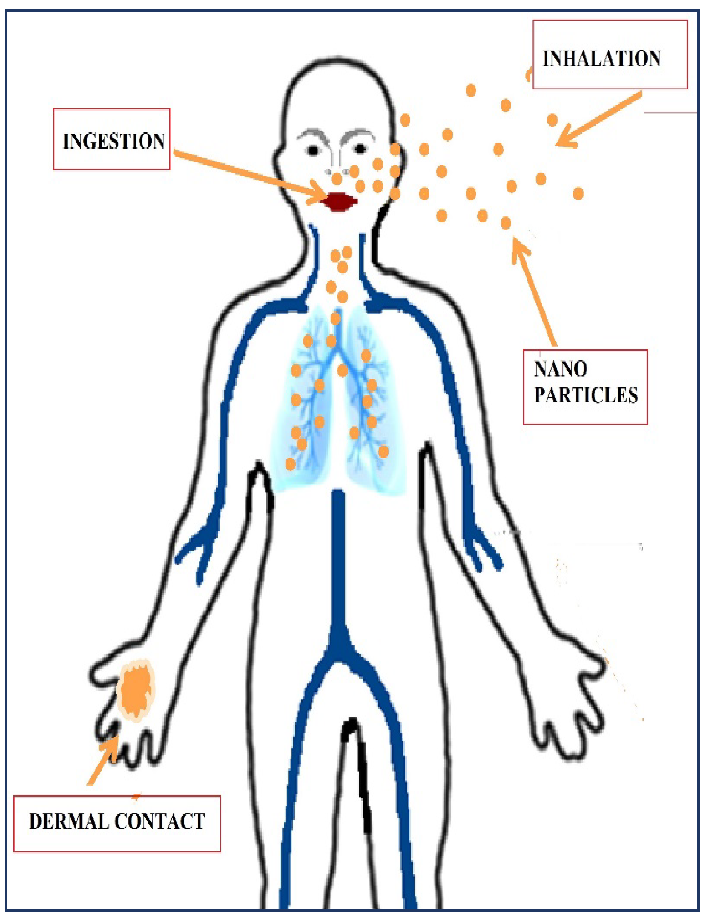
- Bergin, I.L.; Witzmann, F.A. Nanoparticle toxicity by the gastrointestinal route: Evidence and knowledge gaps. Int. J. Biomed. Nanosci. Nanotechnol. 2013, 3, 163. [Google Scholar] [CrossRef]
- Buzea, C.; Pacheco, I.I.; Robbie, K. Nanomaterials and nanoparticles: Sources and toxicity. Biointerphases 2007, 2, MR17–MR71. [Google Scholar] [CrossRef]
- Moore, C.; Movia, D.; Smith, R.J.; Hanlon, D.; Lebre, F.; Lavelle, E.C.; Byrne, H.J.; Coleman, J.N.; Volkov, Y.; McIntyre, J. Industrial grade 2D molybdenum disulphide (MoS2): An in vitro exploration of the impact on cellular uptake, cytotoxicity, and inflammation. 2D Mater. 2017, 4, 025065. [Google Scholar] [CrossRef]
- Oberdörster, G.; Sharp, Z.; Atudorei, V.; Elder, A.; Gelein, R.; Kreyling, W.; Cox, C. Translocation of inhaled ultrafine particles to the brain. Inhal. Toxicol. 2004, 16, 437–445. [Google Scholar] [CrossRef]
- Braakhuis, H.M.; Gosens, I.; Krystek, P.; Boere, J.A.F.; Cassee, F.R.; Fokkens, P.H.B.; Post, J.A.; Van Loveren, H.; Park, M.V.D.Z. Particle size dependent deposition and pulmonary inflammation after short-term inhalation of silver nanoparticles. Part. Fibre Toxicol. 2014, 11, 49. [Google Scholar] [CrossRef]
- Mak, K.F.; Lee, C.; Hone, J.; Shan, J.; Heinz, T.F. Atomically thin MoS2: A new direct-gap semiconductor. Phys. Rev. Lett. 2010, 105, 136805. [Google Scholar] [CrossRef] [PubMed]
- Teo, W.Z.; Chng, E.L.K.; Sofer, Z.; Pumera, M. Cytotoxicity of exfoliated transition-metal dichalcogenides (MoS2, WS2, and WSe2) is lower than that of graphene and its analogues. Chem. A Eur. J. 2014, 20, 9627–9632. [Google Scholar] [CrossRef]
- Chng, E.L.K.; Sofer, Z.; Pumera, M. MoS2 exhibits stronger toxicity with increased exfoliation. Nanoscale 2014, 6, 14412–14418. [Google Scholar] [CrossRef] [PubMed]
- Pardo, M.; Shuster-Meiseles, T.; Levin-Zaidman, S.; Rudich, A.; Rudich, Y. Low cytotoxicity of inorganic nanotubes and fullerene-like nanostructures in human bronchial epithelial cells: Relation to inflammatory gene induction and antioxidant response. Environ. Sci. Technol. 2014, 48, 3457–3466. [Google Scholar] [CrossRef] [PubMed]
- Appel, J.H.; Li, D.O.; Podlevsky, J.D.; Debnath, A.; Green, A.A.; Wang, Q.H.; Chae, J. Low Cytotoxicity and Genotoxicity of Two-Dimensional MoS2 and WS2. ACS Biomater. Sci. Eng. 2016, 2, 361–367. [Google Scholar] [CrossRef]
- Liu, S.; Shen, Z.; Wu, B.; Yu, Y.; Hou, H.; Zhang, X.X.; Ren, H.Q. Cytotoxicity and Efflux Pump Inhibition Induced by Molybdenum Disulfide and Boron Nitride Nanomaterials with Sheetlike Structure. Environ. Sci. Technol. 2017, 51, 10834–10842. [Google Scholar] [CrossRef]
- Shang, E.; Niu, J.; Li, Y.; Zhou, Y.; Crittenden, J.C. Comparative toxicity of Cd, Mo, and W sulphide nanomaterials toward E. coli under UV irradiation. Environ. Pollut. 2017, 224, 606–614. [Google Scholar] [CrossRef]
- Zou, W.; Zhang, X.; Zhao, M.; Zhou, Q.; Hu, X. Cellular proliferation and differentiation induced by single-layer molybdenum disulfide and mediation mechanisms of proteins via the Akt-mTOR-p70S6K signaling pathway. Nanotoxicology 2017, 11, 781–793. [Google Scholar] [CrossRef]
- Zapór, L. Cytotoxicity Elicited by Molybdenum Disulphide in Different Size of Particles in Human Airway Cells. Rocz. Ochr. Sr. 2019, 21, 794–809. [Google Scholar]
- Roy, D.; Das, A.K.; Saini, R.; Singh, P.K.; Kumar, P.; Hussain, M.; Mandal, A.; Dixit, A.R. Pulse current co-deposition of Ni–WS2 nano-composite film for solid lubrication. Mater. Manuf. Processes 2017, 32, 365–372. [Google Scholar] [CrossRef]
- Goldman, E.B.; Zak, A.; Tenne, R.; Kartvelishvily, E.; Levin-Zaidman, S.; Neumann, Y.; Stiubea-Cohen, R.; Palmon, A.; Hovav, A.-H.; Aframian, D.J. Biocompatibility of tungsten disulfide inorganic nanotubes and fullerene-like nanoparticles with salivary gland cells. Tissue Eng. Part A 2015, 21, 1013–1023. [Google Scholar] [CrossRef]
- Garcia-Hevia, L.; Roehrer, I.; Mazzocchi, T.; Menciassi, A.; Ricotti, L. Cytotoxicity of pristine and functionalized tungsten disulfide particles in the urinary system. J. Nanopart. Res. 2020, 22, 273. [Google Scholar] [CrossRef]
- Domi, B.; Bhorkar, K.; Rumbo, C.; Sygellou, L.; Martin, S.M.; Quesada, R.; Yannopoulos, S.N.; Tamayo-Ramos, J.A. Toxicological assessment of commercial monolayer tungsten disulfide nanomaterials aqueous suspensions using human A549 cells and the model fungus Saccharomyces cerevisiae. Chemosphere 2021, 272, 129603. [Google Scholar] [CrossRef]
- Pakdel, A.; Bando, Y.; Golberg, D. Nano boron nitride flatland. Chem. Soc. Rev. 2014, 43, 934–959. [Google Scholar] [CrossRef]
- Horváth, L.; Magrez, A.; Golberg, D.; Zhi, C.; Bando, Y.; Smajda, R.; Horváth, E.; Forró, L.; Schwaller, B. In vitro investigation of the cellular toxicity of boron nitride nanotubes. ACS Nano 2011, 5, 3800–3810. [Google Scholar] [CrossRef]
- Del Turco, S.; Ciofani, G.; Cappello, V.; Gemmi, M.; Cervelli, T.; Saponaro, C.; Nitti, S.; Mazzolai, B.; Basta, G.; Mattoli, V. Cytocompatibility evaluation of glycol-chitosan coated boron nitride nanotubes in human endothelial cells. Colloids Surf. B Biointerfaces 2013, 111, 142–149. [Google Scholar] [CrossRef]
- Ciofani, G.; Raffa, V.; Menciassi, A.; Cuschieri, A. Cytocompatibility, interactions, and uptake of polyethyleneimine-coated boron nitride nanotubes by living cells: Confirmation of their potential for biomedical applications. Biotechnol. Bioeng. 2008, 101, 850–858. [Google Scholar] [CrossRef]
- Ciofani, G.; del Turco, S.; Rocca, A.; de Vito, G.; Cappello, V.; Yamaguchi, M.; Li, X.; Mazzolai, B.; Basta, G.; Gemmi, M.; et al. Cytocompatibility evaluation of gum Arabic-coated ultra-pure boron nitride nanotubes on human cells. Nanomedicine 2014, 9, 773–788. [Google Scholar] [CrossRef]
- Xin, X.; Barger, M.; Roach, K.A.; Bowers, L.; Stefaniak, A.B.; Kodali, V.; Glassford, E.; Dunn, K.H.L.; Dunn, K.H.L.; Wolfarth, M.; et al. Toxicity evaluation following pulmonary exposure to an as-manufactured dispersed boron nitride nanotube (BNNT) material in vivo. NanoImpact 2020, 19, 100235. [Google Scholar] [CrossRef]
- Campatelli, G.; Lorenzini, L.; Scippa, A. Optimization of process parameters using a Response Surface Method for minimizing power consumption in the milling of carbon steel. J. Clean. Prod. 2014, 66, 309–316. [Google Scholar] [CrossRef]
- Rasel, M.A.I.; Li, T.; Nguyen, T.D.; Gu, Y.T. The assessment of toxicity of boron nitride nanoparticle using atomic forced microscopy. IFMBE Proc. 2015, 52, 31–34. [Google Scholar] [CrossRef]
- Kodali, V.K.; Roberts, J.R.; Shoeb, M.; Wolfarth, M.G.; Bishop, L.; Eye, T.; Barger, M.; Roach, K.A.; Friend, S.; Schwegler-Berry, D.; et al. Acute in vitro and in vivo toxicity of a commercial grade boron nitride nanotube mixture. Nanotoxicology 2017, 11, 1040–1058. [Google Scholar] [CrossRef]
- Wang, N.; Wang, H.; Tang, C.; Lei, S.; Shen, W.; Wang, C.; Wang, G.; Wang, Z.; Wang, L. Toxicity evaluation of boron nitride nanospheres and water-soluble boron nitride in caenorhabditis elegans. Int. J. Nanomed. 2017, 12, 5941–5957. [Google Scholar] [CrossRef]
- Kıvanç, M.; Barutca, B.; Koparal, A.T.; Göncü, Y.; Bostancı, S.H.; Ay, N. Effects of hexagonal boron nitride nanoparticles on antimicrobial and antibiofilm activities, cell viability. Mater. Sci. Eng. C 2018, 91, 115–124. [Google Scholar] [CrossRef]
- Kar, F.; Hacıoğlu, C.; Göncü, Y.; Söğüt, İ.; Şenturk, H.; Dönmez, D.B.; Kanbak, G.; Ay, N. In Vivo Assessment of the Effect of Hexagonal Boron Nitride Nanoparticles on Biochemical, Histopathological, Oxidant and Antioxidant Status. J. Clust. Sci. 2021, 32, 517–529. [Google Scholar] [CrossRef]
- Sliwinska, A.; Kwiatkowski, D.; Czarny, P.; Milczarek, J.; Toma, M.; Korycinska, A.; Szemraj, J.; Sliwinski, T. Genotoxicity and cytotoxicity of ZnO and Al2O3 nanoparticles. Toxicol. Mech. Methods 2015, 25, 176–183. [Google Scholar] [CrossRef]
- Zhang, X.Q.; Yin, L.H.; Tang, M.; Pu, Y.P. ZnO, TiO2, SiO2, and Al2O3 nanoparticles-induced toxic effects on human fetal lung fibroblasts. Biomed. Environ. Sci. 2011, 24, 661–669. [Google Scholar] [CrossRef]
- Lin, W.; Stayton, I.; Huang, Y.W.; Zhou, X.D.; Ma, Y. Cytotoxicity and cell membrane depolarization induced by aluminum oxide nanoparticles in human lung epithelial cells A549. Toxicol. Environ. Chem. 2008, 90, 983–996. [Google Scholar] [CrossRef]
- Nogueira, D.J.; Arl, M.; Köerich, J.S.; Simioni, C.; Ouriques, L.C.; Vicentini, D.S.; Matias, W.G. Comparison of cytotoxicity of α-Al2O3 and η-Al2O3 nanoparticles toward neuronal and bronchial cells. Toxicol. Vitr. 2019, 61, 104596. [Google Scholar] [CrossRef]
- Jeng, H.A.; Swanson, J. Toxicity of metal oxide nanoparticles in mammalian cells. J. Environ. Sci. Health Part A 2006, 41, 2699–2711. [Google Scholar] [CrossRef]
- Zhu, X.; Zhu, L.; Duan, Z.; Qi, R.; Li, Y.; Lang, Y. Comparative toxicity of several metal oxide nanoparticle aqueous suspensions to Zebrafish (Danio rerio) early developmental stage. J. Environ. Sci. Health Part A 2008, 43, 278–284. [Google Scholar] [CrossRef]
- Chen, L.; Yokel, R.A.; Hennig, B.; Toborek, M. Manufactured aluminum oxide nanoparticles decrease expression of tight junction proteins in brain vasculature. J. Neuroimmune Pharmacol. 2008, 3, 286–295. [Google Scholar] [CrossRef]
- Oesterling, E.; Chopra, N.; Gavalas, V.; Arzuaga, X.; Lim, E.J.; Sultana, R.; Butterfield, D.A.; Bachas, L.; Hennig, B. Alumina nanoparticles induce expression of endothelial cell adhesion molecules. Toxicol. Lett. 2008, 178, 160–166. [Google Scholar] [CrossRef]
- Sadiq, I.M.; Pakrashi, S.; Chandrasekaran, N.; Mukherjee, A. Studies on toxicity of aluminum oxide (Al2O3) nanoparticles to microalgae species: Scenedesmus sp. and Chlorella sp. J. Nanopart. Res. 2011, 13, 3287–3299. [Google Scholar] [CrossRef]
- Ates, M.; Demir, V.; Arslan, Z.; Daniels, J.; Farah, I.O.; Bogatu, C. Evaluation of alpha and gamma aluminum oxide nanoparticle accumulation, toxicity, and depuration in Artemia salina larvae. Environ. Toxicol. 2015, 30, 109–118. [Google Scholar] [CrossRef]
- Shirazi, A.; Shariati, F.; Keshavarz, A.K.; Ramezanpour, Z. Toxic Effect of Aluminum Oxide Nanoparticles on Green Micro-Algae dunaliella salina. Int. J. Environ. Res. 2015, 9, 585–594. [Google Scholar]
- Srikanth, K.; Mahajan, A.; Pereira, E.; Duarte, A.C.; Rao, J.V. Aluminium oxide nanoparticles induced morphological changes, cytotoxicity and oxidative stress in Chinook salmon (CHSE-214) cells. J. Appl. Toxicol. 2015, 35, 1133–1140. [Google Scholar] [CrossRef]
- Park, E.J.; Sim, J.; Kim, Y.; Han, B.S.; Yoon, C.; Lee, S.; Cho, M.H.; Lee, B.S.; Kim, J.H. A 13-week repeated-dose oral toxicity and bioaccumulation of aluminum oxide nanoparticles in mice. Arch. Toxicol. 2015, 89, 371–379. [Google Scholar] [CrossRef]
- Rajiv, S.; Jerobin, J.; Saranya, V.; Nainawat, M.; Sharma, A.; Makwana, P.; Gayathri, C.; Bharath, L.; Singh, M.; Kumar, M.; et al. Comparative cytotoxicity and genotoxicity of cobalt (II, III) oxide, iron (III) oxide, silicon dioxide, and aluminum oxide nanoparticles on human lymphocytes in vitro. Hum. Exp. Toxicol. 2016, 35, 170–183. [Google Scholar] [CrossRef]
- Benavides, M.; Fernández-Lodeiro, J.; Coelho, P.; Lodeiro, C.; Diniz, M.S. Single and combined effects of aluminum (Al2O3) and zinc (ZnO) oxide nanoparticles in a freshwater fish, Carassius auratus. Environ. Sci. Pollut. Res. 2016, 23, 24578–24591. [Google Scholar] [CrossRef]
- Murali, M.; Suganthi, P.; Athif, P.; Bukhari, A.S.; Mohamed, H.S.; Basu, H.; Singhal, R. Histological alterations in the hepatic tissues of Al2O3 nanoparticles exposed freshwater fish Oreochromis mossambicus. J. Trace Elem. Med. Biol. 2017, 44, 125–131. [Google Scholar] [CrossRef]
- Akbaba, G.B.; Türkez, H. Investigation of the Genotoxicity of Aluminum Oxide, β-Tricalcium Phosphate, and Zinc Oxide Nanoparticles In Vitro. Int. J. Toxicol. 2018, 37, 216–222. [Google Scholar] [CrossRef] [PubMed]
- Kim, Y.S.; Chung, Y.H.; Seo, D.S.; Choi, H.S.; Lim, C.H. Twenty-eight-day repeated inhalation toxicity study of aluminum oxide nanoparticles in male Sprague-Dawley rats. Toxicol. Res. 2018, 34, 343–354. [Google Scholar] [CrossRef]
- Yousef, M.I.; Mutar, T.F.; Kamel, M.A.E.N. Hepato-renal toxicity of oral sub-chronic exposure to aluminum oxide and/or zinc oxide nanoparticles in rats. Toxicol. Rep. 2019, 6, 336–346. [Google Scholar] [CrossRef] [PubMed]
- Yousef, M.I.; Al-Hamadani, M.Y.I.; Kamel, M.A. Reproductive Toxicity of Aluminum Oxide Nanoparticles and Zinc Oxide Nanoparticles in Male Rats. Nano Part. 2019, 1, 1–10. [Google Scholar] [CrossRef]
- Anand, A.S.; Gahlot, U.; Prasad, D.N.; Kohli, E. Aluminum oxide nanoparticles mediated toxicity, loss of appendages in progeny of Drosophila melanogaster on chronic exposure. Nanotoxicology 2019, 13, 977–989. [Google Scholar] [CrossRef]
- Boran, H.; Şaffak, S. Transcriptome alterations and genotoxic influences in zebrafish larvae after exposure to dissolved aluminum and aluminum oxide nanoparticles. Toxicol. Mech. Methods 2020, 30, 546–554. [Google Scholar] [CrossRef]
- Lin, W.; Xu, Y.; Huang, C.-C.; Ma, Y.; Shannon, K.B.; Chen, D.-R.; Huang, Y.-W. Toxicity of nano- and micro-sized ZnO particles in human lung epithelial cells. J. Nanopart. Res. 2009, 11, 25–39. [Google Scholar] [CrossRef]
- Brunner, T.J.; Wick, P.; Manser, P.; Spohn, P.; Grass, R.N.; Limbach, L.K. In vitro cytotoxicity of oxide nanoparticles: Comparison to asbestos, silica, and the effect of particle solubility. Environ. Sci. Technol. 2006, 40, 4374–4381. [Google Scholar] [CrossRef]
- Aruoja, V.; Dubourguier, H.C.; Kasemets, K.; Kahru, A. Toxicity of nanoparticles of CuO, ZnO and TiO2 to microalgae Pseudokirchneriella subcapitata. Sci. Total Environ. 2009, 407, 1461–1468. [Google Scholar] [CrossRef]
- Kasemets, K.; Ivask, A.; Dubourguier, H.C.; Kahru, A. Toxicity of nanoparticles of ZnO, CuO and TiO2 to yeast Saccharomyces cerevisiae. Toxicol. Vitr. 2009, 23, 1116–1122. [Google Scholar] [CrossRef]
- Guan, R.; Kang, T.; Lu, F.; Zhang, Z.; Shen, H.; Liu, M. Cytotoxicity, oxidative stress, and genotoxicity in human hepatocyte and embryonic kidney cells exposed to ZnO nanoparticles. Nanoscale Res. Lett. 2012, 7, 602. [Google Scholar] [CrossRef] [PubMed]
- Pasupuleti, S.; Alapati, S.; Ganapathy, S.; Anumolu, G.; Pully, N.R.; Prakhya, B.M. Toxicity of zinc oxide nanoparticles through oral route. Toxicol. Ind. Health 2012, 28, 675–686. [Google Scholar] [CrossRef] [PubMed]
- Park, H.-S.; Kim, S.-J.; Lee, T.-J.; Kim, G.-Y.; Meang, E.; Hong, J.-S.; Kim, Y.-H.; Koh, S.-B.; Hong, S.-G.; Sun, Y.-S.; et al. A 90-day study of sub-chronic oral toxicity of 20nm positively charged zinc oxide nanoparticles in Sprague Dawley rats. Intern. J. Nanomed. 2014, 9, 93–107. [Google Scholar]
- Davoren, M.; Herzog, E.; Casey, A.; Cottineau, B.; Chambers, D.; Byrne, H.J.; Lyng, F.M. In vitro toxicity evaluation of single walled carbon nanotubes on human A549 lung cells. Toxicol. Vitr. 2007, 21, 438–448. [Google Scholar] [CrossRef] [PubMed]
- Zhang, L.W.; Zeng, L.; Barron, A.R.; Monteiro-Riviere, N.A. Biological interactions of functionalized single-wall carbon nanotubes in human epidermal keratinocytes. Int. J. Toxicol. 2007, 26, 103–113. [Google Scholar] [CrossRef]
- Poland, C.A.; Duffin, R.; Kinloch, I.; Maynard, A.; Wallace, W.A.; Seaton, A.; Stone, V.; Brown, S.; MacNee, W.; Donaldson, K. Carbon nanotubes introduced into the abdominal cavity of mice show asbestos-like pathogenicity in a pilot study. Nat. Nanotechnol. 2008, 3, 423–428. [Google Scholar] [CrossRef]
- Clichici, S.; Biris, A.R.; Catoi, C.; Filip, A.; Tabaran, F. Short-term splenic impact of single-strand DNA functionalized multi-walled carbon nanotubes intraperitoneally injected in rats. J. Appl. Toxicol. 2014, 34, 332–344. [Google Scholar] [CrossRef]
- Reddy, A.R.N.; Krishna, D.R.; Himabindu, V.; Reddy, Y.N. Single walled carbon nanotubes induce cytotoxicity and oxidative stress in HEK293 cells. Toxicol. Environ. Chem. 2014, 96, 931–940. [Google Scholar] [CrossRef]
- Dal Bosco, L.; Weber, G.E.; Parfitt, G.M.; Paese, K.; Gonçalves, C.O.; Serodre, T.M.; Furtado, C.A.; Santos, A.P.; Monserrat, J.M.; Barros, D.M. PEGylated carbon nanotubes impair retrieval of contextual fear memory and alter oxidative stress parameters in the rat hippocampus. BioMed Res. Int. 2015, 2015, 104135. [Google Scholar] [CrossRef]
- Shang, S.; Yang, S.Y.; Liu, Z.M.; Yang, X. Oxidative damage in the kidney and brain of mice induced by different nano-materials. Front. Biol. 2015, 10, 91–96. [Google Scholar] [CrossRef]
- Hosseinpour, M.; Azimirad, V.; Alimohammadi, M.; Shahabi, P.; Sadighi, M.; Nejad, G.G. The cardiac effects of carbon nanotubes in rat. BioImpacts 2016, 6, 79–84. [Google Scholar] [CrossRef] [PubMed]
- Stoddart, M.J. Cell Viability Assays: Introduction; Humana Press: Totowa, NJ, USA, 2011. [Google Scholar] [CrossRef]
- Nouzil, I.; Pervaiz, S.; Kannan, S. Role of jet radius and jet location in cryogenic machining of Inconel 718: A finite element method based approach. Int. J. Interact. Des. Manuf. 2021, 15, 1–19. [Google Scholar] [CrossRef]
| Process | Workpiece | Nanoparticle Used | Conclusion | Reference |
|---|---|---|---|---|
| Grinding | 45 steel | MoS2 | NMQL provided a better surface finish than MQL and dry cooling. Ra and SGE provided almost the same as flood cooling. | [12]. |
| Silicon nitride | MoS2-WS2, WS2-hBN, MoS2-hBN, AL2O3, ZnO and B4C | NMQL performed best with lower SGE and cutting forces compared to wet and MQL cooling. MoS2 performed best among the mono NPs. Among the hybrids, MoS2- WS2 and MoS2-hBN provided the best lubrication. | [13]. | |
| Inconel 718 | AL2O3 | NMQL provided a better Ra and lower energy compared to pure MQL. | [14]. | |
| Turning | Inconel | hBN and AL2O3 | 0.5% vol. hBN performed better than pure MQL and dry cooling. It yielded a low tool wear and roughness. | [20,21]. |
| 0.5% hBN + LN2 provided the best results in terms of interface temperature and Ra. | ||||
| Nimonic 90 | AL2O3 | Cryogenic cooling was concluded to be superior to NMQL. | [22]. | |
| Milling | Inconel X750 | hBN, MoS2 and graphite | 0.5% hBN was found to have superior performance. | [23]. |
| Ferritic stainless steel ASI 430 | Graphene | NMQL performed better. It improved the initial flank wear. | [18]. | |
| TiAlN-coated carbide surface | Graphite (xGnP) and hBN (XGS) | A reduction in the friction coefficient was observed due to an expanding processing envelope of MQL due to the nanoplatelets. | [19]. |
| Type of Study | Concentration | Diameter (nm) | Time of Exposure | Cell Line/Organism | Major Outcomes |
|---|---|---|---|---|---|
| In vitro | 100 µg/mL | 24 h | Human lung epithelial cells | TMDs resulted in a higher cell viability than their organic analogs, such as graphene oxide. Furthermore, among the TMDs tested, WSe2 resulted in the least cell viability while WS2 and MoS2 had very low toxicological effects on the cell [34]. | |
| In vitro | 0–400 µg/mL | 24 h | Human lung carcinoma epithelial cells | The toxicity of the nanoparticle was shown to increase with an increasing degree of exfoliation and was also dependent on the intercalating agents used. MeLi-exfoliated MoS2 showed the least toxicity [35]. | |
| In vitro | 0–100 µg/mL | 80–100 | 24 h | Human bronchial cells (NL-20) | The research stated that compared to silica dioxide and carbon black nanoparticles, up to a concentration of 100 µg mL−1, WS2 and MoS2 nanoparticles exhibited a very low toxicity. At the highest concentration, the cell viability was above 85% for both WS2 and MoS2 [36]. |
| In vitro | 0.1–100 µg/mL | 200–300 | 48 h | Human epithelial kidney cells (HEK293f) | WS2 and MoS2 both exhibited high cell survival rates of over 90% over a 48 h exposure. The authors concluded they are safe for biomedical applications [37]. |
| In vitro | 0.5–30 µg/mL | 50 | 24 h | Human hepatoma HepG2 cells | At 30 μg/mL, MoS2 and BN nanoparticles reduced cell viability [38]. |
| In vitro | 0–50 mg/L | 50 and 90 | 4 h | Escherichia coli (E. coli) | MoS2 resulted in a mortality rate of 38.5%, while WS2 caused a mortality rate of 31.2%. The study aimed to research the effects of NPs in natural water under UV irradiation [39]. |
| In vitro | 0.1–25 µg/mL | 1–1.12 | 24 h | Human embryonic lung fibroblasts (HELFs). | The cell viability reduced to about 80% at a concentration above 10 µg/mL [40]. |
| In vitro | 0.5, 2 and 10 µg/mL | 50, 117 and 177 | 24, 48 and 72 h | Human acute monocytic leukemia (THP-1) Human lung adenocarcinoma (A-549) Human gastric adenocarcinoma (AGS) | The authors concluded that these three sizes of MoS2 nanoparticles are non-toxic at a concentration of 100 µg mL−1 in the three cell lines that were studied [30]. |
| In vitro | 2.5–200 µg/mL | <100 | 24, 48 and 72 h | Normal bronchial epithelium cells BEAS-2B (CRL-9609) | MoS2 NPs and MoS2 MPs exhibit a similar toxicity. After a 72 h exposure, the cell viability reduced to about 50% at all concentration levels. No dose-dependent increase in toxicity was observed [41]. |
| Type of Study | Concentration | Diameter (nm) | Time of Exposure | Cell Line/Organism | Major Outcomes |
|---|---|---|---|---|---|
| In vitro | 100 µg/mL | 24 h | Human lung epithelial cells | TMDs resulted in a higher cell viability than their organic analogs, such as graphene oxide. WSe2 resulted in the least cell viability while WS2 and MoS2 had very low toxicological effects on the cell [34]. | |
| In vitro | 0–100 µg/mL | 80–100 | 24 h | Human bronchial cells (NL-20) | The research stated that compared to silica dioxide and carbon black nanoparticles, up to a concentration of 100 µg mL−1, WS2 and MoS2 nanoparticles exhibited a very low toxicity. At the highest concentration, the cell viability was above 85% for both WS2 and MoS2 [36]. |
| In vitro | 0.22, 3.52 and 35.2 µg/mL | 120–150 | 24 h | Salivary gland cells | The cell viability and cell morphology were unaffected by WS2 uptakes [43]. |
| In vitro | 0.1–100 µg/mL | 200–300 | 48 h | Human epithelial kidney cells (HEK293f) | WS2 and MoS2 both exhibited high cell survival rates of over 90% over a 48 h exposure. The authors concluded they are safe for biomedical applications [37]. |
| In vitro | 0–50 mg/L | 50 and 90 | 4 h | Escherichia coli (E. coli) | MoS2 resulted in a mortality rate of 38.5%, while WS2 caused a mortality rate of 31.2%. The study aimed to research the effect of NPs in natural water under UV irradiation [39]. |
| In vitro | 0–100 µg/mL | ≈500 | 24, 48 and 72 h | Human urinary bladder cells | An 87% cell viability was observed at the highest concentration after a 72 h exposure [44]. |
| In vitro | 20–160 mg/L | 20–500 | 24 h | Human A549 cells Fungus Saccharomyces cerevisiae | The viability of A549 cells was unaffected at all tested concentrations. However, the cell vitality of the fungus reduced to about 70% at the highest concentration [45]. |
| Type of Study | Concentration | Diameter (nm) | Time of Exposure | Cell Line/Organism | Major Outcomes |
|---|---|---|---|---|---|
| In vitro | 10–100 µg/mL | 40–47 | 48 h | Mouse neuroblastoma (neuro-2A) | The cell viability was unaffected at concentrations up to 100 µg/mL−1 during a 48 h exposure [62]. |
| Aqueous suspension | 0.1–50 mg/L | 80 | 96 h | Zebrafish larvae/embryo | Even at the highest concentrations, no effect on hatching rate or survival was observed. The effect was non-toxic [63]. |
| In vitro | 1 µM–10 mM | 8 | 24 h | Human brain microvascular endothelial cells (HBMEC) | It resulted in low cell vilability of 20% at a concentration of 10 mM. No change in viability was observed for up to 0.01 mM [64]. |
| In vivo | 29 mg/Kg b.w. | 8 | 24 h | Fisher 344 rats | It resulted in alterations of mitochondrial functions and decreased expression of tight junction proteins [64]. |
| In vitro | 1–250 µg/mL | 10–20 | 8 h | Porcine pulmonary artery endothelial cells/human umbilical vein endothelial cells | Inflammation was observed. The results indicated a probable cardiovascular disease risk [65]. |
| Aqueous suspension | 3–192 mg/L | 50 | 72 h | Scenedesmus sp./Chlorella sp. | A 50% mortality rate was observed for chlorella at 45.4 mg/L and 39.35 mg/L for Scenedesmus [66]. |
| Aqueous suspension | 5–100 mg/L | 5 and 50 | 24 and 96 h | Artemia salina (crustacean filter feeder) larvae | γ-AL2O3 NPs were more toxic than α-AL2O3 NPs at all concentrations. The highest mortality rate of 34% was observed at 100 mg/L for a 96 h exposure [67]. |
| Aqueous suspension | 0.005–3.8 mg/L | 20 | 72 h | Green micro-algae Dunaliella salina | A swelling and enlargement of Dunaleilla cells was observed. It resulted in a significant impact on the shape and topography [68]. |
| In vitro | 10–100 µg/mL | 50 | 6, 12 and 24 h | Chinook salmon (CHSE-214) | A dose-dependent reduction in cell viability was observed. At 10 µg/mL, the cell viability was found to be 80%, while at 100 µg/mL, it was about 60% for a 24 h exposure [69]. |
| In vivo | 1.5, 3 and 6 mg/kg b.w. | 50 | 13 weeks | ICR mice | The kidney, liver and immune systems were impacted. There was a development of a pathological lesion in the kidney and liver [70]. |
| In vitro | 100 µg/L | 39.4 | 24 h | Human lymphocytes | AL2O3 did not cause DNA damage when compared to other metal oxide NPs (Co3O4, Fe2O3 and SiO2) studied [71]. |
| In vivo | 10 and 100 µg/L | 20 | 7, 14 and 21 days | Freshwater fish (Carassius auratus) | Liver degeneration and gill hyperplasia were observed when exposed to both aluminium oxide and ZnO nanoparticles [72]. |
| In vivo | 120–300 ppm | 96 h | Freshwater fish Oreochromis mossambicus | A 50% mortality rate was observed at a concentration of about 235–245 ppm. Accumulations of NPs were found in the fish liver, affecting the health conditions of the fish [73]. | |
| In vitro | 1–2000 ppm | 19.8 | 72 h | Human peripheral blood lymphocytes | No genotoxicity was observed in the cells even at the highest concentration [74]. |
| In vivo | 0–5 mg/m3 | 11.94 | 28 days | Sprague Dawley rats | Lungs were the most impacted organ. An alveolar macrophage accumulation was found in the lungs. The level of no observed adverse effects of AL2O3 NPs in male rats was suggested to be about 1 mg/m3 [75]. |
| In vivo | 70 mg/kg b.w. | 50 | 75 days | Wistar male albino rats | Liver and kidney damage were observed in the rats. A weight reduction occurred when compared to a control group without nanoparticle injections [76]. |
| In vivo | 70 mg/kg b.w. | 50 | 75 days | Wistar male albino rats | It caused changes on the testicular architecture and caused fertility problems through different pathways, including cell death and oxidative stress [77]. |
| In vivo | 0.1 and 1 mM | 50 | Lifespan—chronic exposure | Flies—Drosophila melanogaster | Wing blisters, malformed legs and a segmented thorax were observed in progeny flies. Behavioural defects in climbing were also noticed [78]. |
| Aqueous suspension | 0–500 mg/L | 50 | 96 h | Zebrafish larvae | At a concentration of 130 mg/L, 50% of the larvae died. It was also noted that at sub-lethal concentrations, AL2O3-NPs can produce DNA damage and change stress-related gene expressions in zebrafish larvae [79]. |
| Type of Study | Concentration | Diameter (nm) | Time of Exposure | Cell Line/Organism | Major Outcomes |
|---|---|---|---|---|---|
| In vitro | 10–100 µg/mL | 50–70 | 48 h | Mouse neuroblastoma (neuro-2A) | Cell viability was unaffected at concentrations below 25 µg/mL. At higher concentrations, 15–50% of the cells died during a 48 h exposure. Mitochondrial function was also severely impacted [62]. |
| In vitro | 0–30 ppm and 0–15 ppm | 3–6 days | Human mesothelioma/rodent fibroblast cell | Above 15 ppm, all cells died over a 3 day exposure to ZnO nanoparticles [81]. | |
| Aqueous suspension | 0.1–0.16 mg/L | 50–70 | 72 h | Algae—Pseudokirchneriella subcapitata | Algae growth inhibition was observed at 0.1 mg/L. Total growth observation occurred at 0.16 mg/L [82]. |
| Aqueous suspension | 0.1–50 mg/L | 20 | 96 h | Zebrafish larvae/embryo | At concentrations less than 0.5 mg/L, no toxicity was observed. At higher concentrations, a dose-dependent increase in toxicity on survival and hatching rate was recorded [63]. |
| In vitro | 31.25–1000 mg/L | 50–70 | 16–18 h | Yeast—Saccharomyces cerevisiae | An 80% inhibition of growth occurred at 250 mg/L [83]. |
| In vitro | 5–100 µg/mL | 50 | 24 h | Human hepatocyte cell (L02)/human embryonic kidney cell (HEK293) | Reduced mitochondrial function occurred at concentrations of 10 µg/mL. ZnO resulted in DNA damage, cell membrane disruption and subsequent cell death [84]. |
| In vivo | 5–2000 mg/kg body weight | 20 | 14 days | Sprague Dawley rats | Toxicity was observed at low doses. Higher liver damage was observed at low does of 5 mg/kg b.w. compared to 2000 mg/kg b.w. Cell inflammation and clotting were observed [85]. |
| In vivo | 125–250–500 mg/kg of body weight | 20 | 90 days | Sprague Dawley rats | The pancreas, eye and stomach were affected at a low concentration of 125 mg/kg b.w. At 250 and 500 mg/kg b.w., significant changes in terms of anemia in the hematological and blood chemical analysis occurred [86]. |
| In vivo | 10/100 µg/L | 50 | 7/14/21 days | Freshwater fish (Carassius auratus) | Liver degeneration and gill hyperplasia were observed when exposed to both aluminium oxide and ZnO nanoparticles [72]. |
| In vitro | 1–2000 ppm | 19.8 | 72 h | Human peripheral blood lymphocytes | Genotoxity was observed at even a low concentration of 12.5 ppm. A high concentration of 500 ppm resulted in a mortality of blood cells [74]. |
| In vivo | 100 mg/kg b.w. | 100 | 75 days | Wistar male albino rats | Liver and kidney damage were observed in the rats. A weight reduction occurred when compared to a control group without nanoparticle injections [76]. |
| In vivo | 100 mg/kg b.w. | 100 | 75 days | Wistar male albino rats | ZnO caused changes to the testicular architecture and caused fertility problems through different pathways, including cell death and oxidative stress [77]. |
| Type of Study | Concentration | Diameter (nm) | Time of Exposure | Cell Line/Organism | Major Outcomes |
|---|---|---|---|---|---|
| In vitro | 1.56–800 µg/mL | 0.8–1.2 | 24 h | A549 human lung cell | A significant cytotoxicity was recorded at concentrations above 400 µg/mL [87]. |
| In vitro | 0.05 µg/mL–0.05 mg/mL | 1 | 24 and 48 h | Human epidermal keratinocytes (HEKs) | A low concentration of 0.05 µg/mL maintains cell viability. A dose-dependent decrease in cell viability was observed. At a high concentration of 0.05 mg/mL, cell viability was reduced to about 50% during a 48 h exposure [88]. |
| In vivo | 50 µg | 15–100 | 24 h | Mice | Similar to aesbestos, MWCNTs result in the formation of a scar-like structure (lesion) called granuloma. Great caution is advised in the use of CNTs [89]. |
| In vivo | 2 mg/1.5 mL/b.w. | 15–25 | 1–144 h | Male Wistar rats | MWCNT translocates progressively in the spleen, with a peak of concentration after 48 h. Transient alterations due to oxidative stress and inflammation were present and need further investigation [90]. |
| In vitro | 3–300 µg/mL | 60–300 | 48 h | Human embryonic kidney cell line (HEK293) | A 50% reduction in cell viabilty was observed at 87.58 µg/mL. A dose-dependent increase in cell membrane damage was observed at 10–100 µg/mL [91]. |
| In vivo | 0.5, 1 and 2.1 mg/mL | 100 | 24 h and 30 min | Male Wistar rats | It caused a short-term impairment in fear memory retrieval. However, the effect was transient and overcome in 24 h [92]. |
| In vivo | 4 mg/kg b.w. | 7 days | Male BALB/c mice | The organ coefficient and GSH levels in the brain and kidney decreased when compared to a control group. Morphological changes were also seen in the experimentation [93]. | |
| In vivo | 1 mg/kg b.w. | 5–10 | 0.5 h | Male Wistar rats | An increased heart rate was observed in rats after injections of CNTs. A possible blockage of potassium channels may be the cause [94]. |
| In Vitro Toxicity Test Parameters | Nanoparticle Exposure Route | |||||||||
|---|---|---|---|---|---|---|---|---|---|---|
| Nanoparticle | Exposure (h) | Concentration | Inhalation | Ingestion | Dermal | |||||
| A549 Human Lung Epithelial Cells | NL-20 Human Bronchial Epithelial Cells | AGS Human Gastric Cells | Human Epidermal Keratinocytes (HEKs) | HepG2 Human Liver-Derived Cells | Human Endothelial Cells | Human Peripheral Blood Cells | ||||
| Cell Viability % | ||||||||||
| MoS2 | 24 | 100 µg mL−1 | 87% | [34] | ||||||
| 24 | 100 µg mL−1 | 70–95% | [35] | |||||||
| 24 | 10 µg mL−1 | 98% | 98–100% | [36] | ||||||
| 24 | 10 µg mL−1 | >90% | <50% | [30] | ||||||
| WS2 | 24 | 85% | [34] | |||||||
| 24 | 90% | 98–100% | [36] | |||||||
| hBN | 120 | 20 µg mL−1 | 30% | [47] | ||||||
| 72 | 20 µg mL−1 | 100% | [50] | |||||||
| AL2O3 | 24 | 10 mM | 70% | [47] | ||||||
| 24 | 25 µg mL−1 | 80% | [60] | |||||||
| ZnO | 24 | 10 mM | 44% | [58] | ||||||
| 24 | 18 µg mL−1 | 20% | [80] | |||||||
| CNT | 24 | 0.05 mg/mL | 50% | [88] | ||||||
| 48 | 87.58 µg mL−1 | 50% | [91] | |||||||
Publisher’s Note: MDPI stays neutral with regard to jurisdictional claims in published maps and institutional affiliations. |
© 2022 by the authors. Licensee MDPI, Basel, Switzerland. This article is an open access article distributed under the terms and conditions of the Creative Commons Attribution (CC BY) license (https://creativecommons.org/licenses/by/4.0/).
Share and Cite
Nouzil, I.; Eltaggaz, A.; Pervaiz, S.; Deiab, I. Toxicity Analysis of Nano-Minimum Quantity Lubrication Machining—A Review. Lubricants 2022, 10, 176. https://doi.org/10.3390/lubricants10080176
Nouzil I, Eltaggaz A, Pervaiz S, Deiab I. Toxicity Analysis of Nano-Minimum Quantity Lubrication Machining—A Review. Lubricants. 2022; 10(8):176. https://doi.org/10.3390/lubricants10080176
Chicago/Turabian StyleNouzil, Ibrahim, Abdelkrem Eltaggaz, Salman Pervaiz, and Ibrahim Deiab. 2022. "Toxicity Analysis of Nano-Minimum Quantity Lubrication Machining—A Review" Lubricants 10, no. 8: 176. https://doi.org/10.3390/lubricants10080176
APA StyleNouzil, I., Eltaggaz, A., Pervaiz, S., & Deiab, I. (2022). Toxicity Analysis of Nano-Minimum Quantity Lubrication Machining—A Review. Lubricants, 10(8), 176. https://doi.org/10.3390/lubricants10080176