A Clinician-Oriented Approach to Plaque Pathology in ACS: Implications for Personalized Cardiovascular Medicine—A Comprehensive Review
 , , ,
, , ,  ,
,  and
and 
Abstract
1. Introduction
2. Methods
2.1. Study Design
2.2. Literature Identification
2.3. Selection of Relevant Literature
2.4. Data Extraction and Thematic Synthesis
2.5. Conceptual Framework
3. Pathophysiology of Myocardial Infarction
3.1. Plaque Rupture
3.1.1. Molecular Features of Plaque Rupture
3.1.2. Inflammatory and Non-Inflammatory Pathways of Plaque Rupture
3.1.3. Imaging Features and Diagnostic Assessment
3.1.4. Clinical Perspectives
3.2. Plaque Erosion
3.2.1. Prevalence and Clinical Relevance
3.2.2. Morphological and Histopathological Features
3.2.3. Molecular and Pathophysiological Mechanisms
3.2.4. Imaging Features and Diagnostic Assessment
3.2.5. Clinical Perspectives
3.3. Calcified Nodules
3.3.1. Morphological and Histopathological Features
3.3.2. Molecular and Pathophysiological Mechanisms
3.3.3. Imaging Features and Diagnostic Assessment
3.3.4. Clinical Perspectives

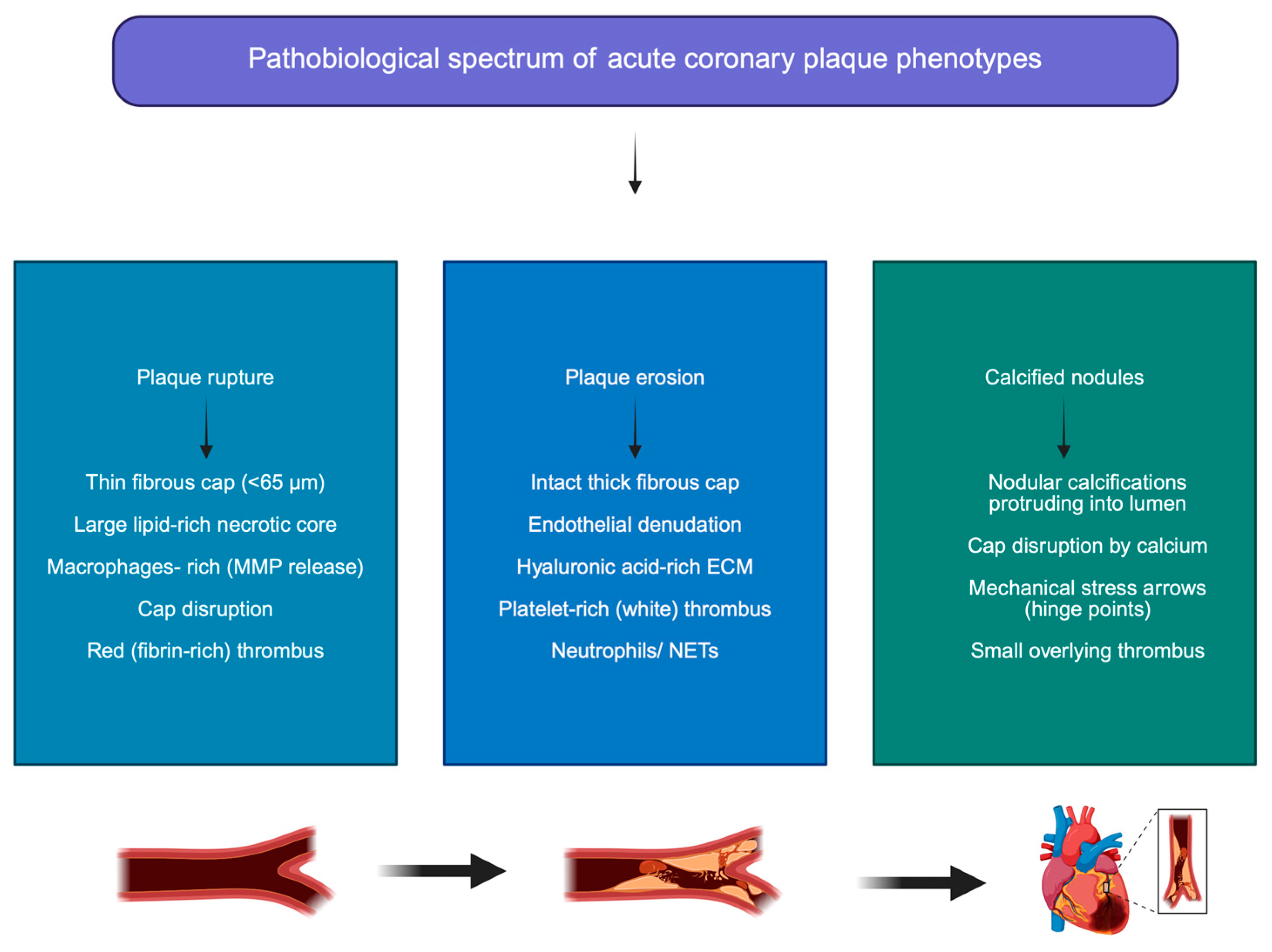
| Feature | Plaque Rupture (PR) | Plaque Erosion (PE) | Calcified Nodules (CNs) | References |
|---|---|---|---|---|
| Prevalence in ACS | Most common substrate (~60–70%) | Intermediate (~25–35%) | Rare (~2–7%) | [7,31,42] |
| Typical plaque morphology | Thin-cap fibroatheroma with large lipid-rich necrotic core | Intact and relatively thick fibrous cap with endothelial denudation | Nodular calcific deposits protruding into the lumen | [8,9,10,11,26,42] |
| Fibrous cap characteristics | Thin (<65 μm), disrupted | Thick and intact but endothelial layer lost | Disrupted by eruptive calcific fragments | [8,9,10,11,26,42] |
| Lipid core | Large lipid-rich necrotic core | Small or absent lipid core | Variable; often associated with heavily calcified plaques | [8,26,42] |
| Dominant cellular components | Macrophages, foam cells, inflammatory infiltrate | VSMCs, proteoglycans, hyaluronic acid | Osteogenic VSMCs, calcified extracellular matrix | [11,12,13,14,26,29,44] |
| Main molecular mechanisms | Matrix degradation by macrophage-derived MMPs; inflammatory activation | Endothelial dysfunction, HA accumulation, TLR2 signaling, NET formation | Osteogenic differentiation of VSMCs, microfracture of calcified plates, vascular mineralization | [11,12,13,14,29,30,31,32,33,47,48,49] |
| Thrombus composition | Fibrin- and erythrocyte-rich (“red thrombus”) | Platelet-rich (“white thrombus”) | Mixed thrombus, often associated with calcific protrusion | [27,32,42] |
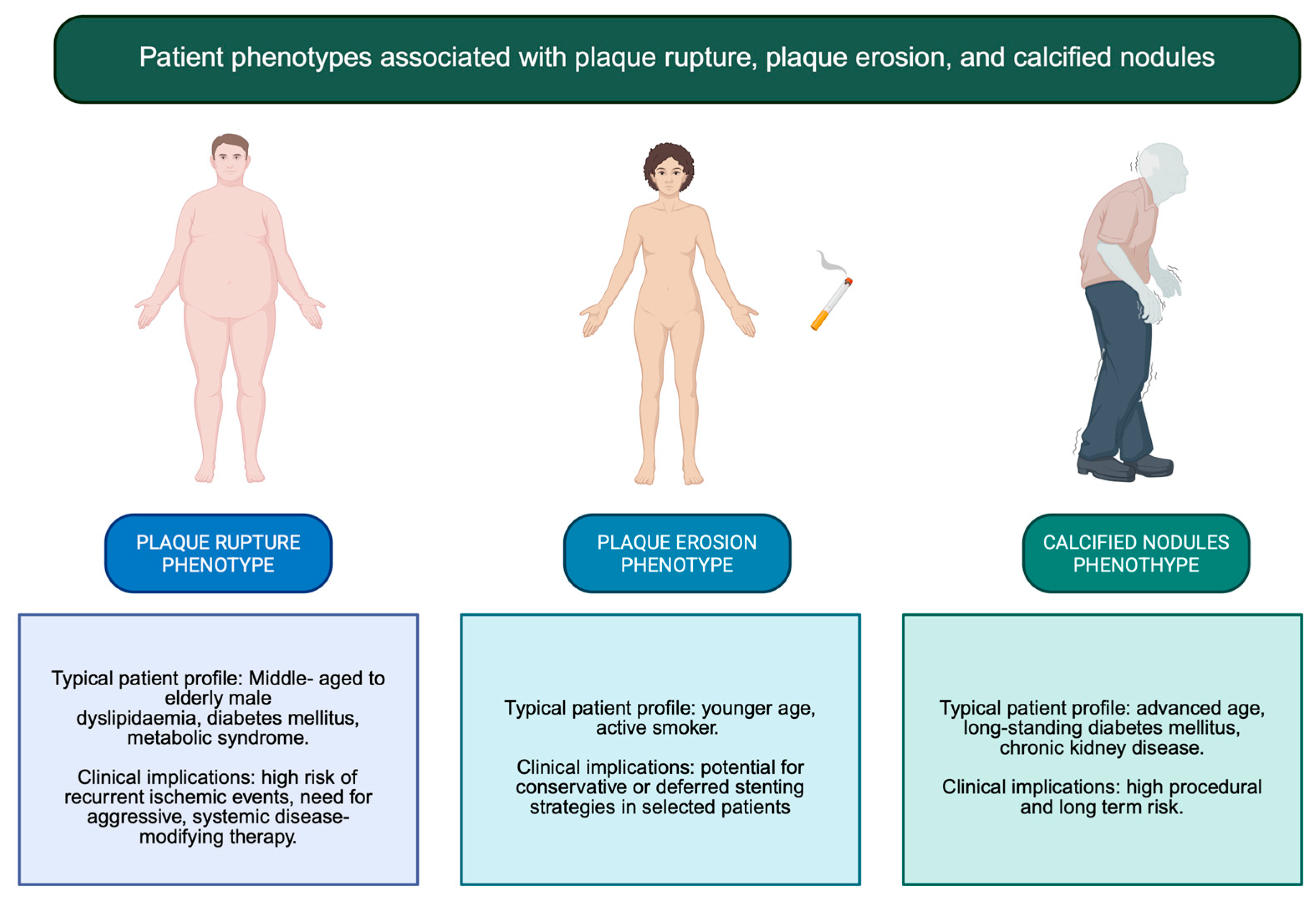
| Typical patient profile | Older patients with high inflammatory burden and lipid-rich plaques | Younger patients, frequently smokers; more common in women | Elderly patients with severe calcification and multiple comorbidities | [30,39,44] |
| Common clinical presentation | STEMI or high-risk ACS | Often NSTEMI with preserved distal flow | ACS with complex calcified lesions | [30,40,44] |
| Imaging features (OCT/IVUS/CCTA) | Cap disruption, cavity formation, lipid-rich plaque | Intact fibrous cap with luminal thrombus, surface irregularity | Protruding calcific nodules with irregular luminal surface | [20,24,37,38,48,49,50] |
| Therapeutic implications | Culprit lesion PCI + aggressive systemic therapy | Selected cases may be managed conservatively with antithrombotic therapy | Often requires plaque modification (e.g., lithotripsy, atherectomy) before stenting | [26,27,28,40,46] |
| Prognostic implications | Marker of diffuse coronary vulnerability | Generally smaller infarcts and potentially better short-term prognosis | Higher risk of procedural complications and target lesion failure | [26,27,28,40,46] |
3.4. Adding Determinants of High-Risk Plaques
4. From Mechanism to Management: A Clinician-Oriented Framework
4.1. Comparison Between European and American Guidelines
4.1.1. Acute Phase: Clinical Phenotyping Beyond Stenosis Severity
4.1.2. Antithrombotic Therapy: Tailoring Intensity and Duration
4.1.3. Disease-Modifying Therapy: Stabilizing the Entire Coronary Tree
4.1.4. Long-Term Follow-Up and Secondary Prevention
4.1.5. New Perspectives and the Potential Role of Artificial Intelligence
5. Conclusions
Author Contributions
Funding
Institutional Review Board Statement
Informed Consent Statement
Data Availability Statement
Conflicts of Interest
Abbreviations
| AI | artificial intelligence |
| ALP | alkaline phosphatase |
| ACS | acute coronary syndrome |
| AMI | acute myocardial infarction |
| BMP-1 | bone morphogenic protein-1 |
| CAD | coronary artery disease |
| CCTA | coronary computed tomography angiography |
| CD4+ | cluster of differentiation 4+ |
| CKD | chronic kidney disease |
| CN | calcified nodules |
| CTE | coronary thrombotic event |
| CRP | C reactive protein |
| CTE | cardiac thrombotic events |
| CV | cardiovascular |
| DM | diabetes mellitus |
| ECM | extracellular matrix |
| HA | hyaluronic acid |
| HYALs | hyaluronidases |
| HMG-CoA | 3-hydroxy-3-methylglutaryl coenzyme A |
| IL-8 | interleukin 8 |
| IL-10 | interleukin 10 |
| IVUS | intravascular ultrasound |
| LCBI | lipid core burden index |
| LRP | lipid-rich plaque |
| MI | myocardial infarction |
| MLA | minimal lumen area |
| MMPs | metalloproteinases |
| MPO | myelopexoxidase |
| NET | neutrophil extracellular traps |
| NIRS | near infrared spectroscopy |
| NE | neutrophil elastase |
| OCT | optical coherence tomography |
| PCI | percutaneous angioplasty |
| PE | plaque erosion |
| PR | plaque rupture |
| ROS | reactive oxygen species |
| Runx-2 | Runt-related transcription factor-2 |
| STEMI | ST elevation myocardial infarction |
| TCFA | thin-cap fibroatheromas |
| TGFβ1 | transforming growth factor 1 |
| Th17 | T helper 17 |
| TLR-8 | Toll-like receptor 8 |
| Treg | lymphocyte T regulatory cells |
| VH-IVUS | virtual histology intravascular ultrasound |
| VSMCs | vascular smooth muscle cells |
References
- Martin, S.S.; Aday, A.W.; Allen, N.B.; Almarzooq, Z.I.; Anderson, C.A.M.; Arora, P.; Avery, C.L.; Baker-Smith, C.M.; Bansal, N.; Beaton, A.Z.; et al. 2025 Heart Disease and Stroke Statistics: A Report of US and Global Data from the American Heart Association. Circulation 2025, 151, 8. [Google Scholar] [CrossRef]
- Byrne, R.A.; Rossello, X.; Coughlan, J.J.; Barbato, E.; Berry, C.; Chieffo, A.; Claeys, M.J.; Dan, G.A.; Dweck, M.R.; Galbraith, M.; et al. 2023 ESC Guidelines for the management of acute coronary syndromes. Eur. Heart J. 2023, 38, 3720–3826. [Google Scholar] [CrossRef]
- Liu, T.; Hao, Y.; Zhang, Z.; Zhou, H.; Peng, S.; Zhang, D.; Li, K.; Chen, Y.; Chen, M. Advanced Cardiac Patches for the Treatment of Myocardial Infarction. Circulation 2024, 149, 2002–2020. [Google Scholar] [CrossRef]
- Zaman, S.; Wasfy, J.H.; Kapil, V.; Ziaeian, B.; Parsonage, W.A.; Sriswasdi, S.; Chico, T.J.A.; Capodanno, D.; Colleran, R.; Sutton, N.R.; et al. The Lancet Commission on Rethinking Coronary Artery Disease: Moving from Ischaemia to Atheroma. Lancet 2025, 405, 1264–1312. [Google Scholar] [CrossRef]
- Salari, N.; Morddarvanjoghi, F.; Abdolmaleki, A.; Rasoulpoor, S.; Khaleghi, A.A.; Hezarkhani, L.A.; Shohaimi, S.; Mohammadi, M. The Global Prevalence of Myocardial Infarction: A Systematic Review and Meta-Analysis. BMC Cardiovasc. Disord. 2023, 23, 206. [Google Scholar] [CrossRef]
- Page, M.J.; McKenzie, J.E.; Bossuyt, P.M.; Boutron, I.; Hoffmann, T.C.; Mulrow, C.D.; Shamseer, L.; Tetzlaff, J.M.; Akl, E.A.; Brennan, S.E.; et al. The PRISMA 2020 Statement: An Updated Guideline for Reporting Systematic Reviews. BMJ 2021, 372, n71. [Google Scholar] [CrossRef]
- Vergallo, R.; Park, S.-J.; Stone, G.W.; Erlinge, D.; Porto, I.; Waksman, R.; Mintz, G.S.; D’Ascenzo, F.; Seitun, S.; Saba, L.; et al. Vulnerable or High-Risk Plaque. JACC Cardiovasc. Imaging 2025, 18, 709–740. [Google Scholar] [CrossRef]
- Baaten, C.C.F.M.J.; Nagy, M.; Bergmeier, W.; Spronk, H.M.H.; van der Meijden, P.E.J. Platelet Biology and Function: Plaque Erosion vs. Rupture. Eur. Heart J. 2024, 45, 18–31. [Google Scholar] [CrossRef]
- Hafiane, A. Vulnerable Plaque, Characteristics, Detection, and Potential Therapies. J. Cardiovasc. Dev. Dis. 2019, 6, 26. [Google Scholar] [CrossRef]
- Delwarde, C.; Aikawa, M. Novel Mouse Model of Late-Stage Coronary Atherosclerosis With Features of Plaque Rupture and Stroke. Circulation 2024, 150, 706–709. [Google Scholar] [CrossRef]
- Terada, K.; Kubo, T.; Kameyama, T.; Matsuo, Y.; Ino, Y.; Emori, H.; Higashioka, D.; Katayama, Y.; Khalifa, A.K.M.; Takahata, M.; et al. NIRS-IVUS for Differentiating Coronary Plaque Rupture, Erosion, and Calcified Nodule in Acute Myocardial Infarction. JACC Cardiovasc. Imaging 2021, 7, 1440–1450. [Google Scholar] [CrossRef]
- Jansen, I.; Cahalane, R.; Hengst, R.; Akyildiz, A.; Farrell, E.; Gijsen, F.; Aikawa, E.; van der Heiden, K.; Wissing, T. The Interplay of Collagen, Macrophages, and Microcalcification in Atherosclerotic Plaque Cap Rupture Mechanics. Basic Res. Cardiol. 2024, 119, 193–213. [Google Scholar] [CrossRef]
- Nurmohamed, N.S.; Gaillard, E.L.; Malkasian, S.; de Groot, R.J.; Ibrahim, S.; Bom, M.J.; Kaiser, Y.; Earls, J.P.; Min, J.K.; Kroon, J.; et al. Lipoprotein(a) and Long-Term Plaque Progression, Low-Density Plaque, and Pericoronary Inflammation. JAMA Cardiol. 2024, 9, 826. [Google Scholar] [CrossRef]
- De Caterina, R.; Libby, P.; Peng, H.B.; Thannickal, V.J.; Rajavashisth, T.B.; Gimbrone, M.A.; Shin, W.S.; Liao, J.K. Nitric Oxide Decreases Cytokine-Induced Endothelial Activation. Nitric Oxide Selectively Reduces Endothelial Expression of Adhesion Molecules and Proinflammatory Cytokines. J. Clin. Investig. 1995, 96, 60–68. [Google Scholar] [CrossRef]
- Parma, L.; Duchene, J.; Weber, C. Breaking Point: How Intraplaque Hemorrhage Propels Plaque Rupture. Circ. Res. 2024, 135, 317–319. [Google Scholar] [CrossRef]
- Hirose, T.; Fujii, K.; Hashimoto, K.; Bando, K.; Morishita, S.; Taniichi, M.; Horitani, K.; Shiojima, I. Plaque Rupture and Calcified Nodules: Major Contributors to out-of-Hospital Cardiac Arrest in Acute Myocardial Infarction. Cardiovasc. Interv. Ther. 2025, 40, 788–795. [Google Scholar] [CrossRef]
- Fujimoto, D.; Kinoshita, D.; Suzuki, K.; Niida, T.; Yuki, H.; McNulty, I.; Lee, H.; Otake, H.; Shite, J.; Ferencik, M.; et al. Relationship Between Calcified Plaque Burden, Vascular Inflammation, and Plaque Vulnerability in Patients With Coronary Atherosclerosis. JACC Cardiovasc. Imaging 2024, 17, 1214–1224. [Google Scholar]
- Crea, F.; Libby, P. Acute Coronary Syndromes. Circulation 2017, 136, 1155–1166. [Google Scholar]
- Ishii, Y.; Kure, M.; Kawasumi, H.; Numaziri, Y.; Tanizaki, Y.; Takei, Y.; Sone, H.; Tashiro, K.; Sato, T.; Suzuki, H.; et al. Endothelial Dysfunction in Plaque Rupture and Plaque Erosion. Heart Vessel. 2025, 41, 143–149. [Google Scholar] [CrossRef]
- Milzi, A.; Landi, A.; Dettori, R.; Burgmaier, K.; Reith, S.; Burgmaier, M. Multimodal Intravascular Imaging of the Vulnerable Coronary Plaque. Echocardiography 2024, 41, e70035. [Google Scholar] [CrossRef]
- Mintz, G.S.; Matsumura, M.; Ali, Z.; Maehara, A. Clinical Utility of Intravascular Imaging. JACC Cardiovasc. Imaging 2022, 15, 1799–1820. [Google Scholar] [PubMed]
- Yang, X.; Yang, J.; Kashima, Y.; Hachinohe, D.; Sugie, T.; Xu, S.; Guo, X.; Li, X.; Hu, X.; Sun, B.; et al. The Influence between Plaque Rupture and Non-Plaque Rupture on Clinical Outcomes in Patients with ST-Segment Elevation Myocardial Infarction after Primary Percutaneous Coronary Intervention: A Prospective Cohort Study. J. Thorac. Dis. 2024, 16, 7771–7786. [Google Scholar] [CrossRef]
- Hormel, T.T.; Huang, D.; Jia, Y. Advances in OCT Angiography. Transl. Vis. Sci. Technol. 2025, 14, 6. [Google Scholar] [CrossRef]
- Kubo, T.; Terada, K.; Ino, Y.; Shiono, Y.; Tu, S.; Tsao, T.-P.; Chen, Y.; Park, D.-W. Combined Use of Multiple Intravascular Imaging Techniques in Acute Coronary Syndrome. Front. Cardiovasc. Med. 2022, 8, 824128. [Google Scholar] [CrossRef]
- Vergallo, R.; Jia, H.; Soeda, T.; Tian, J.; Jang, I.-K. Interpretation of Optical Coherence Tomography Images. Lancet 2014, 383, 1887. [Google Scholar]
- Stone, G.W.; Maehara, A.; Lansky, A.J.; de Bruyne, B.; Cristea, E.; Mintz, G.S.; Mehran, R.; McPherson, J.; Farhat, N.; Marso, S.P.; et al. A Prospective Natural-History Study of Coronary Atherosclerosis. N. Engl. J. Med. 2011, 364, 226–235. [Google Scholar]
- Erlinge, D.; Maehara, A.; Ben-Yehuda, O.; Bøtker, H.E.; Maeng, M.; Kjøller-Hansen, L.; Engstrøm, T.; Matsumura, M.; Crowley, A.; Dressler, O.; et al. Identification of Vulnerable Plaques and Patients by Intracoronary Near-Infrared Spectroscopy and Ultrasound (PROSPECT II): A Prospective Natural History Study. Lancet 2021, 397, 985–995. [Google Scholar] [CrossRef]
- Araki, M.; Park, S.-J.; Dauerman, H.L.; Uemura, S.; Kim, J.-S.; Di Mario, C.; Johnson, T.W.; Guagliumi, G.; Kastrati, A.; Joner, M.; et al. Author Correction: Optical Coherence Tomography in Coronary Atherosclerosis Assessment and Intervention. Nat. Rev. Cardiol. 2024, 21, 348. [Google Scholar]
- Jiang, S.; Fang, C.; Xu, X.; Xing, L.; Sun, S.; Peng, C.; Yin, Y.; Lei, F.; Wang, Y.; Li, L.; et al. Identification of High-Risk Coronary Lesions by 3-Vessel Optical Coherence Tomography. J. Am. Coll. Cardiol. 2023, 81, 1217–1230. [Google Scholar] [PubMed]
- Yonetsu, T.; Lee, T.; Murai, T.; Suzuki, M.; Matsumura, A.; Hashimoto, Y.; Kakuta, T. Plaque Morphologies and the Clinical Prognosis of Acute Coronary Syndrome Caused by Lesions with Intact Fibrous Cap Diagnosed by Optical Coherence Tomography. Int. J. Cardiol. 2016, 203, 766–774. [Google Scholar] [CrossRef] [PubMed]
- Wight, T.N. A Role for Extracellular Matrix in Atherosclerotic Plaque Erosion. J. Am. Coll. Cardiol. 2018, 72, 1504–1505. [Google Scholar] [CrossRef]
- Waksman, R.; Di Mario, C.; Torguson, R.; Ali, Z.A.; Singh, V.; Skinner, W.H.; Artis, A.K.; Cate, T.T.; Powers, E.; Kim, C.; et al. Identification of Patients and Plaques Vulnerable to Future Coronary Events with Near-Infrared Spectroscopy Intravascular Ultrasound Imaging: A Prospective, Cohort Study. Lancet 2019, 394, 1629–1637. [Google Scholar] [CrossRef]
- Garcia-Garcia, H.M.; Sanz-Sanchez, J.; Pinilla-Echeverri, N.; Blanco, P.J.; Bourantas, C.; Alfonso, F. Advances in Coronary Imaging of Atherosclerotic Plaques. EuroIntervention 2025, 21, e778–e795. [Google Scholar] [CrossRef]
- Khatana, C.; Saini, N.K.; Chakrabarti, S.; Saini, V.; Sharma, A.; Saini, R.V.; Saini, A.K. Mechanistic Insights into the Oxidized Low-Density Lipoprotein-Induced Atherosclerosis. Oxid. Med. Cell. Longev. 2020, 2020, 5245308. [Google Scholar] [CrossRef]
- Li, J.; Li, Y.; Zhou, X.; Yang, S.; Liu, D.; Wen, H.; Chen, X.; Duan, C.; Yu, M.; Zhang, M.; et al. Adipocytes Orchestrate Obesity-Related Chronic Inflammation through Β2-Microglobulin. Signal Transduct. Target. Ther. 2025, 10, 394. [Google Scholar] [CrossRef] [PubMed]
- Wensveen, F.M.; Valentić, S.; Šestan, M.; Turk Wensveen, T.; Polić, B. The “Big Bang” in Obese Fat: Events Initiating Obesity-Induced Adipose Tissue Inflammation. Eur. J. Immunol. 2015, 45, 2446–2456. [Google Scholar] [CrossRef]
- Meteva, D.; Pedicino, D.; Abdelwahed, Y.S.; Kränkel, N.; Anwari, E.; Leistner, D.M.; den Ruijter, H.; Gerhardt, T.; Joner, M.; Libby, P.; et al. Acute Coronary Syndrome with Plaque Erosion: Clinical Characteristics, Mechanisms, and Unmet Needs. Eur. Heart J. 2025, 47, 1403–1414. [Google Scholar] [CrossRef]
- Shetty, S.; Subramanian, M. Neutrophil Extracellular Traps (NETs) as Drivers of Atherosclerosis: Pathogenic Mechanisms and Therapeutic Opportunities. Pharmacol. Ther. 2025, 274, 108917. [Google Scholar] [CrossRef]
- Bentzon, J.F.; Falk, E. Plaque Erosion: New Insights from the Road Less Travelled. Circ. Res. 2017, 121, 8–10. [Google Scholar] [CrossRef] [PubMed]
- Wietzorrek, G.; Drexel, M.; Trieb, M.; Santos-Sierra, S. Anti-inflammatory activity of small-molecule antagonists of Toll-like receptor 2 (TLR2) in mice. Immunobiology 2019, 224, 1–9. [Google Scholar] [CrossRef]
- Shin, D.; Karimi Galougahi, K.; Spratt, J.C.; Maehara, A.; Collet, C.; Barbato, E.; Ribichini, F.L.; Gonzalo, N.; Sakai, K.; Mintz, G.S.; et al. Calcified Nodule in Percutaneous Coronary Intervention. JACC Cardiovasc. Interv. 2024, 17, 1187–1199. [Google Scholar] [CrossRef]
- Kondo, S.; Mizukami, T.; Kobayashi, N.; Wakabayashi, K.; Mori, H.; Yamamoto, M.H.; Sambe, T.; Yasuhara, S.; Hibi, K.; Nanasato, M.; et al. Diagnosis and Prognostic Value of the Underlying Cause of Acute Coronary Syndrome in Optical Coherence Tomography-Guided Emergency Percutaneous Coronary Intervention. J. Am. Heart Assoc. 2023, 12, e030412. [Google Scholar] [CrossRef] [PubMed]
- Ashikaga, T.; Lee, T.; Miyazaki, R.; Nagase, M.; Misawa, T.; Kaneko, M.; Nozato, T. Methods for Treating Coronary Eruptive Calcified Nodules. Catheter. Cardiovasc. Interv. 2024, 104, 899–906. [Google Scholar] [CrossRef]
- Ali, Z.A.; Kereiakes, D.; Hill, J.; Saito, S.; Di Mario, C.; Honton, B.; Gonzalo, N.; Riley, R.; Maehara, A.; Matsumura, M.; et al. Safety and Effectiveness of Coronary Intravascular Lithotripsy for Treatment of Calcified Nodules. JACC Cardiovasc. Interv. 2023, 16, 1122–1124. [Google Scholar]
- Sato, T.; Matsumura, M.; Yamamoto, K.; Shlofmitz, E.; Moses, J.W.; Khalique, O.K.; Thomas, S.V.; Tsoulios, A.; Cohen, D.J.; Mintz, G.S.; et al. Impact of Eruptive vs. Noneruptive Calcified Nodule Morphology on Acute and Long-Term Outcomes After Stenting. JACC Cardiovasc. Interv. 2023, 16, 1024–1035. [Google Scholar] [CrossRef] [PubMed]
- Katsaros, O.; Sagris, M.; Karakasis, P.; Ktenopoulos, N.; Soulaidopoulos, S.; Theofilis, P.; Apostolos, A.; Tzoumas, A.; Patsourakos, N.; Toutouzas, K.; et al. The Role of Calcified Nodules in Acute Coronary Syndrome: Diagnosis and Management. Int. J. Mol. Sci. 2025, 26, 2581. [Google Scholar] [CrossRef]
- Onnis, C.; Virmani, R.; Kawai, K.; Nardi, V.; Lerman, A.; Cademartiri, F.; Scicolone, R.; Boi, A.; Congiu, T.; Faa, G.; et al. Coronary Artery Calcification: Current Concepts and Clinical Implications. Circulation 2024, 149, 251–266. [Google Scholar] [CrossRef] [PubMed]
- Nagasawa, A.; Otake, H.; Kawamori, H.; Toba, T.; Sugizaki, Y.; Takeshige, R.; Nakano, S.; Tanimura, K.; Takahashi, Y.; Fukuyama, Y.; et al. Relationship among Clinical Characteristics, Morphological Culprit Plaque Features, and Long-Term Prognosis in Patients with Acute Coronary Syndrome. Int. J. Cardiovasc. Imaging 2021, 37, 2827–2837. [Google Scholar] [CrossRef]
- Duissailant, G.R.; Mintz, G.S.; Pichard, A.D.; Kent, K.M.; Salter, L.F.; Popma, J.J.; Griffin, J.; Leon, M.B. Intravascular Ultrasound Identification of Calcified Intraluminal Lesions Misdiagnosed as Thrombi by Coronary Angiography. Am. Heart J. 1996, 132, 687–689. [Google Scholar] [CrossRef]
- Westman, P.C.; Lipinski, M.J.; Luger, D.; Waksman, R.; Bonow, R.O.; Wu, E.; Epstein, S.E. Inflammation as a Driver of Adverse Left Ventricular Remodeling After Acute Myocardial Infarction. J. Am. Coll. Cardiol. 2016, 67, 2050–2060. [Google Scholar]
- Rao, S.V.; O’Donoghue, M.L.; Ruel, M.; Rab, T.; Tamis-Holland, J.E.; Alexander, J.H.; Baber, U.; Baker, H.; Cohen, M.G.; Cruz-Ruiz, M.; et al. 2025 ACC/AHA/ACEP/NAEMSP/SCAI Guideline for the Management of Patients With Acute Coronary Syndromes: A Report of the American College of Cardiology/American Heart Association Joint Committee on Clinical Practice Guidelines. J. Am. Coll. Cardiol. 2025, 85, 2135–2237, Erratum in J. Am. Coll. Cardiol. 2025, 85, 1800. https://doi.org/10.1016/j.jacc.2025.03.500. Erratum in J. Am. Coll. Cardiol. 2025, 86, 2723. https://doi.org/10.1016/j.jacc.2025.10.050. [Google Scholar] [CrossRef]
- Frangogiannis, N.G. Pathophysiology of Myocardial Infarction. Compr. Physiol. 2015, 5, 1841–1875. [Google Scholar] [CrossRef]
- Bhatt, D.L.; Lopes, R.D.; Harrington, R.A. Diagnosis and Treatment of Acute Coronary Syndromes: A Review. JAMA 2022, 327, 662–675, Erratum in JAMA 2022, 327, 1710. https://doi.org/10.1001/jama.2022.6185. [Google Scholar] [CrossRef]
- Kim, S.; Nam, H.S.; Kang, D.O.; Han, J.; Kim, H.; Song, J.W.; Park, E.J.; Kim, R.H.; Kim, H.J.; Kim, J.H.; et al. Intracoronary Structural-Molecular Imaging for Multitargeted Characterization of High-Risk Plaque: First-in-Human OCT-FLIm. JAMA Cardiol. 2025, 7, 708–717. [Google Scholar] [CrossRef]
- Niccoli, G.; Montone, R.A.; Cataneo, L.; Cosentino, N.; Gramegna, M.; Refaat, H.; Porto, I.; Burzotta, F.; Trani, C.; Leone, A.M.; et al. Morphological-Biohumoral Correlations in Acute Coronary Syndromes: Pathogenetic Implications. Int. J. Cardiol. 2014, 171, 463–466. [Google Scholar] [CrossRef] [PubMed]
- Gerhardt, T.; Seppelt, C.; Abdelwahed, Y.S.; Meteva, D.; Wolfram, C.; Stapmanns, P.; Erbay, A.; Zanders, L.; Nelles, G.; Musfeld, J.; et al. OPTICO-ACS study group. Culprit plaque morphology determines inflammatory risk and clinical outcomes in acute coronary syndrome. Eur. Heart J. 2023, 38, 3911–3925. [Google Scholar] [CrossRef] [PubMed]
- Hu, S.; Wang, C.; Zhe, C.; Zhu, Y.; Yonetsu, T.; Jia, H.; Hou, J.; Zhang, S.; Jang, I.; Yu, B. Plaque Erosion Delays Vascular Healing after Drug Eluting Stent Implantation in Patients with Acute Coronary Syndrome. Catheter. Cardiovasc. Interv. 2017, 89, 592–600. [Google Scholar] [CrossRef]
- Gallone, G.; Bellettini, M.; Gatti, M.; Tore, D.; Bruno, F.; Scudeler, L.; Cusenza, V.; Lanfranchi, A.; Angelini, A.; de Filippo, O.; et al. Coronary Plaque Characteristics Associated With Major Adverse Cardiovascular Events in Atherosclerotic Patients and Lesions: A Systematic Review and Meta-Analysis. JACC Cardiovasc. Imaging 2023, 12, 1584–1604. [Google Scholar] [CrossRef] [PubMed]
- Jia, H.; Dai, J.; Hou, J.; Xing, L.; Ma, L.; Liu, H.; Xu, M.; Yao, Y.; Hu, S.; Yamamoto, E.; et al. Effective anti-thrombotic therapy without stenting: Intravascular optical coherence tomography-based management in plaque erosion (the EROSION study). Eur. Heart J. 2017, 11, 792–800. [Google Scholar] [CrossRef]
- Souteyrand, G.; Viallard, L.; Combaret, N.; Pereira, B.; Clerfond, G.; Malcles, G.; Barber-Chamoux, N.; Prati, F.; Motreff, P. Innovative Invasive Management without Stent Implantation Guided by Optical Coherence Tomography in Acute Coronary Syndrome. Arch. Cardiovasc. Dis. 2018, 111, 666–677. [Google Scholar] [CrossRef]
- Galli, M.; Angiolillo, D.J. De-Escalation of Antiplatelet Therapy in Acute Coronary Syndromes: Why, How and When? Front. Cardiovasc. Med. 2022, 9, 975969. [Google Scholar] [CrossRef]
- Pertiwi, K.; van der Wal, A.; Pabittei, D.; Mackaaij, C.; van Leeuwen, M.; Li, X.; de Boer, O. Neutrophil Extracellular Traps Participate in All Different Types of Thrombotic and Haemorrhagic Complications of Coronary Atherosclerosis. Thromb. Haemost. 2018, 118, 1078–1087. [Google Scholar] [CrossRef] [PubMed]
- Tan, Y.; Yang, S.; Chen, R.; Sheng, Z.; Zhou, P.; Liu, C.; Zhao, H.; Song, L.; Li, J.; Zhou, J.; et al. High Plasma Myeloperoxidase Is Associated with Plaque Erosion in Patients with ST-Segment Elevation Myocardial Infarction. J. Cardiovasc. Transl. Res. 2020, 13, 908–915. [Google Scholar] [CrossRef]
- Libby, P. Superficial Erosion and the Precision Management of Acute Coronary Syndromes: Not One-Size-Fits-All. Eur. Heart J. 2017, 38, 801–803. [Google Scholar] [CrossRef][Green Version]
- Fan, J.; Watanabe, T. Atherosclerosis: Known and unknown. Pathol. Int. 2022, 3, 151–160. [Google Scholar] [CrossRef]
- Russo, G.; Pedicino, D.; Chiastra, C.; Vinci, R.; Lodi Rizzini, M.; Genuardi, L.; Sarraf, M.; d’Aiello, A.; Bologna, M.; Aurigemma, C.; et al. Coronary artery plaque rupture and erosion: Role of wall shear stress profiling and biological patterns in acute coronary syndromes. Int. J. Cardiol. 2023, 370, 356–365. [Google Scholar] [CrossRef]
- Nissen, S.E.; Tuzcu, E.M.; Schoenhagen, P.; Crowe, T.; Sasiela, W.J.; Tsai, J.; Orazem, J.; Magorien, R.D.; O’Shaughnessy, C.; Ganz, P. Statin Therapy, LDL Cholesterol, C-Reactive Protein, and Coronary Artery Disease. N. Engl. J. Med. 2005, 352, 29–38. [Google Scholar] [CrossRef] [PubMed]
- Räber, L.; Ueki, Y.; Otsuka, T.; Losdat, S.; Häner, J.D.; Lonborg, J.; Fahrni, G.; Iglesias, J.F.; van Geuns, R.-J.; Ondracek, A.S.; et al. Effect of Alirocumab Added to High-Intensity Statin Therapy on Coronary Atherosclerosis in Patients with Acute Myocardial Infarction. JAMA 2022, 327, 1771. [Google Scholar] [CrossRef]
- Nicholls, S.J. PCSK9 Inhibitors and Reduction in Cardiovascular Events: Current Evidence and Future Perspectives. Kardiol. Pol. 2023, 81, 115–122. [Google Scholar] [CrossRef]
- Libby, P.; Ridker, P.M.; Hansson, G.K. Inflammation in Atherosclerosis. J. Am. Coll. Cardiol. 2009, 54, 2129–2138. [Google Scholar] [CrossRef] [PubMed]
- Hamana, T.; Shah, P.; Grogan, A.; Kawakami, R.; Williams, D.; Diaz, K.M.; Virmani, R.; Finn, A.V. Risk Factors Favoring Plaque Erosion. Curr. Atheroscler. Rep. 2025, 27, 17. [Google Scholar] [CrossRef]
- Gomes, P.M.; Almeida, B.O.; Marinelli Pedrini, S.; Freitas, B.P.; Júnior, J.M.; Lemos, P.A.; Fonseca, F.H.; Mintz, G.S.; Caixeta, A. Morphology and phenotype characteristics of atherosclerotic plaque in patients with acute coronary syndrome: Contemporary optical coherence tomography findings. Coron. Artery Dis. 2021, 8, 698–705. [Google Scholar] [CrossRef] [PubMed]
- Scheiber, A.; Mank, V. Anti-Inflammatory Diets. In StatPearls; StatPearls Publishing: Treasure Island, FL, USA, 2023. [Google Scholar]
- Khan, Z.; Rakhit, R. Secondary Prevention Lipid Management Following ACS: A Missed Opportunity? Br. J. Cardiol. 2022, 29, 35. [Google Scholar] [CrossRef] [PubMed]
- De la Rosa, A.; Arrington, K.; Desai, R.; Acharya, P.C. Polypill Strategy in Secondary Cardiovascular Prevention. Curr. Cardiol. Rep. 2024, 26, 443–450. [Google Scholar] [CrossRef] [PubMed]
- Mathews, R.; Wang, T.Y.; Honeycutt, E.; Henry, T.D.; Zettler, M.; Chang, M.; Fonarow, G.C.; Peterson, E.D. Persistence with Secondary Prevention Medications after Acute Myocardial Infarction: Insights from the TRANSLATE-ACS Study. Am. Heart J. 2015, 170, 62–69. [Google Scholar] [CrossRef]
- Kwiecinski, J. Artificial Intelligence-Based Quantitative Coronary Plaque Analysis. JACC Cardiovasc. Imaging 2024, 3, 281–283. [Google Scholar] [CrossRef]
- Scicolone, R.; Vacca, S.; Pisu, F.; Benson, J.C.; Nardi, V.; Lanzino, G.; Suri, J.S.; Saba, L. Radiomics and Artificial Intelligence: General Notions and Applications in the Carotid Vulnerable Plaque. Eur. J. Radiol. 2024, 176, 111497. [Google Scholar] [CrossRef]
- Motwani, M.; Dey, D.; Berman, D.S.; Germano, G.; Achenbach, S.; Al-Mallah, M.H.; Andreini, D.; Budoff, M.J.; Cademartiri, F.; Callister, T.Q.; et al. Machine Learning for Prediction of All-Cause Mortality in Patients with Suspected Coronary Artery Disease: A 5-Year Multicentre Prospective Registry Analysis. Eur. Heart J. 2016, 38, 500–507. [Google Scholar] [CrossRef]
- van Timmeren, J.E.; Cester, D.; Tanadini-Lang, S.; Alkadhi, H.; Baessler, B. Radiomics in Medical Imaging—“How-to” Guide and Critical Reflection. Insights Imaging 2020, 11, 91. [Google Scholar] [CrossRef]
- Benjamin, M.M.; Rabbat, M.G. Machine Learning-Based Advances in Coronary Computed Tomography Angiography. Quant. Imaging Med. Surg. 2021, 11, 2208–2213. [Google Scholar] [CrossRef]
- Ambale-Venkatesh, B.; Yang, X.; Wu, C.O.; Liu, K.; Hundley, W.G.; McClelland, R.; Gomes, A.S.; Folsom, A.R.; Shea, S.; Guallar, E.; et al. Cardiovascular Event Prediction by Machine Learning. Circ. Res. 2017, 121, 1092–1101. [Google Scholar] [CrossRef] [PubMed]
- Koo, B.K.; Yang, S.; Jung, J.W.; Zhang, J.; Lee, K.; Hwang, D.; Lee, K.S.; Doh, J.H.; Nam, C.W.; Kim, T.H.; et al. Artificial Intelligence-Enabled Quantitative Coronary Plaque and Hemodynamic Analysis for Predicting Acute Coronary Syndrome. JACC Cardiovasc. Imaging 2024, 9, 1062–1076. [Google Scholar] [CrossRef] [PubMed]
- Volleberg, R.H.J.A.; Luttikholt, T.J.; van der Waerden, R.G.A.; Cancian, P.; van der Zande, J.L.; Gu, X.; Mol, J.Q.; Roleder, T.; Prokop, M.; Sánchez, C.I.; et al. Artificial intelligence-based identification of thin-cap fibroatheromas and clinical outcomes: The PECTUS-AI study. Eur. Heart J. 2025, 46, 5032–5041. [Google Scholar] [CrossRef] [PubMed]


Disclaimer/Publisher’s Note: The statements, opinions and data contained in all publications are solely those of the individual author(s) and contributor(s) and not of MDPI and/or the editor(s). MDPI and/or the editor(s) disclaim responsibility for any injury to people or property resulting from any ideas, methods, instructions or products referred to in the content. |
© 2026 by the authors. Licensee MDPI, Basel, Switzerland. This article is an open access article distributed under the terms and conditions of the Creative Commons Attribution (CC BY) license.
Share and Cite
Pala, B.; Piscione, M.; Cribari, F.; Gualtieri, P.; Perrone, M.A.; Di Renzo, L. A Clinician-Oriented Approach to Plaque Pathology in ACS: Implications for Personalized Cardiovascular Medicine—A Comprehensive Review. J. Pers. Med. 2026, 16, 240. https://doi.org/10.3390/jpm16050240
Pala B, Piscione M, Cribari F, Gualtieri P, Perrone MA, Di Renzo L. A Clinician-Oriented Approach to Plaque Pathology in ACS: Implications for Personalized Cardiovascular Medicine—A Comprehensive Review. Journal of Personalized Medicine. 2026; 16(5):240. https://doi.org/10.3390/jpm16050240
Chicago/Turabian StylePala, Barbara, Mariagrazia Piscione, Francesco Cribari, Paola Gualtieri, Marco Alfonso Perrone, and Laura Di Renzo. 2026. "A Clinician-Oriented Approach to Plaque Pathology in ACS: Implications for Personalized Cardiovascular Medicine—A Comprehensive Review" Journal of Personalized Medicine 16, no. 5: 240. https://doi.org/10.3390/jpm16050240
APA StylePala, B., Piscione, M., Cribari, F., Gualtieri, P., Perrone, M. A., & Di Renzo, L. (2026). A Clinician-Oriented Approach to Plaque Pathology in ACS: Implications for Personalized Cardiovascular Medicine—A Comprehensive Review. Journal of Personalized Medicine, 16(5), 240. https://doi.org/10.3390/jpm16050240