


Abstract
Background: Lung cancer (LC) remains the leading cause of cancer-related mortality worldwide. Soluble receptor for advanced glycation end products (sRAGE) has emerged as a candidate biomarker in metabolic, inflammatory, and malignant diseases, although its prognostic significance in LC remains uncertain. Methods: Serum sRAGE levels were prospectively measured at baseline and prior to the second cycle of treatment in patients with non-small-cell lung cancer (NSCLC) and small-cell lung cancer (SCLC). Associations of sRAGE with overall survival (OS), progression-free survival (PFS), clinical features, and other biomarkers were analyzed. Results: In total, 42 patients were enrolled in this study. sRAGE was detected in 16 patients (38.1%) at baseline and in 15 patients (37.5%) after the first cycle of treatment. Pre-treatment sRAGE levels were strongly correlated with post-treatment levels (Pearson’s r = 0.78; 95% CI, 0.61–0.88; p = 4.1 × 10⁻⁹) and moderately correlated with PD-L1 tumor proportion score in NSCLC patients (Spearman’s ρ = 0.4, p = 0.049). Pre-treatment sRAGE levels tended to be higher in patients with indeterminate/high risk of liver fibrosis compared to patients with low risk (Wilcoxon rank-sum test, p = 0.041). Post-treatment change in sRAGE levels was correlated with whole blood cell count-derived inflammatory markers. A preliminary association between decreased sRAGE and overall survival in SCLC patients was observed. Conclusions: Serum sRAGE shows potential as a blood-based biomarker reflecting metabolic, immune, and inflammatory status in lung cancer, warranting further investigation to clarify its prognostic and therapeutic relevance.
1. Introduction
Lung cancer (LC) is the leading cause of cancer mortality worldwide, with approximately 1.8 million deaths every year [1]. It is broadly classified into two categories: non-small-cell lung cancer (NSCLC), which accounts for approximately 85% of all LCs, and small-cell lung cancer (SCLC), a more aggressive and rapidly advancing form [2]. The major risk factors include advanced age, cigarette smoking, environmental pollutants, occupational exposures, and genetic predisposition [3,4]. The introduction of immunotherapy in LC treatment has increased survival in patients treated with immune checkpoint inhibitors (ICIs), although most patients with metastatic disease will, at some point, develop resistance and disease progression [5].
More recently, attention has turned to the roles of advanced glycation end products (AGEs), either endogenously formed or diet-derived, in cellular metabolism, inflammation, and immune response, with potential implications for cancer patients in general, and those receiving checkpoint immunotherapy in particular [6]. AGEs are a heterogeneous group of irreversible adducts generated through non-enzymatic glycation and glycoxidation of proteins, lipids, and nucleic acids during prolonged exposure to reducing sugars (the Maillard reaction) [7]. AGEs are constantly formed in the body, but their formation is greatly accelerated under conditions of increased inflammation, oxidative stress, insulin resistance, hyperglycemia, and dyslipidemia, such as diabetes, obesity, renal failure, aging, neurodegeneration, and cancer, contributing to the pathogenesis of the diseases and their complications [8,9,10,11,12]. In addition to tobacco smoke, diet-derived AGEs, produced during cooking foods as the result of application of heat, are an important exogenous source, contributing significantly to the total AGE burden and exerting their actions in a similar way to those endogenously formed [9,13]. Once formed, intracellular protective systems, tissue macrophages, and other cellular systems endocytose and degrade AGEs via receptor- and non-receptor-mediated pathways, through which they undergo a variable degree of reabsorption, further catabolism in the proximal nephron, and the rest is excreted in the urine [14]. However, in cases of increased AGE formation and/or consumption, these systems are overwhelmed and cannot degrade AGEs in an efficient way.
In particular, the interaction of AGEs with the receptor for advanced glycation end products (RAGE), a multiligand immunoglobulin superfamily receptor abundantly expressed in the lung and on mononuclear phagocytes [15], activates several pathogenic intracellular pathways, including NADPH oxidase-dependent ROS production, NF-κB activation, and MAPK signaling cascades [16], leading to transcription of pro-inflammatory cytokines, adhesion molecules, and survival/proliferation genes. This sustains a self-perpetuating inflammatory loop, as the RAGE promoter itself contains NF-κB-responsive elements, thereby amplifying RAGE expression with continued ligand exposure [17]. The AGE–RAGE axis, therefore, functions as a significant driver of oxidative stress, chronic inflammation, tissue remodeling, and tumor progression [10,18]. To counterbalance these effects, circulating soluble RAGE (sRAGE), composed of cleaved RAGE (cRAGE) and alternatively spliced endogenous secreted RAGE (esRAGE), acts as a decoy receptor, sequestering AGEs and preventing downstream signaling [17,19]. Conversely, high AGE/sRAGE ratios more reliably mark dysregulated AGE–RAGE biology, reflecting increased ligand availability relative to the protective decoy pool [7]. As RAGE and sRAGE are closely linked to inflammation and immune regulation, sRAGE may help identify patients with alterations in inflammatory signaling.
With the expanding use of immunotherapy, biomarkers that reflect the metabolic and inflammatory states of the tumor microenvironment are becoming increasingly important. The leading prognostic biomarker in the field of immunotherapy is Programmed Death Ligand 1 (PD-L1) expression in tumor cells, as detected by immunohistochemistry. However, PD-L1 expression alone presents several problems as a biomarker, as patients without PD-L1 expression still derive benefit from ICIs, while many patients with high PD-L1 expression will eventually develop resistance [20,21]. Furthermore, PD-L1 expression from T lymphocytes located close to the tumor, as well as tumor heterogeneity, may complicate interpretation [21]. Recently, sRAGE was directly associated with upregulation of PD-L1 through the JAK2/STAT3 signaling pathway in models of myocardial ischemia [22]. Therefore, the relationship between sRAGE and PD-L1 expression in NSCLC patients may be important for predicting treatment response with ICIs.
Circulating inflammatory markers, including metabolic and/or circulating whole blood cell count-based parameters, have demonstrated significant promise in observational studies of ICI-treated patients, although they have not yet entered into routine clinical practice [23,24]. Another immunometabolic score, the fibrosis index (FIB-4), which was developed to assess the risk of fibrosis in patients with chronic liver disease [25], has been associated with increased LC risk and may be related to ICI treatment response in this population [26]. Liver inflammation and non-alcoholic fatty liver disease (NAFLD)/non-alcoholic steatohepatitis (NASH) may alter hepatic or systemic immune surveillance in patients receiving ICIs [27]. The AGE/RAGE axis, including serum sRAGE levels, has been associated with the development of NAFLD [28]. Notably, elevated FIB-4 index may be directly associated with circulating sRAGE [29] and PD-L1 expression levels [30], although this has not been previously evaluated in LC patients.
In this study, we evaluated serum sRAGE levels in patients with LC (NSCLC and SCLC) and investigated their associations with patient demographics, tumor characteristics, the presence of circulating inflammatory markers, and survival outcomes.
2. Materials and Methods
In this study, we included patients aged over 18 years with histological or cytological confirmation of advanced or metastatic LC that received systemic therapy at the Third Department of Internal Medicine, “Sotiria” General Hospital for Chest Diseases, School of Medicine, National and Kapodistrian University of Athens, Athens, Greece. Patients had to be treatment-naïve in the advanced/metastatic setting, although prior adjuvant or neoadjuvant therapy that had been completed more than 6 months prior to study enrollment was permitted. Information about disease characteristics and patient demographics was collected at study enrollment. Follow-up information about survival outcomes was prospectively collected at regular intervals with in-person or remote patient interviews.
Serum samples were collected from 42 patients at baseline, before treatment initiation, and from 40 patients at the following patient visit, prior to the second treatment cycle. Samples were centrifuged at room temperature at 1100–1300 g for 20 min and subsequently stored at −80 °C. Serum sRAGE levels were measured using the Human sRAGE ELISA Kit (FineTest®, Cat. No. EH0408, Wuhan, China; sensitivity: <31.25 pg/mL, Intra-Assay coefficient of variation (CV): <6%, Inter-Assay CV: <8%, Recovery range: 93%). Immunohistochemminical PD-L1 expression was determined using the assays VENTANA PD-L1 (SP263) or Agilent PD-L1 IHC 22C3 pharmDx, as part of routine clinical practice, in NSCLC patients. The FIB-4 index was calculated using the formula: FIB-4 = (Age in years × AST level)/[Platelet count × √(ALT level)] (AST, Aspartate Aminotransferase; ALT, Alanine Aminotransferase).
Tumor response was assessed according to the Response Evaluation Criteria in Solid Tumors (RECIST) version 1.1. Progression-free survival (PFS) was measured from the start of first-line treatment until either disease progression or death from any cause. Overall survival (OS) was measured from the start of first-line treatment to death from any cause. The data cutoff date was 14 November 2025.
All statistical analyses were conducted in R (version 4.2.1) with the survival package. Descriptive statistics were used to summarize patient demographics and clinical features. PFS and OS were estimated with Kaplan–Meier methods. Associations between clinical variables and survival outcomes were examined using Cox proportional hazards models. Optimal sRAGE cut-point values were derived by Receiver Operating Characteristic (ROC) curve analysis. The Wilcoxon rank-sum test was used to analyze differences in categorical variables. Pearson’s and Spearman’s correlation coefficients were used to evaluate linear and monotonic associations in continuous variables, respectively. A two-sided alpha level of 0.05 was applied for all statistical tests.
3. Results
Of the 42 patients that were included in this study, 32 patients (76.2%) were male and 10 patients (26.8%) were female. Regarding the LC type, 30 patients (71.4%) were diagnosed with NSCLC, while 12 patients (28.6%) were diagnosed with SCLC. Most patients (81.0%) had metastatic disease at the time of treatment initiation. Smoking history was provided by 38 patients; of those, approximately two thirds (65.8%) were current smokers, and the rest (34.2%) were former smokers. All patients enrolled in this study received chemotherapy, with or without ICIs. Seven patients (16.7%) did not receive ICIs: six had stage II–III disease for which ICI therapy was not indicated at the time of study enrollment, and one experienced rapid deterioration in performance status and received salvage chemotherapy. Patient demographics are summarized in Table 1.
Table 1.
Patient demographics.
Serum sRAGE levels were detectable at baseline in 11 patients with NSCLC (36.7%) and 5 patients with SCLC (41.7%), while post-treatment sRAGE was detectable in 10 patients with NSCLC (35.7%) and 5 patients with SCLC (41.7%). Serum sRAGE measurements for the study population are summarized in Table 2.
Table 2.
Serum sRAGE measurements in the study population.
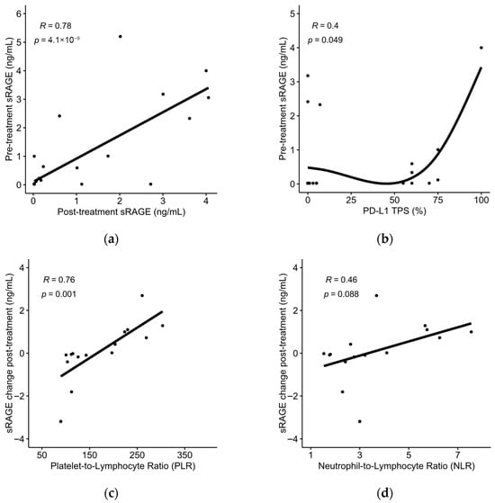
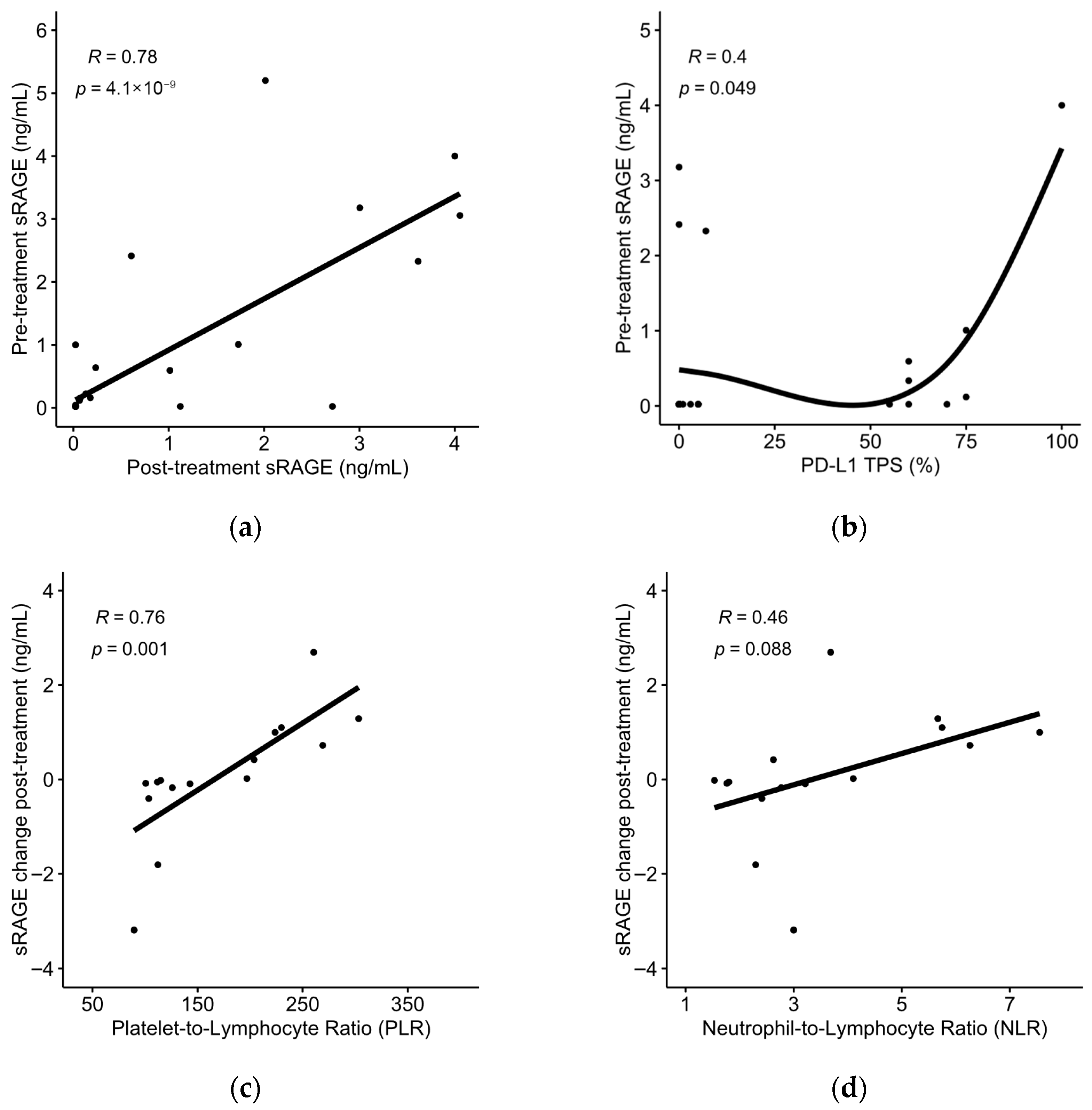
Pre-treatment sRAGE demonstrated a strong positive correlation with post-treatment sRAGE (Pearson’s r = 0.78; 95% CI, 0.61–0.88; p = 4.1 × 10⁻⁹) (Figure 1a) and a weak positive correlation with PD-L1 tumor proportion score (TPS) in NSCLC patients (Spearman’s ρ = 0.4, p = 0.049, n = 25) (Figure 1b). The absolute change in sRAGE levels post-treatment demonstrated a strong positive correlation with the platelet-to-lymphocyte ratio (PLR) at baseline (Pearson’s r = 0.76; 95% CI, 0.40–0.92; p = 0.001) (Figure 1c) and a non-significant correlation with the neutrophil-to-lymphocyte ratio (NLR) at baseline (Pearson’s r = 0.46; 95% CI; −0.07–0.78; p = 0.088) (Figure 1d).
Figure 1.
(a) Associations between pre-treatment sRAGE and post-treatment sRAGE levels or (b) PD-L1 tumor proportion score (TPS); (c) associations between absolute change in sRAGE levels and baseline platelet-to-lymphocyte ratio (PLR) or (d) baseline neutrophil-to-lymphocyte ratio (NLR).
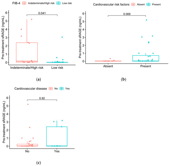
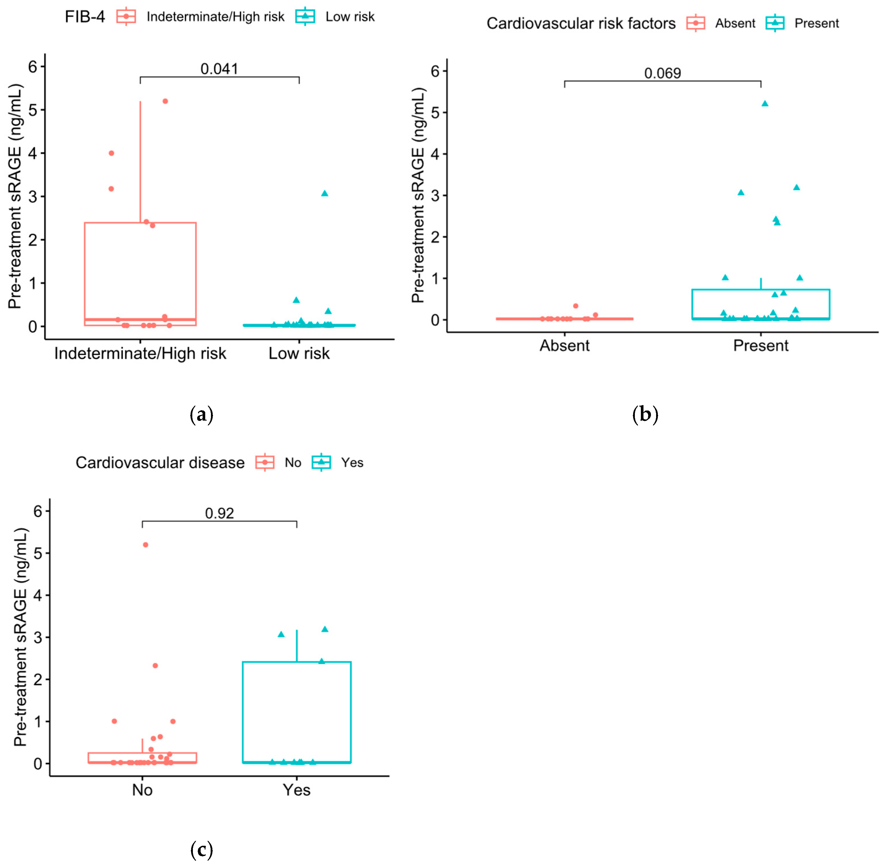
Patients with indeterminate/high risk of liver fibrosis, as defined by FIB-4 index ≥ 1.45, tended to exhibit higher pre-treatment sRAGE levels compared to patients with low risk of fibrosis (Wilcoxon rank-sum test, p = 0.041) (Figure 2a). There was a trend towards higher pre-treatment sRAGE levels in patients with cardiovascular risk factors (Wilcoxon rank-sum test, p = 0.069), although no significant association was observed between established cardiovascular disease and serum sRAGE levels (p = 0.92) (Figure 2b,c). Seven patients (16.7%) received oral corticosteroid therapy at the time of treatment initiation. No significant association was observed between oral corticosteroid therapy and serum sRAGE levels (p = 0.8).
Figure 2.
Baseline sRAGE levels in patients with low or indeterminate/high risk of liver fibrosis: (a) as defined by FIB-4 index; (b) in patients with or without cardiovascular risk factors; (c) in patients with or without established cardiovascular disease.
Driver alterations were identified in 8 of 19 patients (42.1%) with non-squamous NSCLC, including KRAS mutations in five patients, TP53 mutations in two patients, and an FGFR2–TACC2 fusion in one patient. No significant association was observed between the presence of driver alterations and serum sRAGE levels (p = 0.25).
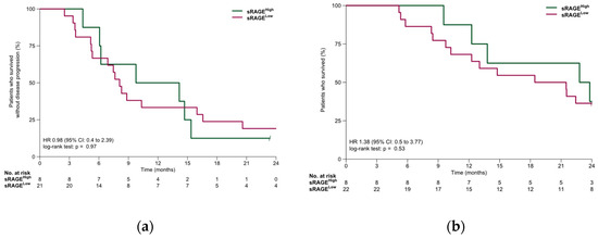
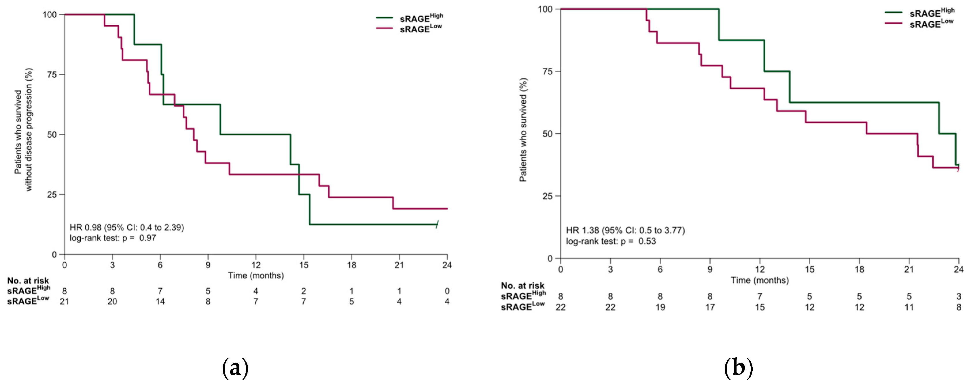
In the NSCLC cohort, median PFS was 8.3 months (95% CI: 6.9–15.4 months). PFS was similar in the sRAGE-low and sRAGE-high subgroups (8.1 vs. 12.0 months, HR, 0.98; 95% CI, 0.4–2.39; p = 0.97). Median OS was 21.5 months (95% CI: 13.0 months—NR). OS was similar in the sRAGE-low and sRAGE-high subgroups (20.0 vs. 23.3 months, HR, 1.38 95% CI 0.5–3.77; p = 0.53). Survival outcomes for the NSCLC cohort are summarized in Figure 3.
Figure 3.
(a) Progression-free survival and (b) overall survival in the NSCLC cohort.
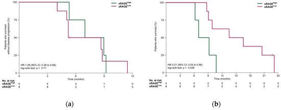
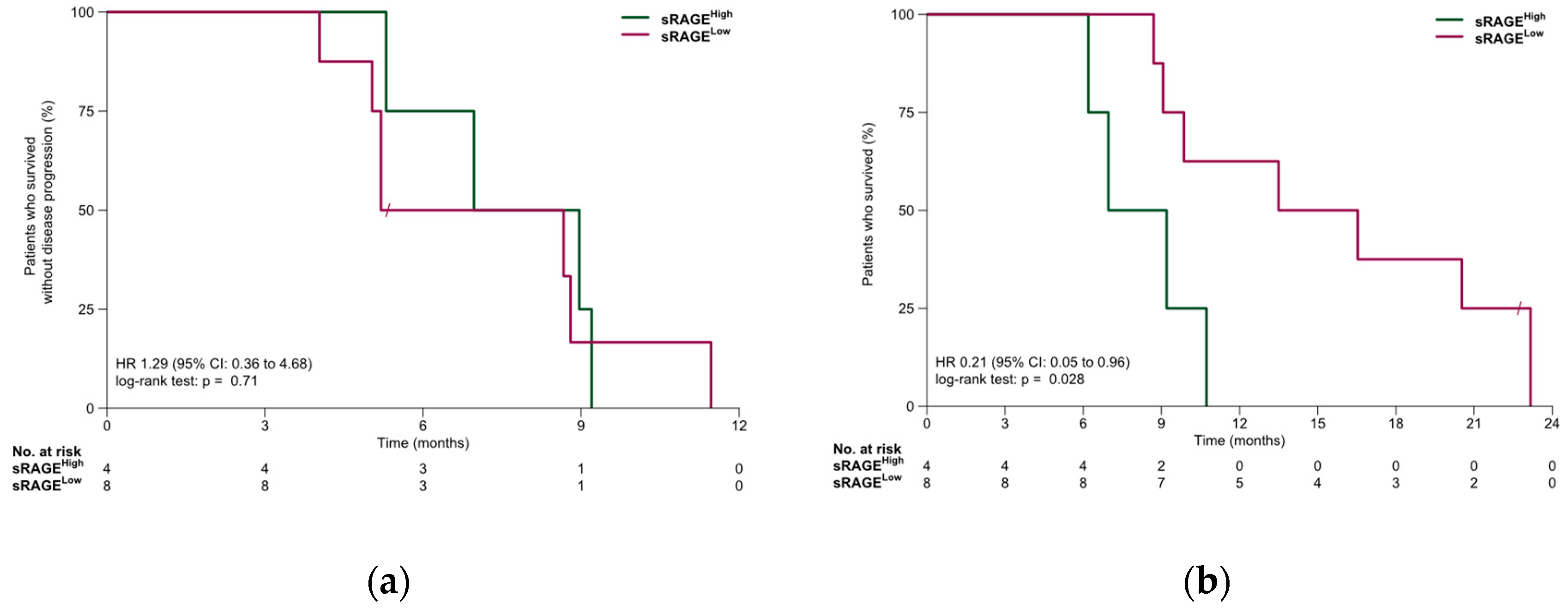
In the SCLC cohort, median PFS was 7.0 months (95% CI: 5.2 months—NR). PFS was similar in the sRAGE-low and sRAGE-high subgroups (6.9 vs. 8.0 months, HR, 1.29; 95% CI, 0.36–4.68; p = 0.71). Median OS was 10.3 months (95% CI: 9.1 months—NR). OS was significantly longer in the sRAGE-low subgroup compared to the sRAGE-high subgroup (15.0 vs. 8.1 months, HR, 0.21; 95% CI 0.05–0.96; p = 0.028). Survival outcomes for the SCLC cohort are summarized in Figure 4.
Figure 4.
(a) Progression-free survival and (b) overall survival in the SCLC cohort.
4. Discussion
In this study, we found that circulating sRAGE is frequently low or undetectable in patients with advanced LC and is variably associated with markers of systemic inflammation, tumor characteristics, and clinical outcomes. Baseline sRAGE levels showed distinct patterns between NSCLC and SCLC, with a prognostic association observed only in the SCLC cohort, while, in NSCLC, sRAGE appeared to be more closely linked to inflammatory indices and PD-L1 expression, rather than survival. Collectively, these findings suggest that sRAGE may reflect underlying immunometabolic and inflammatory processes in LC, with potentially different biological and clinical implications across histological subtypes.
The deregulation of cellular metabolism towards aerobic glycolysis (the Warburg effect) and the development of tumor-promoting inflammation have been widely recognized as hallmarks of cancer [31]. The AGE–RAGE interaction represents a key regulator of glucose metabolism and glycation-associated inflammation [32] and, therefore, constitutes a potential oncogenic driver, which may be important for disease prognosis and therapeutic drug targeting. As RAGE is highly expressed in lung tissue, disruptions in RAGE expression and circulating sRAGE levels are common in different lung diseases [33]. Interestingly, alterations in sRAGE appear to be differentiated in LC compared to other lung diseases. In a study of a Chinese cohort, sRAGE levels were decreased in LC patients compared to tuberculosis patients and healthy controls [34]. Similarly, bronchial sRAGE levels were lower in patients with LC compared to patients with lung infections [35].
A key observation is that, in many patients in our cohort, sRAGE levels were below the assay’s detection limit. Low sRAGE levels are widely reported in conditions of heightened inflammatory stress, including hypertension, coronary artery disease, hyperthyroidism, and rheumatoid arthritis [36,37,38,39], suggesting consumption or impaired generation of decoy receptors when AGE burden is high. Reduced sRAGE has also been reported in various malignancies, such as gastric, colorectal, pancreatic and breast cancer, melanoma, and in patients with bone metastases, where low sRAGE correlates with disease burden, advanced stage, or poor survival [40,41,42,43,44,45]. In addition, genetic alterations in the RAGE gene associated with decreased levels of circulating sRAGE have been linked to increased cancer susceptibility [46]; specific RAGE polymorphisms may be associated with NSCLC development [47]. Furthermore, NSCLC has been linked to deregulation of cellular metabolism and reduced formation of AGEs, due to changes in oxidative stress exposure, which may reduce circulating sRAGE [48]. Therefore, the fact that many NSCLC patients in our cohort exhibited sRAGE levels below the assay’s detection limit is biologically plausible and consistent with its known suppression in high-inflammatory and high-tumor-burden states. Another possible explanation may relate to differences in the production of sRAGE through proteolytic cleavage and endogenous secretion of RAGE isoforms in LC patients [33,49]; the sensitivity of the ELISA may be different depending on the specific cleavage site or alternative splice variants.
The association between sRAGE and outcomes in LC patients has been less studied compared with its role in other systemic diseases. Previous studies in acute lung injury and acute respiratory distress syndrome (ARDS) suggested sRAGE as a marker of alveolar epithelial injury and poor outcomes [50]. Similarly, in critical illness and sepsis, elevated sRAGE has been linked to disease severity and mortality [51]. To our knowledge, this is the first study to report the potential association of sRAGE with survival outcomes of chemoimmunotherapy in SCLC patients. In the SCLC cohort, increased baseline sRAGE was associated with worse overall survival. While this finding should be interpreted with caution, given the limited number of patients, the potential association with overall survival warrants further evaluation in larger studies of SCLC patients. In contrast, we did not observe an association between serum sRAGE and survival outcomes in NSCLC patients. In one previous study, increased sRAGE at baseline was associated with superior progression-free survival and overall survival in NSCLC patients treated with immunotherapy, with or without chemotherapy [52]. Differences between NSCLC and SCLC may, in part, be attributable to variations in metabolic characteristics in the tumor immune microenvironment [53].
To date, few studies have evaluated sRAGE dynamics or association with prognosis in patients with LC receiving chemoimmunotherapy. In the study performed by Giglio et al., after 3 months of first-line immunotherapy, with or without chemotherapy, sRAGE levels were significantly increased from baseline in patients who responded to treatment compared to non-responders [52]. Interestingly, in another study of patients with lung adenocarcinoma receiving carboplatin and pemetrexed with or without camrelizumab, RAGE mRNA levels decreased after treatment in both cohorts, while patients with radiological response or stable disease had lower post-treatment mRNA RAGE levels than patients with disease progression [54]. While we did not observe any association of early sRAGE dynamics with response rates or survival outcomes in our cohort, post-treatment changes in serum sRAGE were significantly correlated with baseline PLR and trended towards correlation with baseline NLR, suggesting a potential association with inflammatory status, which may merit further investigation. Inflammatory markers derived from peripheral whole blood cell counts have been previously correlated with sRAGE levels in NSCLC patients and in patients with COPD [52,55].
The positive correlation between circulating sRAGE and PD-L1 expression in LC may reflect shared inflammatory mechanisms driven by RAGE ligands, including NF-κB and STAT3 [56], transcription factors integral to PD-L1 expression and regulation [57,58]. In melanocytes and melanoma cells, RAGE has been shown to mediate PD-L1 expression as a response to ultraviolet radiation in a manner dependent on NF-κB [59]. Furthermore, the RAGE ligand High-Mobility Group Box 1 (HMGB1) can activate the PI3K/AKT signaling pathway through interaction with RAGE and, subsequently, promote PD-L1 expression in breast cancer cells [60]. Through its NF-κB-dependent feedback loop, RAGE signaling sustains chronic inflammation, while HMGB1-mediated dendritic-cell activation and chemokine induction help generate an immunologically “hot” microenvironment where PD-L1 is typically elevated [61,62]. Because sRAGE acts as a decoy receptor released in response to increased ligand burden, higher circulating levels may serve as a surrogate marker of intensified RAGE–ligand activity and, consequently, a PD-L1-high inflammatory tumor environment [63]. Thus, sRAGE could indirectly capture RAGE-driven immune activation with potential relevance to predicting immunotherapy responsiveness.
The role of sRAGE in the development and prognosis of cardiovascular disease is debated and may be variable depending on the presence of other aggravating factors. Increased sRAGE has been consistently associated with cardiovascular complications in patients with diabetes mellitus and/or established coronary artery disease [64], but may have a protective role in non-diabetic populations [36,37]. We observed a non-significant trend towards higher serum sRAGE levels in patients with cardiovascular risk factors in our LC cohort. LC-associated inflammation may activate the AGE/RAGE axis in a similar way to diabetes mellitus and other systemic diseases [65]. As a result, elevated sRAGE may be involved in the development of cardiovascular complications in LC patients, which represent a significant cause of mortality in this patient population, including in patients treated with ICIs [66,67]. The potential association between sRAGE and cardiovascular disease in LC patients, including treatment-related cardiotoxicity, warrants further evaluation in larger prospective studies.
We observed increased serum sRAGE levels in patients with intermediate/high risk of liver fibrosis, as assessed by the FIB-4 index, compared to patients with low risk of fibrosis. Similar observations have been made in non-cancer populations, such as participants in the Atherosclerosis Risk in Communities Study [29]. The AGE/RAGE axis, including polymorphisms in the RAGE gene, has been implicated in the development of NAFLD [68,69]. The effect of liver inflammation and/or NAFLD on the efficacy of immune checkpoint inhibitors in LC patients is debated, although it may be significant in patients with liver metastases [70,71].
This study has several limitations. Μany sRAGE values were below the assay detection limit, producing zero-inflated distributions and potentially inflating correlation coefficients. Second, the sample size was limited, particularly for subgroup analyses. Third, this study was not powered to evaluate interactions with immunotherapy, which would be of high clinical relevance given the association with tissue PD-L1 expression. Future work should employ assays with greater analytical sensitivity to examine these associations in larger, independent cohorts and explore mechanistic links between the RAGE axis and immune checkpoint regulation.
5. Conclusions
In this exploratory study, we investigated serum sRAGE as a potential biomarker in patients with advanced LC. Our findings suggest that baseline sRAGE levels are frequently low or undetectable in LC patients, consistent with high-inflammatory and high-tumor-burden states. In SCLC, elevated baseline sRAGE was associated with worse overall survival, highlighting a possible prognostic role that warrants further validation. In NSCLC, no significant associations with survival outcomes were observed, although sRAGE showed a modest correlation with PD-L1 expression and inflammatory markers, suggesting a potential link to tumor-associated immune activity. Additionally, higher sRAGE levels were observed in patients with intermediate/high liver fibrosis risk and, potentially, those with cardiovascular risk factors, underscoring the broader systemic involvement of the AGE–RAGE axis.
Author Contributions
Conceptualization, E.P. and M.P.; methodology, E.P.; validation, E.P. and E.G.; formal analysis, E.P.; investigation, E.P., A.G. and E.G.; resources, E.K. (Elias Kotteas) and M.P.; data curation, E.P.; writing—original draft preparation, E.P., A.G., E.K. (Eleftheria Karaviti), D.K., A.K. and M.P.; writing—review and editing, E.P., A.G., E.G., S.C., E.K. (Elias Kotteas), N.S. and M.P.; visualization, E.P.; supervision, E.K. (Elias Kotteas) and M.P. All authors have read and agreed to the published version of the manuscript.
Funding
This research received no external funding.
Institutional Review Board Statement
This study was conducted in accordance with the Declaration of Helsinki, and it was approved by the Institutional Review Board of Sotiria General Hospital for Chest Diseases (protocol number 24938/4.10.2023, date of approval 4 October 2023) and the Ethics Committee of the School of Medicine, National and Kapodistrian University of Athens (protocol number 803, date of approval 23 October 2023).
Informed Consent Statement
Informed consent was obtained from all patients involved in this study.
Data Availability Statement
The data presented in this study are available on reasonable request from the corresponding author, E.P., due to privacy restrictions.
Conflicts of Interest
The authors declare no conflicts of interest.
Abbreviations
The following abbreviations are used in this manuscript:
| LC | Lung cancer |
| NSCLC | Non-small-cell lung cancer |
| SCLC | Small-cell lung cancer |
| ICI | Immune checkpoint inhibitor |
| AGEs | Advanced glycation end products |
| RAGE | Receptor for advanced glycation end products |
| sRAGE | Soluble receptor for advanced glycation end products |
| cRAGE | Cleaved receptor for advanced glycation end products |
| esRAGE | Endogenous secreted receptor for advanced glycation end products |
| PD-L1 | Programmed death ligand 1 |
| NAFLD | Non-alcoholic fatty liver disease |
| NASH | Non-alcoholic steatohepatitis |
| CV | Coefficient of variation |
| AST | Aspartate Aminotransferase |
| ALT | Alanine Aminotransferase |
| RECIST | Response Evaluation Criteria in Solid Tumors |
| PFS | Progression-free survival |
| OS | Overall survival |
| ROC | Receiver operating characteristic |
| NLR | Neutrophil-to-lymphocyte ratio |
| PLR | Platelet-to-lymphocyte ratio |
| ARDS | Acute respiratory distress syndrome |
| HMGB1 | High-Mobility Group Box 1 |
References
- Ji, Y.; Zhang, Y.; Liu, S.; Li, J.; Jin, Q.; Wu, J.; Duan, H.; Liu, X.; Yang, L.; Huang, Y. The Epidemiological Landscape of Lung Cancer: Current Status, Temporal Trend and Future Projections Based on the Latest Estimates from GLOBOCAN 2022. J. Natl. Cancer Cent. 2025, 5, 278–286. [Google Scholar] [CrossRef] [Scilit]
- Smolarz, B.; Łukasiewicz, H.; Samulak, D.; Piekarska, E.; Kołaciński, R.; Romanowicz, H. Lung Cancer—Epidemiology, Pathogenesis, Treatment and Molecular Aspect (Review of Literature). Int. J. Mol. Sci. 2025, 26, 2049. [Google Scholar] [CrossRef] [Scilit] [PubMed]
- Chaitanya Thandra, K.; Barsouk, A.; Saginala, K.; Sukumar Aluru, J.; Barsouk, A. Epidemiology of Lung Cancer. Contemp. Oncol./Współczesna Onkol. 2021, 25, 45–52. [Google Scholar] [CrossRef] [Scilit]
- Panagiotou, E.; Vathiotis, I.A.; Makrythanasis, P.; Hirsch, F.; Sen, T.; Syrigos, K. Biological and Therapeutic Implications of the Cancer-Related Germline Mutation Landscape in Lung Cancer. Lancet Respir. Med. 2024, 12, 997–1005. [Google Scholar] [CrossRef] [Scilit]
- Lahiri, A.; Maji, A.; Potdar, P.D.; Singh, N.; Parikh, P.; Bisht, B.; Mukherjee, A.; Paul, M.K. Lung Cancer Immunotherapy: Progress, Pitfalls, and Promises. Mol. Cancer 2023, 22, 40. [Google Scholar] [CrossRef] [Scilit]
- Dong, H.; Zhang, Y.; Huang, Y.; Deng, H. Pathophysiology of RAGE in Inflammatory Diseases. Front. Immunol. 2022, 13, 931473. [Google Scholar] [CrossRef] [Scilit]
- Prasad, K. Is There Any Evidence That AGE/sRAGE Is a Universal Biomarker/Risk Marker for Diseases? Mol. Cell. Biochem. 2019, 451, 139–144. [Google Scholar] [CrossRef] [Scilit]
- Kim, D.; Choi, K.-N.; Park, J.-I.; Kim, E.-H.; Majid, A.; Bae, O.-N.; Kim, D.; Choi, K.-N.; Park, J.-I.; Kim, E.-H.; et al. Role of Advanced Glycation End Products and Mitohormesis in Cancer Development and Progression. Antioxidants 2025, 14, 1165. [Google Scholar] [CrossRef] [Scilit] [PubMed]
- Uribarri, J.; Cai, W.; Sandu, O.; Peppa, M.; Goldberg, T.; Vlassara, H. Diet-Derived Advanced Glycation End Products Are Major Contributors to the Body’s AGE Pool and Induce Inflammation in Healthy Subjects. Ann. N. Y. Acad. Sci. 2005, 1043, 461–466. [Google Scholar] [CrossRef] [Scilit]
- Peppa, M.; Mavroeidi, I. Experimental Animal Studies Support the Role of Dietary Advanced Glycation End Products in Health and Disease. Nutrients 2021, 13, 3467. [Google Scholar] [CrossRef] [Scilit] [PubMed]
- Pascual-Morena, C.; Garrido-Miguel, M.; Martínez-García, I.; Lucerón-Lucas-Torres, M.; Rodríguez-Gutiérrez, E.; Berlanga-Macías, C.; Fernández-Bravo-Rodrigo, J.; Patiño-Cardona, S. Association of Dietary Advanced Glycation End Products with Overall and Site-Specific Cancer Risk and Mortality: A Systematic Review and Meta-Analysis. Nutrients 2025, 17, 1638. [Google Scholar] [CrossRef] [Scilit] [PubMed]
- Peppa, M.; Uribarri, J.; Vlassara, H. The Role of Advanced Glycation End Products in the Development of Atherosclerosis. Curr. Diabetes Rep. 2004, 4, 31–36. [Google Scholar] [CrossRef] [Scilit]
- Goldberg, T.; Cai, W.; Peppa, M.; Dardaine, V.; Baliga, B.S.; Uribarri, J.; Vlassara, H. Advanced Glycoxidation End Products in Commonly Consumed Foods. J. Am. Diet. Assoc. 2004, 104, 1287–1291. [Google Scholar] [CrossRef] [Scilit] [PubMed]
- Peppa, M. Dietary Advanced Glycation End Products: Animal Studies. In Dietary AGEs and Their Role in Health and Disease; Uribarri, J., Ed.; CRC Press: Boca Raton, FL, USA, 2017. [Google Scholar]
- Fritz, G. RAGE: A Single Receptor Fits Multiple Ligands. Trends Biochem. Sci. 2011, 36, 625–632. [Google Scholar] [CrossRef] [Scilit]
- Soman, S.; Raju, R.; Sandhya, V.K.; Advani, J.; Khan, A.A.; Harsha, H.C.; Prasad, T.S.K.; Sudhakaran, P.R.; Pandey, A.; Adishesha, P.K. A Multicellular Signal Transduction Network of AGE/RAGE Signaling. J. Cell Commun. Signal. 2013, 7, 19–23. [Google Scholar] [CrossRef] [Scilit]
- Wautier, M.-P.; Chappey, O.; Corda, S.; Stern, D.M.; Schmidt, A.M.; Wautier, J.-L. Activation of NADPH Oxidase by AGE Links Oxidant Stress to Altered Gene Expression via RAGE. Am. J. Physiol.-Endocrinol. Metab. 2001, 280, E685–E694. [Google Scholar] [CrossRef] [Scilit]
- Zhou, M.; Zhang, Y.; Shi, L.; Li, L.; Zhang, D.; Gong, Z.; Wu, Q. Activation and Modulation of the AGEs-RAGE Axis: Implications for Inflammatory Pathologies and Therapeutic Interventions—A Review. Pharmacol. Res. 2024, 206, 107282. [Google Scholar] [CrossRef] [Scilit]
- Tam, X.H.L.; Shiu, S.W.M.; Leng, L.; Bucala, R.; Betteridge, D.J.; Tan, K.C.B. Enhanced Expression of Receptor for Advanced Glycation End-Products Is Associated with Low Circulating Soluble Isoforms of the Receptor in Type 2 Diabetes. Clin. Sci. 2011, 120, 81–89. [Google Scholar] [CrossRef] [Scilit]
- Paz-Ares, L.; Luft, A.; Vicente, D.; Tafreshi, A.; Gümüş, M.; Mazières, J.; Hermes, B.; Çay Şenler, F.; Csőszi, T.; Fülöp, A.; et al. Pembrolizumab plus Chemotherapy for Squamous Non–Small-Cell Lung Cancer. N. Engl. J. Med. 2018, 379, 2040–2051. [Google Scholar] [CrossRef] [Scilit] [PubMed]
- Pabst, L.; Lopes, S.; Bertrand, B.; Creusot, Q.; Kotovskaya, M.; Pencreach, E.; Beau-Faller, M.; Mascaux, C. Prognostic and Predictive Biomarkers in the Era of Immunotherapy for Lung Cancer. Int. J. Mol. Sci. 2023, 24, 7577. [Google Scholar] [CrossRef] [Scilit]
- Liu, J.; Zhang, J.; Chang, J.; Chen, L.; Wang, H.; Liu, Y.; Huang, M.; Zeng, X.; Guo, C. sRAGE Inhibits Myocardial Ischemia/Reperfusion Injuries via Regulating Treg Cells. Cell Biosci. 2025, 15, 144. [Google Scholar] [CrossRef] [Scilit]
- Zhang, C.; Gao, M.; Jiang, X.; Pan, X.; Zhang, X.; Li, Y.; Shen, Q.; Chen, Y.; Pang, B. Research Progress and Value of Albumin-Related Inflammatory Markers in the Prognosis of Non-Small Cell Lung Cancer: A Review of Clinical Evidence. Ann. Med. 2023, 55, 1294–1307. [Google Scholar] [CrossRef] [Scilit]
- Platini, H.; Ferdinand, E.; Kohar, K.; Prayogo, S.A.; Amirah, S.; Komariah, M.; Maulana, S.; Platini, H.; Ferdinand, E.; Kohar, K.; et al. Neutrophil-to-Lymphocyte Ratio and Platelet-to-Lymphocyte Ratio as Prognostic Markers for Advanced Non-Small-Cell Lung Cancer Treated with Immunotherapy: A Systematic Review and Meta-Analysis. Medicina 2022, 58, 1069. [Google Scholar] [CrossRef] [Scilit]
- Xu, X.; Jiang, L.; Wu, C.; Pan, L.; Lou, Z.; Peng, C.; Dong, Y.; Ruan, B. The Role of Fibrosis Index FIB-4 in Predicting Liver Fibrosis Stage and Clinical Prognosis: A Diagnostic or Screening Tool? J. Formos. Med. Assoc. 2022, 121, 454–466. [Google Scholar] [CrossRef] [Scilit]
- Haimi, M.; Baker, F.A.; Vinker, S.; Safadi, R.; Israel, A. Association Between High FIB-4 Score and the Risk of Malignancy Development and Mortality: A Retrospective Longitudinal Case–Control Study. Dig. Dis. Sci. 2025, 70, 3525–3537. [Google Scholar] [CrossRef] [Scilit] [PubMed]
- Pfister, D.; Núñez, N.G.; Pinyol, R.; Govaere, O.; Pinter, M.; Szydlowska, M.; Gupta, R.; Qiu, M.; Deczkowska, A.; Weiner, A.; et al. NASH Limits Anti-Tumour Surveillance in Immunotherapy-Treated HCC. Nature 2021, 592, 450–456. [Google Scholar] [CrossRef] [Scilit] [PubMed]
- Ivancovsky-Wajcman, D.; Zelber-Sagi, S.; Fliss Isakov, N.; Webb, M.; Zemel, M.; Shibolet, O.; Kariv, R. Serum Soluble Receptor for AGE (sRAGE) Levels Are Associated With Unhealthy Lifestyle and Nonalcoholic Fatty Liver Disease. Clin. Transl. Gastroenterol. 2019, 10, e00040. [Google Scholar] [CrossRef] [Scilit]
- Laudenslager, M.; Lazo, M.; Wang, D.; Selvin, E.; Chen, P.-H.; Pankow, J.S.; Clark, J.M. Association between the Soluble Receptor for Advanced Glycation End Products (sRAGE) and NAFLD in Participants in the Atherosclerosis Risk in Communities Study. Dig. Liver Dis. 2021, 53, 873–878. [Google Scholar] [CrossRef] [Scilit] [PubMed]
- Ke, M.; Xu, T.; Fang, Y.; Ye, Y.; Li, Z.; Ren, F.; Lu, S.; Zhang, X.; Wu, R.; Lv, Y.; et al. Liver Fibrosis Promotes Immune Escape in Hepatocellular Carcinoma via GOLM1-Mediated PD-L1 Upregulation. Cancer Lett. 2021, 513, 14–25. [Google Scholar] [CrossRef] [Scilit]
- Hanahan, D. Hallmarks of Cancer: New Dimensions. Cancer Discov. 2022, 12, 31–46. [Google Scholar] [CrossRef] [Scilit]
- Palanissami, G.; Paul, S.F.D. AGEs and RAGE: Metabolic and Molecular Signatures of the Glycation-Inflammation Axis in Malignant or Metastatic Cancers. Explor. Target. Anti-Tumor Ther. 2023, 4, 812–849. [Google Scholar] [CrossRef] [Scilit] [PubMed]
- Buckley, S.T.; Ehrhardt, C. The Receptor for Advanced Glycation End Products (RAGE) and the Lung. BioMed Res. Int. 2010, 2010, 917108. [Google Scholar] [CrossRef] [Scilit]
- Jing, R.; Cui, M.; Wang, J.; Wang, H. Receptor for Advanced Glycation End Products (RAGE) Soluble Form (sRAGE): A New Biomarker for Lung Cancer. Neoplasma 2010, 57, 55–61. [Google Scholar] [CrossRef] [Scilit] [PubMed]
- Kim, T.; Kim, S.J.; Choi, H.; Shin, T.R.; Sim, Y.S.; Kim, T.; Kim, S.J.; Choi, H.; Shin, T.R.; Sim, Y.S. Diagnostic Utility and Tendency of Bronchial and Serum Soluble Receptor for Advanced Glycation EndProducts (sRAGE) in Lung Cancer. Cancers 2023, 15, 2819. [Google Scholar] [CrossRef] [Scilit]
- Geroldi, D.; Falcone, C.; Emanuele, E.; D’Angelo, A.; Calcagnino, M.; Buzzi, M.P.; Scioli, G.A.; Fogari, R. Decreased Plasma Levels of Soluble Receptor for Advanced Glycation End-Products in Patients with Essential Hypertension. J. Hypertens. 2005, 23, 1725–1729. [Google Scholar] [CrossRef] [Scilit] [PubMed]
- Falcone, C.; Emanuele, E.; D’Angelo, A.; Buzzi, M.P.; Belvito, C.; Cuccia, M.; Geroldi, D. Plasma Levels of Soluble Receptor for Advanced Glycation End Products and Coronary Artery Disease in Nondiabetic Men. Arterioscler. Thromb. Vasc. Biol. 2005, 25, 1032–1037. [Google Scholar] [CrossRef] [Scilit]
- Caspar-Bell, G.; Dhar, I.; Prasad, K. Advanced Glycation End Products (AGEs) and Its Receptors in the Pathogenesis of Hyperthyroidism. Mol. Cell. Biochem. 2016, 414, 171–178. [Google Scholar] [CrossRef] [Scilit]
- Pullerits, R.; Bokarewa, M.; Dahlberg, L.; Tarkowski, A. Decreased Levels of Soluble Receptor for Advanced Glycation End Products in Patients with Rheumatoid Arthritis Indicating Deficient Inflammatory Control. Arthritis Res. Ther. 2005, 7, R817. [Google Scholar] [CrossRef] [Scilit]
- Li, T.; Qin, W.; Liu, Y.; Li, S.; Qin, X.; Liu, Z. Effect of RAGE Gene Polymorphisms and Circulating sRAGE Levels on Susceptibility to Gastric Cancer: A Case–Control Study. Cancer Cell Int. 2017, 17, 19. [Google Scholar] [CrossRef] [Scilit]
- Kuniyasu, H.; Chihara, Y.; Takahashi, T. Co-Expression of Receptor for Advanced Glycation End Products and the Ligand Amphoterin Associates Closely with Metastasis of Colorectal Cancer. Oncol. Rep. 2003, 10, 445–448. [Google Scholar] [CrossRef] [Scilit]
- Wittwer, C.; Boeck, S.; Heinemann, V.; Haas, M.; Stieber, P.; Nagel, D.; Holdenrieder, S. Circulating Nucleosomes and Immunogenic Cell Death Markers HMGB1, sRAGE and DNAse in Patients with Advanced Pancreatic Cancer Undergoing Chemotherapy: HMGB1, sRAGE, DNAse and Circulating Nucleosomes in PC. Int. J. Cancer 2013, 133, 2619–2630. [Google Scholar] [CrossRef] [Scilit]
- Coser, M.; Neamtu, B.M.; Pop, B.; Cipaian, C.R.; Crisan, M. RAGE and Its Ligands in Breast Cancer Progression and Metastasis. Oncol. Rev. 2025, 18, 1507942. [Google Scholar] [CrossRef] [Scilit]
- Wagner, N.B.; Weide, B.; Reith, M.; Tarnanidis, K.; Kehrel, C.; Lichtenberger, R.; Pflugfelder, A.; Herpel, E.; Eubel, J.; Ikenberg, K.; et al. Diminished Levels of the Soluble Form of RAGE Are Related to Poor Survival in Malignant Melanoma. Int. J. Cancer 2015, 137, 2607–2617. [Google Scholar] [CrossRef] [Scilit] [PubMed]
- Galliera, E.; Marazzi, M.G.; Vianello, E.; Drago, L.; Luzzati, A.; Bendinelli, P.; Maroni, P.; Tacchini, L.; Desiderio, M.A.; Corsi Romanelli, M.M. Circulating sRAGE in the Diagnosis of Osteolytic Bone Metastasis. J. Biol. Regul. Homeost. Agents 2016, 30, 1203–1208. [Google Scholar]
- Peng, Y.; Liu, F.; Qiao, Y.; Wang, P.; Du, H.; Si, C.; Wang, X.; Chen, K.; Song, F.; Peng, Y.; et al. Genetically Modified Circulating Levels of Advanced Glycation End-Products and Their Soluble Receptor (AGEs-RAGE Axis) with Risk and Mortality of Breast Cancer. Cancers 2022, 14, 6124. [Google Scholar] [CrossRef] [Scilit] [PubMed]
- Wang, H.; Li, Y.; Yu, W.; Ma, L.; Ji, X.; Xiao, W. Expression of the Receptor for Advanced Glycation End-Products and Frequency of Polymorphism in Lung Cancer. Oncol. Lett. 2015, 10, 51–60. [Google Scholar] [CrossRef] [Scilit]
- Tomin, T.; Honeder, S.E.; Liesinger, L.; Gremel, D.; Retzl, B.; Lindenmann, J.; Brcic, L.; Schittmayer, M.; Birner-Gruenberger, R. Increased Antioxidative Defense and Reduced Advanced Glycation End-Product Formation by Metabolic Adaptation in Non-Small-Cell-Lung-Cancer Patients. Nat. Commun. 2025, 16, 5157. [Google Scholar] [CrossRef] [Scilit] [PubMed]
- Raucci, A.; Cugusi, S.; Antonelli, A.; Barabino, S.M.; Monti, L.; Bierhaus, A.; Reiss, K.; Saftig, P.; Bianchi, M.E. A Soluble Form of the Receptor for Advanced Glycation Endproducts (RAGE) Is Produced by Proteolytic Cleavage of the Membrane-bound Form by the Sheddase a Disintegrin and Metalloprotease 10 (ADAM10). FASEB J. 2008, 22, 3716–3727. [Google Scholar] [CrossRef] [Scilit]
- Creagh-Brown, B.C.; Burke-Gaffney, A.; Evans, T.W. sRAGE: A Useful Biomarker in Acute Lung Injury? Crit. Care Med. 2011, 39, 589–590. [Google Scholar] [CrossRef] [Scilit]
- Matsumoto, H.; Matsumoto, N.; Ogura, H.; Shimazaki, J.; Yamakawa, K.; Yamamoto, K.; Shimazu, T. The Clinical Significance of Circulating Soluble RAGE in Patients with Severe Sepsis. J. Trauma Acute Care Surg. 2015, 78, 1086. [Google Scholar] [CrossRef] [Scilit]
- Giglio, A.D.; Conte, M.; Matteis, S.D.; Galuppi, F.; Bianco, F.L.; Naddeo, M.; Ricciotti, I.; Federico, A.D.; Trofarello, L.; Melotti, B.; et al. P2.06.92 Host-Derived Biomarkers of Inflammation and Body Composition Predict Immunotherapy Outcomes in Advanced NSCLC. J. Thorac. Oncol. 2025, 20, S334–S335. [Google Scholar] [CrossRef] [Scilit]
- Chen, Y.; Li, H.; Fan, Y. Shaping the Tumor Immune Microenvironment of SCLC: Mechanisms, and Opportunities for Immunotherapy. Cancer Treat. Rev. 2023, 120, 102606. [Google Scholar] [CrossRef] [Scilit]
- Li, X.; Wang, G.; Sun, X.; Feng, Y.; Wang, L.; Sun, G.; Qiao, H. Effect of AGEs-RAGE System on the Efficacy of PD-1 Inhibitors in the Treatment of Driver-Gene Mutation Negative Advanced Non-Squamous Non-Small Cell Lung Cancer. Cell. Mol. Biol. 2023, 69, 155–160. [Google Scholar] [CrossRef] [Scilit] [PubMed]
- Seri, V.C.; Ullah, M.K.; Siddaiah, J.B.; Chaya, S.K.; Lokesh, K.S.; Khan, S.A.; Aladakatti, A.R.; Shahul, S.; Vasanthan, V.; Karnik, M.; et al. Reduced Serum sRAGE Levels Identify COPD and Reflect Disease Severity: Findings from a Cross-Sectional Study in India. Diagnostics 2025, 15, 2910. [Google Scholar] [CrossRef] [Scilit]
- El-Far, A.H.; Sroga, G.; Jaouni, S.K.A.; Mousa, S.A.; El-Far, A.H.; Sroga, G.; Jaouni, S.K.A.; Mousa, S.A. Role and Mechanisms of RAGE-Ligand Complexes and RAGE-Inhibitors in Cancer Progression. Int. J. Mol. Sci. 2020, 21, 3613. [Google Scholar] [CrossRef] [Scilit]
- Antonangeli, F.; Natalini, A.; Garassino, M.C.; Sica, A.; Santoni, A.; Di Rosa, F. Regulation of PD-L1 Expression by NF-κB in Cancer. Front. Immunol. 2020, 11, 584626. [Google Scholar] [CrossRef] [Scilit]
- Zhang, X.; Zeng, Y.; Qu, Q.; Zhu, J.; Liu, Z.; Ning, W.; Zeng, H.; Zhang, N.; Du, W.; Chen, C.; et al. PD-L1 Induced by IFN-γ from Tumor-Associated Macrophages via the JAK/STAT3 and PI3K/AKT Signaling Pathways Promoted Progression of Lung Cancer. Int. J. Clin. Oncol. 2017, 22, 1026–1033. [Google Scholar] [CrossRef] [Scilit] [PubMed]
- Wang, W.; Chapman, N.M.; Zhang, B.; Li, M.; Fan, M.; Laribee, R.N.; Zaidi, M.R.; Pfeffer, L.M.; Chi, H.; Wu, Z.-H. Upregulation of PD-L1 via HMGB1-Activated IRF3 and NF-κB Contributes to UV Radiation-Induced Immune Suppression. Cancer Res. 2019, 79, 2909–2922. [Google Scholar] [CrossRef] [Scilit]
- Amornsupak, K.; Thongchot, S.; Thinyakul, C.; Box, C.; Hedayat, S.; Thuwajit, P.; Eccles, S.A.; Thuwajit, C. HMGB1 Mediates Invasion and PD-L1 Expression through RAGE-PI3K/AKT Signaling Pathway in MDA-MB-231 Breast Cancer Cells. BMC Cancer 2022, 22, 578. [Google Scholar] [CrossRef] [Scilit]
- Waghela, B.N.; Vaidya, F.U.; Ranjan, K.; Chhipa, A.S.; Tiwari, B.S.; Pathak, C. AGE-RAGE Synergy Influences Programmed Cell Death Signaling to Promote Cancer. Mol. Cell. Biochem. 2021, 476, 585–598. [Google Scholar] [CrossRef] [Scilit] [PubMed]
- Wang, L.; Geng, H.; Liu, Y.; Liu, L.; Chen, Y.; Wu, F.; Liu, Z.; Ling, S.; Wang, Y.; Zhou, L. Hot and Cold Tumors: Immunological Features and the Therapeutic Strategies. MedComm 2023, 4, e343. [Google Scholar] [CrossRef] [Scilit]
- Zoccali, C.; Tripepi, G.; Stel, V.; Fu, E.L.; Mallamaci, F.; Dekker, F.; Jager, K.J. Decoy Receptors as Biomarkers for Exploring Aetiology and Designing New Therapies. Clin. Kidney J. 2024, 17, sfae222. [Google Scholar] [CrossRef] [Scilit]
- Katakami, N. Can Soluble Receptor for Advanced Glycation End-Product (sRAGE) Levels in Blood Be Used as a Predictor of Cardiovascular Diseases? Atherosclerosis 2017, 266, 223–225. [Google Scholar] [CrossRef] [Scilit]
- Ahmad, S.; Khan, M.Y.; Rafi, Z.; Khan, H.; Siddiqui, Z.; Rehman, S.; Shahab, U.; Khan, M.S.; Saeed, M.; Alouffi, S.; et al. Oxidation, Glycation and Glycoxidation—The Vicious Cycle and Lung Cancer. Semin. Cancer Biol. 2018, 49, 29–36. [Google Scholar] [CrossRef] [Scilit]
- Sun, J.-Y.; Zhang, Z.-Y.; Qu, Q.; Wang, N.; Zhang, Y.-M.; Miao, L.-F.; Wang, J.; Wu, L.-D.; Liu, Y.; Zhang, C.-Y.; et al. Cardiovascular Disease-Specific Mortality in 270,618 Patients with Non-Small Cell Lung Cancer. Int. J. Cardiol. 2021, 330, 186–193. [Google Scholar] [CrossRef] [Scilit]
- Charpidou, A.; Gerotziafas, G.; Popat, S.; Araujo, A.; Scherpereel, A.; Kopp, H.-G.; Bironzo, P.; Massard, G.; Jiménez, D.; Falanga, A.; et al. Lung Cancer Related Thrombosis (LCART): Focus on Immune Checkpoint Blockade. Cancers 2024, 16, 450. [Google Scholar] [CrossRef] [Scilit] [PubMed]
- Asadipooya, K.; Lankarani, K.B.; Raj, R.; Kalantarhormozi, M. RAGE Is a Potential Cause of Onset and Progression of Nonalcoholic Fatty Liver Disease. Int. J. Endocrinol. 2019, 2019, 2151302. [Google Scholar] [CrossRef] [Scilit] [PubMed]
- Mehta, R.; Shaw, G.; Masschelin, P.; Felix, S.; Otgonsuren, M.; Baranova, A.; Goodman, Z.; Younossi, Z. Polymorphisms in the Receptor for Advanced Glycation End-Products (RAGE) Gene and Circulating RAGE Levels as a Susceptibility Factor for Non-Alcoholic Steatohepatitis (NASH). PLoS ONE 2018, 13, e0199294. [Google Scholar] [CrossRef] [Scilit] [PubMed]
- Li, Y.; Gong, B.; Guo, Y.; Liu, W.; Chen, C.; Jiang, S.; Pan, F.; Song, J.; Yang, L.; Zhou, G. Non–Small Cell Lung Cancer and Immune Checkpoint Inhibitor Therapy: Does Non-Alcoholic Fatty Liver Disease Have an Effect? BMC Cancer 2024, 24, 535. [Google Scholar] [CrossRef] [Scilit]
- Zhou, J.; Zhou, F.; Chu, X.; Zhao, J.; Wu, Y.; Zhao, W.; Xu, C.; Su, C. Non-Alcoholic Fatty Liver Disease Is Associated with Immune Checkpoint Inhibitor-Based Treatment Response in Patients with Non-Small Cell Lung Cancer with Liver Metastases. Transl. Lung Cancer Res. 2020, 9, 316–324. [Google Scholar] [CrossRef] [Scilit]

Disclaimer/Publisher’s Note: The statements, opinions and data contained in all publications are solely those of the individual author(s) and contributor(s) and not of MDPI and/or the editor(s). MDPI and/or the editor(s) disclaim responsibility for any injury to people or property resulting from any ideas, methods, instructions or products referred to in the content. |
© 2026 by the authors. Licensee MDPI, Basel, Switzerland. This article is an open access article distributed under the terms and conditions of the Creative Commons Attribution (CC BY) license.