You are currently viewing a new version of our website. To view the old version click .
Journal of Personalized Medicine
  • Systematic Review
  • Open Access

28 August 2025

The Role of the Microbiome in Oropharyngeal Squamous Cell Carcinoma: A Systematic Review

1
Department of Surgery, UMONS Research Institute for Health Sciences and Technology, University of Mons (UMons), B7000 Mons, Belgium
2
Department of Otolaryngology Head Neck Surgery, CHU Saint-Pierre, B1000 Brussels, Belgium
3
Department of Otolaryngology, Elsan Hospital, F75008 Paris, France
This article belongs to the Section Personalized Therapy in Clinical Medicine

Abstract

Objective: This systematic review aimed to investigate existing evidence regarding the implications of the microbiome in the initiation and progression of oropharyngeal squamous cell carcinoma (OPSCC). Methods: PubMed, Scopus, and Cochrane Library systematic searches were conducted according to the PRISMA statements to identify the relevant studies examining microbiome signatures, underlying molecular mechanisms, and their associations with clinical and oncological outcomes in OPSCC. The bias analysis was conducted with the MINORS. Results: Of the 83 identified papers, 12 met the inclusion criteria (298 OPSCC patients). Spirochaetes and most Bacteroidetes may be predominant in OPSCC versus control specimens, while Proteobacteria may be predominant in control tissues compared to tumor. Leptotrichia, Selenomonas, and Treponema trended to be overrepresented in OPSCC compared to control specimens. Neisseria, Porphyromonas, Rothia, Streptococcus, and Veillonela were predominantly reported in normal compared to OPSCC patient specimens. Microbiome compositional shifts were associated with chemoradiation response, HPV status, and addictions. Methodological heterogeneity was noted in sampling protocols, control selection, and analytical approaches, with limited statistical power due to small cohort sizes. Conclusions: OPSCC demonstrates different microbiome signatures from healthy tissues, influenced by HPV status and addictions. A microbiome shift is plausible from pre- to post-chemoradiotherapy, with the baseline microbiome acting as a predictive response factor; however, the low number of studies and substantial methodological heterogeneity across investigations limit the drawing of valid conclusions. The identification of key species is important in the development of OPSCC for developing personalized medicine considering bacterial mediators in terms of prevention, and targeted therapy using the microbiome–tumor–host interaction pathways.

1. Introduction

Head and neck squamous cell carcinoma (HNSCC) is the 6th most common adult cancer worldwide, accounting for 5.3% of all cancers [,]. Global HNSCC incidence has decreased significantly over the past three decades, primarily attributed to reduced tobacco and alcohol consumption [,]. Among HNSCC, the incidence of oropharyngeal squamous cell carcinoma (OPSCC) has progressively increased with annual incidence rates rising by 2.7% and 0.5% in males and females in the United States []. The rising incidence of OPSCC can be attributed to human papillomavirus (HPV) infection, which increased in the past few decades []. While HPV-associated OPSCC has become predominant in the United States, it is noteworthy that in many regions outside the US, particularly where anti-tobacco campaigns have been less successful or implemented more recently, tobacco and alcohol-related OPSCC continues to predominate, with significantly lower proportions of HPV-induced cases in countries across Europe, Asia, and South America []. The mechanisms underlying the interactions between HPV and the host environment in the OPSCC development are incompletely elucidated []. The number of studies dedicated to the role of microbial communities in respiratory and gastrointestinal pathophysiology increased over the past decade regarding the development of culture-independent metagenomic techniques []. In OPSCC, research has identified differential microbial signatures between HPV+, HPV− malignant, and healthy tissues, suggesting that dysbiosis-associated alterations influence tumor-promoting inflammation, carcinogenic metabolism, and chemoradiotherapy response patterns [].
This systematic review aimed to investigate existing evidence regarding the implications of the microbiome in OPSCC tumorigenesis, progression, clinical phenotypes, and treatment-related endpoints.

2. Materials and Methods

Two researchers conducted the review using the Preferred Reporting Items for a Systematic Review and Meta-analysis (PRISMA) checklist []. Note that the protocol of review was not registered. Study selection criteria were defined with the PICOTS (Population, Intervention, Comparison, Outcome, Timing, and Setting) framework [].
Studies: Studies published in peer-reviewed English-language journals between January 2000 and January 2025 were considered, including prospective and retrospective cohorts, cross-sectional investigations of cancer registries, and controlled trials exploring microbiome findings in OPSCC. Case reports, conference papers, preprints, and experimental animal studies were excluded.
Participants and inclusion criteria: Eligible studies provided OPSCC-specific patient data. Microbiome assessments derived from oropharyngeal or oral fluid or tumor specimens were considered eligible. There was no selection criteria based on the treatments, microbiome characterization methods, or demographic factors.
Outcomes: The primary outcomes consisted of the diversity and composition of the microbiome (taxonomic classification: phylum, class, order, family, genus, and species) and their associations with OPSCC. Secondary outcomes included study features (design, evidence-based level), demographics (mean/median age, sex ratio), oncological findings (grade, cTNM staging, treatments), and methodological approaches for microbiome evaluations (DNA extraction, amplification, quantification, and sequencing).
Intervention and comparison: There was no criterion for intervention. The data related to the type of treatments (surgery, chemo/radiotherapy) were extracted in studies investigating the prognostic value of microbiome findings.
Timing and Setting: There were no criteria for specific timing in the disease process.

2.1. Search Strategy

A University librarian and the author of the paper independently conducted the systematic literature search using PubMed, Scopus, and Cochrane Library databases. The following keywords were used for the literature research: Oropharynx; Oropharyngeal; Cancer; Squamous Cell Carcinoma; Oncological; Microbiome; Microbiota; Bacteria; and Outcomes. The investigators considered research reporting database abstracts, available full-texts, or titles with the search terms. The reference list of some articles, particularly reviews or meta-analyses, were considered for additional valuable studies. The studies were evaluated for the number of subjects, study design, inclusion and exclusion criteria, quality of trial, evidence-based level [], demographics, and outcomes. Cohort studies from the same research team were carefully investigated for potential overlaps. Ethics committee approval was not required.

2.2. Bias Analysis

The bias analysis was conducted using the methodological index for non-randomized studies (MINORS), a validated tool for assessing study quality []. MINORS evaluates key methodological aspects on a scale of 0 (absent), 1 (inadequate/partial), or 2 (adequate). Items include study aim clarity, consecutive patient inclusion, prospective data collection, endpoint appropriateness, adequate follow-up period (for predictive studies), and acceptable lost-to-follow-up rates (<5%). For prospective studies, sample size calculation was also evaluated. The optimal MINORS score is 16 for non-comparative studies and 24 for comparative studies [].

3. Results

Twelve studies were included, accounting for 298 OPSCC patients (Figure 1) [,,,,,,,,,,,]. There were 8 prospective controlled studies (evidence-based level (EBL) = 3C) and 4 uncontrolled/cross-sectional studies (EBL = 4) (Table 1). Six studies were excluded because authors reported data on oral/hypopharyngeal and oropharyngeal squamous cell carcinomas in a single group without specifically differentiating the anatomical locations [,,,,,]. There was no participant overlap in the included studies.
Figure 1. PRISMA flowchart.
Table 1. Demographic, clinical, and microbiome outcomes.

3.1. Demographics, Patients, and Tumor Stages

The data of 298 OPSCC patients were reported (Table 1, Figure 1). The gender ratio was specified in 7 studies [,,,,,,]. There were 33 females (17.8%) and 152 males (82.2%), respectively. The mean age of patients ranged from 57.7 to 66.9 years (Table 1). The OPSCC stage, HPV status, and anatomical location information are described in Table 2. The cTNM information was reported in 5 studies [,,,,]. The microbiome findings were investigated in cT1-T2 (n = 80), and cT3-T4 (n = 23) OPSCC patients. The majority of patients were cN1+ (n = 66; 64.7%). There were no data about patients with distant metastasis. The HPV status has been reported in 10 studies [,,,,,,,,,], accounting for 184 HPV+ (69.4%), 56 HPV− (21.1%), and 25 undetermined status (9.4%) patients. There was heterogeneity across studies for the HPV detection method, i.e., p16 immunostaining or PCR-based detection. The anatomical location was available in only four studies [,,,] and primarily consisted of tonsil OPSCC (Table 2). Two studies investigated the predictive value of the microbiome on the chemoradiation response [,].
Table 2. Oncological features.

3.2. Microbiome Outcomes

The microbiome outcomes are summarized in Table 1 and Table 3. The microbiome analyses were carried out on the following material: oral saliva (n = 7) [,,,,,,], tumor/non-tumor tissue (n = 8) [,,,,,,,], and the stool (n = 1) []. Details about the methodology of microbiome analyses (e.g., DNA extraction, amplification, quantification, sequencing) are provided in Table 4. Among controlled studies, authors used the following material as controls: the saliva of healthy individuals [,,,], tissues of patients with obstructive sleep apnea syndrome [,], tissues of healthy individuals [], or normal OPSCC-adjacent tissue []. De Martin et al. did not report significant differences in terms of α and β-diversities between OPSCC, NAT/CT [].
Table 3. Species.
Table 4. Microbiome analysis methods.
Table 3 summarizes the statistically significant findings reported in the studies. Most authors used standardized statistical approaches, such as PERMANONA. Considering phylum transversal observations, Spirochaetes and most Bacteroidetes may be predominant in OPSCC versus control specimens, while Proteobacteria may be predominant in control tissues compared to tumor. No substantial trends can be extracted for other phyla due to controversial results across studies or lack of reported data.
The taxonomic profiling of at least two studies revealed that the following genera have been considered as predominant in OPSCC compared to control specimens: Leptotrichia [,,], Selenomonas [,], and Treponema [,]. Moreover, Neisseria [,], Porphyromonas [,], Rothia [,], Streptococcus [,], and Veillonela [,,] were predominantly reported in normal compared to OPSCC patient specimens.
Actinomyces was predominant in the salivary specimens of healthy controls in two studies [,], while being predominant in OPSCC tissue samples in two other studies [,]. Similar observations were found for Dialister [,], Gemella [,], Tannarella [,].

3.3. Influencing Factors on Microbiome Profile

The number of species detected in oral samples significantly decreased after chemoradiation [], with controversial results about the α-diversity evolution from pre- to post-treatment [,]. Bahig et al. reported a potential predictive value of the microbiome on chemoradiation response, with higher baseline oral saliva and tumor tissue α-diversity in complete responders versus partial responders [].
The potential influence of HPV infection on the microbiome profile has been primarily investigated in two studies [,]. Zakrzewski et al. observed higher microbial diversity in HPV− compared to HPV+ patients, while they did not report group differences in the salivary microbiome []. Bornigen et al. reported that some phyla and genera (e.g., Actinomyces, Granulicatella, Campylobacter, Oribacterium, Rothia, Haemophilus parainfluenzae, Veillonella dispar) were predominant in HPV+ versus HPV− OPSCC, while others (e.g., Streptococcus anginosus, Mycoplasma, Peptoniphilus) were predominant in HPV− OPSCC. Bornigen et al. [] observed Rothia predominance in HPV-positive oral samples, whereas Zakrzewski et al. [] reported a higher proportion of Rothia in HPV-negative samples compared to HPV-positive specimens. In the same vein, age, alcohol, and tobacco consumptions have been identified as factors influencing the microbiome profile [,] (Table 1).

3.4. Epidemiological Analysis

The mean MINORS was 12.0 ± 4.8, indicating substantial methodological limitations among the included studies (Table 5). There were no studies considering the inclusion of consecutive patients, while six teams clearly reported prospective data collection [,,,,,]. Endpoints (microbiome analyses) were potentially biased in two studies [,,]. Precisely, Castaneda et al. assessed tobacco use, oral sexual behavior, alcohol consumption, and reflux; however, the distribution of these factors was asymmetrical between patient and control groups. Chan et al. analyzed the microbiome in surgical specimens without documenting pre- or perioperative antibiotic administration []. De Martin et al.’s study exhibited similar limitations regarding antibiotic documentation []. Study size calculation was provided in one study []. Most studies investigating microbiome patterns in OPSCC patients were limited by inadequate sample sizes, which limited subgroup analyses (HPV, alcohol, tobacco). In three studies, patient and control groups were matched for demographic, addiction, and infection findings [,,]. Note that three studies lacked the specific documentation of alcohol and tobacco exposure [,,,].
Table 5. Bias analysis.
The missing information related to the OPSCC stage [,,,,,,], or HPV status [,] is an additional bias, limiting the finding interpretation. The selection of controls is an additional limitation of most studies with the consideration of obstructive sleep apnea syndrome tissues [,] and normal OPSCC-adjacent tissue [] as healthy. Finally, some studies reported heterogeneity in the methods used for DNA extraction, amplification, quantification, and sequencing (Table 5).

4. Discussion

Multi-omic analysis and microbiome dynamics characterization are emerging in medicine and surgery due to the accessibility of metagenomic shotgun sequencing and microbiome functional analyses [].
The present systematic review identified specific phyla/bacteria that may be significantly associated with the development/progression of OPSCC. Precisely, the transversal analysis of the literature demonstrated a predominance of Spirochaetes and Bacteroidetes, while Proteobacteria were predominant in control tissues compared to tumor specimens. Among the genera, Leptotrichia, Selenomonas, and Treponema showed higher representation in OPSCC compared to control specimens, whereas Neisseria, Porphyromonas, Rothia, Streptococcus, and Veillonella were more abundant in normal versus OPSCC tissue specimens.
The overrepresentation of Bacteroidetes in carcinoma tissues corroborates the findings found for laryngeal squamous cell carcinoma (LSCC) [,], with the detection of Bacteroidetes genera in approximately 15% of LSCC tissues. In oral squamous cell carcinomas, a recent review reported that Bacteroidetes was predominantly found in 13 of the 27 studies exploring microbiome features in oral squamous cell carcinoma []. Bacteroidetes was similarly involved in the development of gastrointestinal malignancies through multiple mechanistic pathways, including the modulation of WNT/β-catenin signaling, the activation of pro-inflammatory cytokine releases such as IL-8, and the upregulation of MAPK and WNT signaling cascades []. A better understanding of the mechanisms linking the tumor and Bacteroidetes may lead to the identification of transversal biomarkers across HNSCC. The relative abundance of Bacteroidetes, Fusobacteria, Proteobacteria, and Actinobacteria was inversely correlated with Firmicutes in LSCC [,]. In the present review, Streptococcus (Firmicutes) was consistently overrepresented in healthy tissues compared to OPSCC specimens, corroborating findings in LSCC and OSCC. Beyond its potential role as a biomarker for HNSCC, Streptococcus demonstrates prognostic value, as disease-free patients exhibited higher Streptococcus abundance in HNSCC []. Similarly to other phyla and genera, the mechanistic role of Streptococcus as protective genera in carcinogenesis remains largely unknown. In the oral cavity, the genus Streptococcus constitutes approximately 80% of the oral biofilm, where perturbations in oral streptococcal composition can lead to dysbiosis, altering host–pathogen interactions and resulting in oral inflammation []. The mechanistic relationship between decreased Streptococcus abundance in the upper aerodigestive tract mucosa and the development of chronic inflammation, related DNA damage, and carcinogenesis warrants further investigation. Similar observations can be made for Rothia genera that was transversally identified as abundant in healthy tissues compared to carcinoma specimens in HNSCC [,].
Despite the limited available literature, findings from the present review indicate potential distinct microbiome compositional shifts associated with chemoradiation response, HPV status, and tobacco-alcohol intoxications. Oliva et al. observed that the number of species detected in oral samples significantly decreased after chemoradiation [], while Bahig et al. reported a potential predictive value of the microbiome on chemoradiation response, with higher baseline oral saliva and tumor tissue α-diversity in complete responders versus partial responders []. The predictive value of microbiome features in chemoradiotherapy response was investigated in studies considering all HNSCC sublocations [,]. Torozan et al. reported that patients with HNSCC exhibited significantly reduced alpha diversity compared to controls before and after chemoradiotherapy with an increase in the relative abundance of Staphylococcus aureus and Escherichia coli during chemoradiotherapy []. In a cohort of 52 HNSCC patients with stool samples, Hes et al. demonstrated that gut microbiome composition had predictive value for oral mucositis development, revealing a significant correlation between severe mucositis and reduced overall survival []. To date, the limited number of studies investigating oral or OPSCC microbiome dynamics during chemoradiotherapy and follow-up precludes definitive conclusions. However, such future investigations are important given the rising incidence of OPSCC and the substantial proportion of patients receiving chemoradiation therapy.
Some conflicting results across studies in the present review may be attributed to heterogeneity in inclusion criteria and confounding factors, which represent the primary limitation of the present review. Given the small number of patients, most studies did not investigate the impact of HPV status, laryngopharyngeal reflux disease, tobacco and alcohol consumption on the microbial composition and diversity, although these factors may influence the microbial composition [,,]. In OSCC, alcohol consumption leads to chronic inflammation, dysbiosis, and an increased acetaldehyde level, leading to a tumor-promoting environment [,]. In LSCC, tobacco consumption was found to be significantly associated with the global community structure, specifically at lower taxonomic levels [].
The potential heterogeneity across the studies in the inclusion criteria is an additional limitation. This limitation particularly concerns antibiotic exposure criteria at enrollment. Some authors did not exclude antibiotic use in the days preceding sampling or during surgery (sample collection time) [,], while others documented antibiotic consumption in the week before sample collection, which can significantly impact microbiome assessment []. Although most authors used saliva samples for microbiome analyses, some assessed the microbiome using tumor samples [,,,,], which can limit comparisons across studies. The use of tissue from patients with identified diseases as controls (e.g., obstructive sleep apnea tonsils, tumor-adjacent tissue) represents a limitation due to potential dysbiosis related to the underlying disease. Finally, the full understanding of the role of the microbiome in the development of OSCC may require additional examinations, such as secretome and metaproteomic analyses, which provide the specific activity of species, while identifying the mediators influencing the specific activities. Spatial metagenomic and metaproteomic approaches represent another pathway for improvement, as they can determine the three-dimensional relationships among host tissues (tumor, peritumoral tissue) and microbial species. A summary of the key limitations of the literature and related considerations for future studies are provided in Table 6.
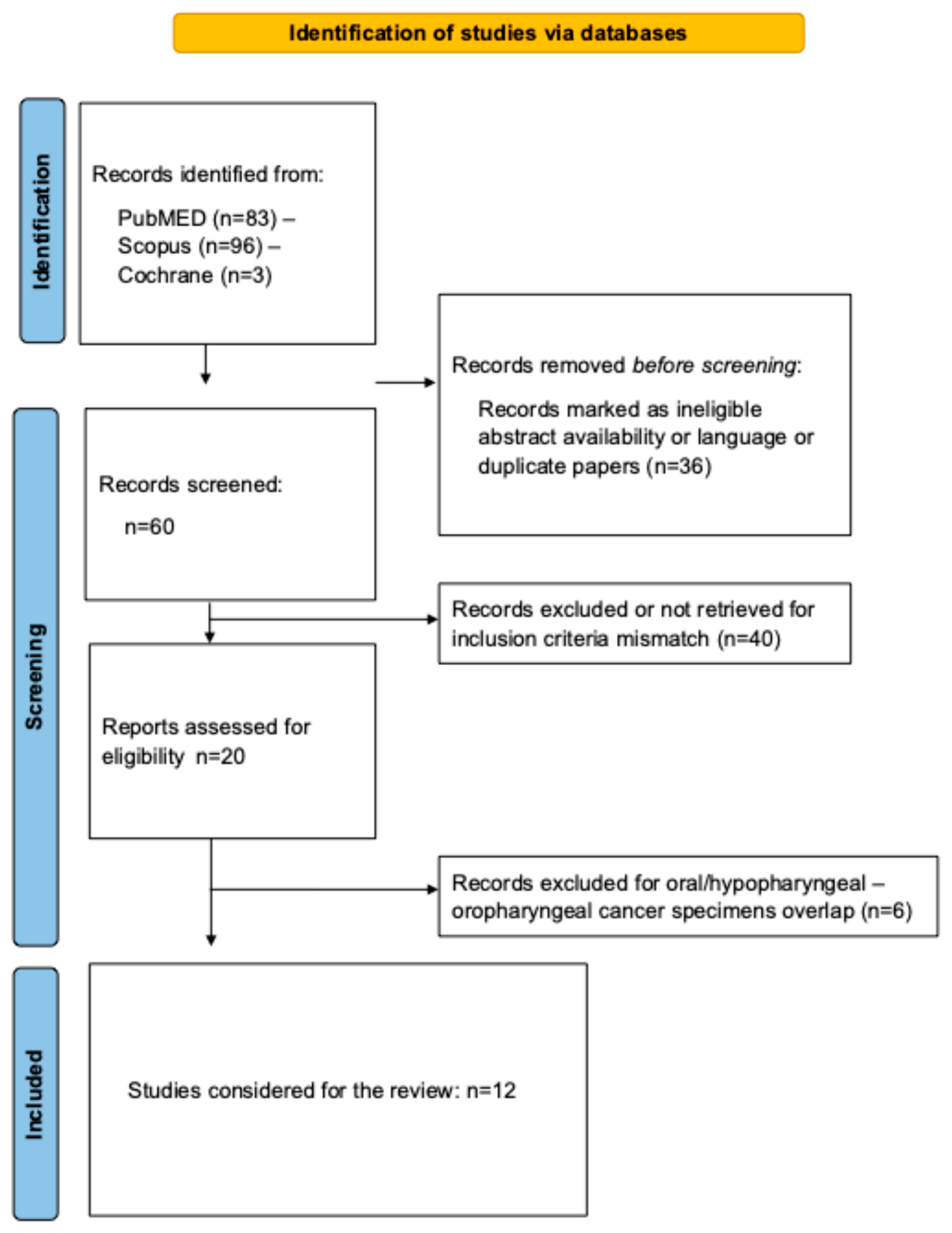
Table 6. Key findings for future studies.

5. Conclusions

The current literature supports potential distinct microbiome signatures between OPSCC and non-cancerous tissues, with an overrepresentation of Spirochaetes and Bacteroidetes in OPSCC, and Proteobacteria and some Firmicutes (Streptococcus) in healthy tissues—features influenced by HPV. A microbiome shift is possible from pre- to post-chemoradiotherapy, with baseline microbiome acting as a predictive response factor; however, the low number of studies and substantial methodological heterogeneity across investigations limit the drawing of valid conclusions. The identification of key species is important in the development of OPSCC for developing personalized medicine considering bacterial mediators in terms of prevention, and targeted therapy using the microbiome–tumor–host interaction pathways.

Funding

This research received no external funding.

Institutional Review Board Statement

Not applicable.

Data Availability Statement

The data presented in this study are available on request from the corresponding author.

Conflicts of Interest

The author declares no conflicts of interest.

Abbreviations

The following abbreviations are used in this manuscript:
OPSCCoropharyngeal squamous cell carcinoma
PSCCoral squamous cell carcinoma

References

  1. Global Burden of Disease Cancer Collaboration. Global, Regional, and National Cancer Incidence, Mortality, Years of Life Lost, Years Lived With Disability, and Disability-Adjusted Life-Years for 29 Cancer Groups, 1990 to 2017: A Systematic Analysis for the Global Burden of Disease Study. JAMA Oncol. 2019, 5, 1749–1768. [Google Scholar]
  2. Aupérin, A. Epidemiology of head and neck cancers: An update. Curr. Opin. Oncol. 2020, 32, 178–186. [Google Scholar] [CrossRef]
  3. Damgacioglu, H.; Sonawane, K.; Zhu, Y.; Li, R.; Balasubramanian, B.A.; Lairson, D.R.; Giuliano, A.R.; Deshmukh, A.A. Oropharyngeal Cancer Incidence and Mortality Trends in All 50 States in the US, 2001–2017. JAMA Otolaryngol. Head Neck Surg. 2022, 148, 155–165. [Google Scholar] [CrossRef]
  4. Pinkiewicz, M.; Dorobisz, K.; Zatoński, T. Human Papillomavirus-Associated Head and Neck Cancers. Where are We Now? A Systematic Review. Cancer Manag. Res. 2022, 14, 3313–3324. [Google Scholar] [CrossRef]
  5. Gallus, R.; Nauta, I.H.; Marklund, L.; Rizzo, D.; Crescio, C.; Mureddu, L.; Tropiano, P.; Delogu, G.; Bussu, F. Accuracy of p16 IHC in Classifying HPV-Driven OPSCC in Different Populations. Cancers 2023, 15, 656. [Google Scholar] [CrossRef] [PubMed]
  6. Hes, C.; Desilets, A.; Tonneau, M.; El Ouarzadi, O.; De Figueiredo Sousa, M.; Bahig, H.; Filion, É.; Nguyen-Tan, P.F.; Christopoulos, A.; Benlaïfaoui, M.; et al. Gut microbiome predicts gastrointestinal toxicity outcomes from chemoradiation therapy in patients with head and neck squamous cell carcinoma. Oral Oncol. 2024, 148, 106623. [Google Scholar] [CrossRef]
  7. Chung, R.S.; Wong, S.; Lin, D.; Kokot, N.C.; Sinha, U.K.; Han, A.Y. Mechanisms of crosstalk between the oropharyngeal microbiome and human papillomavirus in oropharyngeal carcinogenesis: A mini review. Front. Oncol. 2024, 14, 1425545. [Google Scholar] [CrossRef]
  8. McInnes, M.D.; Moher, D.; Thombs, B.D.; McGrath, T.A.; Bossuyt, P.M.; Clifford, T.; Cohen, J.F.; Deeks, J.J.; Gatsonis, C.; Hooft, L.; et al. Preferred Reporting Items for a Systematic Review and Meta-analysis of Diagnostic Test Accuracy Studies: The PRISMA-DTA statement. JAMA 2018, 319, 388–396. [Google Scholar]
  9. Thompson, M.; Tiwari, A.; Fu, R.; Moe, E.; Buckley, D.I. A Frame-Work to Facilitate the Use of Systematic Reviews and Meta-Analyses in the Design of Primary Research Studies; Agency for Healthcare Research and Quality: Rockville, MD, USA, 2012. [Google Scholar]
  10. Burns, P.B.; Rohrich, R.J.; Chung, K.C. The levels of evidence and their role in evidence-based medicine. Plast. Reconstr. Surg. 2011, 128, 305–310. [Google Scholar] [CrossRef]
  11. Slim, K.; Nini, E.; Forestier, D.; Kwiatkowski, F.; Panis, Y.; Chipponi, J. Methodological index for non-randomized studies (minors): Development and validation of a new instrument. ANZ J. Surg. 2003, 73, 712–716. [Google Scholar] [CrossRef] [PubMed]
  12. Börnigen, D.; Ren, B.; Pickard, R.; Li, J.; Ozer, E.; Hartmann, E.M.; Xiao, W.; Tickle, T.; Rider, J.; Gevers, D.; et al. Alterations in oral bacterial communities are associated with risk factors for oral and oropharyngeal cancer. Sci. Rep. 2017, 7, 17686. [Google Scholar] [CrossRef]
  13. Castañeda-Corzo, G.J.; Infante-Rodríguez, L.F.; Villamil-Poveda, J.C.; Bustillo, J.; Cid-Arregui, A.; García-Robayo, D.A. Association of Prevotella intermedia with oropharyngeal cancer: A patient-control study. Heliyon 2023, 9, e14293. [Google Scholar] [CrossRef]
  14. Chan, J.Y.K.; Cheung, M.K.; Lan, L.; Ng, C.; Lau, E.H.L.; Yeung, Z.W.C.; Wong, E.W.Y.; Leung, L.; Qu, X.; Cai, L.; et al. Characterization of oral microbiota in HPV and non-HPV head and neck squamous cell carcinoma and its association with patient outcomes. Oral Oncol. 2022, 135, 106245. [Google Scholar] [CrossRef]
  15. De Martin, A.; Lütge, M.; Stanossek, Y.; Engetschwiler, C.; Cupovic, J.; Brown, K.; Demmer, I.; Broglie, M.A.; Geuking, M.B.; Jochum, W.; et al. Distinct microbial communities colonize tonsillar squamous cell carcinoma. Oncoimmunology 2021, 10, 1945202. [Google Scholar] [PubMed]
  16. Dhakal, A.; Upadhyay, R.; Wheeler, C.; Hoyd, R.; Karivedu, V.; Gamez, M.E.; Valentin, S.; Vanputten, M.; Bhateja, P.; Bonomi, M.; et al. Association between Tumor Microbiome and Hypoxia across Anatomic Subsites of Head and Neck Cancers. Int. J. Mol. Sci. 2022, 23, 15531. [Google Scholar] [CrossRef]
  17. Hayes, R.B.; Ahn, J.; Fan, X.; Peters, B.A.; Ma, Y.; Yang, L.; Agalliu, I.; Burk, R.D.; Ganly, I.; Purdue, M.P.; et al. Association of Oral Microbiome With Risk for Incident Head and Neck Squamous Cell Cancer. JAMA Oncol. 2018, 4, 358–365. [Google Scholar] [CrossRef]
  18. Oliva, M.; Schneeberger, P.H.H.; Rey, V.; Cho, M.; Taylor, R.; Hansen, A.R.; Taylor, K.; Hosni, A.; Bayley, A.; Hope, A.J.; et al. Transitions in oral and gut microbiome of HPV+ oropharyngeal squamous cell carcinoma following definitive chemoradiotherapy (ROMA LA-OPSCC study). Br. J. Cancer 2021, 124, 1543–1551. [Google Scholar] [CrossRef]
  19. Guerrero-Preston, R.; White, J.R.; Godoy-Vitorino, F.; Rodríguez-Hilario, A.; Navarro, K.; González, H.; Michailidi, C.; Jedlicka, A.; Canapp, S.; Bondy, J.; et al. High-resolution microbiome profiling uncovers Fusobacterium nucleatum, Lactobacillus gasseri/johnsonii, and Lactobacillus vaginalis associated to oral and oropharyngeal cancer in saliva from HPV positive and HPV negative patients treated with surgery and chemo-radiation. Oncotarget 2017, 8, 110931–110948. [Google Scholar]
  20. Rajasekaran, K.; Carey, R.M.; Lin, X.; Seckar, T.D.; Wei, Z.; Chorath, K.; Newman, J.G.; O’Malley, B.W.; Weinstein, G.S.; Feldman, M.D.; et al. The microbiome of HPV-positive tonsil squamous cell carcinoma and neck metastasis. Oral Oncol. 2021, 117, 105305. [Google Scholar] [CrossRef] [PubMed]
  21. Wolf, A.; Moissl-Eichinger, C.; Perras, A.; Koskinen, K.; Tomazic, P.V.; Thurnher, D. The salivary microbiome as an indicator of carcinogenesis in patients with oropharyngeal squamous cell carcinoma: A pilot study. Sci. Rep. 2017, 7, 5867. [Google Scholar] [CrossRef] [PubMed]
  22. Zakrzewski, M.; Gannon, O.M.; Panizza, B.J.; Saunders, N.A.; Antonsson, A. Human papillomavirus infection and tumor microenvironment are associated with the microbiota in patients with oropharyngeal cancers-pilot study. Head Neck 2021, 43, 3324–3330. [Google Scholar] [CrossRef]
  23. Bahig, H.; Fuller, C.D.; Mitra, A.; Yoshida-Court, K.; Solley, T.; Ping Ng, S. Longitudinal characterization of the tumoral microbiome during radiotherapy in HPV-associated oropharynx cancer. Clin. Transl. Radiat. Oncol. 2021, 26, 98–103. [Google Scholar] [CrossRef] [PubMed]
  24. Banerjee, S.; Tian, T.; Wei, Z.; Peck, K.N.; Shih, N.; Chalian, A.A.; O’Malley, B.W.; Weinstein, G.S.; Feldman, M.D.; Alwine, J.; et al. Microbial Signatures Associated with Oropharyngeal and Oral Squamous Cell Carcinomas. Sci. Rep. 2017, 7, 4036. [Google Scholar] [CrossRef] [PubMed]
  25. Lim, Y.; Fukuma, N.; Totsika, M.; Kenny, L.; Morrison, M.; Punyadeera, C. The Performance of an Oral Microbiome Biomarker Panel in Predicting Oral Cavity and Oropharyngeal Cancers. Front. Cell. Infect. Microbiol. 2018, 8, 267. [Google Scholar] [CrossRef] [PubMed]
  26. Lim, Y.; Tang, K.D.; Karpe, A.V.; Beale, D.J.; Totsika, M.; Kenny, L.; Morrison, M.; Punyadeera, C. Chemoradiation therapy changes oral microbiome and metabolomic profiles in patients with oral cavity cancer and oropharyngeal cancer. Head Neck 2021, 43, 1521–1534. [Google Scholar] [CrossRef]
  27. Escobar Marcillo, D.I.; Privitera, G.F.; Rollo, F.; Latini, A.; Giuliani, E.; Benevolo, M.; Giuliani, M.; Pichi, B.; Pellini, R.; Donà, M.G. Microbiome analysis in individuals with human papillomavirus oral infection. Sci. Rep. 2025, 15, 2953. [Google Scholar] [CrossRef]
  28. Oberste, M.; Böse, B.E.; Dos Anjos Borges, L.G.; Junca, H.; Plumeier, I.; Kahl, S. Effects of squamous cell carcinoma and smoking status on oropharyngeal and laryngeal microbial communities. Head Neck 2024, 46, 145–160. [Google Scholar]
  29. Panda, M.; Rai, A.K.; Rahman, T.; Das, A.; Das, R.; Sarma, A.; Kataki, A.C.; Chattopadhyay, I. Alterations of salivary microbial community associated with oropharyngeal and hypopharyngeal squamous cell carcinoma patients. Arch. Microbiol. 2020, 202, 785–805. [Google Scholar]
  30. Díaz-Rivera, J.; Rodríguez-Rivera, M.A.; Meléndez-Vázquez, N.M.; Godoy-Vitorino, F.; Dorta-Estremera, S.M. Immune and Microbial Signatures Associated with PD-1 Blockade Sensitivity in a Preclinical Model for HPV+ Oropharyngeal Cancer. Cancers 2024, 16, 2065. [Google Scholar] [CrossRef]
  31. Lechien, J.R. Dysbiosis Patterns in Glottic and Laryngeal Cancers: A Systematic Review of Microbiome Alterations. J. Voice 2025, submitted. [CrossRef]
  32. Gong, H.; Wang, B.; Shi, Y.; Shi, Y.; Xiao, X.; Cao, P.; Tao, L.; Wang, Y.; Zhou, L. Composition and abundance of microbiota in the pharynx in patients with laryngeal carcinoma and vocal cord polyps. J. Microbiol. 2017, 55, 648–654. [Google Scholar] [CrossRef]
  33. Crispino, A.; Varricchio, S.; Esposito, A.; Marfella, A.; Cerbone, D.; Perna, A.; Petronio, G.; Staibano, S.; Merolla, F.; Ilardi, G. The oral microbiome and its role in oral squamous cell carcinoma: A systematic review of microbial alterations and potential biomarkers. Pathologica 2024, 116, 338–357. [Google Scholar] [CrossRef]
  34. Dadgar-Zankbar, L.; Shariati, A.; Bostanghadiri, N.; Elahi, Z.; Mirkalantari, S.; Razavi, S.; Kamali, F.; Darban-Sarokhalil, D. Evaluation of enterotoxigenic Bacteroides fragilis correlation with the expression of cellular signaling pathway genes in Iranian patients with colorectal cancer. Infect. Agents Cancer 2023, 18, 48. [Google Scholar] [CrossRef] [PubMed]
  35. de Freitas Neiva Lessa, A.; da Silva Amâncio, A.M.T.; de Oliveira, A.C.R.; de Sousa, S.F.; Caldeira, P.C.; De Aguiar, M.C.F.; Bispo, P.J.M. Assessing the oral microbiome of head and neck cancer patients before and during radiotherapy. Support Care Cancer 2024, 32, 752. [Google Scholar] [CrossRef]
  36. Senthil Kumar, S.; Johnson, M.D.L.; Wilson, J.E. Insights into the enigma of oral streptococci in carcinogenesis. Microbiol. Mol. Biol. Rev. 2024, 88, e0009523. [Google Scholar] [CrossRef] [PubMed]
  37. Torozan, D.A.; Laczny, C.C.; Roomp, K.; Wilmes, P.; Fleckenstein, J.; Schneider, J.G. Metagenomic Profiling of Oral Microbiome Dynamics During Chemoradiotherapy in Head and Neck Squamous Cell Carcinoma Patients. Cancer Med. 2025, 14, e70589. [Google Scholar] [CrossRef]
  38. Chakladar, J.; John, D.; Magesh, S.; Uzelac, M.; Li, W.T.; Dereschuk, K.; Apostol, L.; Brumund, K.T.; Rodriguez, J.W.; Ongkeko, W.M. The Intratumor Bacterial and Fungal Microbiome Is Characterized by HPV, Smoking, and Alcohol Consumption in Head and Neck Squamous Cell Carcinoma. Int. J. Mol. Sci. 2022, 23, 13250. [Google Scholar] [CrossRef] [PubMed]
  39. O’Grady, I.; Anderson, A.; O’Sullivan, J. The interplay of the oral microbiome and alcohol consumption in oral squamous cell carcinomas. Oral Oncol. 2020, 110, 105011. [Google Scholar] [CrossRef]
Disclaimer/Publisher’s Note: The statements, opinions and data contained in all publications are solely those of the individual author(s) and contributor(s) and not of MDPI and/or the editor(s). MDPI and/or the editor(s) disclaim responsibility for any injury to people or property resulting from any ideas, methods, instructions or products referred to in the content.

Article Metrics

Citations

Article Access Statistics

Multiple requests from the same IP address are counted as one view.