Review

A Systematic Review of Chest Imaging Findings in Long COVID Patients

by
Somayeh Bazdar
1,2,3,
Anastasia K. A. L. Kwee
4,
Laura Houweling
1,5,
Yolanda de Wit-van Wijck
1,2,3,
Firdaus A. A. Mohamed Hoesein
4,
George S. Downward
5,6,
Esther J. Nossent
1 and
Anke H. Maitland-van der Zee
1,2,3,* on behalf of the P4O2 Consortium
1
Department of Pulmonary Medicine, Amsterdam UMC, 1105 AZ Amsterdam, The Netherlands
2
Amsterdam Institute for Infection and Immunity, 1105 AZ Amsterdam, The Netherlands
3
Amsterdam Public Health, 1105 AZ Amsterdam, The Netherlands
4
Department of Radiology, University Medical Center Utrecht and Utrecht University, 3584 CX Utrecht, The Netherlands
5
Department of Environmental Epidemiology, Institute for Risk Assessment Sciences (IRAS), 3584 CX Utrecht, The Netherlands
6
Julius Center for Health Sciences and Primary Care, University Medical Center Utrecht, 3584 CX Utrecht, The Netherlands
*
Author to whom correspondence should be addressed.
J. Pers. Med. 2023, 13(2), 282; https://doi.org/10.3390/jpm13020282
Submission received: 3 January 2023 / Revised: 25 January 2023 / Accepted: 30 January 2023 / Published: 1 February 2023
(This article belongs to the Section Methodology, Drug and Device Discovery)

Abstract

:
Long COVID is the persistence of one or more COVID-19 symptoms after the initial viral infection, and there is evidence supporting its association with lung damage. In this systematic review, we provide an overview of lung imaging and its findings in long COVID patients. A PubMed search was performed on 29 September 2021, for English language studies in which lung imaging was performed in adults suffering from long COVID. Two independent researchers extracted the data. Our search identified 3130 articles, of which 31, representing the imaging findings of 342 long COVID patients, were retained. The most common imaging modality used was computed tomography (CT) (N = 249). A total of 29 different imaging findings were reported, which were broadly categorized into interstitial (fibrotic), pleural, airway, and other parenchymal abnormalities. A direct comparison between cases, in terms of residual lesions, was available for 148 patients, of whom 66 (44.6%) had normal CT findings. Although respiratory symptoms belong to the most common symptoms in long COVID patients, this is not necessarily linked to radiologically detectable lung damage. Therefore, more research is needed on the role of the various types of lung (and other organ) damage which may or may not occur in long COVID.

1. Introduction

The worldwide crisis of Coronavirus disease 2019 (COVID-19) pandemic caused by the severe acute respiratory syndrome, coronavirus 2 (SARS-CoV-2), has influenced people’s lives globally [1,2]. On 30 January 2020, the WHO declared the outbreak of SARS-CoV-2 a Public Health Emergency of International Concern and, since then, COVID-19 has caused (as of 23 December 2022) at least 6,651,415 deaths and 650,879,143 confirmed cases. [3,4]
Although COVID-19 is primarily a respiratory disease, it may affect extra-pulmonary organs and cause systemic complications [5,6]. These complications can occur not just in the acute phase but also after the initial infection has been overcome [7]. These long-term (or chronic) complications are collectively described as “long COVID” and represent a variety of complaints. Presently, long COVID is defined as the persistence of one or more COVID-19 symptoms weeks or months after the initial viral infection [8,9]. Long COVID is probably one of the most important complications of SARS-CoV-2 infection; it can affect both the mental and physical ability of patients and have significant effects on society’s work force [10,11].
Although the etiology of long COVID is currently unknown, several studies have indicated that lung damage may play a significant role in its development [12]. This is supported by reports that the most common symptoms among long COVID patients are respiratory in nature [13]. During the initial phase of COVID-19, lung abnormalities are related to the course and prognosis of the disease. In the initial two weeks, the most common imaging finding is ground-glass opacity (GGO) followed by consolidation. From the second week of infection, however, imaging abnormalities suggestive of fibrosis might start to appear [14]. Radiological abnormalities can also be observed long after the initial infection has resolved. For example, CT scans of COVID-19 survivors who were admitted to the ICU during their acute phase showed that residual lung abnormalities can still be radiologically detected after three months. However, whether findings are clinically relevant has not yet been elucidated; these changes represent an ongoing recovery; thus, to distinguish them from irreversible fibrotic changes, further follow-up imaging after at least one year has been recommended [15].
Further evidence supporting the association between lung damage and the development of long COVID is detecting radiological abnormalities in patients who were infected with other coronaviruses (SARS and MERS) even years after infection. [16]. Serial lung high-resolution computed tomography (HRCT) scans of SARS-CoV-1 patients demonstrated that some patients still demonstrated fibrotic changes, and even ground-glass-like changes, in their long-term serial follow-ups after 12 months [17]. Moreover, in a one-year assessment of post-SARS-CoV-1 patients, a significant correlation between the percentage of lung imaging abnormalities and certain lung function parameters (specifically, the total lung capacity and diffusing capacity of the lungs for carbon monoxide) was reported [18].
The key question for patients and their pulmonologists is whether performing lung imaging in patients with long COVID is illuminative. As the COVID-19 pandemic has now been present for more than two years, sufficient time since initial infection has occurred to allow closer examinations of radiological changes in those with long COVID. However, there is presently no collective overview of the relationship between imaging findings and their outcomes. Therefore, the aim of this systematic review is to provide an overview of lung imaging findings in long COVID patients.

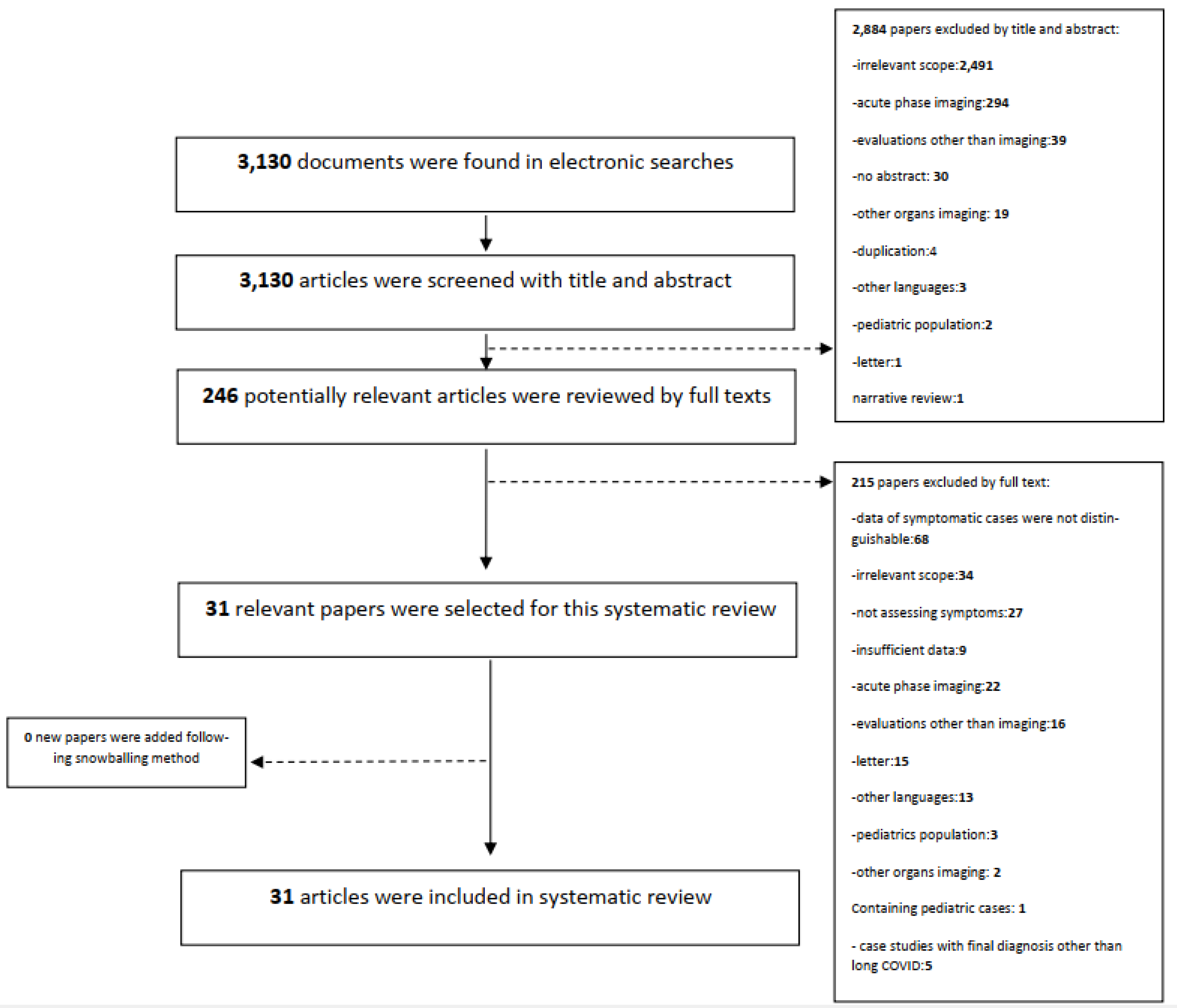
2. Methods and Materials

This systematic review was conducted as per our registered protocol (PROSPERO Record ID = CRD42021292358) (https://www.crd.york.ac.uk/prospero/display_record.php?RecordID=292358, access on 26 November 2021) and findings are reported as per Preferred Reporting Items for Systematic Reviews and Meta-Analyses statement (PRISMA) guidelines. On 29 September 2021, we performed a PubMed search to identify articles examining lung imaging among adults suffering from long COVID. No explicit date was included in the search, although only studies published during the COVID-19 pandemic, from 30 January 2019, were considered. The search terms (see below) were selected based on the authors’ knowledge and on previously identified publications. Snowballing sampling was performed where the references of any identified review articles were also examined to identify any additional relevant articles.
Search terms: (((long COVID) OR (long COVID) OR (post acute sequelae of COVID) OR (pacs) OR (chronic COVID syndrome) OR (chronic COVID) OR (ccs) OR (long haul COVID) OR (long COVID haul*) OR (post COVID) OR (COVID surviv*) OR (late effect* COVID) OR (COVID late complication*) OR (post acute COVID) OR (long term COVID) OR (longterm COVID) OR (persist* COVID) OR (late COVID) OR (ongoing COVID) OR (Enduring COVID) OR (longlast* COVID) OR (lengthy COVID) OR (residual COVID) OR (relaps* COVID) OR (COVID remission*) OR (linger* COVID) OR (permanent* COVID) OR (subacute COVID) OR (sub acute COVID) OR (permanent* COVID)) AND ((chest) OR (lung) OR (thorax) OR (thorac*) OR (pulm*)) AND ((imag*) OR (radiolog*) OR (graph*) OR (scan) OR (computed tomography scan) OR (ct scan) OR (ct) OR (CAT) OR (computer assisted tomography) OR (hrct) OR (X ray) OR (cxr) OR (magnetic resonance imaging) OR (mri) OR (ultrasonography) OR (us)))
Inclusion criteria: English language studies in which any modality of lung imaging was performed on adults suffering from long COVID were eligible for inclusion, regardless of whether lung imaging was the only intervention in that study or one among several interventions. Preprints and letters to the editor were not considered valid for inclusion, nor were papers in which we were unable to distinguish between asymptomatic post-COVID patients and those with long COVID symptoms.
Long COVID was defined following the Centers for Disease Control and Prevention’s (CDC) definition as patients with a confirmed diagnosis of COVID-19 (whether based on PCR test, serology test, any other laboratory test, clinic-radiological diagnosis, history of close contact with proven case) who continue to experience COVID-19 symptoms for more than 28 days.
Screening: Title and abstract screening of the papers identified in our search was performed by SB. All articles identified through title and abstract underwent full text screening to ensure they matched our inclusion criteria prior to data extraction. As a quality control step, 10% of the identified articles (selected randomly) were evaluated by a second reviewer (AK). Disagreements in evaluations (which occurred three times) were discussed with a third reviewer (LH). All final determinations aligned with those of the main reviewer (SB). The flowchart of the screening process is provided in Figure 1.
Data extraction: Data from the identified articles were extracted into a customized spreadsheet containing study information (study design, publication year, number of participants, inclusion criteria, country and continent of the performed study, study period), demographic data (age, sex, ethnicity, nationality) and imaging/outcome information. Specific imaging and outcome information were as follows: percentage of long COVID cases with lung imaging abnormalities, the type of lung imaging abnormalities, lesion distribution, imaging interpreters, imaging modalities, imaging evaluation time, imaging evaluation setting, long COVID symptoms, medical interventions, probable risk factors for developing long COVID complications, such as smoking, obesity, comorbidities, and intubation. According to the anatomical positioning and physiology, we categorized the extracted imaging findings into the four groups: interstitial (fibrotic) abnormalities, other interstitial abnormalities, airway abnormalities, and parenchymal abnormalities. However, it should be noted that some specified abnormalities can fit into different categories.
Risk of bias (quality) assessment: The National Heart, Lung, and Blood Institute (NHLBI) risk of bias assessment tool was used to evaluate the risk of bias and was performed by two reviewers (SB, AK) independently. There was complete agreement between these reviewers regarding quality assessment.

3. Results

Our literature search identified a total of 3130 articles. After title and abstract screening, we removed 2884 papers, while a further 215 articles were removed after full text screening, leaving 31 articles for data extraction. Primary reasons for paper exclusion during the title and abstract screening stage were as follows: non-COVID-19 diseases, addressing the technical aspects of artificial intelligence usage in COVID-19 lung imaging, imaging findings being reported only from the acute phase of COVID-19, and non-lung imaging findings. During full text screening, several articles were excluded as data from symptomatic patients could not be distinguished from the data of asymptomatic ones (N = 68). Other reasons for exclusion during the full text screening step included: topics irrelevant to the aim and scope of our systematic review (N = 34), not assessing symptoms in the follow-up evaluation (N = 27), insufficient information about participants or intervention such as, participants age range, evaluation time, COVID-19 diagnosis method, etc., that caused them not to fulfill our inclusion criteria clearly (N = 9), and case-reports with final diagnoses other than long COVID (N = 5). The flowchart of the screening process, as well as the exclusion reasons for each step, is provided in Figure 1.
After title, abstract, and full-text screening, 31 articles were identified that met our inclusion criteria, representing the imaging findings of 342 patients who were suffering from long COVID. Of these 31 articles, 9 represented cohort/case-control studies, covering 309 participants; 5 were case series, covering 16 patients; and 17 were individual case reports. The study population tended to be male (62.2%) with a mean age of 59.5 years. The study design of the original studies included in this systematic review were either cohort or case-control (Table 1). With the exception from one case-report, the quality of all studies was fair based on the NHLBI checklist. More extensive information, including the outcome of quality assessment, is presented in the Supplement (Table S1).
The number of patients undergoing imaging ranged from four to 91 in the identified studies. Imaging was typically performed in one of three settings: follow-up evaluation, re-admission, and prolonged hospitalization. Case reports and series occurred exclusively in the re-admission and prolonged hospitalization setting whereas, with a single exception (Gaspardone et.al.) all of the original articles occurred in the follow-up evaluation setting.
Disease severity and/or used interventions were inconsistently reported across studies and not explicitly linked to radiological findings. For example, the intubation status of patients studied was not mentioned for 201 cases. In the remaining 141 cases, 63 were intubated and 78 were not (Table S2). While medication use was similarly inconsistently reported, among the medications that were used during the acute phase of COVID-19 infection, hydroxychloroquine was the most commonly used, followed by antiviral medications such as remdesivir, oral antibiotics such as azithromycin, and interlukin-1 (IL-1) inhibitors such as anakinra (Table S4). Among the 111 patients where comorbidities were reported, hypertension was the most common comorbidity (n = 61), followed by diabetes (n = 28) (Table S3).
Dyspnea was the most common Long-COVID symptom in patients, followed by fatigue. Supplement shows the frequency of all long COVID symptoms for each study (Table S4).
Across all settings and study types, the most common imaging modality used was CT scan (N = 249), followed by USS (N = 73), CXR (N = 25), positron emission tomography (PET) scan (N = 23), and magnetic resonance imaging (MRI) (N = 2) (Table 1 and Table S5). Across these modalities, a total of 29 different imaging findings were reported, with a variety of reporting techniques and terminology. On the basis of anatomical positioning and physiology, we categorized these findings into the following groups: interstitial (fibrotic) abnormalities, pleural abnormalities, airway abnormalities, and other parenchymal abnormalities (Table 2). In two articles, the findings were reported as general terms of COVID pneumonia (Malik et al.) or bilateral pneumonia/viral pneumonitis (Mitrani et al.), and thus could not be categorized into any group. Several of the cohort/case-control articles (Armange et al., Bai et al., Cesarone et al., Rinaldo et al., Sollini et al., Sollini et al., Miwa et al.) also reported the number of long COVID patients without any residual lesions on their imaging. These articles represented 148 patients, of whom 82 (55.4%) had positive findings on their imaging (Table 3). All case studies and reports had positive imaging findings (Table S5).
When categorizing specific radiological findings, other parenchymal abnormalities were the most reported finding (112 times), of which ground-glass opacity (reported 93 times) was the most common specific abnormality. The second most common abnormality grouping was interstitial (fibrotic) abnormalities, reported 105 times. Within this group, reticulation (reported 78 times) and fibrotic changes (reported 14 times) were the most common specific abnormalities. Airway and pleural abnormalities were reported relatively less than the other categories (28 and 7 times, respectively (Table 2)). Only two studies (Armange et al. and Yin et al.) assessed the relationship between symptoms and radiological findings, with contradictory findings. Armange et al. reported that there was no relationship between post-COVID symptoms and CT scan findings [19]; however, Yin et al. found that there was a relationship between post-COVID symptoms and imaging findings, and that dyspneic long COVID patients had larger lesions, a higher residual lesion rate, and incompletely absorbed lesions [26].
While none of the patients that were categorized into the re-admission and prolonged hospitalization groups had negative imaging findings, 46% of the patients evaluated in the outpatient follow-up setting had negative imaging findings (Table 4, Table S6). In the prolonged hospitalization group other parenchymal abnormalities were more frequent; while in the follow-up evaluation and re-admission groups, interstitial (fibrotic) abnormalities followed by other parenchymal abnormalities were the most frequent lung imaging abnormalities (Table 4).
When examining only the cohort/case-control studies to assess their findings based on the evaluation setting, we found that their imaging was all performed in the follow-up setting, except for the study conducted by Gaspardo et al., whose patients were evaluated by LUSS in a prolonged hospitalization setting. Moreover, while Gaspardo et al. did not report on specific findings or on positive versus negative findings, they commented that the LUSS severity score at discharge was higher among those with worse disease at admission. Table 5 illustrates the imaging findings of the cohort/case-control studies, which were conducted in the follow-up setting. In these studies, the most reported finding was interstitial (fibrotic) abnormalities (58.2%), followed by other parenchymal abnormalities (53.8%) and airway abnormalities (13.9%). No pleural abnormalities were reported in these studies.
There were eight studies in this systematic review which compared lung imaging from two different modalities (Table 6). Sollini et al. compared CT vs. PET in two studies, with somewhat contradictory findings. In a study of ten patients, fibrotic changes were reported on CT for six of them, while only two cases had mild [18F]FDG lung uptake [24]. However, in an evaluation of a different group of thirteen patients, [18F]FDG uptake was detected in six patients, with bilateral findings being observed on CT for only four patients who only had mild [18F]FDG uptake [25].
Garg et al. and Heiss et al. both compared CT and MRI scans for single patients, reporting identical findings in both cases [28,29]. Tung Chen et al. compared USS to CT and found anatomically correlated radiological residuals in both imaging techniques; however, they could not specify the type of abnormality in USS [30]. Finally, four papers [31,32,33,34] compared plain CXR findings to CT scan, finding that while radiological residuals were visible on CXR, they lacked the more precise definition of CT scan.
Table 6. Comparison of various imaging modalities.
First AuthorCompared Modalities and Performing Days *Imaging ModalityImaging Finding
Sollini et al. [24]30 daysCTFibrotic change in 6 cases out of 10 population of study
PET[18F]FDG lung uptake in 2 cases out of total 10 population of study
Sollini et al. [25]132 daysCTbilateral lung abnormalities, as typically observed in recovered COVID-19 pneumonia, was detected in 4 cases out of 13 in the total population of study
PETMild [18F]FDG PET/CT uptake was detected in of Post-pneumonia lung abnormalities in 4 cases out of 13in the total population of study, and 2 other patients presented moderate/high [18F]FDG uptake in the lung, mediastinal lymph nodes, soft tissue, and breast tissue related to their comorbidities.
Garg et al. [28]CT at 53 days
and
MRI at 55 days
CTinterlobular septal thickening, parenchymal band, Fibrotic change, bronchiectasis, bronchiolectasis, crazy-paving pattern
Garg et al. [28]MRIinterlobular septal thickening, parenchymal band, Fibrotic change, bronchiectasis, bronchiolectasis, crazy-paving pattern
Heiss et al. [29]97 daysCTGGO, Consolidation
Heiss et al. [29]MRIGGO, Consolidation
Tung-Chen et al. [30]56 daysCTpleural thickening
Tung-Chen et al. [30]USSirregular pleural line in the right lateral area of the chest, which correlated with an area of pleural thickening on chest CT
Tung-Chen et al. [30]CTGGO
Tung-Chen et al. [30]USSa mild irregular pleural line and B-lines in the right anterior area of the chest, which correlated with ground-glass opacities on chest CT with no abnormal findings in the pleura.
Tung-Chen et al. [30]CTfibrotic change
Tung-Chen et al. [30]USSa marked irregular pleural line and multiple B lines, especially in the posterior inferior area of the chest, which correlated with fibrotic changes on chest CT
Alhiyari et al. [32]130 daysCXRair space opacity
Alhiyari et al. [32]CTGGO, bronchiectasis, interlobular septal thickening, reticulation, honeycomb-like appearance, interstitial pneumonia pattern
Hamad et al. [33]CT at 35 days
and
CXR at 36 days
CXRpneumothorax, air space opacity
Hamad et al. [33]CTpleural effusion, atelectasis, pulmonary edema, cystic airspace
Aesif et al. [31]CT at 59 days
and
CXR at 122 days
CTpneumothorax, Consolidation
Aesif et al. [31]CXRcomplete opacification, volume loss
Malik et al. [34]42 daysCXRConsolidation
Malik et al. [34]CTGGO, Fibrotic change, bronchiectasis, architectural distortion, linear scaring, airspace disease, COVID pneumonia
Footnote: * The performance day refers to the number of days that have passed since the onset of symptoms and in which the imaging evaluation was performed.
Regarding the imaging interpreter, there were only three studies, performed by Sollini et al. and Yin et al. [23,24,26], which mentioned the interpretation procedure and specialist who reported the imaging findings. In both studies of Sollini et al., it was noted that interpretation was performed by nuclear medicine physicians, and details about the experience of the interpreter or any available guidelines that they might have used were not reported. Yin et al. reported that two radiologists with 15 and 10 years of experience in cardiothoracic imaging reported the radiologic imaging. However, information related to the guidelines on which they based their interpretations and reports was not included.

4. Discussion

The role of lung imaging in the care of long COVID patients is an area of uncertainty for physicians and patients. While physicians should consider precise evaluation of the respiratory system through imaging, exposing patients to unnecessary doses of radiation and straining health care resources should be also avoided. Therefore, obtaining a comprehensive insight, including indications for lung imaging in long COVID patients, is essential. This systematic review aimed to develop an understanding of the current role of imaging (and its findings) among those with long COVID.
A major finding of this systematic review was that although the dominant symptoms in long COVID patients are respiratory, this is not necessarily related to lung imaging abnormalities. Overall, approximately 60% of patients included in the cohort and case-control studies in this review had radiological residuals, the most common of which was parenchymal abnormalities other than interstitial (fibrotic) abnormalities. Moreover, the findings of the only two studies assessing the relationship between the findings and the extent of symptoms [20,26] were not in accordance with each other. Together, this indicates that chest imaging alone is not sufficient for the evaluation of long COVID, and that it should instead form one part of overall patient care.
This review identified that CT scans were the most commonly used modality for imaging in long COVID. However, there were studies using modalities other than CT scans, such as CXR, US sonography, MRI, and even PET scans in long COVID patients. These imaging techniques are quite novel, unusual, and clinically irrelevant in this area, and in clinical practice setting, the common imaging technique was firstly the CT scan and secondly CXR. Almost all of the studies using modalities other than the CT scan and CXT were performed in an experimental setting, while the studies employing CT scans and CXR as imaging modalities were mostly conducted in a setting of routine follow-up evaluation of patients.
Studies comparing MRI versus CT [28,29] reported consistent findings between the two. However, since the identified articles only covered the findings from two patients, further investigations in larger scale settings are needed to better understand the relative role of these imaging modalities. In routine medical practice, CT is generally considered superior to MRI for imaging of areas prone to movement, such as the lung [35]. It should also be considered that MRI is the more expensive and time-consuming modality, and any potential benefits need to be weighed against the higher costs and lower availability.
US sonography is mostly used in the acute phase, and especially for obstetric or pediatric patients [36,37]. However, Gaspardone et al. and Tung-Chen et al. found that this modality would also be helpful in detecting the long-term chest findings of COVID-19 patients [22,30]. While US sonography is among the safest imaging modalities since it is a radiation-free technique [38], the precision of this modality, especially for the respiratory system, will never be as high as a CT scan, and the detailed evaluation of parenchymal is necessary for patients with prolonged respiratory symptoms. Therefore, some believe that the clinical application of this technique in long COVID is to be the first-line assessment and guide the physicians to perform CT scans for cases that had USS findings [39]. Furthermore, since US sonography is an operator-based modality [40], providing a standard protocol for post-COVID US sonography is also necessary.
The PET scan was utilized in two studies of this systematic review [24,25]. This modality is typically used for detecting areas of activity in the body, such as cancer and inflammation [41]. However, the role of this method in lung evaluation of long COVID patients is less clear, since in some abnormalities no active inflammation is happening (e.g., a fibrotic lesion) and cannot thus be detected in this way [42]. Moreover, pre-existing disease or comorbidities may result in [18F]FDG uptake, causing a false positive finding. Despite these limitations, this imaging technique may be appropriate in terms of searching for the systemic etiology of long COVID, as well as tracking the treatment response to the anti-inflammatory medications that has been administrated for long COVID patients with lung inflammation.
Comparing the imaging findings that were reported through CT scan and CXR showed that the abnormalities can be reported more precisely through CT scan compared with CXR. This may indicate that CXR is a sub-optimal modality for providing detailed lung evaluation in long COVID cases. However, this low-dose radiation lung imaging technique [43] can be a screening method among long COVID patients to determine the proper candidates for a CT scan.
Air trapping, identified as a diffuse lung injury that might be developed by the inflammatory obstruction of small airways [44], was not reported in any of included articles. Neither was it reported whether it was even examined during the imaging. This finding is a sign of the obstructive problems in small air ways and has been reported in long COVID patients [45,46]. Therefore, this radiological finding should be examined in long COVID lung imaging.
COVID-19, and by extension long COVID, are novel diseases, meaning that the ability of imaging devices and interpreters to accurately identify relevant findings is more limited than for other diseases. Using either a computer-based or a human-based method to determine the findings affects the precision of the imaging reports. In human-based reports, the expertise and experience of the interpreter are important, an importance which is likely to be increased in the context of a new and emerging condition [47,48]. Although one of our aims was to evaluate what the imaging interpretations method is, only three studies [23,24,26] mentioned this point.
An additional challenge is that while we sought to examine long COVID, this condition was rarely (if ever) being explicitly examined. Most of the available articles were assessing all post-COVID patients, and, where possible, we focused on only symptomatic cases, extracting information specifically related to long COVID. This highlights a need for research more specifically focusing on long COVID. Another limitation of this study is that there are many different radiological terms, such as "interstitial pneumonia pattern” and “volume loss of lung” used in the included article, for which there are no clear definitions but some potential overlap in what the term means, which allow some specified abnormalities to fit into different categories.

5. Conclusions

In this review of imaging studies, we identified a variety of radiological findings among patients with long COVID, including scans where no abnormalities were found. This indicates that in order to properly evaluate long COVID, a holistic view is required, which will consider both the individual patient and the potential role of other organs and systems.
The small number of included articles indicates that lung imaging findings in long COVID patients is an area that requires more research.
Considering the burden of long COVID syndrome and the remaining uncertainties, more research which specifically examines long COVID is urgently required. Furthermore, the correlation between the imaging findings of long COVID patients and their underlying diseases, predisposing factors, medications that were consumed during the acute phase, and their symptoms should be studied.

Supplementary Materials

The following supporting information can be downloaded at: https://www.mdpi.com/article/10.3390/jpm13020282/s1, Table S1. Details of included studies; Table S2. Risk factors and comorbidities of long COVID patients in the included articles; Table S3. Frequency of administrated medications during the acute phase in participants of included studies; Table S4. Frequency of long COVID symptoms in participants of included studies; Table S5. Imaging modalities and imaging findings of included case-studies; Table S6. Frequency of lung imaging abnormalities per different evaluation setting. References [49,50,51,52,53,54,55,56,57,58,59,60,61,62,63,64] are cited in the Supplementary Materials

Author Contributions

Conceptualization, S.B., A.H.M.-v.d.Z., G.S.D. and F.A.A.M.H.; Methodology, S.B., A.H.M.-v.d.Z., G.S.D. and F.A.A.M.H.; Validation, S.B., A.H.M.-v.d.Z., G.S.D., F.A.A.M.H., A.K.A.L.K., L.H. and Y.d.W.-v.W.; Formal Analysis, S.B., A.H.M.-v.d.Z., G.S.D., F.A.A.M.H., Y.d.W.-v.W. and E.J.N.; Investigation, S.B., A.K.A.L.K. and L.H.; Resources, S.B., A.H.M.-v.d.Z., G.S.D. and Y.d.W.-v.W.; Data Curation, S.B.; Writing—Original Draft Preparation, S.B.; Writing—Review & Editing, all authors; Visualization, S.B.; Supervision, A.H.M.-v.d.Z., G.S.D., F.A.A.M.H., Y.d.W.-v.W. and E.J.N.; Project Administration, S.B., A.H.M.-v.d.Z., G.S.D. and Y.d.W.-v.W.; Funding Acquisition, A.H.M.-v.d.Z. All authors have read and agreed to the published version of the manuscript.

Funding

This study is funded by the P4O2 consortium. Partners in the Precision Medicine for more Oxygen (P4O2) consortium are the Amsterdam UMC, Leiden University Medical Center, Maastricht UMC+, Maastricht University, UMC Groningen, UMC Utrecht, Utrecht University, TNO, Aparito, Boehringer Ingelheim, Breathomix, Clear, Danone Nutricia Research, Fluidda, MonitAir, Ncardia, Ortec Logiqcare, Philips, Proefdiervrij, Quantib-U, RespiQ, Roche, Smartfish, SODAQ, Thirona, TopMD, Lung Alliance Netherlands (LAN) and the Lung Foundation Netherlands (Longfonds). The consortium is additionally funded by the PPP Allowance made available by Health~Holland, Top Sector Life Sciences & Health (grant numbers LSHM20104 and LSHM20068), to stimulate public-private partnerships and by Novartis.

Institutional Review Board Statement

Not applicable.

Informed Consent Statement

Not applicable.

Data Availability Statement

Not applicable.

Conflicts of Interest

Prof. Dr. Anke-Hilse Maitland-van der Zee has received research grants outside the submitted work from GSK, Boehringer Ingelheim and Vertex, she is the PI of P4O2 (Precision Medicine for more Oxygen), a public private partnership co-funded by Health~Holland involving many private partners that contribute in-cash and/or in-kind (Aparito, Boehringer Ingelheim, Breathomix, Clear, Danone Nutricia Research, Fluidda, MonitAir, Ncardia, Ortec Logiqcare, Philips, Quantib-U, RespiQ, Roche, Smartfish, SODAQ, Thirona, TopMD and Novartis), and she has served in advisory boards for AstraZeneca, GSK, and Boehringer Ingelheim with money paid to her institution. Moreover Dr. George Downward’s, Anastasia K.A.L. Kwee’s, Laura Houweling’s, Yolanda de Wit-van Wijck’s, and Dr. Firdaus A.A. Mohamed Hoesein’s Conflicts of interest relates to the funding structure of P4O2 (public-private).

References

  1. Grover, S.; Dua, D.; Sahoo, S.; Mehra, A.; Nehra, R.; Chakrabarti, S. Why all COVID-19 hospitals should have mental health professionals: The importance of mental health in a worldwide crisis! Asian J. Psychiatry 2020, 51, 102147. [Google Scholar] [CrossRef] [PubMed]
  2. Jara, A.; Undurraga, E.A.; González, C.; Paredes, F.; Fontecilla, T.; Jara, G.; Pizarro, A.; Acevedo, J.; Leo, K.; Leon, F.; et al. Effectiveness of an inacti-vated SARS-CoV-2 vaccine in Chile. N. Engl. J. Med. 2021, 385, 875–884. [Google Scholar] [CrossRef] [PubMed]
  3. Zheng, Y.-Y.; Ma, Y.-T.; Zhang, J.-Y.; Xie, X. COVID-19 and the cardiovascular system. Nat. Rev. Cardiol. 2020, 17, 259–260. [Google Scholar] [CrossRef] [PubMed]
  4. WHO. 2022. Available online: https://covid19.who.int/ (accessed on 23 December 2022).
  5. Harb, J.G.; Noureldine, H.; Chedid, G.; Eldine, M.N.; Abdallah, D.A.; Chedid, N.F.; Nour-Eldine, W. SARS, MERS and COVID-19: Clinical manifestations and organ-system complications: A mini review. Pathog. Dis. 2020, 78, ftaa033. [Google Scholar] [CrossRef] [PubMed]
  6. Louis, T.J.; Qasem, A.; Abdelli, L.S.; Naser, S.A. Extra-Pulmonary Complications in SARS-CoV-2 Infection: A Comprehensive Multi Organ-System Review. Microorganisms 2022, 10, 153. [Google Scholar] [CrossRef]
  7. Higgins, V.; Sohaei, D.; Diamandis, E.P.; Prassas, I. COVID-19: From an acute to chronic disease? Potential long-term health consequences. Crit. Rev. Clin. Lab. Sci. 2021, 58, 297–310. [Google Scholar] [CrossRef]
  8. Lancet, T. Facing up to long COVID. Lancet 2020, 396, 1861. [Google Scholar] [CrossRef]
  9. Raveendran, A.V.; Jayadevan, R.; Sashidharan, S. Long COVID: An overview. Diabetes Metab. Syndr. Clin. Res. Rev. 2021, 15, 869–875. [Google Scholar] [CrossRef]
  10. Stephenson, T.; Shafran, R.; De Stavola, B.; Rojas, N.; Aiano, F.; Amin-Chowdhury, Z.; McOwat, K.; Simmons, R.; Zavala, M.; Consortium, C.; et al. Long COVID and the mental and physical health of children and young people: National matched cohort study protocol (the CLoCk study). BMJ Open 2021, 11, e052838. [Google Scholar] [CrossRef]
  11. Kamau, C. Risk of Debilitating Fatigue after COVID-19 Lasting Years and Implications for the UK Workforce: Evidence Report for the House of Lords Select Committee on Risk Assessment and Risk Planning; House of Lords Risk Assessment and Risk Planning Committee Publications. 2021. Available online: https://eprints.bbk.ac.uk/id/eprint/44404/ (accessed on 23 December 2022).
  12. Yong, S.J. Long COVID or post-COVID-19 syndrome: Putative pathophysiology, risk factors, and treatments. Infect. Dis. 2021, 53, 737–754. [Google Scholar] [CrossRef]
  13. Siddiqui, S.; Brightling, C.E. Pathological disease in the lung periphery after acute COVID-19. Lancet Respir. Med. 2021, 9, 1089–1090. [Google Scholar] [CrossRef]
  14. Shi, H.; Han, X.; Jiang, N.; CAo, Y.; Alwalid, O.; Gu, J.; Fan, Y.; Zheng, C. Radiological findings from 81 patients with COVID-19 pneumonia in Wuhan, China: A descriptive study. Lancet Infect. Dis. 2020, 20, 425–434. [Google Scholar] [CrossRef] [PubMed]
  15. Martini, K.; Larici, A.R.; Revel, M.P.; Ghaye, B.; Sverzellati, N.; Parkar, A.P.; Snoeckx, A.; Screaton, N.; Biederer, J.; Prosch, H.; et al. COVID-19 pneumonia imaging follow-up: When and how? A proposition from ESTI and ESR. Eur. Radiol. 2022, 32, 2639–2649. [Google Scholar] [CrossRef] [PubMed]
  16. Nikhra, V. Living with ‘Long COVID-19’: The long-term complications and sequelae. Int. J. Clin. Virol. 2021, 5, 11–17. [Google Scholar]
  17. Xie, L.; Fan, B.; Xiao, Y.; Tian, Q.; Chen, L.; Zhao, H. Dynamic changes of serum SARS-coronavirus IgG, pulmonary function and radiography in patients recovering from SARS after hospital discharge. Respir. Res. 2005, 6, 5. [Google Scholar] [CrossRef] [PubMed]
  18. Hui, D.S.; Wong, K.T.; Ko, F.W.; Tam, L.S.; Chan, D.P.; Woo, J.; Sung, J.J. The 1-year impact of severe acute respiratory syndrome on pulmonary function, exercise capacity, and quality of life in a cohort of survivors. Chest 2005, 128, 2247–2261. [Google Scholar] [CrossRef] [PubMed]
  19. Armange, L.; Bénézit, F.; Picard, L.; Pronier, C.; Guillot, S.; Lentz, P.A.; Carré, F.; Tattevin, P.; Revest, M. Prevalence and characteristics of persistent symptoms after non-severe COVID-19: A prospective cohort study. Eur. J. Clin. Microbiol. Infect. Dis. 2021, 40, 2421–2425. [Google Scholar] [CrossRef]
  20. Bai, Y.; Xu, J.; Chen, L.; Fu, C.; Kang, Y.; Zhang, W.; Fakhri, G.E.; Gu, J.; Shao, F.; Wang, M. Inflammatory response in lungs and extrapulmonary sites detected by [18F] fluorodeoxyglucose PET/CT in convalescing COVID-19 patients tested negative for coronavirus. Eur. J. Nucl. Med. Mol. Imaging 2021, 48, 2531–2542. [Google Scholar] [CrossRef]
  21. Cesarone, M.R.; Hu, S.; Belcaro, G.; Cornelli, U.; Feragalli, B.; Corsi, M.; Bombardelli, E.; Cotellese, R.; Hosoi, M.; Rosenkvist, L. Pycno-genol®-Centellicum® supplementation improves lung fibrosis and Post-COVID-19 lung healing. Minerva Med. 2021, 113, 135–140. [Google Scholar] [PubMed]
  22. Gaspardone, C.; Meloni, C.; Preda, A.; Romagnolo, D.; Brugliera, L.; Castellazzi, P.; Tettamanti, A.; Conte, C.; Secchi, A.; Maranta, F.; et al. Lung ultra-sound in COVID-19 a role beyond the acute phase? J. Ultrasound Med. 2021, 40, 503–511. [Google Scholar] [CrossRef]
  23. Rinaldo, R.F.; Mondoni, M.; Parazzini, E.M.; Baccelli, A.; Pitari, F.; Brambilla, E.; Luraschi, S.; Balbi, M.; Guazzi, M.; Di Marco, F.; et al. Severity does not impact on exercise capacity in COVID-19 survivors. Respir. Med. 2021, 187, 106577. [Google Scholar] [CrossRef]
  24. Sollini, M.; Ciccarelli, M.; Cecconi, M.; Aghemo, A.; Morelli, P.; Gelardi, F.; Chiti, A. Vasculitis changes in COVID-19 survivors with persistent symptoms: An [18F]FDG-PET/CT study. Eur. J. Nucl. Med. 2021, 48, 1460–1466. [Google Scholar] [CrossRef] [PubMed]
  25. Sollini, M.; Morbelli, S.; Ciccarelli, M.; Cecconi, M.; Aghemo, A.; Morelli, P.; Chiola, S.; Gelardi, F.; Chiti, A. Long COVID hallmarks on [18F]FDG-PET/CT: A case-control study. Eur. J. Nucl. Med. 2021, 48, 3187–3197. [Google Scholar] [CrossRef] [PubMed]
  26. Yin, X.; Xi, X.; Min, X.; Feng, Z.; Li, B.; Cai, W.; Fan, C.; Wang, L.; Xia, L. Long-term chest CT follow-up in COVID-19 Survivors: 102–361 days after onset. Ann. Transl. Med. 2021, 9, 1231. [Google Scholar] [CrossRef] [PubMed]
  27. Miwa, M.; Nakajima, M.; Kaszynski, R.H.; Hamada, S.; Ando, H.; Nakano, T.; Shirokawa, M.; Goto, H. Abnormal pulmonary function and imaging studies in critical COVID-19 survivors at 100 days after the onset of symptoms. Respir. Investig. 2021, 59, 614–621. [Google Scholar] [CrossRef]
  28. Garg, M.; Prabhakar, N.; Dhooria, S.; Lamichhane, S. Magnetic resonance imaging (MRI) chest in post-COVID-19 pneumonia. Lung India 2021, 38, 498. [Google Scholar] [CrossRef]
  29. Heiss, R.; Grodzki, D.M.; Horger, W.; Uder, M.; Nagel, A.M.; Bickelhaupt, S. High-performance low field MRI enables visualization of persistent pulmonary damage after COVID-19. Magn. Reson. Imaging 2020, 76, 49–51. [Google Scholar] [CrossRef]
  30. Tung-Chen, Y.; De Gracia, M.M.; Parra-Gordo, M.L.; Díez-Tascón, A.; Agudo-Fernández, S.; Ossaba-Vélez, S. Usefulness of Lung Ultrasound Follow-up in Patients Who Have Recovered from Coronavirus Disease 2019. J. Ultrasound Med. 2021, 40, 1971–1974. [Google Scholar] [CrossRef]
  31. Aesif, S.W.; Bribriesco, A.C.; Yadav, R.; Nugent, S.L.; Zubkus, D.; Tan, C.D.; Mehta, A.C.; Mukhopadhyay, S. Pulmonary Pathology of COVID-19 Following 8 Weeks to 4 Months of Severe Disease: A Report of Three Cases, Including One With Bilateral Lung Transplantation. Am. J. Clin. Pathol. 2021, 155, 506–514. [Google Scholar] [CrossRef]
  32. Alhiyari, M.A.; Ata, F.; Alghizzawi, M.I.; Bilal, A.B.I.; Abdulhadi, A.S.; Yousaf, Z. Post COVID-19 fibrosis, an emerging complicationof SARS-CoV-2 infection. IDCases 2021, 23, e01041. [Google Scholar] [CrossRef]
  33. Hamad, A.-M.M.; El-Saka, H.A. Post COVID-19 large pneumatocele: Clinical and pathological perspectives. Interact. Cardiovasc. Thorac. Surg. 2021, 33, 322–324. [Google Scholar] [CrossRef] [PubMed]
  34. Malik, B.; Abdelazeem, B.; Ghatol, A. Pulmonary Fibrosis after COVID-19 Pneumonia. Cureus 2021, 13, e13923. [Google Scholar] [CrossRef] [PubMed]
  35. Garvey, C.J.; Hanlon, R. Computed tomography in clinical practice. Bmj 2002, 324, 1077–1080. [Google Scholar] [CrossRef] [PubMed]
  36. Moro, F.; Beneduce, G.; Buonsenso, D.; Landolfo, C.; Mascilini, F.; Scambia, G.; Testa, A.C. Lung ultrasound in COVID-19 pregnancies: A literature review. Sechenov. Med. J. 2021, 12, 26–34. [Google Scholar] [CrossRef]
  37. Peixoto, A.O.; Costa, R.M.; Uzun, R.; Fraga, A.D.M.A.; Ribeiro, J.D.; Marson, F.L. Applicability of lung ultra-sound in COVID-19 diagnosis and evaluation of the disease progression: A systematic review. Pulmonology 2021, 27, 529–562. [Google Scholar] [CrossRef]
  38. Caiulo, V.A.; Gargani, L.; Caiulo, S.; Fisicaro, A.; Moramarco, F.; Latini, G.; Picano, E.; Mele, G. Lung ultrasound characteristics of community-acquired pneumonia in hospitalized children. Pediatr. Pulmonol. 2013, 48, 280–287. [Google Scholar] [CrossRef] [PubMed]
  39. Giovannetti, G.; De Michele, L.; De Ceglie, M.; Pierucci, P.; Mirabile, A.; Vita, M.; Palmieri, V.O.; Carpagnano, G.E.; Scardapane, A.; D’Agostino, C. Lung ultrasonography for long-term follow-up of COVID-19 survivors compared to chest CT scan. Respir. Med. 2021, 181, 106384. [Google Scholar] [CrossRef]
  40. Dehbozorgi, A.; Mousavi-Roknabadi, R.S.; Hosseini-Marvast, S.R.; Sharifi, M.; Sadegh, R.; Farahmand, F.; Damghani, F. Correction to: Diagnosing Skull fracture in children with closed head injury using point-of-care ultrasound vs computed tomography scan. Eur. J. Pediatr. 2020, 180, 485–486. [Google Scholar] [CrossRef]
  41. Kubota, R.; Yamada, S.; Kubota, K.; Ishiwata, K.; Tamahashi, N.; Ido, T. Intratumoral distribution of fluorine-18-fluorodeoxyglucose in vivo: High accumulation in macrophages and granulation tissues studied by microautoradiography. J. Nucl. Med. 1992, 33, 1972–1980. [Google Scholar]
  42. Geboes, K. From inflammation to lesion. Acta Gastro-Enterol. Belg. 1994, 57, 273–284. [Google Scholar]
  43. Pezzotti, W. Chest X-ray interpretation: Not just black and white. Nursing2020 2014, 44, 40–47. [Google Scholar] [CrossRef]
  44. Retson, T.A.; Hasenstab, K.A.; Kligerman, S.J.; Jacobs, K.E.; Yen, A.C.; Brouha, S.S.; Hahn, L.D.; Hsiao, A. Reader perceptions and impact of AI on CT assessment of air trapping. Radiol. Artif. Intell. 2021, 4, e210160. [Google Scholar] [CrossRef] [PubMed]
  45. Cho, J.L.; Villacreses, R.; Nagpal, P.; Guo, J.; Pezzulo, A.A.; Thurman, A.L.; Hamzeh, N.Y.; Blount, R.J.; Fortis, S.; Hoffman, E.A.; et al. Small airways disease is a post-acute sequelae of SARS-CoV-2 infection. medRxiv 2021. [Google Scholar] [CrossRef]
  46. Cho, J.L.; Villacreses, R.; Nagpal, P.; Guo, J.; Pezzulo, A.A.; Thurman, A.L.; Hamzeh, N.Y.; Blount, R.J.; Fortis, S.; Hoffman, E.A.; et al. Quantitative chest CT assessment of small airways disease in post-acute SARS-CoV-2 infection. Radiology 2022, 304, 185–192. [Google Scholar] [CrossRef] [PubMed]
  47. Goldberg-Stein, S.; Chernyak, V. Adding value in radiology reporting. J. Am. Coll. Radiol. 2019, 16, 1292–1298. [Google Scholar] [CrossRef] [PubMed]
  48. Nair, A.V.; McInnes, M.; Jacob, B.; Kumar, D.; Soman, D.K.; Subair, H.S.V.; Mahajan, P.S.; Shah, M.A.H.; Sabawi, M.A.S.; Al-Heidous, M. Diagnostic accuracy and inter-observer agreement with the CO-RADS lexicon for CT chest reporting in COVID-19. Emerg. Radiol. 2021, 28, 1045–1054. [Google Scholar] [CrossRef]
  49. Scelfo, C.; Fontana, M.; Casalini, E.; Menzella, F.; Piro, R.; Zerbini, A.; Spaggiari, L.; Ghidorsi, L.; Ghidoni, G.; Facciolongo, N.C. A dangerous conse-quence of the recent pandemic: Early lung fibrosis following Covid-19 pneumonia—Case reports. Ther. Clin. Risk Manag. 2020, 16, 1039. [Google Scholar] [CrossRef] [PubMed]
  50. Lago, V.C.; Prudente, R.A.; Luzia, D.A.; Franco, E.T.; Cezare, T.J.; Peralta, A.; Ferreira, E.V.M.; Albuquerque, A.L.P.; Okoshi, M.P.; Baldi, B.G.; et al. Persistent interstitial lung abnormalities in post-COVID-19 patients: A case series. J. Venom. Anim. Toxins Incl. Trop. Dis. 2021, 27, e20200157. [Google Scholar] [CrossRef] [PubMed]
  51. Rai, D.K.; Kumar, S.; Sahay, N. Post-COVID-19 pulmonary fibrosis: A case series and review of literature. J. Fam. Med. Prim. Care 2021, 10, 2028–2031. [Google Scholar] [CrossRef]
  52. Singh, A.K.; Kumar, O.P.; Bansal, P.; Margekar, S.L.; Aggarwal, R.; Ghotekar, L.R.; Gupta, A. Post-COVID interstitial lung disease—The looming epidemic. J. Assoc. Physicians India 2021, 69, 11–12. [Google Scholar]
  53. Abdelnour, L.H.; Abdalla, M.E. Progression of CXR features on a COVID-19 survivor. Idcases 2020, 21, e00834. [Google Scholar] [CrossRef]
  54. Betelli, M.; De Stefano, F.; Tedeschi, A. Late worsening of COVID-19 pneumonia: Successful treatment with ruxolitinib and steroid. Eur. J. Case Rep. Intern. Med. 2020, 7, 001938. [Google Scholar]
  55. Horii, T.; Fujioka, T.; Takahashi, M.; Mori, M.; Tsuchiya, J.; Yamaga, E.; Yamada, H.; Kimura, M.; Kishino, M.; Tateishi, U. Late-onset pneumothorax in a COVID-19 patient treated with ventilation and ECMO: A case report and literature review. Radiol. Case Rep. 2020, 15, 2560–2564. [Google Scholar] [CrossRef] [PubMed]
  56. Zhou, X.; Zhou, J.; Zhao, J. Recurrent pneumonia in a patient with new coronavirus infection after discharge from hospital for insufficient antibody production: A case report. BMC Infect. Dis. 2020, 20, 500. [Google Scholar] [CrossRef]
  57. Aissaoui, H.; Eskenazi, A.; Suteau, V.; Adenis, A.; Alsibai, K.D. Case Report: Potential Role of Corticosteroids in the Management of Post-COVID-19 Pneumonia. Front. Med. 2021, 8, 686806. [Google Scholar] [CrossRef] [PubMed]
  58. Dayco, J.S.; El-Reda, Z.; Sumbal, N.; Alhusain, R.; Raheem, S. Perpetually Positive: Post-COVID Interstitial Lung Disease in an Immunocompromised Patient With Diffuse Large B-cell Lymphoma. J. Investig. Med. High Impact Case Rep. 2021, 9, 23247096211041207. [Google Scholar] [CrossRef]
  59. Liu, F.; Cai, Z.B.; Huang, J.S.; Niu, H.Y.; Yu, W.Y.; Zhang, Y.; Yan, T.-B.; Chen, C.; Liu, Y.; Xu, A.-F. Repeated COVID-19 relapse during post-discharge surveillance with viral shedding lasting for 67 days in a recovered patient infected with SARS-CoV-2. J. Microbiol. Immunol. Infect. 2021, 54, 101–104. [Google Scholar] [CrossRef] [PubMed]
  60. Mazzolini, M.; Monari, M.; Angeletti, G.; Dalpiaz, G.; Rocca, A. Fatal pulmonary fibrosis complicating COVID-19 infection in preexistent emphysema. Radiol. Case Rep. 2020, 16, 361–363. [Google Scholar] [CrossRef] [PubMed]
  61. Mitrani, M.I.; Bellio, M.A.; Meglin, A.; Khan, A.; Xu, X.; Haskell, G.; Arango, A.; Shapiro, G.C. Treatment of a COVID-19 long hauler with an amniotic fluid-derived extracellular vesicle biologic. Respir. Med. Case Rep. 2021, 34, 101502. [Google Scholar] [CrossRef] [PubMed]
  62. Susanto, A.D.; Triyoga, P.A.; Isbaniah, F.; Fairuz, A.; Cendikiawan, H.; Zaron, F.; Aryanti, I.; Irbah, S.N.; Hidayat, M. Lung Fibrosis Sequelae After Recovery from COVID-19 Infection. J. Infect. Dev. Ctries. 2021, 15, 360–365. [Google Scholar] [CrossRef]
  63. Zhu, M.; Chen, D.; Zhu, Y.; Xiong, X.; Ding, Y.; Guo, F.; Zhu, M.; Zhou, J. Long-term sero-positivity for IgG, sequelae of respiratory symptoms, and abundance of malformed sperms in a patient recovered from severe COVID-19. Eur. J. Clin. Microbiol. Infect. Dis. 2021, 40, 1559–1567. [Google Scholar] [CrossRef] [PubMed]
  64. Boehm, A.; Luger, A.K.; Schmitz, K.; Cima, K.; Patscheider, D.H.; Augustin, F.; Jakob, L.M.; Obermayer, A.; Weiss, G.; Stoiber, W.; et al. A spark of hope: Histopathological and functional recovery after critical COVID-19. Infection 2021, 50, 263–267. [Google Scholar] [CrossRef] [PubMed]
Figure 1. Flowchart of database search and article inclusion.
Jpm 13 00282 g001
Table 1. Details of the original studies in this systematic review.
First Author/Group of ArticlesAll Study ParticipantsLong-COVID Patients Who Underwent ImagingImaging ModalityMean AgeMaleStudy Design
Original articles761309CT, USS, CXR, MRI59.7147 (61%)Cohort/Case-control
Armange et al. [19]21423CT445(21.7%)Cohort
Bai et al. [20]74CT62.752 (50%)Case-control
Cesarone et al. [21]1818CXR56.910(55.5%)Cohort
Gaspardone et al. [22]7070USS6848 (68.5%)Cohort
Rinaldo et al. [23]7568CTNMNMCohort
Sollini et al. [24]10110CT & PET587 (70%)Cohort
Sollini et al. [25]1313CT & PET548 (61.5%)Case-control
Yin et al. [26]33791CT58.6857 (62.6%)Cohort
Miwa et al. [27] *1712CT63.2510 (83.3%)Cohort
Case reports/series3333CT, USS, CXR57.523 (71%)Case report/case series
Abbreviations: CT; Computed Tomography, CXR; Chest X-Ray, PET; Positron Emission Tomography, MRI; Magnetic Resonance Imaging. NM; Not Mentioned, US; Ultra-Sound Sonography. *: For this study the median for age was available and inserted here instead of the mean.
Table 2. Frequency of radiological findings in long COVID patients and their categorization.
Category of AbnormalityFrequency (N)Specified AbnormalityFrequency (N)
Interstitial (fibrotic) abnormalities105Reticulation78
Architectural distortion2
Honeycomb-like appearance2
Linear scarring1
Fibrotic change14
Parenchymal band2
Fibrous stripe3
Scissural deformation1
Interstitial thickening2
Interlobular septal thickening2
Pulmonary edema8
Interstitial pneumonia pattern1
Pleural abnormalities7Pneumothorax4
Pleural effusion6
Hemopneumothorax1
Pleural thickening1
Airway abnormalities28Bronchiectasis4
Bronchiolectasis25
Other parenchymal abnormalities112Ground-glass opacity93
Consolidation22
Complete opacification1
Airspace opacity4
Reticular opacity5
Aeriation1
Volume loss of lung lobe7
Emphysematous changes4
Atelectasis 1
Crazy paving pattern16
Footnote: most of the patients had multiple specified abnormalities and all of them belonged to one of the categories; hence, the sum of the specified abnormalities for each category is greater than the frequency of that category.
Table 3. Imaging modalities and imaging findings of the included case-control and cohort studies.
First AuthorEvaluation SettingEvaluation Time (days)Imaging ModalityImaging FindingPositive FU ImagingNegative FU ImagingCategory Interstitial (Fibrotic)Category Pleural Category Airway Category Other Parenchymal
Armange et al. [19]Follow-up Assessment42CTGGO4190004
Bai et al. [20]Follow-up assessment40CTConsolidation, fibrous stripe403001
Cesarone et al. [21]Follow-up assessment60CXRpulmonary edema7117000
Gaspardone et al. [22] *prolonged hsp.43USSNANANANANANANA
Rinaldo et al. [23]Follow-up assessment111CTNM4325NANANANA
Sollini et al. [24]Follow-up assessment30CT, PETFibrotic change646000
Sollini et al. [25]Follow-up assessment132CT, PETin 4 out of 13 long COVID patients, CT images demonstrated bilateral lung abnormalities—as typically observed in recovered COVID-19 pneumonia—presenting mild [18F]FDG uptake, 2 patients presented moderate/high [18F]FDG uptake in the lung, mediastinal lymph nodes, soft tissue, and breast tissue related to their comorbidities.67NANANANA
Yin et al. [26]Follow-up assessment203CTGGO, Consolidation, bronchiectasis, crazy-paving pattern, reticulationNMNM7672268
Miwa et al. [27]Follow-up assessment100CTGGO, Consolidation12000012
Total ---90CT/PET/USS---82668572285
Abbreviations: FU, follow-up; NM, not mentioned; NA, not applicable. * In this study, Lung Ultra-Sound Score (LUSS) was utilized for mentioning the abnormalities and for the study population the mean score was 7.5. For studies in which two imaging modalities were used, the imaging findings are written here are based on both imaging reports.
Table 4. Frequency of lung imaging abnormalities for each different evaluation setting.
Evaluation SettingStudy DesignPositive FU ImagingInterstitial (Fibrotic) AbnormalitiesPleural Abnormalities Airway Abnormalities Other Parenchymal Abnormalities
Follow-up evaluationCohort/Case-control/Case-report/Case-series91/157 (57.9%)95/167 (56.9%)7/167 (4.2%)24/167 (14.4%)94/167(56.3%)
Re-admissionCase-report/Case-series10/10 (100%)3/10 (25.0%)4/10 (25.0%)1/10 (8.3%)8/10 (75.0%)
Prolonged hospitalizationCase-report/Case-series14/14 (100%)7/13 (53.8%)3/13 (23.1%)3/13 (23.1%)10/13 (76.9%)
Several studies did not evaluate or report specific imaging findings. Therefore, the total number of patients for each column, in terms of the way of reporting the imaging finding (residual lesions or the specific imaging findings), may differ. The percentages add up to more than 100% because each patient can have more than one finding.
Table 5. Frequency of lung imaging abnormalities in cohort/case-control studies that were performed In FU setting.
First AuthorPositive FU ImagingNegative FU ImagingCategory Interstitial (Fibrotic) AbnormalitiesCategory Pleural Abnormalities Category Airway Abnormalities Category Other Parenchymal Abnormalities
Armange et al. [19]4190004
Bai et al. [20]403001
Cesarone et al. [21]7117000
Rinaldo et al. [23]4325NANANANA
Sollini et al. [24]646000
Sollin et al. [25]67NANANANA
Yin et al. [26]NMNM7602268
Miwa et al. [27]12000012
Total 82/148 (55.4%)66/148 (44.6%)92/158 (58.2%)0/158 (0.0%)22/158 (14.0%)85/158 (53.8%)
Footnote: Several studies did not evaluate patients in terms of residual lesions and specific imaging findings. Therefore, there may be inconsistencies in total number of patient counts.
Disclaimer/Publisher’s Note: The statements, opinions and data contained in all publications are solely those of the individual author(s) and contributor(s) and not of MDPI and/or the editor(s). MDPI and/or the editor(s) disclaim responsibility for any injury to people or property resulting from any ideas, methods, instructions or products referred to in the content.

