Article

Prognostic Factors for Triple-Negative Breast Cancer with Residual Disease after Neoadjuvant Chemotherapy

Department of Medical Oncology, National Cancer Center/National Clinical Research Center for Cancer/Cancer Hospital, Chinese Academy of Medical Sciences and Peking Union Medical College, Beijing 100021, China
*
Author to whom correspondence should be addressed.
J. Pers. Med. 2023, 13(2), 190; https://doi.org/10.3390/jpm13020190
Submission received: 28 December 2022 / Revised: 18 January 2023 / Accepted: 19 January 2023 / Published: 21 January 2023
(This article belongs to the Special Issue New Advances in Clinical and Translational Breast Cancer Research)

Abstract

:
Valid factors to evaluate the prognosis of triple-negative breast cancer (TNBC) with residual disease after neoadjuvant chemotherapy (NAC) are still lacking. We performed this study to explore prognostic factors focusing on genetic alterations and clinicopathology features in non- pathologic complete response (pCR) TNBC patients. Patients initially diagnosed with early-stage TNBC, treated with NAC, and who had residual disease after primary tumor surgery at the China National Cancer Center during 2016 and 2020 were enrolled. Genomic analyses were performed by targeted sequencing for each tumor sample. Univariable and multivariable analyses were conducted to screen prognostic factors for the survival of patients. Fifty-seven patients were included in our study. Genomic analyses showed that TP53 (41/57, 72%), PIK3CA (12/57, 21%), and MET (7/57, 12%), and PTEN (7/57, 12%) alternations commonly occurred. The clinical TNM (cTNM) stage and PIK3CA status were independent prognostic factors of disease-free survival (DFS) (p < 0.001, p = 0.03). A prognostic stratification indicated that patients with clinical stages I &II possessed the best DFS, followed by those with clinical stage III & wild-type PIK3CA. In contrast, patients with clinical stage III & the PIK3CA mutation had the worst DFS. In TNBC patients with residual disease after NAC, prognostic stratification for DFS was observed by combining the cTNM stage and PIK3CA status.

Graphical Abstract

1. Introduction

Breast cancer is one of the most common malignancies among women, with an estimated 2.3 million new cases each year worldwide [1]. Triple-negative breast cancer (TNBC) represents 15–20% of breast cancer cases and is associated with a lack of estrogen receptor (ER), progesterone receptor (PR), and human epidermal growth factor receptor-2 (HER2) expression [2]. A higher rate of recurrence and mortality is observed in TNBC than in other breast cancer subtypes, especially within the first three years [3]. At the same time, the mainstay therapeutic options are limited to surgery and chemotherapy due to the lack of these therapeutic targets [4,5].
The American Society of Clinical Oncology (ASCO) guidelines recommend neoadjuvant chemotherapy (NAC) regimens for TNBC patients with clinically node-positive or at least T1c disease to obtain surgical treatment opportunities and drug sensitivity information [6]. Approximately 25% to 45% of TNBC patients achieve a pathologic complete response (pCR) after standard anthracycline and taxane NAC [7,8,9]. The remaining patients with residual disease had a significantly worse prognosis and often experienced early recurrence or metastasis. Platinum has been confirmed to be an effective agent for TNBC to increase pCR or objective response rates, especially in those harboring the BRCA1/2 mutation. However, its impact on disease-free survival (DFS) and overall survival (OS) is unclear [10,11,12,13,14,15]. Recently, the exploration of deep learning through convolutional neural networks (CNN) to predict NAC response in breast cancer has achieved good results and is expected to be widely used in clinical practice [16,17].
Inherent heterogeneity exists in TNBC tumors due to various gene somatic mutations. The genomic profile of tumors is shown to be frequently altered during chemotherapy. Several genetic markers that influence prognosis or predict efficacy have been reported prior. TP53, the most frequent mutant gene in TNBC, has been confirmed to be a predictor of chemotherapy resistance but not an effective prognostic marker [18]. Approximately ~15% of TNBC patients harbor a BRCA1/2 germ-line mutation. The products BRCA1/2 proteins were vital in DNA double-strand break repair by homologous recombination [19,20]. The POSH study showed that patients with a BRCA1/2 germ-line mutation had higher OS than those with wild-type BRCA1/2, while the results might be related to the greater sensitivity of BRCA1/2 germ-line carriers to chemotherapy [21].
Valid factors to evaluate prognosis for TNBC patients with residual disease after NAC are still lacking. This study focuses on genetic alterations and clinicopathology features in non-pCR TNBC patients after NAC to explore influential prognostic factors which will guide clinical practice and promote scientific research.

2. Materials and Methods

2.1. Study Population

Patients diagnosed with early-stage TNBC, treated with NAC, and who had residual disease after primary tumor surgery at the China National Cancer Center between January 2016 and December 2020 were enrolled from a total of 2614 patients. Patients who met the following criteria were included in the study: (1) female patients aged 18–80 years; (2) clinical stage I~III breast cancer at initial diagnosis; (3) TNBC: histologically confirmed breast cancer with estrogen-receptor-negative, progesterone-receptor-negative, and HER2-negative breast cancer; (4) NAC with anthracycline plus paclitaxel (AP) or platinum plus paclitaxel (PP) for 6~8 cycles; (5) surgical resection for the primary tumor; and (6) a residual tumor in postoperative pathology and a sufficient tissue sample that could be obtained for genetic testing (at least eight sections of FFPE tumor tissue and surrounding normal tissue with a thickness of 4–5 µm). Patients with primary bilateral breast cancer or other malignancies were excluded.
The study was reviewed and approved by the Institutional Review Board of the National Cancer Center/Cancer Hospital, Chinese Academy of Medical Sciences and Peking Union Medical College in May 2021 (approval number: 21/247-2918). All of the procedures were carried out in accordance with the Declaration of Helsinki.

2.2. DNA Extraction and Capture-Based Targeted DNA Sequencing

Genomic DNA was extracted from formalin-fixed paraffin-embedded (FFPE) tumor tissues with at least 10% tumor content using a QIAamp DNA FFPE tissue kit (Qiagen, Hilden, Germany). Fragments between 200–400 bp from the sheared tissue DNA were purified (Agencourt AMPure XP Kit, Beckman Coulter, Pasadena, CA, USA), hybridized with capture probe baits, selected with magnetic beads, and amplified. Target capture of the tissue DNA samples was performed using a 520-gene panel spanning 1.64 megabases of the human genome. The quality and the size of the fragments were assessed by a high-sensitivity DNA kit using a Bioanalyzer 2100 (Agilent Technologies, Santa Clara, CA, USA). Indexed samples were sequenced on Nextseq 500 (Illumina, Inc., San Diego, CA, USA) with paired-end reads and an average sequencing depth of 1000× for the tissue samples.

2.3. Sequence Data Analysis

Sequence data were mapped to the reference human genome (hg19) using Burrows–Wheeler Aligner version 0.7.10 [22]. Local alignment optimization, duplication marking, and variant calling were performed using Genome Analysis Tool Kit version 3.2 [23] and VarScan version 2.4.3 [24]. To identify somatic variants, tissue samples were compared against their own white blood cell control. Variants were filtered using the VarScan fpfilter pipeline, and loci with depths less than 100 were filtered out. Variants with a population frequency over 0.1% in the ExAC, 1000 Genomes, dbSNP, or ESP6500SI-V2 databases were grouped as single nucleotide polymorphisms and excluded from further analysis. The remaining variants were annotated with ANNOVAR (1 February 2016 release) [25] and SnpEff version 3.6 [26]. Analysis of structural variations was performed using FACTERA version 1.4.3 [27]. Copy number variations (CNVs) were analyzed based on the depth of coverage data of capture intervals. The copy number was calculated based on the ratio between the depth of the coverage in tumor samples and the average range of an adequate number (n > 50) of samples without CNVs as references per capture interval. CNV is called if the coverage data of the gene region is quantitatively and statistically significant from its reference control. The detection limit for CNVs is 1.5 for copy number deletion and 2.64 for copy number amplifications.

2.4. Statistical Analysis

DFS was defined as the time from primary tumor surgery to the first event of either recurrence or death. Clinicopathological features of patients were summarized based on NAC regimens, and differences in category variables were compared using the Wilcoxon test or Fisher’s exact test. The cumulative DFS was estimated using the Kaplan–Meier analysis and compared between groups using the log-rank test. Univariable and multivariable cox regression analyses were conducted to screen potential prognostic factors of the patients. p-values < 0.05 were considered statistically significant. Statistical analysis was performed using R version 4.0.3.

3. Results

3.1. Baseline Characteristics of the Overall Cohort and Treatment Subgroups

The flow chart used to screen eligible patients is presented in Figure 1. Fifty-seven non-PCR TNBC patients treated with NAC and primary tumor surgery were screened for inclusion in this study from 2614 patients with early breast cancer. The median age of general patients was 45 (ranging from 38 to 52). Moreover, the majority of the cohort comprised patients with clinical stage II and III breast cancer (56/57, 98.2%), and only one patient had clinical stage I disease. The cohort was further grouped based on NAC regimens and divided into the AP group (33/57, 57.9%) and the PP group (24/57, 42.1%). No significant difference between the subgroups for all of the variables was observed. Patient characteristics at the baseline of the overall cohort and treatment subgroups are summarized in Table 1.

3.2. Alteration Landscape of the Cohort

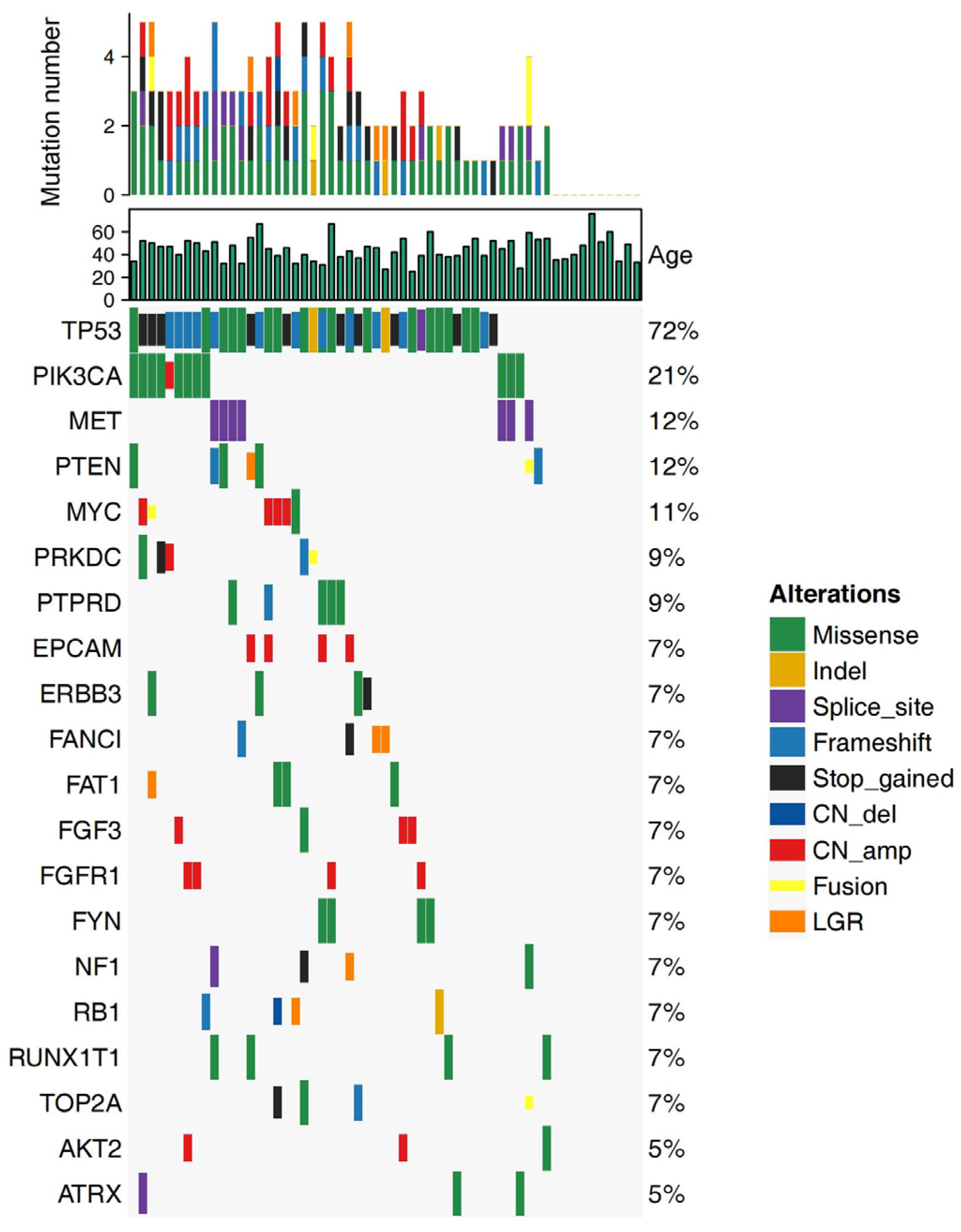
The alteration profiles of 57 breast tumor samples were analyzed by next-generation sequencing (NGS) using a panel of 520 cancer-related genes and are shown in Figure 2. TP53 presented the highest alteration frequency, accounting for approximately 72% (41/57 patients), followed by PIK3CA (12/57 patients, 21%), MET (7/57 patients, 12%), and PTEN (7/57 patients, 12%). Tumors with PTEN and NF1 alteration often occurred in patients with pathologic stage II & III after NAC. Seven tumors with PTEN alteration and four with NF1 alteration were presented in patients with pathologic stage II & III, while none of the patients with pathologic stage I had these two gene alterations.

3.3. Treatment Outcomes

We analyzed the clinical and pathology treatment outcomes of 57 non-pCR TNBC patients who received NAC (Table 1, Supplementary Figure S1). A value of 68.4% of patients had significant tumor shrinkage with Miller–Payne grades 3–4, whereas 31.6% were found to possess minor tumor shrinkage with Miller–Payne grades 1–2, which was evaluated by histopathology. Premenopausal patients had a higher proportion of Miller–Payne grades 3–4 in the PP group (p = 0.02). A value of 84% of premenopausal patients (21/25) and 37.5% of menopausal patients (3/8) had Miller–Payne grades 3–4, respectively. In contrast, the proportion of Miller–Payne grades 3–4 was higher in menopausal patients in the AP group (p = 0.022). A value of 47.1% of premenopausal patients (8/17) had Miller–Payne grades 3–4, compared with 100% (7/7) of the menopausal patients (Supplementary Figure S1A–C).
Regarding the relationship between molecular features and treatment outcomes, we found that patients harboring the BRCA1/2 mutation were insensitive to chemotherapy, especially anthracycline agents (Supplementary Figure S1D–F). Tumors with wild-type TP53 had more sensitivity to chemotherapy than those with the TP53 mutation (p = 0.011), regardless of treatment regimens (Supplementary Figure S1G–I). Miller–Payne grades 3–4 were obtained in 93.8% (15/16) of tumor samples with wild-type TP53, compared to 58.5% (24/41) of tumor samples with mutant TP53.

3.4. Clinical Survival Outcomes

The median follow-up time was 37.1 months (95% CI 33.1–41.1) for the cohort. Nineteen (33.3%) patients developed recurrence or metastasis as of March 2022.

3.4.1. Clinicopathology Features and DFS

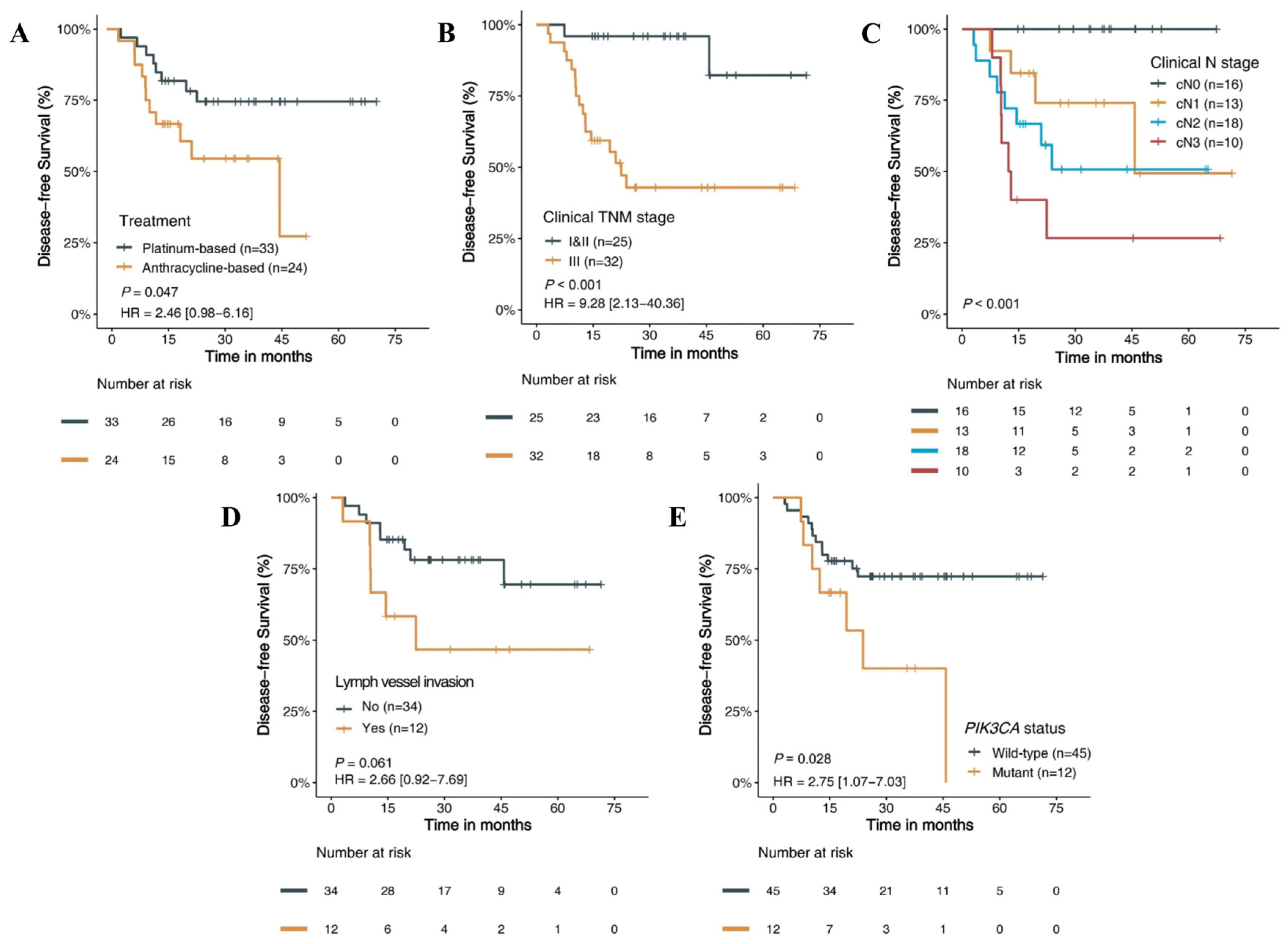
Of the 57 non-pCR TNBC patients, NAC with PP resulted in a significantly improved DFS than AP (p = 0.047) (Figure 3A). The three-year DFS rate was 54.5% compared with 74.5%, and the median DFS was 45.7 and not reached for the AP group and the PP group, respectively. In addition, patients with clinical stage III disease had a significantly shorter DFS than patients with clinical stage I and II disease (HR 9.28, 95% CI 2.13–40.36, p < 0.001) (Figure 3B). The clinical N (cN) stage had the most significant impact on prognosis, among which patients with the cN0 stage had the longest DFS, while patients with the cN3 stage had the shortest DFS (p < 0.001) (Figure 3C). Furthermore, tumors with lymph vessel invasion resulted in worse DFS in patients than non-lymph vessel invasion (HR 2.66, 95% CI 0.92–7.69, p = 0.061) (Figure 3D). However, whether adjuvant chemotherapy or radiotherapy was performed did not affect DFS. (Supplementary Figure S2M,N).
Subgroup analysis of DFS was performed for patients based on the treatment subgroups. The results showed that patients with clinical stage I and II disease had a significantly longer DFS than those with clinical stage III disease both in the PP group (p = 0.004) and the AP group (p = 0.01). (Supplementary Figure S2A–C).

3.4.2. Molecular Features and DFS

The association of molecular features and DFS in TNBC was analyzed (Table 2; Supplementary Figure S2). There were no apparent correlations between TP53 and DFS (Supplementary Figure S2D–F). Patients harboring the PIK3CA mutation showed worse DFS than patients with wild-type PIK3CA (HR 2.75, 95% CI 1.07–7.03, p = 0.028) (Figure 3E; Supplementary Figure S2G–I). The three-year DFS rate was 40.0% compared with 72.3%, and the median DFS was 23.8 and not reached for the mutation and wild-type groups, respectively. However, this difference was more pronounced in patients receiving platinum-containing NAC (HR 4.44, 95% CI 1.05–18.76, p = 0.026) but not in patients receiving anthracycline-containing NAC (HR 1.33, 95% CI 0.38–4.62, p = 0.649). Patients with mutant MYC tended to respond better to PP chemotherapy than those with wild-type MYC (Supplementary Figure S2J–L). Disappointingly, no significant correlations between molecular features and metastatic sites were found.

3.4.3. Prognostic Risk Factors and Stratified Analysis

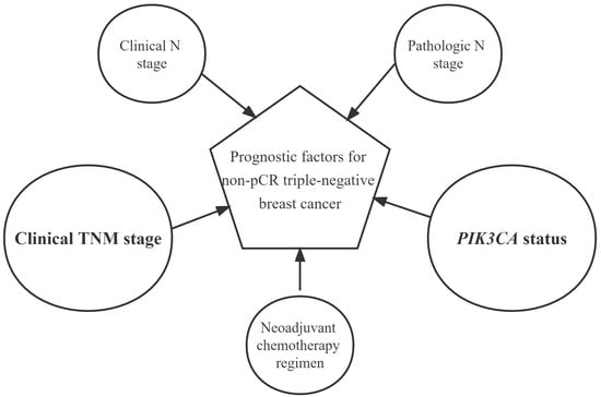
Univariate analysis of clinical characteristics and molecular features in non-pCR TNBC patients was performed. It has been shown that the clinical TNM (cTNM) stage, cN stage, pathologic N (pN) stage, PIK3CA mutation status, and NAC regimens were associated with DFS (p < 0.05). Multivariable Cox analysis confirmed that cTNM stage and PIK3CA mutation status were highly significant independent prognostic factors of DFS in non-pCR TNBC patients (p < 0.05) (Table 2).
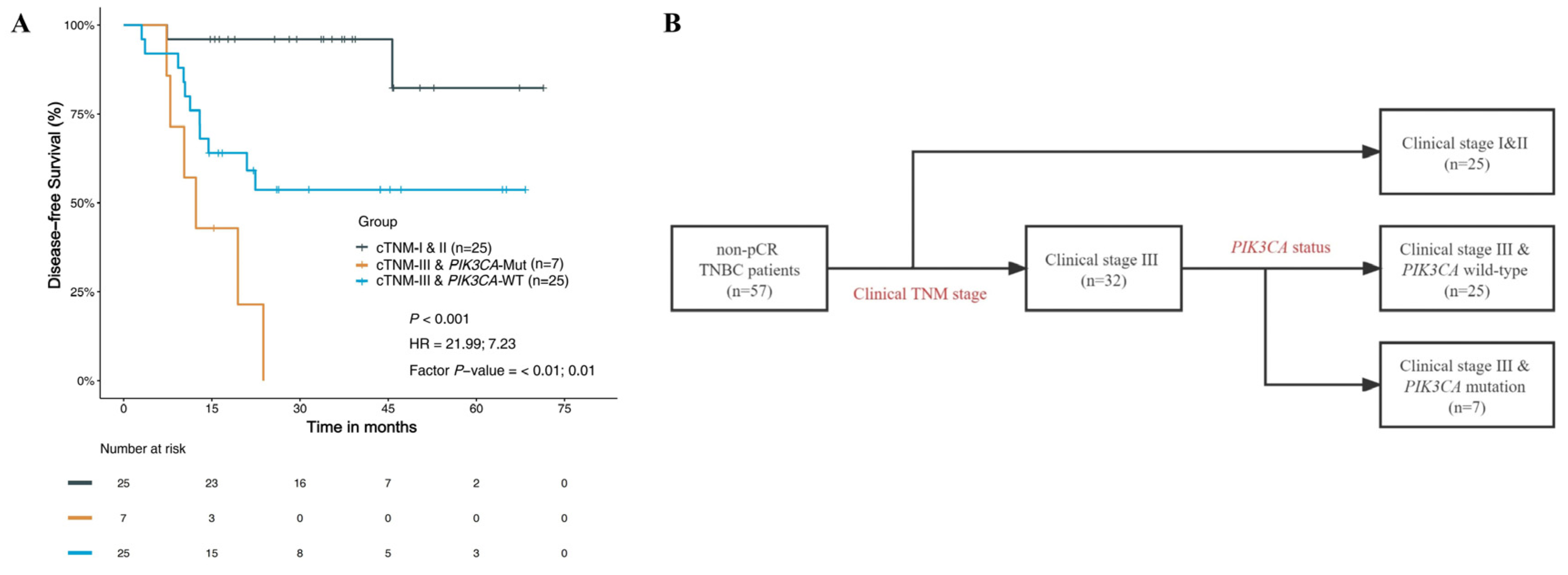
We carried out a prognostic risk assessment in non-pCR TNBC patients by combining these two independent prognostic factors (Figure 4). The results showed that patients with clinical stage I and II had the best DFS (a three-year DFS rate of 96%), followed by those with clinical stage III & wild-type PIK3CA (a three-year DFS rate of 53.7%), while those with clinical stage III & PIK3CA mutation possessed the worst DFS (a three-year DFS rate of 0) (p < 0.001). Therefore, the flow diagram shown in Figure 4 could be a good stratification of DFS for non-pCR TNBC patients.

4. Discussion

In patients with non-pCR TNBC after NAC, the DFS in the PP group was longer than that in the AP group. The three-year DFS rate was 54.5% in the AP group compared with 74.5% in the PP group. Several studies have shown that TNBC patients receiving platinum-containing NAC achieved a higher pCR rate than those without platinum regimens. In the GeparSixto trial, 315 patients of stage II & III TNBC were assigned to receive paclitaxel + liposomal doxorubicin + bevacizumab with or without carboplatin as neoadjuvant treatment. The pCR rate was increased from 36.9% to 53.2% after adding carboplatin to NAC (p = 0.005) [13]. While event-free survival (EFS) and OS have been controversial in prior research, studies have indicated similar, even worse results in platinum-containing regimens than platinum-free regimens of NAC [8,28]. However, the differences in the above survival results cannot rule out the strong inherent heterogeneity in TNBC. Therefore, we tried to explore prognostic differences at the gene signatures level with a view to finding effective prognostic factors.
Our data demonstrated divergent sensitivity to different chemotherapeutic agents when patients were grouped according to their menopausal status. Premenopausal patients were sensitive to PP treatment, while menopausal patients responded well to AP treatment. Our prior study showed that premenopausal patients were more likely to have homologous recombination deficiency (HRD). As we know, patients with HRD are more responsive to platinum-containing therapy [29]. Therefore, the relationship between menopausal status and chemotherapy sensitivity might primarily be attributable to HRD. Moreover, we observed that all patients with PTEN or NF1 alteration remained in pathologic stages II and III despite receiving NAC. As previously reported, patients who possess NF1 mutations or lose the expression of PTEN have worse survival in breast cancer [30,31].
In our study, the PIK3CA mutation frequency in TNBC patients accounted for 15%, which is consistent with previous reports [32]. Our results showed that PIK3CA mutation might be a poor prognostic indicator for DFS. Patients harboring the PIK3CA mutation had a worse three-year DFS rate than those with wild-type PIK3CA, for 40.0% and 72.3% of patients, respectively (p = 0.028). Similarly, previous studies have confirmed that the PIK3CA mutation confers resistance to chemotherapy in TNBC by inhibiting apoptosis and activating the PI3K/AKT/mTOR signaling pathway [33], and TNBC patients with the PIK3CA mutation had lower pCR rates than those with wide-type PIK3CA after NAC [34]. Interestingly, we found that the difference in DFS was more pronounced in patients receiving PP NAC (p = 0.026), while no significant difference existed among those undergoing AP treatment. Prior research concluded no predictive effect of PIK3CA mutation for doxorubicin treatment and whether harboring the PIK3CA mutation cannot affect the DFS or OS [35]. However, no studies have reported the impact of platinum agents on DFS in PIK3CA-mutated TNBC patients. Therefore, further research is needed on the mechanism of action and prognostic effect of different chemotherapeutic agents on PIK3CA-mutated TNBC.
It was well known that the TNM stage is closely related to prognosis. In this study, cTNM stage and PIK3CA mutation were confirmed to be highly significant independent prognostic factors of DFS in non-pCR TNBC patients. We performed a prognostic risk assessment using cTNM stage and PIK3CA mutation status in non-pCR TNBC patients according to the univariate and multivariable Cox analysis of clinical characteristics and molecular features. A better prognostic stratification for patients was observed. We found that patients with clinical stages I & II possessed the best DFS, while those with clinical stage III & the PIK3CA mutation had the worst DFS. Therefore, we can further evaluate the prognosis for non-pCR TNBC patients by combining cTNM stage and PIK3CA mutation status.
To our knowledge, this is the first study to explore prognostic factors for patients with non-pCR TNBC by combining clinicopathology and molecular features. However, our study does have several limitations. First, this was a small sample cohort, and targeted sequencing could not be performed in some samples due to insufficient tumor samples. Second, joint analysis of multiple genes could not be performed due to the low incidence of gene mutations. Third, analyses of therapeutic agents such as PI3K inhibitors and PARP inhibitors are lacking.

5. Conclusions

In TNBC patients with residual disease after NAC, a promising prognostic stratification was observed by combining the cTNM stage and PIK3CA mutation status. These findings need to be further verified.

Supplementary Materials

The following supporting information can be downloaded at: https://www.mdpi.com/article/10.3390/jpm13020190/s1, Figure S1: Treatment outcomes based on clinical/molecular features. Miller-Payne grades based on menopausal status A. in overall patients; B. in patients with AP-NAC; C. in patients with PP-NAC. Miller-Payne grades based on BRCA status D. in overall patients; E. in patients with AP-NAC; F. in patients with PP-NAC. Miller-Payne grades based on TP53 status G. in overall patients; H. in patients with AP-NAC; I. in patients with PP-NAC. AP anthracycline plus paclitaxel; PP platinum plus paclitaxel; NAC neoadjuvant chemotherapy; Figure S2. Survival analysis according to different clinicopathologic and molecular features. DFS based on clinical TNM stage A. in overall patients; B. in patients with AP-NAC; C. in patients with PP-NAC. DFS based on TP53 status D. in overall patients; E. in patients with AP-NAC; F. in patients with PP-NAC. DFS based on PIK3CA status G. in overall patients; H. in patients with AP-NAC; I. in patients with PP-NAC. DFS based on MYC status J. in overall patients; K. in patients with AP-NAC; L. in patients with PP-NAC. DFS based on M. adjuvant chemotherapy; N. adjuvant radiotherapy. DFS disease-free survival; AP anthracycline plus paclitaxel; PP platinum plus paclitaxel; NAC neoadjuvant chemotherapy.

Author Contributions

Conception and design, Z.L., J.W. and B.X.; administrative support, B.X.; provision of study materials or patients, Z.L. and J.W.; collection and assembly of data, Z.L. and Y.H.; data analysis and interpretation, Z.L., Y.H. and J.W.; manuscript—writing, all authors. All authors have read and agreed to the published version of the manuscript.

Funding

This research was funded by Chinese Academy of Medical Sciences (CAMS) Innovation Fund for Medical Sciences, grant number 2021-I2M-1-014.

Institutional Review Board Statement

The study was reviewed and approved by the Institutional Review Board of the National Cancer Center/Cancer Hospital, Chinese Academy of Medical Sciences and Peking Union Medical College (21/247-2918). All procedures were carried out in accordance with the Declaration of Helsinki.

Informed Consent Statement

The informed consent of the participants was exempted by the review board due to the retrospective design of this study.

Data Availability Statement

The data used during the current study are available from the corresponding author on reasonable request.

Acknowledgments

We would like to acknowledge the staff of Burning Rock Biotech for their active support.

Conflicts of Interest

The authors declare no conflict of interest.

References

  1. Sung, H.; Ferlay, J.; Siegel, R.L.; Laversanne, M.; Soerjomataram, I.; Jemal, A.; Bray, F. Global Cancer Statistics 2020: GLOBOCAN Estimates of Incidence and Mortality Worldwide for 36 Cancers in 185 Countries. CA Cancer J. Clin. 2021, 71, 209–249. [Google Scholar] [CrossRef]
  2. Vishnubalaji, R.; Alajez, N.M. Transcriptional landscape associated with TNBC resistance to neoadjuvant chemotherapy revealed by single-cell RNA-seq. Mol. Ther. Oncolytics 2021, 23, 151–162. [Google Scholar] [CrossRef]
  3. Singh, D.; Yadav, D. TNBC: Potential Targeting of Multiple Receptors for a Therapeutic Breakthrough, Nanomedicine, and Immunotherapy. Biomedicines 2021, 9, 876. [Google Scholar] [CrossRef]
  4. Lee, K.L.; Kuo, Y.C.; Ho, Y.S.; Huang, Y.H. Triple-Negative Breast Cancer: Current Understanding and Future Therapeutic Breakthrough Targeting Cancer Stemness. Cancers 2019, 11, 1334. [Google Scholar] [CrossRef] [PubMed] [Green Version]
  5. Dent, R.; Trudeau, M.; Pritchard, K.I.; Hanna, W.M.; Kahn, H.K.; Sawka, C.A.; Lickley, L.A.; Rawlinson, E.; Sun, P.; Narod, S.A. Triple-Negative Breast Cancer: Clinical Features and Patterns of Recurrence. Clin. Cancer Res. 2007, 13 Pt 1, 4429–4434. [Google Scholar] [CrossRef] [Green Version]
  6. Korde, L.A.; Somerfield, M.R.; Carey, L.A.; Crews, J.R.; Denduluri, N.; Hwang, E.S.; Khan, S.A.; Loibl, S.; Morris, E.A.; Perez, A.; et al. Neoadjuvant Chemotherapy, Endocrine Therapy, and Targeted Therapy for Breast Cancer: ASCO Guideline. J. Clin. Oncol. 2021, 39, 1485–1505. [Google Scholar] [CrossRef]
  7. Liedtke, C.; Mazouni, C.; Hess, K.R.; André, F.; Tordai, A.; Mejia, J.A.; Symmans, W.F.; Gonzalez-Angulo, A.M.; Hennessy, B.; Green, M.; et al. Response to neoadjuvant therapy and long-term survival in patients with triple-negative breast cancer. J. Clin. Oncol. 2008, 26, 1275–1281. [Google Scholar] [CrossRef]
  8. Poggio, F.; Bruzzone, M.; Ceppi, M.; Pondé, N.F.; La Valle, G.; Del Mastro, L.; de Azambuja, E.; Lambertini, M. Platinum-based neoadjuvant chemotherapy in triple-negative breast cancer: A systematic review and meta-analysis. Ann. Oncol. 2018, 29, 1497–1508. [Google Scholar] [CrossRef]
  9. Gamucci, T.; Pizzuti, L.; Sperduti, I.; Mentuccia, L.; Vaccaro, A.; Moscetti, L.; Marchetti, P.; Carbognin, L.; Michelotti, A.; Iezzi, L.; et al. Neoadjuvant chemotherapy in triple-negative breast cancer: A multicentric retrospective observational study in real-life setting. J. Cell. Physiol. 2017, 233, 2313–2323. [Google Scholar] [CrossRef] [PubMed]
  10. Tutt, A.; Tovey, H.; Cheang, M.; Kernaghan, S.; Kilburn, L.; Gazinska, P.; Owen, J.; Abraham, J.; Barrett, S.; Barrett-Lee, P.; et al. Carboplatin in BRCA1/2-mutated and triple-negative breast cancer BRCAness subgroups: The TNT Trial. Nat. Med. 2018, 24, 628–637. [Google Scholar] [CrossRef] [PubMed]
  11. Sikov, W.M.; Berry, D.A.; Perou, C.M.; Singh, B.; Cirrincione, C.T.; Tolaney, S.M.; Kuzma, C.S.; Pluard, T.J.; Somlo, G.; Port, E.R.; et al. Impact of the addition of carboplatin and/or bevacizumab to neoadjuvant once-per-week paclitaxel followed by dose-dense doxorubicin and cyclophosphamide on pathologic complete response rates in stage II to III triple-negative breast cancer: CALGB 40603 (Alliance). J. Clin. Oncol. 2015, 33, 13–21. [Google Scholar] [PubMed] [Green Version]
  12. Ando, M.; Yamauchi, H.; Aogi, K.; Shimizu, S.; Iwata, H.; Masuda, N.; Yamamoto, N.; Inoue, K.; Ohono, S.; Kuroi, K.; et al. Randomized phase II study of weekly paclitaxel with and without carboplatin followed by cyclophosphamide/epirubicin/5-fluorouracil as neoadjuvant chemotherapy for stage II/IIIA breast cancer without HER2 overexpression. Breast Cancer Res. Treat. 2014, 145, 401–409. [Google Scholar] [CrossRef]
  13. von Minckwitz, G.; Schneeweiss, A.; Loibl, S.; Salat, C.; Denkert, C.; Rezai, M.; Blohmer, J.U.; Jackisch, C.; Paepke, S.; Gerber, B.; et al. Neoadjuvant carboplatin in patients with triple-negative and HER2-positive early breast cancer (GeparSixto; GBG 66): A randomised phase 2 trial. Lancet Oncol. 2014, 15, 747–756. [Google Scholar] [CrossRef] [PubMed]
  14. Alba, E.; Chacon, J.I.; Lluch, A.; Anton, A.; Estevez, L.; Cirauqui, B.; Carrasco, E.; Calvo, L.; Segui, M.A.; Ribelles, N.; et al. A randomized phase II trial of platinum salts in basal-like breast cancer patients in the neoadjuvant setting. Results from the GEICAM/2006-03, multicenter study. Breast Cancer Res. Treat. 2012, 136, 487–493. [Google Scholar] [CrossRef] [PubMed]
  15. Iwase, M.; Ando, M.; Aogi, K.; Aruga, T.; Inoue, K.; Shimomura, A.; Tokunaga, E.; Masuda, N.; Yamauchi, H.; Yamashita, T.; et al. Long-term survival analysis of addition of carboplatin to neoadjuvant chemotherapy in HER2-negative breast cancer. Breast Cancer Res. Treat. 2020, 180, 687–694. [Google Scholar] [CrossRef] [PubMed]
  16. Ha, R.; Chin, C.; Karcich, J.; Liu, M.Z.; Chang, P.; Mutasa, S.; Pascual Van Sant, E.; Wynn, R.T.; Connolly, E.; Jambawalikar, S. Prior to Initiation of Chemotherapy, Can We Predict Breast Tumor Response? Deep Learning Convolutional Neural Networks Approach Using a Breast MRI Tumor Dataset. J. Digit. Imaging 2019, 32, 693–701. [Google Scholar] [CrossRef]
  17. Liu, M.Z.; Mutasa, S.; Chang, P.; Siddique, M.; Jambawalikar, S.; Ha, R. A novel CNN algorithm for pathological complete response prediction using an I-SPY TRIAL breast MRI database. Magn. Reson. Imaging 2020, 73, 148–151. [Google Scholar] [CrossRef]
  18. Kim, J.-Y.; Park, K.; Jung, H.H.; Lee, E.; Cho, E.Y.; Lee, K.H.; Bae, S.Y.; Lee, S.K.; Kim, S.W.; Lee, J.E.; et al. Association between Mutation and Expression of TP53 as a Potential Prognostic Marker of Triple-Negative Breast Cancer. Cancer Res. Treat. 2016, 48, 1338–1350. [Google Scholar] [CrossRef] [Green Version]
  19. Cancer Genome Atlas Network. Comprehensive molecular portraits of human breast tumours. Nature 2012, 490, 61–70. [Google Scholar] [CrossRef] [Green Version]
  20. Sporikova, Z.; Koudelakova, V.; Trojanec, R.; Hajduch, M. Genetic Markers in Triple-Negative Breast Cancer. Clin. Breast Cancer 2018, 18, e841–e850. [Google Scholar] [CrossRef]
  21. Copson, E.R.; Maishman, T.C.; Tapper, W.J.; Cutress, R.I.; Greville-Heygate, S.; Altman, D.G.; Eccles, B.; Gerty, S.; Durcan, L.T.; Jones, L.; et al. Germline BRCA mutation and outcome in young-onset breast cancer (POSH): A prospective cohort study. Lancet Oncol. 2018, 19, 169–180. [Google Scholar] [CrossRef] [PubMed] [Green Version]
  22. Li, H.; Durbin, R. Fast and accurate short read alignment with Burrows—Wheeler transform. Bioinformatics 2009, 25, 1754–1760. [Google Scholar] [CrossRef] [PubMed] [Green Version]
  23. McKenna, A.; Hanna, M.; Banks, E.; Sivachenko, A.; Cibulskis, K.; Kernytsky, A.; Garimella, K.; Altshuler, D.; Gabriel, S.; Daly, M.; et al. The Genome Analysis Toolkit: A MapReduce framework for analyzing next-generation DNA sequencing data. Genome Res. 2010, 20, 1297–1303. [Google Scholar] [CrossRef] [PubMed] [Green Version]
  24. Koboldt, D.C.; Zhang, Q.; Larson, D.E.; Shen, D.; McLellan, M.D.; Lin, L.; Miller, C.A.; Mardis, E.R.; Ding, L.; Wilson, R.K. VarScan 2: Somatic mutation and copy number alteration discovery in cancer by exome sequencing. Genome Res. 2012, 22, 568–576. [Google Scholar] [CrossRef] [PubMed] [Green Version]
  25. Wang, K.; Li, M.; Hakonarson, H. ANNOVAR: Functional annotation of genetic variants from high-throughput sequencing data. Nucleic Acids Res. 2010, 38, e164. [Google Scholar] [CrossRef]
  26. Cingolani, P.; Platts, A.; Wang, L.L.; Coon, M.; Nguyen, T.; Wang, L.; Land, S.J.; Lu, X.; Ruden, D.M. A program for annotating and predicting the effects of single nucleotide polymorphisms, SnpEff: SNPs in the genome of Drosophila melanogaster strain w1118; iso-2; iso-3. Fly 2012, 6, 80–92. [Google Scholar] [CrossRef] [Green Version]
  27. Newman, A.; Bratman, S.; Stehr, H.; Lee, L.J.; Liu, C.L.; Diehn, M.; Alizadeh, A.A. FACTERA: A practical method for the discovery of genomic rearrangements at breakpoint resolution. Bioinformatics 2014, 30, 3390–3393. [Google Scholar] [CrossRef] [Green Version]
  28. Shepherd, J.H.; Ballman, K.; Polley, M.-Y.C.; Campbell, J.D.; Fan, C.; Selitsky, S.; Fernandez-Martinez, A.; Parker, J.S.; Hoadley, K.A.; Hu, Z.; et al. CALGB 40603 (Alliance): Long-Term Outcomes and Genomic Correlates of Response and Survival After Neoadjuvant Chemotherapy with or Without Carboplatin and Bevacizumab in Triple-Negative Breast Cancer. J. Clin. Oncol. 2022, 40, 1323–1334. [Google Scholar] [CrossRef]
  29. Telli, M.L.; Timms, K.M.; Reid, J.; Hennessy, B.; Mills, G.B.; Jensen, K.C.; Szallasi, Z.; Barry, W.T.; Winer, E.P.; Tung, N.M.; et al. Homologous Recombination Deficiency (HRD) Score Predicts Response to Platinum-Containing Neoadjuvant Chemotherapy in Patients with Triple-Negative Breast Cancer. Clin. Cancer Res. 2016, 22, 3764–3773. [Google Scholar] [CrossRef] [Green Version]
  30. Wu, S.-Y.; Yan, M.-D.; Wu, A.T.; Yuan, K.S.-P.; Liu, S.H. Brown Seaweed Fucoidan Inhibits Cancer Progression by Dual Regulation of mir-29c/ADAM12 and miR-17-5p/PTEN Axes in Human Breast Cancer Cells. J. Cancer 2016, 7, 2408–2419. [Google Scholar] [CrossRef]
  31. Uusitalo, E.; Kallionpää, R.A.; Kurki, S.; Rantanen, M.; Pitkäniemi, J.; Kronqvist, P.; Härkönen, P.; Huovinen, R.; Carpen, O.; Pöyhönen, M.; et al. Breast cancer in neurofibromatosis type 1: Overrepresentation of unfavourable prognostic factors. Br. J. Cancer 2017, 116, 211–217. [Google Scholar] [CrossRef] [PubMed] [Green Version]
  32. Martínez-Sáez, O.; Chic, N.; Pascual, T.; Adamo, B.; Vidal, M.; González-Farré, B.; Sanfeliu, E.; Schettini, F.; Conte, B.; Brasó-Maristany, F.; et al. Frequency and spectrum of PIK3CA somatic mutations in breast cancer. Breast Cancer Res. 2020, 22, 45. [Google Scholar] [CrossRef] [PubMed]
  33. Hu, H.; Zhu, J.; Zhong, Y.; Geng, R.; Ji, Y.; Guan, Q.; Hong, C.; Wei, Y.; Min, N.; Qi, A.; et al. PIK3CA mutation confers resistance to chemotherapy in triple-negative breast cancer by inhibiting apoptosis and activating the PI3K/AKT/mTOR signaling pathway. Ann. Transl. Med. 2021, 9, 410. [Google Scholar] [CrossRef] [PubMed]
  34. Guo, S.; Loibl, S.; von Minckwitz, G.; Darb-Esfahani, S.; Lederer, B.; Denkert, C. PIK3CA H1047R Mutation Associated with a Lower Pathological Complete Response Rate in Triple-Negative Breast Cancer Patients Treated with Anthracycline-Taxane-Based Neoadjuvant Chemotherapy. Cancer Res. Treat. 2020, 52, 689–696. [Google Scholar] [CrossRef] [PubMed] [Green Version]
  35. Chen, L.; Yang, L.; Yao, L.; Kuang, X.Y.; Zuo, W.J.; Li, S.; Qiao, F.; Liu, Y.R.; Cao, Z.G.; Zhou, S.L.; et al. Characterization of PIK3CA and PIK3R1 somatic mutations in Chinese breast cancer patients. Nat. Commun. 2018, 9, 1357. [Google Scholar] [CrossRef] [PubMed]
Figure 1. Flowchart of the screening procedure.
Jpm 13 00190 g001
Figure 2. Alteration profiles of the top 20 genes from 57 patients with non-pCR TNBC. pCR, pathologic complete response; TNBC, triple-negative breast cancer.
Jpm 13 00190 g002
Figure 3. Kaplan–Meier survival curves for DFS of patients with non-pCR TNBC. DFS based on (A) NAC regimens; (B) clinical TNM stage; (C) clinical N stage; (D) lymph vessel invasion; (E) PIK3CA status. DFS, disease-free survival; pCR, pathologic complete response; TNBC, triple-negative breast cancer; NAC, neoadjuvant chemotherapy; cTNM, clinical TNM stage; cN, clinical N stage; HR, hazard ratio.
Jpm 13 00190 g003
Figure 4. Prognostic risk assessment based on clinical and molecular features of patients with non-pCR TNBC. (A) DFS based on clinical TNM stage and PIK3CA status; (B) flowchart of prognostic risk assessment. DFS, disease-free survival; pCR, pathologic complete response; TNBC, triple-negative breast cancer; cTNM, clinical TNM stage; Mut, mutation; WT, wild-type; HR, hazard ratio.
Jpm 13 00190 g004
Table 1. Baseline characteristics and treatment outcomes of the overall cohort and treatment subgroups.
VariableOverall
(n = 57)
Platinum–Paclitaxel Regimen
(n = 33)
Anthracycline–Paclitaxel Regimen
(n = 24)
p-Value
Age 0.312
Median [range]45.0 [38.0, 52.0]43.0 [36.0, 51.0]46.5 [38.0, 54.0]
Menopausal status 0.765
Premenopause42 (73.7%)25 (75.8%)17 (70.8%)
Postmenopause15 (26.3%) 8 (24.2%) 7 (29.2%)
Family history of cancer 0.584
No38 (66.7%)23 (69.7%)15 (62.5%)
Yes19 (33.3%)10 (30.3%) 9 (37.5%)
Ki-67(%) 0.416
Median [range]55.0 [21.3, 70.0]50.0 [10.0, 70.0]60.0 [30.0, 75.0]
Missing 1 (1.8%) 0 (0.0%) 1 (4.2%)
Clinical T stage 0.417
cT1 6 (10.5%) 2 (6.1%) 4 (16.7%)
cT236 (63.2%)23 (69.7%)13 (54.2%)
cT312 (21.1%) 7 (21.2%) 5 (20.8%)
cT4 3 (5.3%) 1 (3.0%) 2 (8.3%)
Clinical N stage 0.836
cN016 (28.1%) 9 (27.3%) 7 (29.2%)
cN113 (22.8%) 9 (27.3%) 4 (16.7%)
cN218 (31.6%)10 (30.3%) 8 (33.3%)
cN310 (17.5%) 5 (15.2%) 5 (20.8%)
Clinical TNM stage 0.794
I and II25 (43.9%)15 (45.5%)10 (41.7%)
III32 (56.4%)18 (54.5%)14 (58.3%)
Pathologic T stage 1.000
ypT139 (68.4%)23 (69.7%)16 (66.7%)
ypT218 (31.6%)10 (30.3%) 8 (33.3%)
Pathologic N stage 0.497
ypN026 (45.6%)14 (42.4%)12 (50.0%)
ypN118 (31.6%)13 (39.4%) 5 (20.8%)
ypN2 6 (10.5%) 3 (9.1%) 3 (12.5%)
ypN3 7 (12.3%) 3 (9.1%) 4 (16.7%)
Pathologic TNM stage 0.376
I21 (36.8%)11 (33.3%)10 (41.7%)
II23 (40.4%)16 (48.5%) 7 (29.2%)
III13 (22.8%) 6 (18.2%) 7 (29.2%)
Miller–Payne 0.565
G1 and G218 (31.6%) 9 (27.3%) 9 (37.5%)
G3 and G439 (68.4%)24 (72.7%)15 (62.5%)
Histological grade 0.732
Grade II11 (19.30%) 7 (21.21%) 4 (16.67%)
Grade III35 (61.40%)19 (57.58%)16 (66.67%)
Missing11 (19.30%) 7 (21.21%) 4 (16.67%)
Lymph vessel invasion 0.738
No34 (59.7%)20 (60.6%)14 (58.3%)
Yes12 (21.1%) 6 (18.2%) 6 (25.0%)
Missing11 (19.3%) 7 (21.2%) 4 (16.7%)
Adjuvant chemotherapy 0.787
No24 (42.1%)13 (39.4%)11 (45.8%)
Yes33 (57.9%)20 (60.6%)13 (54.2%)
Adjuvant radiotherapy 1.000
No 9 (15.8%) 5 (15.2%) 4 (16.7%)
Yes48 (84.2%)28 (84.9%)20 (83.3%)
Initial metastatic sites 0.168
Bone/soft tissue only12 (21.1%) 6 (18.2%) 6 (25.0%)
Visceral 7 (12.3%) 2 (6.1%) 5 (20.8%)
None38 (66.7%)25 (75.8%)13 (54.2%)
Number of metastatic sites 1.000
112 (21.1%) 5 (15.2%) 7 (29.2%)
2 7 (12.3%) 3 (9.1%) 4 (16.7%)
None38 (66.7%)25 (75.8%)13 (54.2%)
BRCA1/2 status 0.073
Mutant 6 (10.5%) 1 (3.0%) 5 (20.8%)
Wild-type51 (89.5%)32 (97.0%)19 (79.2%)
cT, clinical T; cN, clinical N; pT, pathologic T; pN, pathologic N.
Table 2. Univariate and multivariate survival analysis of 57 non-pathologic complete response triple-negative breast cancer patients.
VariableUnivariateMultivariate
HR (95% CI)p-Value HR (95% CI)p-Value
Treatment 0.047
Platinum–Paclitaxel (n = 33)Ref.
Anthracycline–Paclitaxel (n = 24)2.46 (0.98–6.16)
Clinical N stage <0.001
cN0 and cN1 (n = 29)Ref.
cN2 (n = 18)4.41 (1.32–14.72)
cN3 (n = 10)8.07 (2.33–27.98)
Clinical TNM stage <0.001 <0.001
I and II (n = 25)Ref. Ref.
III (n = 32)9.28 (2.13–40.36) 11.02 (3.02–40.2)
Pathologic N stage <0.001
pN0& and N1 (n = 44)Ref.
pN2 (n = 6)0.68 (0.09–5.25)
pN3 (n = 7)8.58 (2.78–26.51)
PIK3CA status 0.028 0.03
Wild-type (n = 45)Ref. Ref.
Mutant (n = 12)2.75 (1.07–7.03) 2.70 (1.10–6.6)
cN clinical N; pN pathologic N.
Disclaimer/Publisher’s Note: The statements, opinions and data contained in all publications are solely those of the individual author(s) and contributor(s) and not of MDPI and/or the editor(s). MDPI and/or the editor(s) disclaim responsibility for any injury to people or property resulting from any ideas, methods, instructions or products referred to in the content.