Review

Application of Reactive Oxygen Species in Dental Treatment

1
Department of Laboratory Medicine and Dentistry for the Compromised Patient, Nihon University School of Dentistry at Matsudo, Chiba 271-8587, Japan
2
Department of Oral Function and Fixed Prothodontics, Nihon University School of Dentistry at Matsudo, Chiba 271-8587, Japan
3
Department of Endodontics, Nihon University School of Dentistry at Matsudo, Chiba 271-8587, Japan
*
Author to whom correspondence should be addressed.
J. Pers. Med. 2022, 12(9), 1531; https://doi.org/10.3390/jpm12091531
Submission received: 15 August 2022 / Revised: 7 September 2022 / Accepted: 13 September 2022 / Published: 18 September 2022
(This article belongs to the Special Issue Precision Medicine for Oral Diseases)

Abstract

:
Reactive oxygen species (ROS) and free radicals, which have been implicated in inflammation, pain, carcinogenesis, and aging, are actually used in dental treatments such as tooth bleaching and composite resin polymerization. Recently, numerous studies have investigated the application of ROS in the medical and dental fields. In previous studies, ROS were generated intentionally through pathways such as photolysis, photocatalytic methods, and photodynamic therapy, which are used in the medical field to target cancer. In the field of dentistry, generated ROS are applied mainly for periodontal treatment and sterilization of the root canal, and its effectiveness as an antibacterial photodynamic therapy has been widely reported.. Given this background, the present article aimed to review the basic effects of ROS in dental medicine, especially endodontic therapy, and to discuss future applications of ROS.

1. Introduction

The terms “reactive oxygen species (ROS)” and “free radicals” are frequently used not only in medicine and dentistry, but also in other fields. However, for general clinical dentists, these terms are still unfamiliar and, thus, assumed to be poorly understood. Research on ROS and free radicals has made great strides, and their involvement in systemic diseases such as inflammation [1,2], pain [3,4], cancer [5,6,7], aging [7,8], adult diseases [9,10], and numerous other diseases of various organs [11,12] has been demonstrated. These include, of course, oral diseases such as periodontitis [13] and squamous cell carcinoma [14]. At the cellular level, ROS are also known to play important roles with respect to the second messengers and stem cell differentiation. That is, controlling ROS with antioxidants is expected to induce stem cells to differentiate into osteoblasts and contribute to bone regeneration [15]. Thus, ROS is an important and interesting element for living organisms in a variety of situations.
On the other hand, many novel methods for intentionally generating ROS and incorporating them into treatment in medical and dental medicine have also been studied. These methods, which generate ROS by irradiating photosensitizers (or photocatalysts) with appropriate excitation light, are known as photodynamic therapy (PDT) and antimicrobial photodynamic therapy (a-PDT), and are particularly effective against cancer [16,17,18,19] and bacteria-infected tissue [19,20,21], respectively.
The present review aims to examine ROS and free radicals by focusing on a-PDT in endodontic treatment, as well as the advantages and disadvantages of a-PDT and its potential for the future.

2. Materials and Methods

An electronic search was conducted for English language articles published between 1934 and 2021. Search terms for the general theory of endodontic therapy and ROS, and especially a-PDT and ROS in endodontic therapy, were entered into the following databases: PubMed, Ichushi-Web, and Google Scholar. The inclusion criteria were narrative reviews; in vitro, ex vivo, and clinical studies; and case reports written in English. The exclusion criteria were studies with conflicts of interest. The retrieved literature was identified for all titles, abstracts, and full texts. A total of 100 articles were included in this narrative review, including our previous papers. This paper provides a great deal of information on the application of ROS to dentistry, referring to the many findings reported to date, and provides researchers and practitioners with scientifically based knowledge.

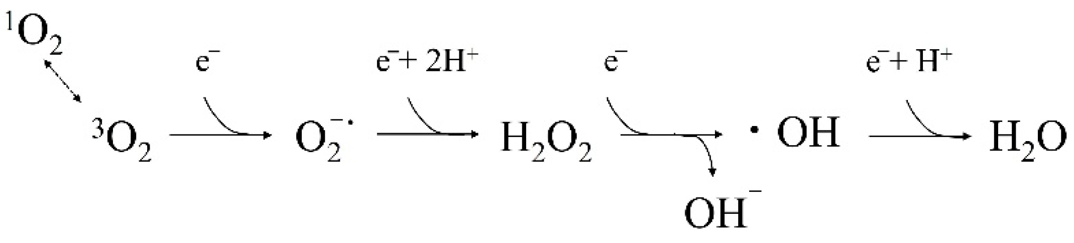
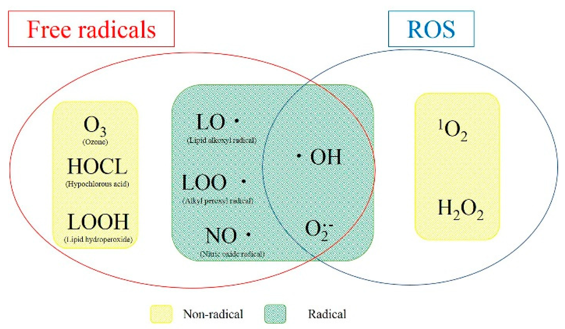
3. What Are ROS and Free Radicals?

Oxygen comprises about 20% of the atmosphere. The oxygen breathed by humans is called triplet oxygen (3O2) in its stable (ground) state; it enters the body through respiration, and then binds to red blood cells, which allows for transport throughout the body. The concentration of 3O2 transported to peripheral tissues is low, about 1/150 of that in the atmosphere, and is taken up by cells. Then, 3O2 that has passed through the cell membrane undergoes various enzymatic reactions in the mitochondria to generate energy, after which, it acts as an oxidant, and the oxygen itself is reduced to eventually become H2O. When atoms, molecules, or ions lose electrons, they are defined as oxidized, and when they gain electrons, they are defined as reduced.
ROS are the products of reactions that occur during the oxidation–reduction process of oxygen in cells (Figure 1). Unlike 3O2, superoxide anions (O2), hydrogen peroxide (H2O2), hydroxyl radicals (OH), and singlet oxygen (1O2), are highly reactive and commonly referred to as ROS. Radicals, on the other hand, are a general term for highly reactive and unstable substances with one or more “unpaired electrons.” When these radicals damage biological tissues, the tissues deprived of electrons become free radicals, and this process causes a chain reaction.
Figure 2 shows the relationship between ROS and free radicals, which are closely related to the organisms in which they occur. Therefore, radicals with unpaired electrons, ROS (radical or non-radical), and including the radical reserve, are widely defined as “free radicals”.
Among all ROS, O2−· and OH are free radicals, and OH is known to be the most reactive [22]. OH is produced by the Fenton reaction [23] between transition metal ions such as Cu2+ and Fe2+ and H2O2, and by irradiation of H2O2 with light in the ultraviolet (UV) to blue light range [24]. Therefore, the OH produced by photolysis of H2O2 is applied in dentistry for sterilization [25,26,27,28,29,30,31] and tooth bleaching [32,33]. By contrast, 1O2 and H2O2 are classified as non-radical ROS. 1O2 is the most important factor in the mechanism of action of a-PDT, which is discussed below. Previous findings have shown that the mechanisms of a-PDT in endodontics are closely related to ROS, especially OH and 1O2.

4. Root Canal Cleaning by ROS

Endodontic treatment is the clinical management of bacterial disease, and the bacteria present in the root canal are the primary target of treatment [30,31]. However, the complexity of root canal structures makes complete debridement and bacterial elimination by instrumentation, irrigation, and intracanal medication virtually impossible [34]. In addition, another factor, the smear layer created during root canal shaping, seals the dentin tubules.
Enterococcus faecalis and Candida albicans are the species most frequently associated with refractory apical periodontitis [35]. E. faecalis has been reported to be naturally resistant to common disinfectants such as Ca (OH)2, iodine-containing tinctures, and antibiotics [36,37]. C. albicans is a fungus with a stronger cytoskeleton than bacteria, which makes it resistant to drugs [38], and its ability to adapt to various environments due to its dimorphic morphology reduces the success rate of root canal treatment [35,39]. Chemical disinfectants such as H2O2, ethylenediaminetetraacetic acid (EDTA), and sodium hypochlorite (NaClO) have conventionally been used for root canal irrigation for disinfection and removal of the smear layer [40,41]. However, conventional chemical disinfectants sometimes cause other problems such as tissue damage and accidents due to leakage [42,43]. Therefore, many researchers have been keenly interested in developing alternative sterilization methods with selective sterilizing power that do not cause damage to the organism.
Recently, various photochemically applied sterilization methods, especially a-PDT, have attracted increasing attention as alternative sterilization methods for bacterial oral diseases [19,20,44,45,46,47,48]. A-PDT requires three elements: 3O2, photosensitizing agents (PS), and excitation light. Then, the bacteria are effectively sterilized in the following manner via the a-PDT mechanism (a-PDT action type 2): (i) PS attach to the cell membrane of microorganisms, (ii) irradiation with light at a specific wavelength matched to the peak absorption of PS leads to the generation of 1O2, and (iii) bacterial death via destruction of the bacteria walls is induced by 1O2 [19,20,49,50] (Figure 3).
Despite the fact that the mechanisms of a-PDT are closely related to ROS, many studies have reported that the use of a-PDT for bactericidal effects requires the consideration of numerous variables when developing an a-PDT protocol, including light parameters, PS, and light delivery techniques [19,20,51,52,53,54]. Therefore, our laboratory used the electron spin resonance (ESR) spin-trapping method to supplement ROS and investigated the relationship between ROS generated by a-PDT and the bactericidal effect on microorganisms [55,56,57]. We also reviewed the findings of other researchers in regard to root canal cleaning with OH obtained by the photolysis reaction of H2O2 [25,26,27,28,29,30,31,58,59].

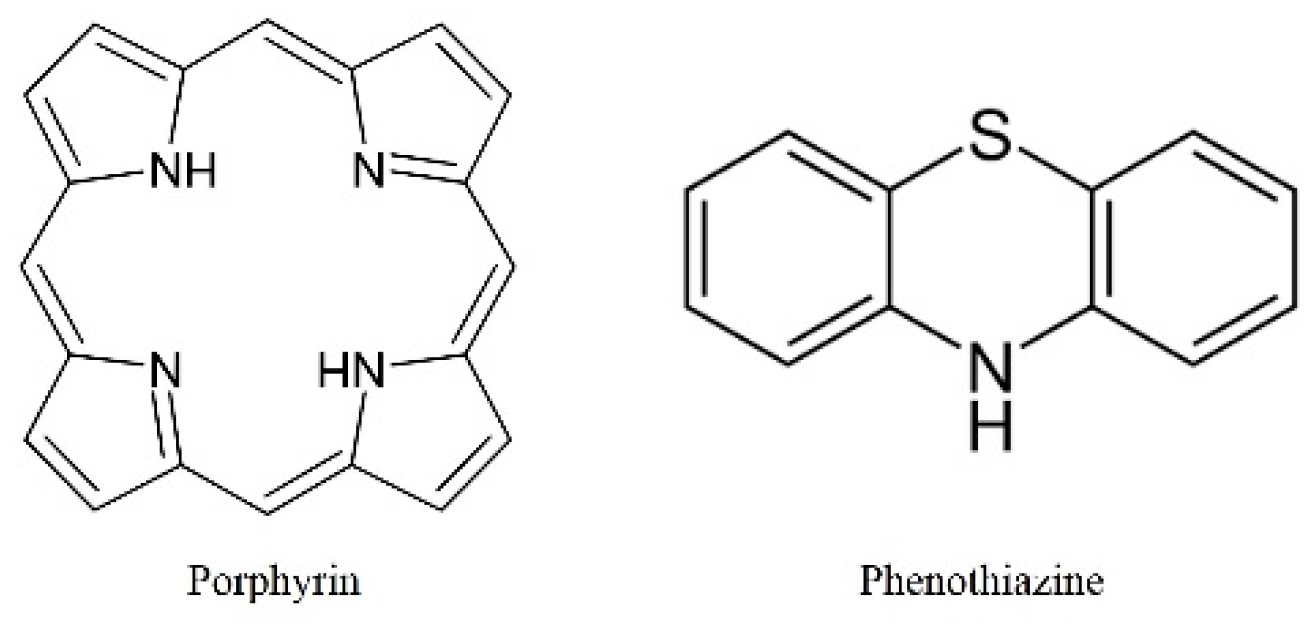
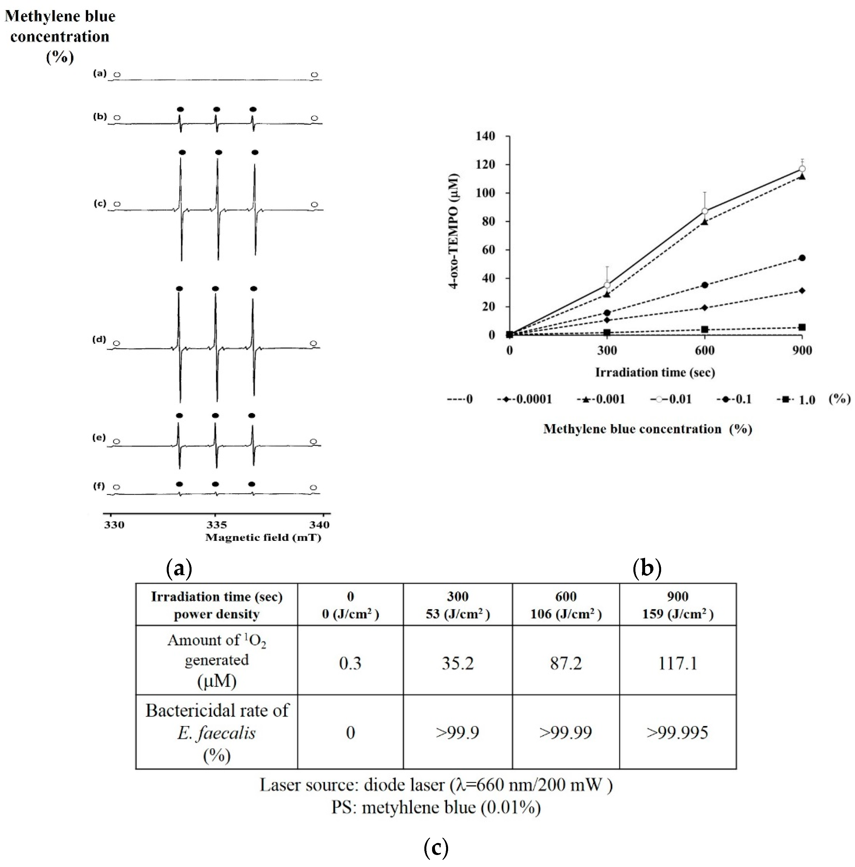
4.1. Bactericidal Efficacy for Bacteria Associated with Refractory Apical Periodontitis and the Appropriate PS Concentration for 1O2 Generation

The bactericidal effect of a-PDT on refractory periodontitis-associated bacteria, such as E. faecalis, C. albicans, Propionibacterium, Porphyromonas, and Prevotella spp., has been reported in numerous papers, which suggests its efficacy even with different PS and light sources [19,20,44,45,46,47,48]. Although those previous reports were very significant, they were not uniform in terms of PS concentration or light source output. It is important to understand the minimum 1O2 required for a bactericidal effect to provide safer treatment. As the absorption of excitation light by PS triggers a series of mechanisms, there may be appropriate PS concentrations depending on the output of the excitation light. Many PS often use substances having the basic skeleton of porphyrins and phenothiazines [19,20], and many change color when exposed to solvent (Figure 4).
Therefore, the irradiation of excitation light should not be considered to produce much 1O2 in either case or to have a bactericidal effect because low PS concentrations result in little PS excitation, whereas high PS concentrations result in no excitation light penetrating the PS. Actually, in our previous report [55] using a diode laser (660 nm, 200 mW) and methylene blue (MB) as the light source and PS, respectively, we were able to determine the appropriate concentration of MB to generate 1O2. Figure 5a shows the typical ESR-spectra of 4-oxo-TEMPO, which is the 1O2-specific oxidation from 4-oxo-TMP to 4-oxo-TEMPO, and Figure 5b shows the amount of 1O2 generated from each concentration of excited MB. The amount of generated 1O2 increased in the following order: 0.01% > 0.001% > 0.1% > 0.0001% > 1.0%. The most efficient generation of a large amount of 1O2 was from 0.01% excited MB, which is the appropriate PS concentration for this output. Further, the amount of generated 1O2 necessary to kill E. faecalis (>99.9%) was at least 35.2 μM (Figure 5c).
Tanaka et al. [60] used a diode laser (λ = 664 nm, 20 J/cm2) as a light source and compared MB concentrations of 1, 5, 20, 100, and 400 μM. When a-PDT was applied to the methicillin-resistant Staphylococcus aureus bactericidal test, a biphasic dose response was observed with the highest bactericidal effect at 100 μM and a lower effect at 1 or 400 μM. In addition, Chan et al. [61] observed a sterilization effect when a diode laser (λ = 665 nm, 21.2 J/cm2) with 0.01% MB was used to irradiate oral bacteria. Therefore, it is considered that 0.001%–0.01% MB on a-PDT corresponds precisely to the appropriate PS concentration of MB, which is between 26.7 and 267 μM, and that the bactericidal effect is due to the efficient generation of 1O2. However, these reports also differ in terms of irradiation conditions, such as the irradiation distance or width; therefore, irradiation conditions should be standardized for clinical applications in the future. The appropriate concentration for PS other than MB from the perspective of 1O2 generation also needs to be considered.

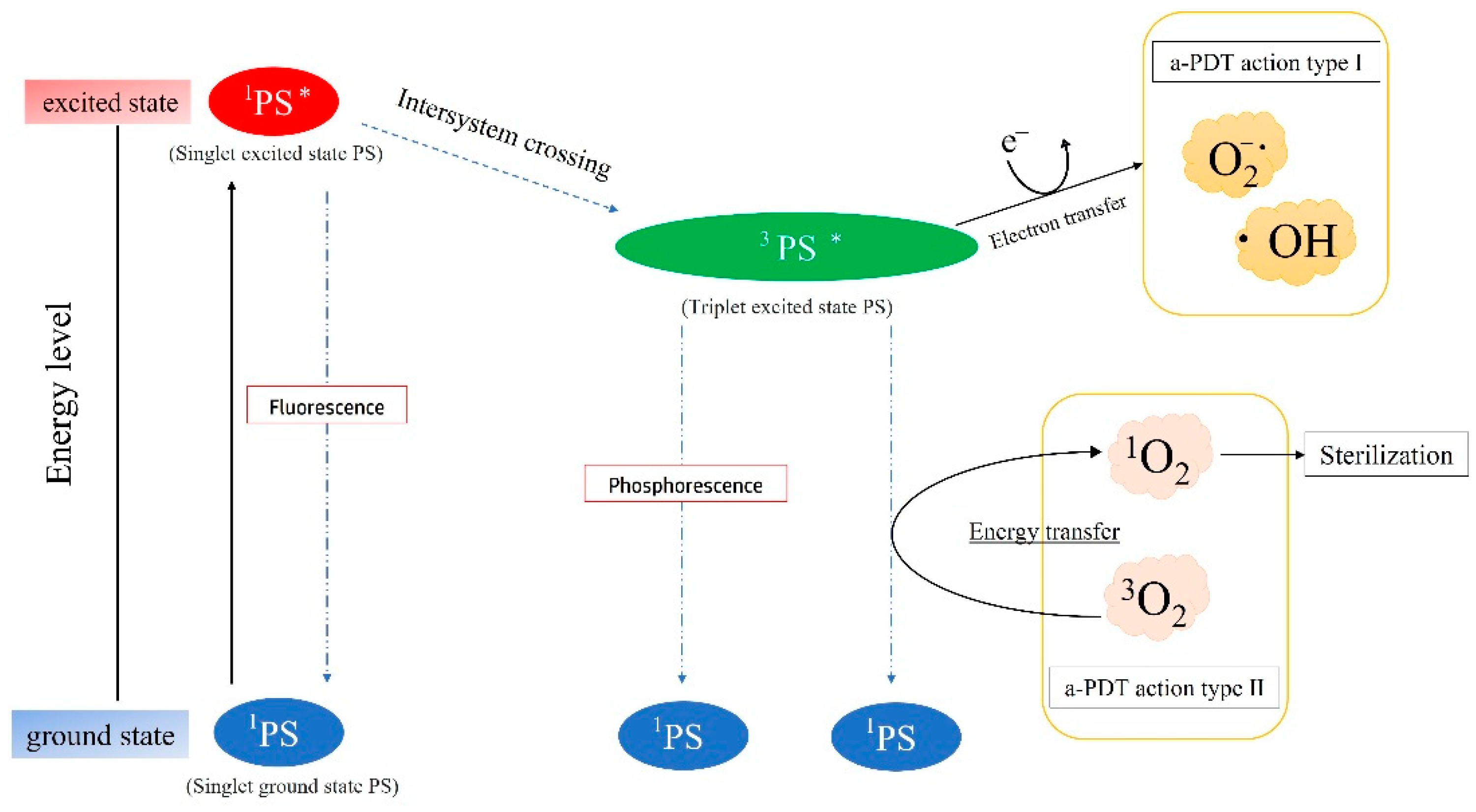
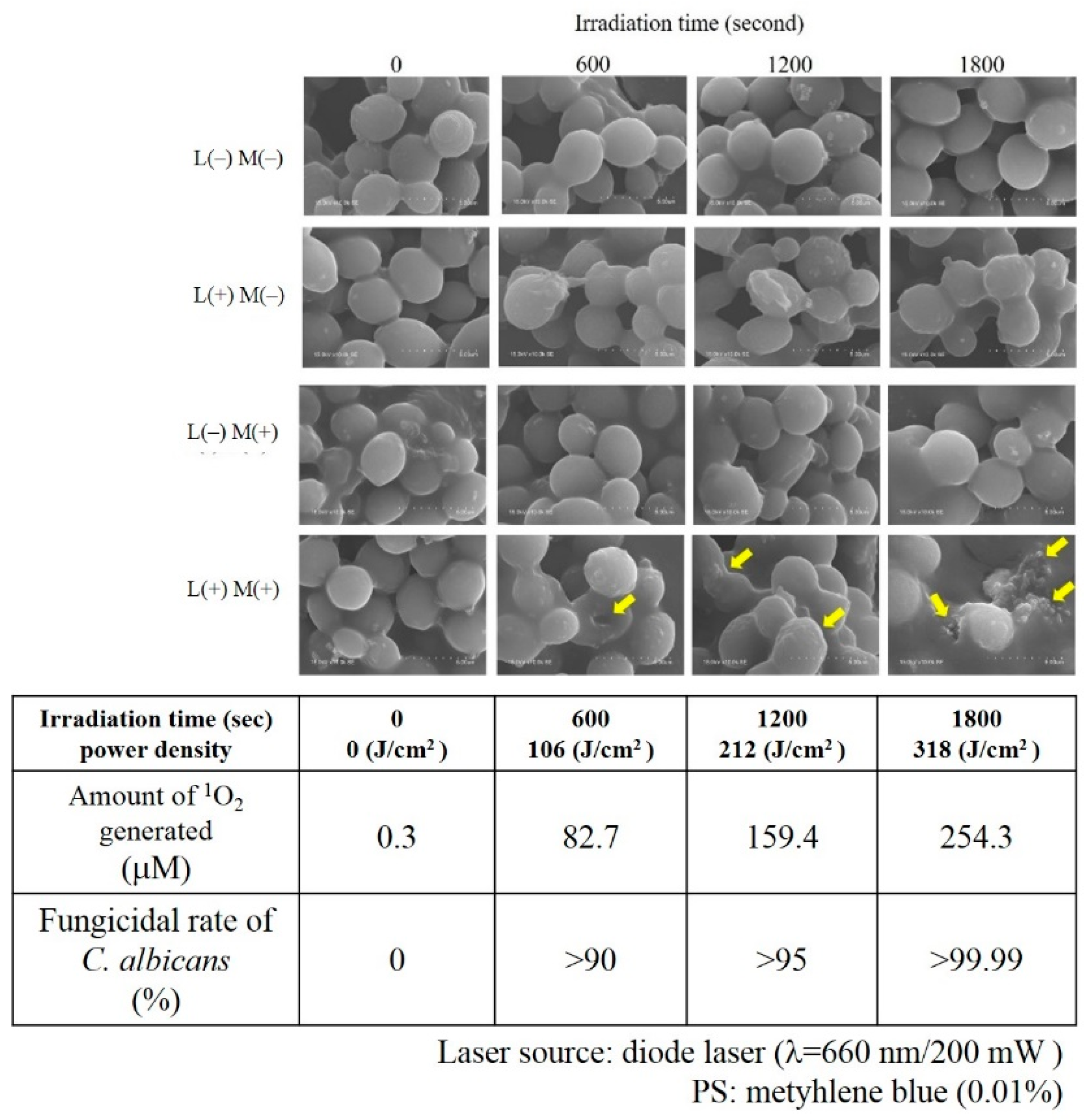
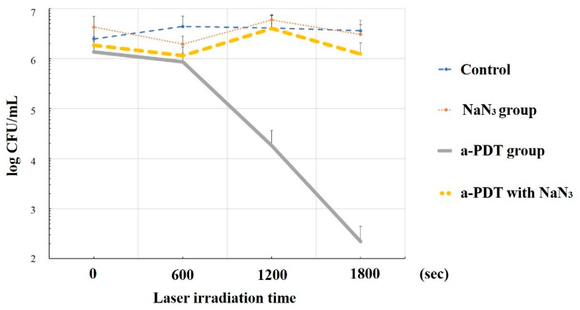
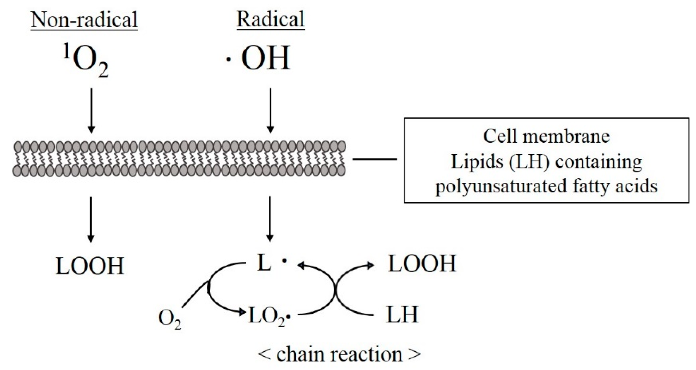
4.2. Sterilization Mechanism of a-PDT by 1O2
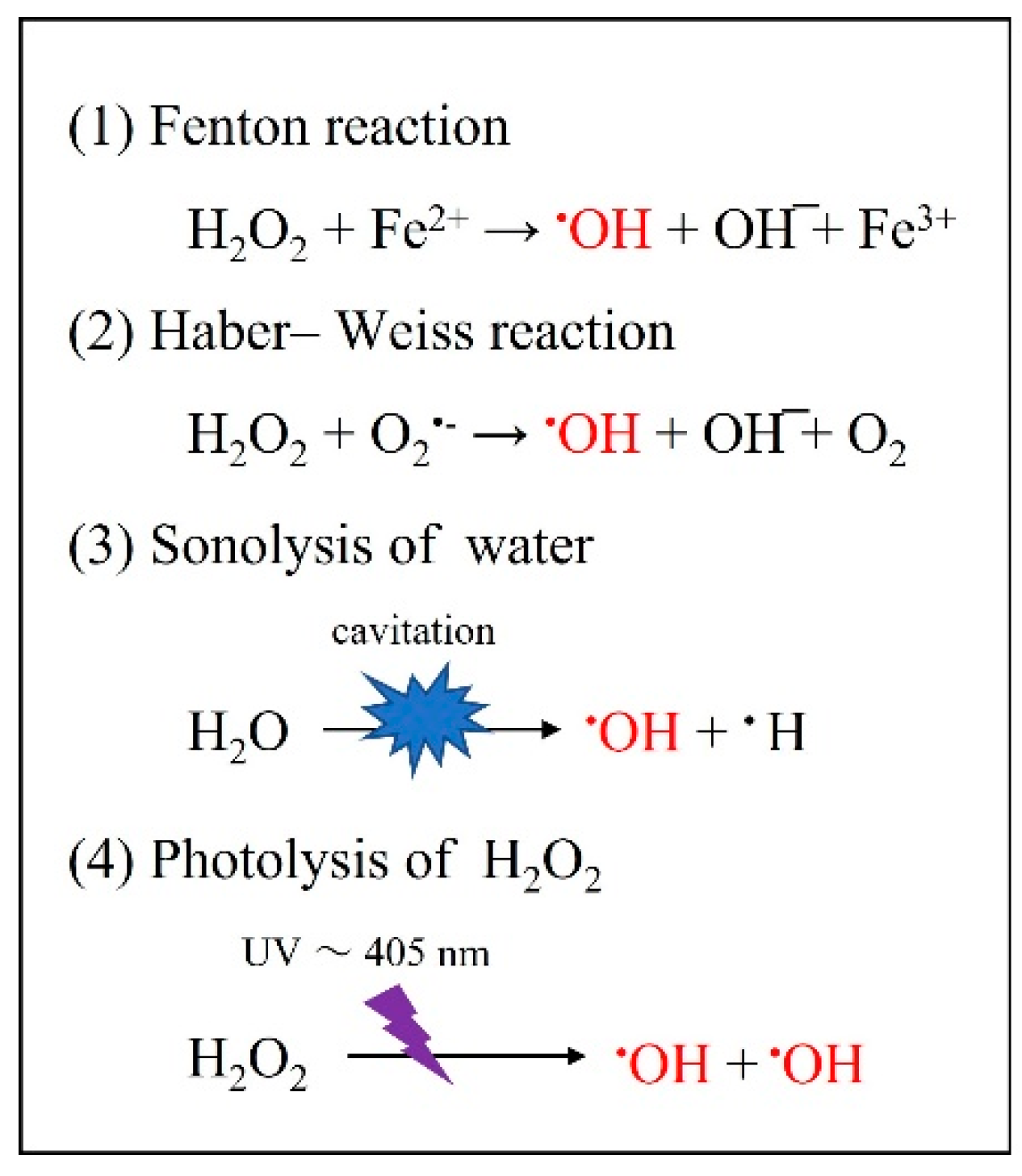
Considering bactericidal action and 1O2, Allen et al. [62] first reported that neutrophils may produce 1O2 via the catalysis of myeloperoxidase during respiratory bursts. Subsequently, Krinsky [63] suggested that neutrophil-derived 1O2 mediates bacterial killing. Further, Tatsuzawa et al. [64] suggested that phagocytic leukocytes produce 1O2 as a major bactericidal oxidant in the phagosome, and may also interact with the cytoplasmic membrane of microorganisms, especially E. coli and other Gram-negative bacteria, and damage respiratory enzymes while inhibiting ATP formation. That is, the small amount of 1O2 generated in vivo is not toxic to eukaryotic cells. Due to its short half-life (10−6 s), 1O2 only inactivates enzyme activity on the surface of nearby prokaryotic cells. In the a-PDT mechanism, on the other hand, 1O2 generated by PS and excitation light from outside the cell destroys the cell wall and/or membrane, and then PS is transferred into the cell. Next, 1O2 generated by photoexcitation destroys intracellular organelles, leading to cell death. A large amount of 1O2 is produced by PS excitation, and, unlike in vivo, the amount of 1O2 produced increases with the irradiation time of the excitation light. Additionally, the 1O2 produced is a nonspecific oxidant for which there is no defense [65,66]. Importantly, the amount of 1O2 used for treatment should be minimized to help prevent excessive oxidative damage to normal cells [67].
Therefore, our laboratory used scanning electron microscopy (SEM) to evaluate visually the mechanism of 1O2 in the fungicidal action of C. albicans, a eukaryote with a cell wall, and further examined the amount of 1O2 produced along with its fungicidal effect [56]. The results of a-PDT against C. albicans using 0.01% MB as PS and a diode laser (660 nm, 200 mW) as the light source showed that at least 245.3 μM of 1O2 was required to achieve >99.99% fungicidal activity. That is, compared with bacteria, which are prokaryotic cells, C. albicans requires a very large amount of 1O2. The reason for this may be attributed to the differences in the cytoskeleton mentioned previously. Figure 6 shows irradiation time-dependent fusion, i.e., fusion that is dependent on the amount of 1O2 generated, as well as the loss of the normal morphology of single independent cells as they begin to fuse with each other. In SEM images exposed to 245.3 μM of 1O2, the fused cells further lose their morphology and become amorphous lumps (Figure 6).
Conversely, when sodium azide (NaN3, 10 mM), which is a specific scavenger of 1O2 and at a concentration not toxic to C. albicans, was added to the MB solution and a-PDT was performed on C. albicans under the same conditions, no fungicidal effect was observed (Figure 7), which suggests that exposure to large amounts of 1O2 affects not only the target microorganisms but also the host, which is a eukaryotic cell [57].
In summary, our previous reports suggest that a large amount of 1O2 is required to disinfect C. albicans by a-PDT, and that the mechanism underlying the a-PDT’s bactericidal effects on microorganisms is the physical injury caused by 1O2 [56,57].

4.3. Bactericidal Effect of OH Resulting from Other Photochemical Reactions
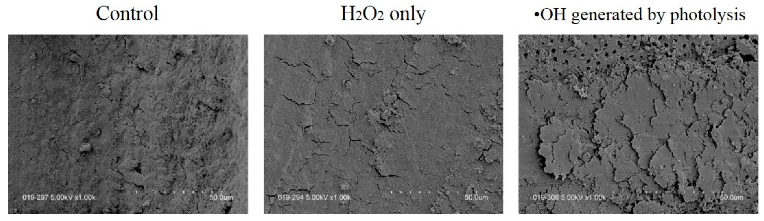
OH is characterized by having the shortest half-life (10−8 s) and the strongest oxidizing power among all ROS [22]. The OH formation systems include (1) the Fenton reaction [23], (2) the Haber–Weiss reaction [68], (3) the sonolysis of water [69], and (4) the photolysis of H2O2 [25,26,27,28,29,30,31,58,59] (Figure 8).
In these systems, OH generated by the photolysis of H2O2 in reactive systems has been applied to oral bacterial infections, such as caries [25,70,71], periodontal disease [25,27,72], periapical disease [25,71], and aspiration pneumonia [25,26,29,59]. The wavelength spectrum used for the photolysis reaction of H2O2 ranges from UV to blue light, which is visible light [24]. Therefore, many reports in the dental field use a wavelength of 405 nm to generate OH [25,26,27,28,29,30,31,58,59,70,71].
The research group of Kanno and Sasaki is a pioneer in the use of this photolysis reaction for sterilization by OH and the effect of OH on dental materials [25,26,27,28,29,30,69,70,71,72,73,74,75]. In particular, they reported that an OH generation rate of approximately 200–300 μM is required to achieve a 4-log reduction in the number of bacteria, i.e., staphylococcus aureus infection (Staphylococcus aureus), dental caries (Streptococcus mutans), periodontal disease (Aggregatibacter actinomycetemcomitans), and refractory apical periodontitis (E. faecalis) [25]. The fact that this disinfection method is possible with 3% H2O2, which is routinely used in dental practice, and 405 nm light-emitting diode (LED), which is used for tooth bleaching, is expected to lead to further developments.

4.4. Effect of OH on Smear Layer Removal

The use of instruments such as reamers, files, and bars can result in the formation of a smear layer on the dentin surface [76]. This smear layer contains dentin components, remnants of the periodontal ligament formation process, pulp tissue, and bacteria [77], and, thus, may interfere with the penetration of the irrigation solution and cause leakage between the root canal wall and filling material [78]. Therefore, our laboratory was interested in the strong oxidizing power of OH, so we investigated not only its bactericidal effect but also its effect on the removal of the smear layer. First, we examined whether OH generated by the Fenton reaction of metal ions with 3% H2O2 could remove the smear layer [79,80,81]. The results showed that H2O2 alone was not effective for removing the smear layer. Although the smear layer was not removed when OH was generated, it was confirmed that the surface of the smear layer was roughened. In other words, it was assumed that OH would damage the inorganic and organic materials that formed the smear layer, and decompose and then clean the smear layer while roughening it. Therefore, the generation and exposure of more OH in the root canal raised expectations for the removal of the smear layer. Next, OH produced by a photolysis reaction using 3% H2O2 and a 405 nm LED was used to remove the smear layer [58]. The amount of OH generated by the photolysis reaction was significantly higher than that generated by the Fenton reaction. Although a large amount of OH is dangerous for living organisms, we consider it to be safe in a closed space only inside the root canal, where the light can reach H2O2. The results showed that when the H2O2 was irradiated with 405 nm of LED light for 3 min, the smear layer was not completely removed but was roughened or partially removed, and some dentin tubules could be observed (Figure 9).
Under this irradiation condition, it is calculated that about 510 μM equivalent of OH is generated. The effect of about 510 μM OH on the smear layer was comparable to that of alternating washes with 5% NaClO or 3% H2O2 [82] and immersion in 1% EDTA for 2 min [83].
Based on the above, OH is an important factor in the removal of the smear layer. Further, Tsujimoto et al. [82] reported that to remove the smear layer more efficiently, it is important to reflux instead of immerse the chemical solution. Therefore, our laboratory is now examining the effect of smear layer removal by generating OH under different immersion and reflux conditions. In addition, the possibility of smear layer removal by 1O2 is also currently under investigation. If an effective smear layer removal method using OH and 1O2 can be established, it may become an alternative cleaning method to NaClO and EDTA. This section may be divided by subheadings. It should provide a concise and precise description of the experimental results, their interpretation, as well as the experimental conclusions that can be drawn.

5. Discussion

Numerous papers have demonstrated that ROS can play an active role in root canal sterilization and smear layer removal. The traditional purpose of applying ROS is to eliminate the unexpected side effects caused by the use of NaClO or EDTA, with an emphasis on safety. Therefore, several points must be considered when applying ROS, such as the difference in post-action effects between the radical OH and the non-radical 1O2. These basic actions occur by directly injuring the substance from hydrogen atom transfer through the oxidizing powers. In general, radicals induce chain reactions [84], whereas non-radicals are quantitative in character [85], so the subsequent effects should be considered greater for the radicals (Figure 10).
In vivo, cells have antioxidant enzymes such as superoxide dismutase and catalase [86,87], and, furthermore, cell membranes damaged by radicals can be stopped in a radical-induced chain reaction by antioxidants such as tocopherol and ascorbic acid [15,88,89]. In root canals, however, there are no antioxidant enzymes or antioxidants to stop the radical-induced chain reaction that damages the organic components of dentin. As a result, even if the objectives of sterilization and smear layer removal are achieved, subsequent dental treatment, i.e., mechanical properties of dentin and bond strength of composite resins, may be adversely affected. Abe et al. [90] reported a significant decrease in bond strength between orthodontic appliances and enamel immediately after tooth bleaching methods involving OH. Therefore, the effect of composite resin on the bond strength to dentin after ROS exposure should also be examined. Conversely, radicals act on the cell membranes of microorganisms, which triggers a chain reaction that may be effective against colonizing bacterial populations. Furthermore, we believe that ROS are particularly effective because anaerobic bacteria have few antioxidant enzymes. Although these ideas are still speculative, they should be considered in the future when using ROS. It should also be noted that ROS inflict nonselective injury on adjacent substances. The optimal application of a-PDT is to let targets take up PS and then selectively kill the targets, similar to PDT in cancer therapy. The selective killing of microorganisms is essential for the future development of a-PDT. In addition, in the case of OH disinfection using the photolysis method, safety considerations such as the use of antioxidants (tocopherol [88], ascorbic acid [89], and catechin [91]) to scavenge radicals and prevent post-action chain reactions are also considered necessary. The control of ROS and free radicals generated in the root canal actually involves not only the a-PDT and photolysis methods but also NaClO, which has an intramolecular hypochlorite radical (OCl) and is routinely used by clinicians. Although it is no longer the gold standard, our laboratory has shown that alternating irrigation with H2O2 generates ROS and free radicals such as O2−·, OH, and 5,5-dimethyl-1-pyrrolidone-(2)-oxyl-(1) (DMPO-X) radical [92,93].
While many dental treatments using ROS have been performed in the past, dental clinicians still tend to have limited knowledge of ROS. ROS and free radicals are associated with inflammation, carcinogenesis, and aging and can not only cause injury to living organisms but also kill microorganisms and viruses depending on how they are applied. On the other hands, various disinfection methods for the application of ROS have not yet standardized experimental conditions, and many have concluded that its bactericidal effect should be used in combination with conventional cleaning by NaClO and EDTA. In other words, it must be noted that there is a limit to sterilization by ROS alone. A further weakness is that this review is a narrative review, not a systematic review, as it does not analyze the data or study group size to determine the validity of the results.
Research on ROS, such as bleaching and disinfection methods, using titanium dioxide as a photocatalyst [94,95] and photofunctionalization of implants [95], is also developing. Furthermore, photochemistry has recently been applied for the detection of oral cancer [96,97,98,99] and tooth decay [100]. Additional findings are expected to be investigated and the development of dental treatment using ROS is expected to continue in the future.

6. Conclusions

The following four points are required for the general clinical application of a-PDT or photolysis methods:
  • The light source and PS should be standardized, taking into account the optimal generation of ROS.
  • The possible disadvantages of dental treatment after ROS exposure should be considered.
  • The advantages and disadvantages to the organism induced by ROS should be familiarized.
  • A-PDT should be used as an adjunct to conventional disinfection methods at this time, and when using NaClO together with a-PDT, it might be reducing the concentration of NaClO, thereby reducing harmful effects on living organisms.

Author Contributions

Conceptualization, C.K. and Y.T.; methodology, C.K., S.U., O.T. and Y.T.; writing—original draft preparation, C.K.; writing—review and editing, Y.T. All authors have read and agreed to the published version of the manuscript.

Funding

This research received no external funding.

Institutional Review Board Statement

Not applicable.

Informed Consent Statement

Not applicable.

Data Availability Statement

The data used to support the findings of this study are included within the article and are available from the corresponding author upon reasonable request.

Conflicts of Interest

The authors declare no potential conflict of interest.

References

  1. Zhao, M.; Wang, Y.; Li, L.; Liu, S.; Wang, C.; Yuan, Y.; Yang, G.; Chen, Y.; Cheng, J.; Lu, Y.; et al. Mitochondrial ROS promote mitochondrial dysfunction and inflammation in ischemic acute kidney injury by disrupting TFAM-mediated mtDNA maintenance. Theranostics 2021, 11, 1845–1863. [Google Scholar] [CrossRef] [PubMed]
  2. Mittal, M.; Siddiqui, M.R.; Tran, K.; Reddy, S.P.; Malik, A.B. Reactive oxygen species in inflammation and tissue injury. Antioxid. Redox Signal. 2014, 20, 1126–1167. [Google Scholar] [CrossRef] [PubMed]
  3. Shi, Y.; Yuan, S.; Tang, S.-J. Reactive Oxygen Species (ROS) are Critical for Morphine Exacerbation of HIV-1 gp120-Induced Pain. J. Neuroimmune Pharmacol. 2021, 16, 581–591. [Google Scholar] [CrossRef] [PubMed]
  4. Ilari, S.; Giancotti, L.A.; Lauro, F.; Dagostino, C.; Gliozzi, M.; Malafoglia, V.; Sansone, L.; Palm, E.; Tafani, M.; Russo, M.A.; et al. Antioxidant modulation of sirtuin 3 during acute inflammatory pain: The ROS control. Pharmacol. Res. 2020, 157, 104851. [Google Scholar] [CrossRef] [PubMed]
  5. Liou, G.-Y.; Storz, P. Reactive oxygen species in cancer. Free Radic. Res. 2010, 44, 479–496. [Google Scholar] [CrossRef] [PubMed]
  6. Srinivas, U.S.; Tan, B.W.Q.; Vellayappan, B.A.; Jeyasekharan, A.D. ROS and the DNA damage response in cancer. Redox Biol. 2019, 25, 101084. [Google Scholar] [CrossRef]
  7. Kudryavtseva, A.V.; Krasnov, G.S.; Dmitriev, A.A.; Alekseev, B.Y.; Kardymon, O.L.; Sadritdinova, A.F.; Fedorova, M.S.; Pokrovsky, A.V.; Melnikova, N.V.; Kaprin, A.D.; et al. Mitochondrial dysfunction and oxidative stress in aging and cancer. Oncotarget 2016, 7, 44879–44905. [Google Scholar] [CrossRef]
  8. Davalli, P.; Mitic, T.; Caporali, A.; Lauriola, A.; D’Arca, D. ROS, Cell Senescence, and Novel Molecular Mechanisms in Aging and Age-Related Diseases. Oxid. Med. Cell Longev. 2016, 2016, 3565127. [Google Scholar] [CrossRef]
  9. Murray, T.V.A.; Ahmad, A.; Brewer, A.C. Reactive oxygen at the heart of metabolism. Trends Cardiovasc. Med. 2014, 24, 113–120. [Google Scholar] [CrossRef]
  10. Togliatto, G.; Lombardo, G.; Brizzi, M.F. The Future Challenge of Reactive Oxygen Species (ROS) in Hypertension: From Bench to Bed Side. Int. J. Mol. Sci. 2017, 18, 1988. [Google Scholar] [CrossRef] [Green Version]
  11. Brieger, K.; Schiavone, S.; Miller, F.J., Jr.; Krause, K.-H. Reactive oxygen species: From health to disease. Swiss Med. Wkly. 2012, 142, w13659. [Google Scholar] [CrossRef] [PubMed]
  12. Jomova, K.; Valko, M. Advances in metal-induced oxidative stress and human disease. Toxicology 2011, 283, 65–87. [Google Scholar] [CrossRef] [PubMed]
  13. Sczepanik, F.C.; Grossi, M.L.; Casati, M.; Goldberg, M.; Glogauer, M.; Fine, N.; Tenenbaum, H.C. Periodontitis is an inflammatory disease of oxidative stress: We should treat it that way. Periodontology 2000 2020, 84, 45–68. [Google Scholar] [CrossRef]
  14. Choudhari, S.K.; Chaudhary, M.; Gadbail, A.R.; Sharma, A.; Tekade, S. Oxidative and antioxidative mechanisms in oral cancer and precancer: A review. Oral Oncol. 2014, 50, 10–18. [Google Scholar] [CrossRef]
  15. Shilpa, B.; Ahmed, A.; Mohammed, M.; Abdulaziz, S.A.; Nassreen, H.M.A.; Apathsakayan, R.; Mazen, F.A.; Ali, R.; Asma, S.A.; Vikrant, R.P.; et al. Effect of Ascorbic Acid on Differentiation, Secretome and Stemness of Stem Cells from Human Exfoliated Deciduous Tooth (SHEDs). J. Pers. Med. 2021, 11, 589. [Google Scholar] [CrossRef]
  16. Khan, S.; Hussain, M.A.B.; Khan, A.P.; Liu, H.; Siddiqui, S.; Mallidi, S.; Leon, P.; Daly, L.; Rudd, G.; Cuckov, F.; et al. Clinical evaluation of smartphone-based fluorescence imaging for guidance and monitoring of ALA-PDT treatment of early oral cancer. J. Biomed. Opt. 2020, 25, 063813. [Google Scholar] [CrossRef]
  17. Zhang, Q.; Li, L. Photodynamic combinational therapy in cancer treatment. J. BUON 2018, 23, 561–567. [Google Scholar]
  18. El-Hussein, A.; Manoto, S.L.; Ombinda-Lemboumba, S.; Alrowaili, Z.A.; Mthunzi-Kufa, P. A Review of Chemotherapy and Photodynamic Therapy for Lung Cancer Treatment. Anticancer Agents Med. Chem. 2021, 21, 149–161. [Google Scholar] [CrossRef]
  19. Konopka, K.; Goslinski, T. Photodynamic therapy in dentistry. J. Dent. Res. 2007, 86, 694–707. [Google Scholar] [CrossRef] [PubMed]
  20. Soukos, N.S.; Goodson, J.M. Photodynamic therapy in the control of oral biofilms. Periodontology 2000 2011, 55, 143–166. [Google Scholar] [CrossRef] [PubMed]
  21. Qi, M.; Chi, M.; Sun, X.; Xie, X.; Weir, M.D.; Oates, T.W.; Zhou, Y.; Wang, L.; Bai, Y.; Xu, H.H. Novel nanomaterial-based antibacterial photodynamic therapies to combat oral bacterial biofilms and infectious diseases. Int. J. Nanomed. 2019, 14, 6937–6956. [Google Scholar] [CrossRef] [PubMed] [Green Version]
  22. Sies, H. Hydrogen peroxide as a central redox signaling molecule in physiological oxidative stress: Oxidative eustress. Redox Biol. 2017, 11, 613–619. [Google Scholar] [CrossRef] [PubMed]
  23. Wardman, P.; Candeias, L.P. Fenton chemistry: An introduction. Radiat. Res. 1996, 145, 523–531. [Google Scholar] [CrossRef] [PubMed]
  24. Nakashima, Y.; Ohta, S.; Wolf, A.M. Blue light-induced oxidative stress in live skin. Free Radic. Biol. Med. 2017, 108, 300–310. [Google Scholar] [CrossRef] [PubMed]
  25. Ikai, H.; Nakamura, K.; Shirato, M.; Kanno, T.; Iwasawa, A.; Sasaki, K.; Niwano, Y.; Kohno, M. Photolysis of hydrogen peroxide, an effective disinfection system via hydroxyl radical formation. Antimicrob. Agents Chemother. 2010, 54, 5086–5091. [Google Scholar] [CrossRef] [PubMed]
  26. Kanno, T.; Nakamura, K.; Ikai, H.; Hayashi, E.; Shirato, M.; Mokudai, T.; Iwasawa, A.; Niwano, Y.; Kohno, M.; Sasaki, K. Novel denture-cleaning system based on hydroxyl radical disinfection. Int. J. Prosthodont. 2012, 25, 376–380. [Google Scholar] [PubMed]
  27. Sato, H.; Niwano, Y.; Nakamura, K.; Mokudai, T.; Ikai, H.; Kanno, T.; Egusa, H. Efficacy and safety of a therapeutic apparatus using hydrogen peroxide photolysis to treat dental and periodontal infectious diseases. Toxicol. Sci. 2016, 41, 793–799. [Google Scholar] [CrossRef]
  28. Toki, T.; Nakamura, K.; Kurauchi, M.; Kanno, T.; Katsuda, Y.; Ikai, H.; Hayashi, E.; Egusa, H.; Sasaki, K.; Niwano, Y. Synergistic interaction between wavelength of light and concentration of H2O2 in bactericidal activity of photolysis of H2O2. J. Biosci. Bioeng. 2015, 119, 358–362. [Google Scholar] [CrossRef]
  29. Hayashi, E.; Mokudai, T.; Yamada, Y.; Nakamura, K.; Kanno, T.; Sasaki, K.; Niwano, Y. In vitro and in vivo anti-Staphylococcus aureus activities of a new disinfection system utilizing photolysis of hydrogen peroxide. J. Biosci. Bioeng. 2012, 114, 193–197. [Google Scholar] [CrossRef]
  30. Nakamura, K.; Shirato, M.; Ikai, H.; Kanno, T.; Sasaki, K.; Kohno, M.; Niwano, Y. Photo-irradiation of proanthocyanidin as a new disinfection technique via reactive oxygen species formation. PLoS ONE 2013, 8, e60053. [Google Scholar] [CrossRef]
  31. Ibi, H.; Hayashi, M.; Yoshino, F.; Tamura, M.; Yoshida, A.; Kobayashi, Y.; Shimizu, K.; Lee, M.-C.; Imai, K.; Ogiso, B. Bactericidal effect of hydroxyl radicals generated by the sonolysis and photolysis of hydrogen peroxide for endodontic applications. Microb. Pathog. 2017, 103, 65–70. [Google Scholar] [CrossRef] [PubMed]
  32. Kawamoto, K.; Tsujimoto, Y. Effects of the hydroxyl radical and hydrogen peroxide on tooth bleaching. J. Endod. 2004, 30, 45–50. [Google Scholar] [CrossRef] [PubMed]
  33. Lee, H.W.; Kim, G.J.; Kim, J.M.; Kil Park, J.; Lee, J.K.; Kim, G.C. Tooth bleaching with nonthermal atmospheric pressure plasma. J. Endod. 2009, 35, 587–591. [Google Scholar] [CrossRef] [PubMed]
  34. Figdor, D. Apical periodontitis: A very prevalent problem. Oral Surg. Oral Med. Oral Pathol. Oral Radiol. Endod. 2002, 94, 651–652. [Google Scholar] [CrossRef]
  35. Nair, P.N.R. Pathogenesis of apical periodontitis and the causes of endodontic failures. Crit. Rev. Oral Biol. Med. 2004, 15, 348–381. [Google Scholar] [CrossRef]
  36. Siqueira, J.F., Jr.; Rôças, I.N.; Paiva, S.S.M.; Magalhães, K.M.; Guimarães-Pinto, T. Cultivable bacteria in infected root canals as identified by 16S rRNA gene sequencing. Oral Microbiol. Immunol. 2007, 22, 266–271. [Google Scholar] [CrossRef]
  37. Sunde, P.T.; Olsen, I.; Debelian, G.J.; Tronstad, L. Microbiota of Periapical Lesions Refractory to Endodontic Therapy. J. Endod. 2002, 28, 304–310. [Google Scholar] [CrossRef]
  38. Lee, Y.; Puumala, E.; Robbins, N.; Cowen, L.E. Antifungal Drug Resistance: Molecular Mechanisms in Candida albicans and Beyond. Chem. Rev. 2021, 121, 3390–3411. [Google Scholar] [CrossRef]
  39. Jacobsen, I.D.; Hube, B. Candida albicans morphology: Still in focus. Expert Rev. Anti-Infect. Ther. 2017, 15, 327–330. [Google Scholar] [CrossRef]
  40. Zehnder, M. Root canal irrigants. J. Endod. 2006, 32, 389–398. [Google Scholar] [CrossRef]
  41. Guivarc’h, M.; Ordioni, U.; Ahmed, H.M.A.; Cohen, S.; Catherine, J.-H.; Bukiet, F. Sodium Hypochlorite Accident: A Systematic Review. J. Endod. 2017, 43, 16–24. [Google Scholar] [CrossRef] [PubMed]
  42. Marshall, M.V.; Cancro, L.P.; Fischman, S.L. Hydrogen peroxide: A review of its use in dentistry. J. Periodontol. 1995, 66, 786–796. [Google Scholar] [CrossRef] [PubMed]
  43. Mehdipour, O.; Kleier, D.J.; Averbach, R.E. Anatomy of sodium hypochlorite accidents. Compend. Contin. Educ. Dent. 2007, 28, 544–550. [Google Scholar] [PubMed]
  44. Takasaki, A.A.; Aoki, A.; Mizutani, K.; Schwarz, F.; Sculean, A.; Wang, C.-Y.; Koshy, G.; Romanos, G.; Ishikawa, I.; Izumi, Y. Application of antimicrobial photodynamic therapy in periodontal and peri-implant diseases. Periodontology 2000 2009, 51, 109–140. [Google Scholar] [CrossRef] [PubMed]
  45. Plotino, G.; Grande, N.M.; Mercade, M. Photodynamic therapy in endodontics. Int. Endod. J. 2019, 52, 760–774. [Google Scholar] [CrossRef]
  46. Prażmo, E.J.; Kwaśny, M.; Łapiński, M.; Mielczarek, A. Photodynamic Therapy as a Promising Method Used in the Treatment of Oral Diseases. Adv. Clin. Exp. Med. 2016, 25, 799–807. [Google Scholar] [CrossRef] [PubMed]
  47. Mohammadi, Z.; Jafarzadeh, H.; Shalavi, S.; Kinoshita, J. Photodynamic Therapy in Endodontics. J. Contemp. Dent. Pract. 2017, 18, 534–538. [Google Scholar] [CrossRef]
  48. Kharkwal, G.B.; Sharma, S.K.; Huang, Y.; Dai, T.; Hamblin, M.R. Photodynamic therapy for infections: Clinical applications. Lasers Surg. Med. 2011, 43, 755–767. [Google Scholar] [CrossRef]
  49. Maisch, T.; Szeimies, R.-M.; Jori, G.; Abels, C. Antibacterial photodynamic therapy in dermatology. Photochem. Photobiol. Sci. 2004, 3, 907–917. [Google Scholar] [CrossRef]
  50. Wainwright, M. Photodynamic antimicrobial chemotherapy (PACT). J. Antimicrob. Chemother. 1998, 42, 13–28. [Google Scholar] [CrossRef]
  51. Maldonado-Carmona, N.; Ouk, T.-S.; Calvete, M.J.F.; Pereira, M.M.; Villandier, N.; Leroy-Lhez, S. Conjugating biomaterials with photosensitizers: Advances and perspectives for photodynamic antimicrobial chemotherapy. Photochem. Photobiol. Sci. 2020, 19, 445–461. [Google Scholar] [CrossRef] [PubMed]
  52. Garin, C.; Alejo, T.; Perez-Laguna, V.; Prieto, M.; Mendoza, G.; Arruebo, M.; Sebastian, V.; Rezusta, A. Chalcogenide nanoparticles and organic photosensitizers for synergetic antimicrobial photodynamic therapy. J. Mater. Chem. B 2021, 9, 6246–6259. [Google Scholar] [CrossRef] [PubMed]
  53. Fimple, J.L.; Fontana, C.R.; Foschi, F.; Ruggiero, K.; Song, X.; Pagonis, T.C.; Tanner, A.C.R.; Kent, R.; Doukas, A.G.; Stashenko, P.P.; et al. Photodynamic treatment of endodontic polymicrobial infection in vitro. J. Endod. 2008, 34, 728–734. [Google Scholar] [CrossRef] [PubMed]
  54. Kranz, S.; Guellmar, A.; Völpel, A.; Gitter, B.; Albrecht, V.; Sigusch, B.W. Photodynamic suppression of Enterococcus faecalis using the photosensitizer mTHPC. Lasers Surg. Med. 2011, 43, 241–248. [Google Scholar] [CrossRef] [PubMed]
  55. Komine, C.; Tsujimoto, Y. A small amount of singlet oxygen generated via excited methylene blue by photodynamic therapy induces the sterilization of Enterococcus faecalis. J. Endod. 2013, 39, 411–413. [Google Scholar] [CrossRef]
  56. Kageyama, M.; Otsuka, I.; Komine, C. Clarification of the Sterilization Mechanism of Antimicrobial Photodynamic Therapy for Candida albicans. JSLD 2019, 29, 141–147. [Google Scholar] [CrossRef]
  57. Komine, C.; Konishi, Y.; Ogura, Y.; Omori, H.; Suzuki, H.; Nakajima, M.; Izawa, M.; Fuchigami, M.; Tsuzukibash, O.; Fukatsu, A.; et al. Fungicidal Effect of Antibacterial Photodynamic Therapy with Suppressing Singlet Oxygen Generation. Nihon Univ. J. Oral Sci. 2019, 45, 164–172. [Google Scholar]
  58. Komine, C.; Konishi, Y.; Ishii, M.; Izawa, M.; Tsujimoto, Y. The smear layer removal effect of hydroxyl radicals generated from hydrogen peroxide irradiated with 405-nm LED light. Int. J. Microdent. 2021, 12, 78–84. [Google Scholar]
  59. Nakanishi, M.; Konishi, Y.; Komine, C. Antifungal effect of hydroxyl radical generated from hydrogen peroxide irradiated with visible light. JJSEDP 2021, 13, 14–19. [Google Scholar] [CrossRef]
  60. Tanaka, M.; Kinoshita, M.; Yoshihara, Y.; Shinomiya, N.; Seki, S.; Nemoto, K.; Hirayama, T.; Dai, T.; Huang, L.; Hamblin, M.R.; et al. Optimal photosensitizers for photodynamic therapy of infections should kill bacteria but spare neutrophils. Photochem. Photobiol. 2012, 88, 227–232. [Google Scholar] [CrossRef]
  61. Chan, Y.; Lai, C.H. Bactericidal effects of different laser wavelengths on periodontopathic germs in photodynamic therapy. Lasers Med. Sci. 2007, 18, 51–55. [Google Scholar] [CrossRef] [PubMed]
  62. Allen, R.C.; Stjernholm, R.L.; Steele, R.H. Evidence for the generation of an electronic excitation state(s) in human polymorphonuclear leukocytes and its participation in bactericidal activity. Biochem. Biophys. Res. Commun. 1972, 47, 679–684. [Google Scholar] [CrossRef]
  63. Krinsky, N.I. Singlet excited oxygen as a mediator of the antibacterial action of leukocytes. Science 1974, 186, 363–365. [Google Scholar] [CrossRef] [PubMed]
  64. Tatsuzawa, H.; Maruyama, T.; Misawa, N.; Fujimori, K.; Hori, K.; Sano, Y.; Kambayashi, Y.; Nakano, M. Inactivation of bacterial respiratory chain enzymes by singlet oxygen. FEBS Lett. 1998, 439, 329–333. [Google Scholar] [CrossRef]
  65. Gonzales, F.; Maisch, T. Photodynamic inactivation for controlling Candida albicans infections. Fungal Biol. 2012, 116, 1–10. [Google Scholar] [CrossRef] [PubMed]
  66. Donnelly, R.F.; McCarron, P.A.; Tunney, M.M. Antifungal photodynamic therapy. Microbiol. Res. 2008, 163, 1–12. [Google Scholar] [CrossRef]
  67. Cló, E.; Snyder, J.W.; Ogilby, P.R.; Gothelf, K.V. Control and selectivity of photosensitized singlet oxygen production: Challenges in complex biological systems. ChemBioChem 2007, 268, 475–481. [Google Scholar] [CrossRef]
  68. Haber, F.; Weiss, J. The catalytic decomposition of hydrogen peroxide by iron salts. Proc. R. Soc. Lond. Ser. A 1934, 147, 332–351. [Google Scholar]
  69. Miyaji, A.; Kohno, M.; Inoue, Y.; Baba, T. Hydroxyl radical generation by dissociation of water molecules during 1.65 MHz frequency ultrasound irradiation under aerobic conditions. Biochem. Biophys. Res. Commun. 2017, 483, 178–182. [Google Scholar] [CrossRef]
  70. Nakamura, K.; Shirato, M.; Kanno, T.; Örtengren, U.; Lingström, P.; Niwano, Y. Antimicrobial activity of hydroxyl radicals generated by hydrogen peroxide photolysis against Streptococcus mutans biofilm. Int. J. Antimicrob. Agents 2016, 48, 373–380. [Google Scholar] [CrossRef]
  71. Shirato, M.; Ikai, H.; Nakamura, K.; Hayashi, E.; Kanno, T.; Sasaki, K.; Kohno, M.; Niwano, Y. Synergistic effect of thermal energy on bactericidal action of photolysis of H2O2 in relation to acceleration of hydroxyl radical generation. Antimicrob. Agents Chemother. 2012, 56, 295–301. [Google Scholar] [CrossRef]
  72. Shirato, M.; Nakamura, K.; Tenkumo, T.; Kano, Y.; Ishiyama, K.; Kanno, T.; Sasaki, K.; Niwano, Y.; Matsuura, H. Oral mucosal irritation potential of antimicrobial chemotherapy involving hydrogen peroxide photolysis with high-power laser irradiation for the treatment of periodontitis. J. Photochem. Photobiol. B 2019, 201, 111633. [Google Scholar] [CrossRef] [PubMed]
  73. Nakamura, K.; Yamada, Y.; Takada, Y.; Mokudai, T.; Ikai, H.; Inagaki, R.; Kanno, T.; Sasaki, K.; Kohno, M.; Niwano, Y. Corrosive effect of disinfection solution containing hydroxyl radicals generated by photolysis of H(2)O(2) on dental metals. Dent. Mater. J. 2012, 31, 941–946. [Google Scholar] [CrossRef] [PubMed]
  74. Nakamura, K.; Shirato, M.; Tenkumo, T.; Kanno, T.; Westerlund, A.; Örtengren, U.; Sasaki, K.; Niwano, Y. Hydroxyl radicals generated by hydrogen peroxide photolysis recondition biofilm-contaminated titanium surfaces for subsequent osteoblastic cell proliferation. Sci. Rep. 2019, 9, 4688. [Google Scholar] [CrossRef] [PubMed] [Green Version]
  75. Nakahara, T.; Harada, A.; Yamada, Y.; Odashima, Y.; Nakamura, K.; Inagaki, R.; Kanno, T.; Sasaki, K.; Niwano, Y. Influence of a new denture cleaning technique based on photolysis of H(2)O(2) the mechanical properties and color change of acrylic denture base resin. Dent. Mater. J. 2013, 32, 529–536. [Google Scholar] [CrossRef]
  76. Gilboe, D.B.; Svare, C.W.; Thayer, K.E.; Drennon, D.G. Dentinal smearing: An investigation of the phenomenon. J. Prosthet. Dent. 1980, 44, 310–316. [Google Scholar] [CrossRef]
  77. McComb, D.; Smith, D.C. A prel1m1nary scanning electron microscopic study of root canals after endodontic procedures. J. Endod. 1975, 1, 238–242. [Google Scholar] [CrossRef]
  78. Torabinejad, M.; Handysides, R.; Khademi, A.A.; Bakland, L.K. Clinical implications of the smear layer in endodontics: A review. Oral Surg. Oral Med. Oral Pathol. Oral Radiol. Endod. 2002, 94, 658–666. [Google Scholar] [CrossRef]
  79. Kozuka, M.; Tsujimoto, Y.; Ishizaki, T.; Aiura, S.; Nakamura, T.; Yamazaki, M. Observation of smear layer after root canal irrigation by Cu2+/H2O2—Investigation of treatment time. J. Jpn. Endod. Assoc. 1999, 20, 11–17. [Google Scholar] [CrossRef]
  80. Kozuka, M.; Kawamoto, K.; Tsujimoto, Y.; Yamazaki, M. Root canal treatment using active oxygen (2 nd report)—Observation of smear layer after root canal irrigation by CuCl2 solutio. J. Jpn. Endod. Assoc. 1999, 20, 87–90. [Google Scholar] [CrossRef]
  81. Kozuka, M.; Tsujimoto, Y.; Miura, H.; Kawamoto, K.; Ueda, I.; Yamazaki, M. The study on root canal irrigation using Cu2+/H2O2 mixed solution—Prevention effect of dye penetration by hyaloid structure. J. Jpn. Endod. Assoc. 2000, 21, 12–16. [Google Scholar]
  82. Tsujimoto, Y.; Kozuka, M.; Hitotsune, N.; Yamazaki, M. Removal capability of smear layer of root canal by NaClO and H2O2 mixed solution. J. Jpn. Endod. Assoc. 1997, 18, 19–24. [Google Scholar] [CrossRef]
  83. Izawa, M.; Tsujimoto, Y.; Matsushima, K. Effective EDTA Concentration for Removing Smear Layer and Smear Plugs After Using Ultrasonic Tips in Microendodontic Therapy. Int. J. Microdent. 2013, 4, 114–120. [Google Scholar]
  84. Unsal, V.; Cicek, M.; Sabancilar, İ. Toxicity of carbon tetrachloride, free radicals and role of antioxidants. Rev. Environ. Health 2020, 36, 279–295. [Google Scholar] [CrossRef]
  85. Frankel, E.N. Chemistry of free radical and singlet oxidation of lipids. Prog. Lipid Res. 1984, 23, 197–221. [Google Scholar] [CrossRef]
  86. Miao, L.; St Clair, D.K. Regulation of superoxide dismutase genes: Implications in disease. Free Radic. Biol. Med. 2009, 47, 344–356. [Google Scholar] [CrossRef] [PubMed] [Green Version]
  87. Goyal, M.M.; Basak, A. Human catalase: Looking for complete identity. Protein Cell. 2010, 1, 888–897. [Google Scholar] [CrossRef]
  88. Engin, K.N. Alpha-tocopherol: Looking beyond an antioxidant. Mol. Vis. 2009, 15, 855–860. [Google Scholar]
  89. Njus, D.; Kelley, P.M.; Tu, Y.-J.; Schlegel, H.B. Ascorbic acid: The chemistry underlying its antioxidant properties. Free Radic. Biol. Med. 2020, 159, 37–43. [Google Scholar] [CrossRef]
  90. Abe, R.; Endo, T.; Shimooka, S. Effects of tooth bleaching on shear bond strength of brackets rebonded with a self-etching adhesive system. Odontology 2011, 99, 83–87. [Google Scholar] [CrossRef] [PubMed]
  91. Kashima, M. Effects of catechins on superoxide and hydroxyl radical. Chem. Pharm. Bull. 1999, 47, 279–283. [Google Scholar] [CrossRef] [PubMed]
  92. Shiozawa, A. Characterization of reactive oxygen species generated from the mixture of NaClO and H2O2 used as root canal irrigants. J. Endod. 2000, 26, 11–15. [Google Scholar] [CrossRef]
  93. Wada, Y.; Tsujimoto, Y. The dissolution effect of sodium hypochlorite and denaturation of the structure of (Pro-Pro-Gly) 10 using electron spin resonance and nuclear magnetic resonance. Int. J. Microdent. 2012, 3, 75–83. [Google Scholar] [CrossRef]
  94. Cuppini, M.; Leitune, V.C.B.; Souza, M.; Alves, A.K.; Samuel, S.M.W.; Collares, F.M. In vitro evaluation of visible light-activated titanium dioxide photocatalysis for in-office dental bleaching. Dent. Mater. J. 2019, 38, 68–74. [Google Scholar] [CrossRef] [PubMed]
  95. Komine, C.; Takahashi, C.; Omori, H.; Ogura, Y.; Konishi, Y.; Suzuki, H.; Issei, O.; Fuchigami, M.; Tsuzukibashi, O.; Fukatsu, A.; et al. Bactericidal Effect of Antimicrobial Photodynamic Therapy Using Visible Light-responsive Titanium Dioxide -the First Report-. Int. J. Oral-Med. Sci. 2020, 18, 155–163. [Google Scholar] [CrossRef]
  96. Ogawa, T. Ultraviolet photofunctionalization of titanium implants. Int. J. Oral Maxillofac. Implant. 2014, 29, e95–e102. [Google Scholar] [CrossRef]
  97. Omori, H.; Komine, C. Development of a Photodynamic Diagnosis Method for Oral Squamous Cell Carcinoma Using 5-Aminolevulinic Acid and a Luminescence Plate Reader. Open J. Stomatol. 2021, 11, 325–340. [Google Scholar] [CrossRef]
  98. Komine, C.; Omori, H.; Ogura, Y.; Konishi-Takahashi, Y.; Asaka, K.; Ono, Y.; Fuchigami, M.; Tsuzukibashi, O.; Fukatsu, A.; Fukumoto, M. Establishment of the method for objective screening of oral malignant tumors using photodynamic technique by a fluorescence plate reader. JJSDEP 2022, 14, 17–26. [Google Scholar] [CrossRef]
  99. Asaka, K.; Fukatsu, A.; Komine, C. The Effectiveness of a Portable Fluorescence Spectrophotometer for Early Detection of Oral Cancer. Open J. Stomatol. 2022, 12, 30–31. [Google Scholar] [CrossRef]
  100. Gomez, J. Detection and diagnosis of the early caries lesion. BMC Oral Health 2015, 15 (Suppl. 1), S1–S3. [Google Scholar] [CrossRef] [Green Version]
Figure 1. Redox reactions and reactive oxygen species (ROS) in vivo. Triplet oxygen (3O2) is reduced (one-electron reduction) and converted to superoxide anion (O2−·). The O2 is then reduced by one more electron, and two H+ are added, leading to the formation of hydrogen peroxide (H2O2). When H2O2 is further reduced by one more electron, the bond between the O atoms can no longer exist stably, and the bond breaks to form the hydroxyl radical (OH) and hydroxide ion (OH). Finally, OH is reduced by one more electron, and one H+ is added, forming H2O. Singlet oxygen (1O2) is generated by an energy transition with 3O2.
Jpm 12 01531 g001
Figure 2. Diagram of radical and non-radical relationships in ROS and free radicals. Radicals are substances with unpaired electrons, and free radicals include non-radicals.
Jpm 12 01531 g002
Figure 3. ROS and the mechanisms of photodynamic therapy (PDT; types 1 and 2). Singlet ground state photosensitizer (1PS) is irradiated with excitation light and changes to singlet excited state PS (1PS*). Subsequently, 1PS* changes to the triplet excited state PS (3PS*) or 1PS via fluorescence, luminescence, and intersystem crossing. The type 1 and 2 reactions occur by electron transfer to 3PS*, and by energy transition when 3PS* changes to 1PS, respectively.
Jpm 12 01531 g003
Figure 4. Typical structure of photosensitizing agents (PS). Photofrin® and 5-amiolevulinic acid are typical examples of porphyrin-based PS, while toluidine blue and methylene blue are typical examples of phenothiazine-based PS.
Jpm 12 01531 g004
Figure 5. Relationship between the appropriate PS concentration and bactericidal effects in terms of ROS generation. (a) Measurement of ROS using an electron spin resonance (ESR) spin-trapping method. Typical ESR signal of 4-oxo-TEMPO generated by a diode laser (λ = 660 nm) with irradiation for each MB concentration (a: 0; b: 0.0001%; c: 0.001%; d: 0.01%; e: 0.1%; f: 1.0%). The 4-oxo-TEMP was used to trap the generated 1O2. The 4-oxo-TEMP traps 1O2 and converts it to 4-oxo-TEMPO. The white and black circles indicate the Mn2+ marker and the nitroxide radical, respectively. (b) Diode laser irradiation time and amount of 1O2 generated. 1O2 occurred at all MB concentrations in a laser irradiation time-dependent manner. The largest amount of 1O2 generated was at 0.01% MB. (c) Relationship between the amount of 1O2 generated and the bactericidal effect of Enterococcus faecalis. Adapted from Ref. [55].
Jpm 12 01531 g005
Figure 6. Morphological evaluation of Candida albicans during 1O2 exposure in scanning electron microscopy. A previous report designated C. albicans as follows: with diode laser irradiation: L(+); without diode laser irradiation: L(−); with MB: M(+); without MB: M(−). These were combined to form four groups: L(+)M(+); L(+)M(−); L(−)M(+); and L(−)M(−). Only L(+)M(+) showed changes in C. albicans morphology. The yellow arrows indicate that the morphology of C. albicans was damaged by 1O2, fused, and underwent further morphological disruption, resulting in an amorphous lump. The fungicidal effect of C. albicans was about 82.7, 159.4, and 254.3 μM with 1O2 generation in >90%, >95%, and >99.99%, respectively. Reprinted/adapted with permission from Ref. [56].
Jpm 12 01531 g006
Figure 7. Fungicidal effect of antimicrobial PDT (a-PDT) in 1O2 scavenging. A-PDT (same conditions as in Ref. [56]) showed reduced bacterial counts of more than 3-log but no reduction at all under the sodium azide (NaN3) combination. Reprinted/adapted with permission from Ref. [57], 2019, Nihon university society of oral science.
Jpm 12 01531 g007
Figure 8. The processes of •OH production.
Jpm 12 01531 g008
Figure 9. Comparison of typical scanning electron microscopy images of root canal surfaces. The control sample is entirely covered by a smear layer, and H2O2 alone causes a smear layer similar to fish scales but no dentin tubules are observed. In photolysis, more smear layers resembling fish scales are observed, and dentin tubules are clearly visible. Reprinted/adapted with permission from Ref. [58], 2021, the Quintessence.
Jpm 12 01531 g009
Figure 10. Differential effects of radical and non-radical reactions. Non-radicals are stoichiometric reactions that oxidize the target molecule and terminate the reaction, whereas radicals oxidize the target molecule and generate new radicals at the same time, and the reaction continues repeatedly.
Jpm 12 01531 g010
Publisher’s Note: MDPI stays neutral with regard to jurisdictional claims in published maps and institutional affiliations.

