Article

The Stages and Grades of Periodontitis Are Risk Indicators for Peri-Implant Diseases—A Long-Term Retrospective Study

1
Department of Endodontics and Periodontics, Graduate School of Dentistry, Ohu University, 31-1 Misumido Tomita-machi, Koriyama 963-8611, Japan
2
Division of Periodontics, Department of Conservative Dentistry, School of Dentistry, Ohu University, 31-1 Misumido Tomita-machi, Koriyama 963-8611, Japan
3
Ohu University Dental Hospital Otorhinolaryngology, School of Dentistry, Ohu University, 31-1 Misumido Tomita-machi, Koriyama 963-8611, Japan
4
Division of Oral Pathology, Department of Oral Medical Science, School of Dentistry, Ohu University, 31-1 Misumido Tomita-machi, Koriyama 963-8611, Japan
5
Department of Periodontology, Academic Centre for Dentistry Amsterdam, University of Amsterdam and Vrije Universiteit, 1081 HV Amsterdam, The Netherlands
*
Author to whom correspondence should be addressed.
J. Pers. Med. 2022, 12(10), 1723; https://doi.org/10.3390/jpm12101723
Submission received: 9 August 2022 / Revised: 4 October 2022 / Accepted: 9 October 2022 / Published: 15 October 2022
(This article belongs to the Special Issue Precision Medicine for Oral Diseases)

Abstract

:
The aim of this study is to evaluate the factors of implant failure in patients with periodontitis and their impact on the prognosis of having a peri-implant disease and/or implant failure. Data regarding 325 implants among 84 patients with periodontitis were retrospectively examined. Patients were classified by Stage (I, II, III, IV) and Grade (A, B, C), implant failures for peri-implant disease and lack of osseointegration. Clinical data, including implant- and patient-related variables were evaluated by principal components analysis (PCA) and two-step cluster analysis (CA). Survival and success rates were 96.3% and 87.1%, respectively. Prevalence of peri-implant disease was significantly higher in Stage IV patients (p < 0.05), and incidence of lost implant due to peri-implantitis was significantly higher in patients with bone augmentation (BA) (p < 0.05). PCA and CA revealed five of eleven variables and four clusters at patient level, and six of fourteen variables and three clusters at implant level. Stage and Grade are useful indicators for the development of peri-implant diseases in which BA and the number of implants are involved.

1. Introduction

Although the pathogenesis of peri-implant disease in patients with periodontitis remains unclear, rehabilitation with implant-supported fixed prostheses in comprehensive periodontal therapy is available. Oral implant treatment is not a panacea and implant failures occur due to many factors, including peri-implantitis, lack of osseointegration (LoO) and others [1,2]. The clinical definition of peri-implantitis has differed among conferences and workshops, suggesting an evidence-based definition of peri-implantitis remains controversial [3].
The prevalence of peri-implantitis, including early and late failures of implant treatment, has been reported [4]. Prevalence of peri-implantitis has been reported as 45% in a large-scale epidemiological study [5]. Canullo et al. have reported that risk of peri-implantitis was classified into three categories: surgical, prosthetic and bacteriological, with surgical skills of dentists being the most significant factor [6]. These results support the possibility that various risk factors regarding patient and/or implant may be involved in peri-implantitis [7,8]. The present study hypothesizes that the reliability of risk assessment of peri-implant disease may be improved by including participants with as few confounding variables as practical.
Peri-implantitis is a multifactorial disease [5,9]. Heterogeneity among study participants, quality of clinical records and patient-related factors, including history and severity of periodontitis, presence or absence of keratinized mucosa and smoking habit, are confounding variables that make it difficult to draw robust conclusions regarding peri-implant disease [10,11].
Based on the 2017 World Workshop on the Classification of Periodontal and Peri-implant Diseases and Conditions [3,12], periodontitis was sub-classified by Stage and Grade. To our knowledge, no study has evaluated the relationship between severity (Stage) and rate of progression (Grade) of periodontitis and peri-implant diseases based on the latest classification.
The aim of this study is to evaluate the factors of implant failure in patients with periodontitis and their impact on the prognosis of having a peri-implant disease and/or implant failure.

2. Materials and Methods

2.1. Study Design

The research protocol for this retrospective study was approved in 2018 by Ethics Board, Ohu University (reference number 222). Exclusion criteria were: (i) incomplete data—lack of radiograph taken after occlusal function; (ii) early failure—implant lost before occlusal function; (iii) non-compliance—behavior demonstrating uncooperative compliance to treatment; (iv) HbA1c ≥ 6.5%—uncontrolled diabetes mellitus. The periodontal conditions of patients were assessed and recorded by periodontists at initial examination, and diagnosed according to the latest classification of periodontal disease based on Stage (I, II, III and IV) and Grade (A, B, C) [3]. Inclusion criteria were: (i) patients were deemed compliant and enrolled as study participants only if they had kept good oral hygiene (O’ Leary’s Plaque Control Record ≤ 20%, routine follow-up visits); (ii) patients accorded with our periodontal therapy.

2.2. Periodontal and Implant Therapy

All patients had been comprehensively treated by experienced periodontists at Ohu University Dental Hospital. After their periodontal condition had been successfully treated non-surgically or surgically, probing pocket depths were reduced to less than 4 mm and plaque scores were <20%, the patients were enrolled for implant treatment and all gave written informed consent. All patients had received their implants between 2006 and 2018 and almost all by a single experienced periodontist (KT) who had at least 20 years’ experience of implant treatment for patients with periodontitis. All reported measurements were made during 2018–2020 by a trained postdoctoral student (MY). Data analysis ensured patient anonymity.
In all cases, treatment plans for implant therapy were designed using dental CT scan images. Two implant systems, POI (Physio Odentram Implants, POI EX®; Kyocera, Kyoto, Japan) and Bmk (Brånemark System® Bmk; Nobel Biocare, Göteborg, Sweden), were used as described (Table 1). All implants were bone-level implants placed using open flap surgery. A two-stage surgical approach was performed in almost all cases, with an abutment placed in the second stage. Implants were placed according to manufacturer guidelines. In the second surgery, apically repositioning flap surgery with partial-thickness flap or free gingival graft procedure were additionally performed if there was lack of keratinized mucosa (<2 mm width) around the inserted implants. At implant sites with an atrophic ridge that required bone augmentation (BA), guided bone regeneration was performed before or at the time of implant placement using autogenous bone, hydroxyapatite and/or β-TCP using a Ti membrane (Jeil Medical, Seoul, Korea) or collagen membrane (Zimmer Biomet, Warsaw, IN, USA). At the atrophic maxilla site, either an osteotome indirect sinus lift without added bone graft or a sinus lift with a lateral window approach was performed. The healing period prior to restoration and loading of the implants was from 3 to 6 months after placement. The prosthetic reconstructions (all screw-retained) were performed by various periodontists.

2.3. Baseline and Follow-Up Measurements

Periodontal examination was performed at first visit (baseline). Intra-oral or panoramic radiographs were obtained at the time of implant placement and after placement of the restoration. Follow-up examinations were conducted by experienced periodontists. Clinical measurements were taken using a plastic periodontal probe (Nihon ShikenKougyou Co., Tokyo, Japan) set to measure under low pressure around implants and record to the nearest millimeter. A full periodontal chart at six points per tooth and implant was completed for each patient, recording probing pocket depths, bleeding on probing (BOP) and suppuration.

2.4. Radiographic Parameters

Intra- or extra-oral radiographs were taken at re-evaluation visits. Radiographic measurements had been made by trained postdoctoral student, MY. Duplicate measurements were taken on radiographs of 15 patients on different days [9]. The mean difference of the two sets of measurements was 0.13 mm ± 0.09 mm, the Pearson correlation was 0.97. Marginal bone loss (MBL) was defined as the mean of mesial and distal bone resorption measured by digital caliper (Niigata Seiki, Niigata, Japan). Distance between the fixture/abutment junction and the marginal bone level at both mesial and distal sites of measured implants on dental and/or panoramic radiographs was recorded. Degree of bone resorption was obtained from the enlargement ratio based on the length of the fixture, as described [13]. The baseline was defined as the time of implant placement.

2.5. Definition of Implant Failure and Success

Implant failure was classified into four categories: loss of implants due to LoO or peri-implantitis (PI (L)); presence of peri-implantitis (PI); and MBL ≥3 mm without inflammatory reaction, with reference to the latest classification [12]. Peri-implantitis was defined as MBL ≥3 mm with a peri-implant pocket depth of >5 mm with BOP and/or suppuration [6,14]. Success of implant treatment was defined as MBL <3 mm. Survival time was defined as time from implant insertion to removal or last follow-up [15]. Implant outcome variables, including MBL and BOP, were recorded at follow-up. Pocket depth measurements may be unreliable because of the difficulty of probing for implants compared to natural teeth, however marginal bone loss can be evaluated by dental and panoramic X-ray imaging. This is the major reason we did not include peri-implant mucositis in this study.

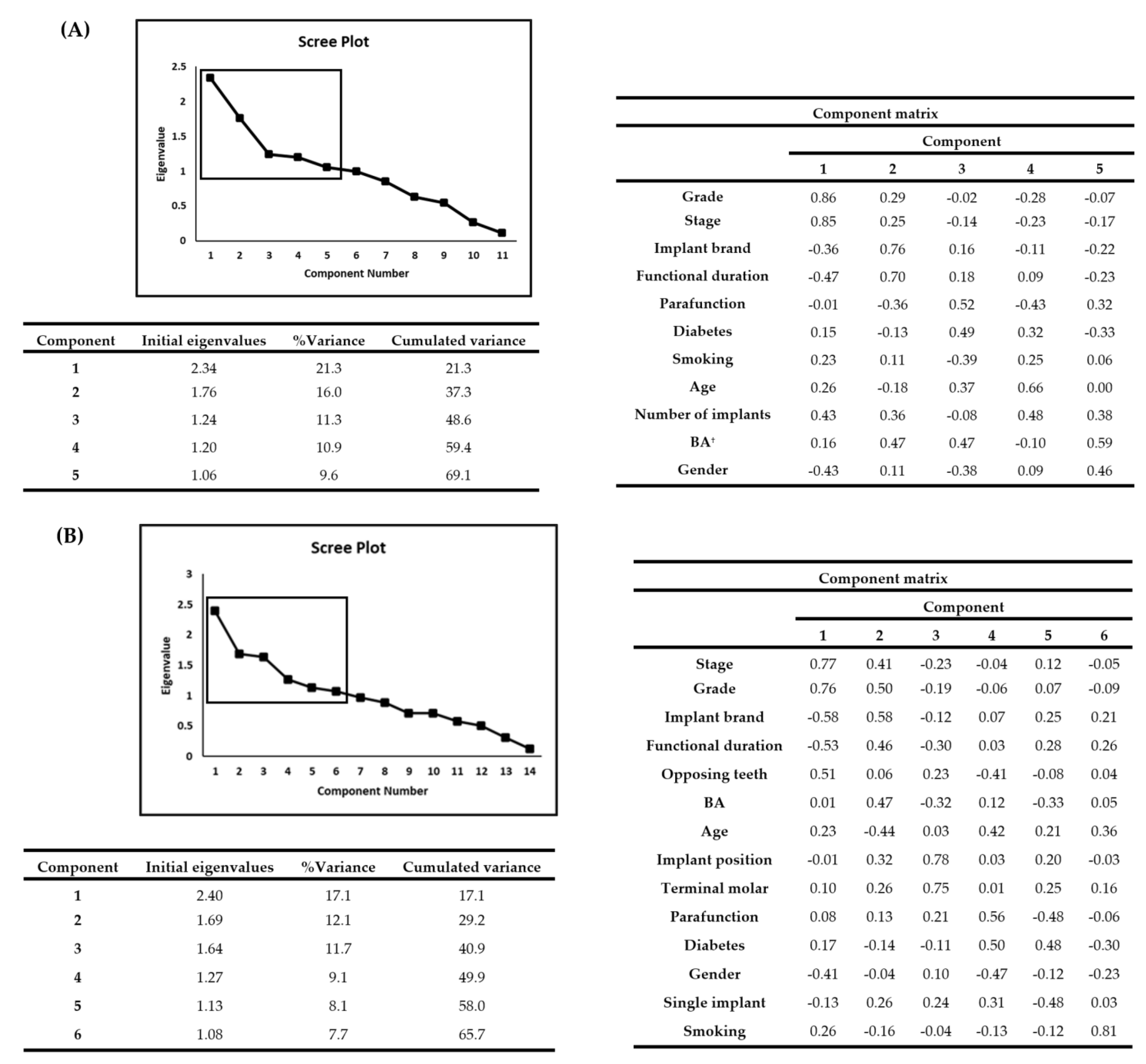
2.6. Statistical Analyses

Results were analyzed using SPSS version 28 (IBM, Chicago, IL, USA) with p < 0.05 deemed significant. Data were evaluated using ANOVA, chi-squared test, Fisher’s exact test, Welch’s test or Student’s t-test, as appropriate. Chi-squared test, Welch’s test, Fisher’s exact test and ANOVA were used to compare related factors among Stage and Grade of periodontitis. Principal components analysis (PCA) and two-step cluster analysis (CA) were performed to identify similarities and differences among observed variables at implant and patient level. PCA examined 15 variables (age, gender, implant brand, function duration, implantation position, number of implants, Stage, Grade, BA, single implant, terminal molar, type of opposite tooth, parafunction, smoking and diabetes). We selected principal components with eigenvalues >1. CA was performed based on the principal component scores. The number of clusters was determined based on Akaike information criterion (AIC) [16].

3. Results

3.1. Study Participants

Of the 98 (383 implants) patients who had received comprehensive treatment for periodontitis between 2006 and 2018 at Ohu University Dental Hospital (Fukushima, Japan), 84 patients (325 implants) were enrolled for this study. For the aforementioned reasons, 14 patients with 58 implants had to be excluded. Data on the 84 enrolled patients and their 325 implants are presented in Table 1. Differences in prevalence of peri-implant disease were assessed at both patient and implant level. There was no significant difference in patient age by Stage or Grade at first visit. Mean follow-up period was significantly longer in patients implanted with POI than in those implanted with Bmk, regardless of Stage or Grade (p < 0.01).
The distribution between maxilla and mandible did not differ significantly among the two groups, although a slightly greater proportion of implants was observed in posterior mandibles. Implant lengths were almost all within the 8–12 mm range, with 10 mm accounting for 90.5% of them. Implant-supported reconstructions included 50 single units and 275 multi-units fixed dentures.

3.2. Treatment Outcomes

3.2.1. Patient Level

(1) Survival and success rates
Survival and success rates were 85.7% and 72.6%, respectively. Of the 84 patients, 4 (4.8%) experienced loss of implants due to peri-implantitis, 7 (8.3%) showed peri-implantitis and 7 showed MBL ≥ 3 mm, 61 (72.6%) showed MBL < 3 mm and 5 (7.1%) showed LoO (Table 2A).
(2) Treatment outcomes
Instances of implant loss due to peri-implantitis and MBL ≥3 mm were significantly higher in Stage IV patients (p < 0.05). Instances of MBL < 3 mm were significantly lower in Stage IV patients (p < 0.05) (Table 3, Tables S1 and S2). Mean MBL differed between smokers (2.19 ± 0.82 mm) and non-smokers (1.57 ± 0.62 mm) (p < 0.01; Student’s t-test). The number of implants and degree of bone resorption were significantly higher in the BA than non-BA group (p < 0.05, t-test). Mean MBL in BA and non-BA were 4.83 ± 3.46 mm and 3.33 ± 2.63 mm, respectively.
(3) Relationship between periodontitis severity and number of implants
The mean numbers of implants placed in Stage IV, III and I patients were 5.71 ± 3.52, 3.24 ± 2.36 and 3.07 ± 1.98, respectively. The number in Stage IV patients was significantly higher than in Stage I and III patients (p < 0.05; ANOVA) and in Grade C (4.50 ± 3.18) than in Grade A (2.50 ± 1.37), (p < 0.05; Welch’s test).
(4) Threshold for increased risk of implant failure by number of implants
The prevalence of peri-implant disease differed by number of implants per patient (p < 0.01) at the rate of MBL ≥3 mm with three or more implants and peri-implantitis with eight or more implants, respectively.
(5) PCA and CA
The results shown in Figure 1A were obtained by PCA. In the subsequent analysis, five principal components with eigenvalues ≥1 were assessed. The % variance of component 1 was 21.3%. Component 1 was strongly influenced by Stage and Grade. Factors other than those were functional duration, number of implants and gender. See Figure 1A for details of the 2nd to 5th principal components.
Four clusters were revealed by CA (Table 4). The number of clusters was determined based on Akaike information criterion (AIC: 282.7).
Subgroups included difficult-case patients with several implants and a high proportion of BA (cluster 1), with a high number of non-compliers with smoking (cluster 4), and compromised hosts with poor prognosis despite few conditions considered high-risk (cluster 2) for developing peri-implant disease. In contrast, there was a low-risk group (cluster 3) with low severity of periodontitis and better treatment outcomes.

3.2.2. LoO

The LoO group had different characteristics from other peri-implant disease groups, and there was effect on neither severity of periodontitis nor number of implants. In contrast, BA combination group (p < 0.05), POI group (p < 0.01) and single implant prosthesis (p < 0.01) were highly significant factors of implant failure (Table 3). Occlusal functional duration of implant was significantly shorter in the LoO group than in the group with MBL < 3 mm (p < 0.05; ANOVA).

3.2.3. Implant Level

(1) Survival and success rates
Survival and success rates were 96.3% and 87.1%, respectively. Of the 325 implants, 5 (1.5%) dropped out due to peri-implantitis, 14 (4.3%) suffered from peri-implantitis, 16 (4.9%) showed MBL ≥3 mm, 283 (87.1%) showed MBL <3 mm and 7 (2.2%) dropped out due to LoO (Table 2B).
(2) Treatment outcomes
Stage IV (p < 0.05) and BA (p < 0.05) were significantly higher in PI (L) group. Stage IV (p < 0.05), implant brand (Bmk, p < 0.01) and smoking (p < 0.01) were significantly higher in PI group. Stage IV (p < 0.05) was significantly higher in MBL ≥3 mm group. BA (p < 0.05), implant brand (POI, p < 0.01), smoking (p < 0.01) and single implant (p < 0.01) were significantly higher, while functional duration was significantly shorter (p < 0.05) in LoO group (Table 3, Tables S3 and S4). In contrast, Stage IV (p < 0.05), single implant (p < 0.01) and smoking (p < 0.01) were significantly lower in MBL <3 mm group. Mean MBL differed between smokers (2.44 ± 1.51 mm) and non-smokers (1.66 ± 0.90 mm) (p < 0.01; Welch’s test). The proportion of patients that received guided bone regeneration was significantly higher in Stage III and in Grade C groups (p < 0.01; chi-square test). Mean MBL differed between Stage I (1.52 ± 0.67 mm) and IV groups (1.92 ± 1.16 mm) (p < 0.05; Welch’s test).
(3) PCA and CA
The results shown in Figure 1B were obtained by PCA. In the subsequent analysis, six principal components with eigenvalues ≥ 1 were used. The % variance of component 1 was 17.1%. Component 1 was strongly influenced by Stage and Grade. Factors other than that were implant brand, functional duration, opposing tooth and gender. See Figure 1B for details of the 2nd to 6th principal components.
Three clusters were revealed by CA (Table 5). The number of clusters was determined based on Akaike information criterion (AIC: 1133.7). Subgroups included difficult-case patients with several implants and a high proportion of BA (cluster 2), with a high number of non-compliers with smoking (cluster 3). In contrast, there was a low-risk group (cluster 1) with low severity of periodontitis and better treatment outcomes.

4. Discussion

Within the limitation of this retrospective study, it tends to show that severity (Stage) of periodontitis is the most critical determinant of increased risk of peri-implant diseases. In contrast, implant loss by LoO occurs regardless of periodontitis severity, suggesting that occlusal overload, parafunction and BA may be involved in the pathogenesis. Implant failure may result from single, combined or multiple factors. It is important to perform patient risk assessment and to establish a suitable preventive regimen for each patient as personalized medicine.
Our finding that patients with severe periodontitis tend to suffer more from peri-implant disease, including peri-implantitis, was similar to findings reported previously [17,18,19]. Kang et al. reported early failure before occlusal function [20]. Low incidence of early failure in the present study suggests surgical skill within our team was sufficient.
The number of patients receiving BA and the number of implants were significantly higher in Stages III, IV and Grade C groups. Possible explanations include fewer remaining teeth, more hopeless teeth and greater bone resorption in Stage III and IV patients. Although BA is an established practice, it is not risk-free, as flap dehiscence and post-operative infection occasionally occur [21]. In addition, artificial bone such as hydroxyapatite or β-TCP may be of poorer quality than real bone [22] and osseointegration between an implant and artificial bone may be weaker than that between an implant and autologous bone.
Canullo et al. reported a higher prevalence of surgically-triggered peri-implantitis than prosthetically- and plaque-induced peri-implantitis, which supports the possibility that the surgical skill of the dentist is a prime factor in implant failure [6]. In fact, the success rate of implant surgery was not 100% in our clinics; if treatment modalities such as BA are involved, the challenge to dentists may increase and treatment success rate drop.
Blood flow around an implant is inferior to that around a natural tooth and may be obstructed, causing increased risk of destruction of hard and soft tissues in patients with multiple implants [23]. In addition, subtle errors during surgery may increase the probability of prosthetically- and surgically-triggered peri-implantitis [6]. If the interface is not tight, a slight gap may be present between the implant and abutment, however even when no gap was observed on dental X-ray radiographs, bacteria were found between the implant and abutment during maintenance [24]. It was reported that implant-abutment assemblies with less tight interfaces lead to inflammatory reactions due to bacterial infection [25].
Papantonopoulos et al. reported cluster patterns and classified patients into two phenotypes: “resistance” and “susceptibility” to peri-implantitis [26]. Likewise, we found similar clusters for peri-implantitis, with a group of clusters characterizing possible risk factors. Therefore, risk assessment of periodontitis in each patient is useful to predict implant failure.
At the patient level, clusters 1, 2 and 4 were a group of patients with poorer outcomes than cluster 3, and included possible risk factors such as high periodontitis severity, BA, smoking and increased number of implants. Similarly, at the implant level, clusters 2 and 3 included possible risk factors that may increase treatment difficulty, such as high periodontitis severity, smoking and opposing teeth.
The worst failure of implant treatment is loss of the implant body, which can be attributed to peri-implantitis, LoO and damage to the implant body [2,27]. In the present study, although the final outcomes of peri-implantitis and LoO were the same, the clinical features of LoO distinctly differed from the former. Time to onset was significantly shorter in LoO group than other groups, and no association with severity of periodontitis was observed. All patients with implant-body detachment due to peri-implantitis were found in Stage IV/Grade C, while LoO was also found in Stage I and Grade A. Occlusal overload was reported to be involved in LoO [28]. Compared to natural teeth, implants without periodontal ligaments may lack osseointegration and detach in a short period of time following excessive occlusal force.
The present study has some limitations. Multivariate analysis to calculate odds ratios requires a sufficient number of samples per factor [5,13], however the number of samples in this study might have been insufficient to calculate the odds ratios robustly. In addition, baseline MBL measurements were not consistently obtained at the same times as previous studies, including at time of implant placement [29], at 1-year after occlusal function [5] and at 1-year after final prosthesis [13]. It was reported that peri-implant bone had been remodeled after implant placement and occlusal function [29], however the observation period in this study was relatively shorter than those in other studies. Accordingly, we took MBL before bone remodeling as the baseline and defined pathological bone resorption as ≥3 mm.
There has been wide heterogeneity in the prevalence of peri-implantitis among previous reports [30,31]. Smoking, history of periodontitis, width of keratinized mucosa and systemic diseases including diabetes mellitus and hypertension may influence implant survival and success [32]. Our results regarding smoking were similar to those of a previous study [13], although this factor might have had minimal impact on implant failure in our study, as relatively few smokers were included (as shown in Table 1). History of periodontal disease has been reported as a possible risk factor for peri-implantitis [13,33], however, in the absence of clinical records, self-reports by patients relying on their memories may be unreliable. This is a main reason why researchers should use Stage and Grade instead of history of periodontal disease.

5. Conclusions

This study demonstrates that severity of periodontitis is a significant risk for development of peri-implant disease at both patient and implant levels. The Stage and Grade classification of periodontitis could be a useful tool to predict the treatment outcome of implant therapy of patients with different types of periodontitis. Further study is required to compare treatment outcome between high- and low-risk groups for peri-implant diseases in a prospective cohort study.

Supplementary Materials

The following supporting information can be downloaded at: https://www.mdpi.com/article/10.3390/jpm12101723/s1, Table S1:【Patient level】Fisher’s exact test (Treatment outcome × Stage); Table S2:【Patient level】Fisher’s exact test (Treatment outcome × Grade); Table S3:【Implant level】Fisher’s exact test (Treatment outcome × Stage); Table S4:【Implant level】Fisher’s exact test (Treatment outcome × Grade).

Author Contributions

Conceptualization, K.T. and M.Y.; methodology, M.Y. and K.T.; writing—original draft preparation, K.T. and M.Y.; writing—review and editing, K.Y., H.I., B.G.L. and Y.B.; supervision, K.T. and B.G.L. All authors have read and agreed to the published version of the manuscript.

Funding

This research was partially funded by Research Funding Granted by Ohu University President, grant number 202103.

Institutional Review Board Statement

The research protocol was approved in 2018 by Ethics Board, Ohu University (reference number 222).

Informed Consent Statement

Informed consent was obtained from all subjects involved in the study.

Data Availability Statement

The data presented in this study are available on request from the corresponding author.

Conflicts of Interest

The authors declare no conflict of interest.

References

  1. Giannobile, W.V.; Lang, N.P. Are dental implants a panacea or should we better strive to save teeth? J. Dent. Res. 2016, 95, 5–6. [Google Scholar] [CrossRef] [PubMed] [Green Version]
  2. Kate, M.A.; Palaskar, S.; Kapoor, P. Implant failure: A dentist’s nightmare. J. Dent. Implant. 2016, 6, 51–56. [Google Scholar] [CrossRef]
  3. Papapanou, P.N.; Sanz, M.; Buduneli, N.; Dietrich, T.; Feres, M.; Fine, D.H.; Flemmig, T.F.; Garcia, R.; Giannobile, W.V.; Graziani, F.; et al. Periodontitis: Consensus report of Workgroup 2 of the 2017 World Workshop on the Classification of Periodontal and Peri-Implant Diseases and Conditions. J. Clin. Periodontol. 2018, 45 (Suppl. 20), S162–S170. [Google Scholar] [CrossRef] [PubMed]
  4. Tarnow, D.P. Increasing prevalence of peri-implantitis: How will we manage? J. Dent. Res. 2016, 95, 7–8. [Google Scholar] [CrossRef] [PubMed]
  5. Derks, J.; Schaller, D.; Håkansson, J.; Wennström, J.L.; Tomasi, C.; Berglundh, T. Effectiveness of implant therapy analyzed in a Swedish population: Prevalence of peri-implantitis. J. Dent. Res. 2016, 95, 43–49. [Google Scholar] [CrossRef]
  6. Canullo, L.; Tallarico, M.; Radovanovic, S.; Delibasic, B.; Covani, U.; Rakic, M. Distinguishing predictive profiles for patient-based risk assessment and diagnostics of plaque induced, surgically and prosthetically triggered peri-implantitis. Clin. Oral Implant. Res. 2016, 27, 1243–1250. [Google Scholar] [CrossRef]
  7. Heitz-Mayfield, L.J.A.; Heitz, F.; Lang, N.P. Implant Disease Risk Assessment IDRA-a tool for preventing peri-implant disease. Clin. Oral Implant. Res. 2020, 31, 397–403. [Google Scholar] [CrossRef] [Green Version]
  8. Sarbacher, A.; Papalou, I.; Vagia, P.; Tenenbaum, H.; Huck, O.; Davideau, J.L. Comparison of Two Risk Assessment Scores in Predicting Peri-Implantitis Occurrence during Implant Maintenance in Patients Treated for Periodontal Diseases: A Long-Term Retrospective Study. J. Clin. Med. 2022, 11, 1720. [Google Scholar] [CrossRef]
  9. Papantonopoulos, G.; Gogos, C.; Housos, E.; Bountis, T.; Loos, B.G. Peri-implantitis: A complex condition with non-linear characteristics. J. Clin. Periodontol. 2015, 42, 789–798. [Google Scholar] [CrossRef]
  10. Schwarz, F.; Derks, J.; Monje, A.; Wang, H.L. Peri-implantitis. J. Periodontol. 2018, 89 (Suppl. 1), S267–S290. [Google Scholar] [CrossRef]
  11. Lindhe, J.; Meyle, J.; Group D of European Workshop on Periodontology. Peri-implant diseases: Consensus Report of the Sixth European Workshop on Periodontology. J. Clin. Periodontol. 2008, 35 (Suppl. 8), 282–285. [Google Scholar] [CrossRef] [Green Version]
  12. Berglundh, T.; Armitage, G.; Araujo, M.G.; Avila-Ortiz, G.; Blanco, J.; Camargo, P.M.; Chen, S.; Cochran, D.; Derks, J.; Figuero, E.; et al. Peri-implant diseases and conditions: Consensus report of workgroup 4 of the 2017 World Workshop on the Classification of Periodontal and Peri-Implant Diseases and Conditions. J. Clin. Periodontol. 2018, 45 (Suppl. 20), S286–S291. [Google Scholar] [CrossRef] [PubMed] [Green Version]
  13. Wada, M.; Mameno, T.; Onodera, Y.; Matsuda, H.; Daimon, K.; Ikebe, K. Prevalence of peri-implant disease and risk indicators in a Japanese population with at least 3 years in function-A multicentre retrospective study. Clin. Oral Implant. Res. 2019, 30, 111–120. [Google Scholar] [CrossRef] [PubMed]
  14. Derks, J.; Schaller, D.; Håkansson, J.; Wennström, J.L.; Tomasi, C.; Berglundh, T. Peri-implantitis-onset and pattern of progression. J. Clin. Periodontol. 2016, 43, 383–388. [Google Scholar] [CrossRef]
  15. Manicone, P.F.; Passarelli, P.C.; Bigagnoli, S.; Pastorino, R.; Manni, A.; Pasquantonio, G.; D’Addona, A. Clinical and radiographic assessment of implant-supported rehabilitation of partial and complete edentulism: A 2 to 8 years clinical follow-up. Eur. Rev. Med. Pharmacol. Sci. 2018, 22, 4045–4052. [Google Scholar] [CrossRef]
  16. Akaike, H. A new look at the statistical model identification. IEEE Trans. Automat. Contr. 1974, 19, 716–723. [Google Scholar] [CrossRef]
  17. Vagia, P.; Papalou, I.; Burgy, A.; Tenenbaum, H.; Huck, O.; Davideau, J.L. Association between periodontitis treatment outcomes and peri-implantitis: A long-term retrospective cohort study. Clin. Oral Implant. Res. 2021, 32, 721–731. [Google Scholar] [CrossRef]
  18. Gatti, C.; Gatti, F.; Chiapasco, M.; Esposito, M. Outcome of dental implants in partially edentulous patients with and without a history of periodontitis: A 5-year interim analysis of a cohort study. Eur. J. Oral Implantol. 2008, 1, 45–51. [Google Scholar]
  19. Pjetursson, B.E.; Helbling, C.; Weber, H.P.; Matuliene, G.; Salvi, G.E.; Brägger, U.; Schmidlin, K.; Zwahlen, M.; Lang, N.P. Peri-implantitis susceptibility as it relates to periodontal therapy and supportive care. Clin. Oral Implant. Res. 2012, 23, 888–894. [Google Scholar] [CrossRef] [Green Version]
  20. Kang, D.Y.; Kim, M.; Lee, S.J.; Cho, I.W.; Shin, H.S.; Caballé-Serrano, J.; Park, J.C. Early implant failure: A retrospective analysis of contributing factors. J. Periodontal Implant Sci. 2019, 49, 287–298. [Google Scholar] [CrossRef]
  21. Briguglio, F.; Falcomatà, D.; Marconcini, S.; Fiorillo, L.; Briguglio, R.; Farronato, D. The use of titanium mesh in guided bone regeneration: A systematic review. Int. J. Dent. 2019, 2019, 1–8. [Google Scholar] [CrossRef]
  22. Zhao, R.; Yang, R.; Cooper, P.R.; Khurshid, Z.; Shavandi, A.; Ratnayake, J. Bone grafts and substitutes in dentistry: A review of current trends and developments. Molecules 2021, 26, 3007. [Google Scholar] [CrossRef]
  23. Berglundh, T.; Lindhe, J.; Jonsson, K.; Ericsson, I. The topography of the vascular systems in the periodontal and peri-implant tissues in the dog. J. Clin. Periodontol. 1994, 21, 189–193. [Google Scholar] [CrossRef]
  24. Broggini, N.; McManus, L.M.; Hermann, J.S.; Medina, R.; Schenk, R.K.; Buser, D.; Cochran, D.L. Peri-implant inflammation defined by the implant-abutment interface. J. Dent. Res. 2006, 85, 473–478. [Google Scholar] [CrossRef]
  25. Assenza, B.; Tripodi, D.; Scarano, A.; Perrotti, V.; Piattelli, A.; Iezzi, G.; D’Ercole, S. Bacterial leakage in implants with different implant-abutment connections: An in vitro study. J. Periodontol. 2012, 83, 491–497. [Google Scholar] [CrossRef] [PubMed]
  26. Papantonopoulos, G.; Gogos, C.; Housos, E.; Bountis, T.; Loos, B.G. Prediction of individual implant bone levels and the existence of implant “phenotypes”. Clin. Oral Implant. Res. 2017, 28, 823–832. [Google Scholar] [CrossRef]
  27. Stoichkov, B.; Kirov, D. Analysis of the causes of dental implant fracture: A retrospective clinical study. Quintessence Int. 2018, 49, 279–286. [Google Scholar] [CrossRef]
  28. Mattheos, N.; Schittek Janda, M.; Zampelis, A.; Chronopoulos, V. Reversible, non-plaque-induced loss of osseointegration of successfully loaded dental implants. Clin. Oral Implant. Res. 2013, 24, 347–354. [Google Scholar] [CrossRef]
  29. Hänggi, M.P.; Hänggi, D.C.; Schoolfield, J.D.; Meyer, J.; Cochran, D.L.; Hermann, J.S. Crestal bone changes around titanium implants. Part I: A retrospective radiographic evaluation in humans comparing two non-submerged implant designs with different machined collar lengths. J. Periodontol. 2005, 76, 791–802. [Google Scholar] [CrossRef] [Green Version]
  30. Rakic, M.; Galindo-Moreno, P.; Monje, A.; Radovanovic, S.; Wang, H.L.; Cochran, D.; Sculean, A.; Canullo, L. How frequent does peri-implantitis occur? A systematic review and meta-analysis. Clin. Oral Investig. 2018, 22, 1805–1816. [Google Scholar] [CrossRef]
  31. Wada, M.; Mameno, T.; Otsuki, M.; Kani, M.; Tsujioka, Y.; Ikebe, K. Prevalence and risk indicators for peri-implant diseases: A literature review. Jpn. Dent. Sci. Rev. 2021, 57, 78–84. [Google Scholar] [CrossRef] [PubMed]
  32. Romandini, M.; Lima, C.; Pedrinaci, I.; Araoz, A.; Soldini, M.C.; Sanz, M. Prevalence and risk/protective indicators of peri-implant diseases: A university-representative cross-sectional study. Clin. Oral Implant. Res. 2021, 32, 112–122. [Google Scholar] [CrossRef]
  33. Arunyanak, S.P.; Sophon, N.; Tangsathian, T.; Supanimitkul, K.; Suwanwichit, T.; Kungsadalpipob, K. The effect of factors related to periodontal status toward peri-implantitis. Clin. Oral Implant. Res. 2019, 30, 791–799. [Google Scholar] [CrossRef] [PubMed]
Figure 1. (A). Parameters on the steep slope of the scree plot are typically selected as principal. Principal components analysis at patient level. As a result of principal components analysis, the 5 components that showed eigenvalues >1 were selected from the 11 evaluated. †: bone augmentation (B). Principal components analysis at implant level. Of the 14 evaluated, 6 components that showed eigenvalues >1 were selected.
Jpm 12 01723 g001
Table 1. Participant and implant characteristic features.
Features at the Patient Level (n = 84)FindingsFeatures at the Implant Level (n = 325)Findings
GenderMale41.7%Severity of periodontitis (Stage)13.2%
Female58.3% 16.0%
Age (Years) 54.7 ± 11.5 33.8%
≤4928.6% 36.9%
50–5933.3%Risk of periodontitis (Grade)A12.3%
60–6929.8% B24.0%
70–798.3% C63.7%
≥800.0%Implant brandPOI48.6%
Diabetes HbA1c ≤ 7%6.0% Bmk51.4%
Smokers 8.3%Implant diameter (mm) 3.3–5.0
≥10 cigarettes/day1.7%Implant length (mm) 7.0–11.5
Severity of periodontitis (Stage)16.7%Functional duration (year) 5.2 ± 2.5
17.9% POI6.7 ± 2.2
40.5% Bmk3.8 ± 1.8
25.0%Type of prosthesisSingle-unit21.5%
Age of patients among four groups (Stage) Ⅰ; 51.1 ± 14.7 (29–74) Multi-unit fixed78.5%
Ⅱ; 59.1 ± 7.5 (48–71)Implant position
Ⅲ; 54.1 ± 11.0 (31–74)Upper jawincisor7.1%
Ⅳ; 54.8 ± 11.9 (26–69) canine1.8%
Risk of periodontitis (Grade)A19.0% premolar15.1%
B26.2% molar26.5%
C54.8%Lower jawincisor2.8%
Age of patients among three groups (Grade) A; 50.9 ± 13.2 (29–74) canine2.5%
B; 59.7 ± 10.0 (31–74) premolar10.8%
C; 53.6 ± 10.9 (26–70) molar33.5%
Implant brandPOI52.4%Opposing teethnatural teeth58.5%
Bmk 47.6% implant41.5%
Functional duration (year) 5.1 ± 2.5Terminal molar 37.5%
POI6.6 ± 2.1Bone augmentation 21.5%
Bmk3.4 ± 1.7Parafunction 73.5%
Number of implants 3.87 ± 3.02
1~359.5%Survival rate 96.3%
4~622.6%Success rate 87.1%
7~914.3%
≥103.6%
Bone augmentation 35.7%
Parafunction 79.8%
Survival rate 85.7%
Success rate 72.6%
: Ex-smokers were counted as non-smokers. : Brånemark system.
Table 2. Distribution of treatment outcomes classified by Stage and Grade. (A). Patient level (n = 84). (B). Implant level (n = 325).
(A)
MBL
PI (L) PI §≥3 mm<3 mmLoO ††SUM
Stage Ⅰ00111214
01113015
03127334
43410021
Grade A00014216
B03217022
C44530346
(B)
MBL
PI (L)PI≥3 mm<3 mmLoOSUM
Stage Ⅰ00239243
02248052
0321014110
5910951120
Grade A00137240
B05370078
C59121765207
: Patient level shows the worst outcomes in implant treatment per patient. : Peri-implantitis (lost). §: Peri-implantitis (surviving). : Marginal bone loss, MBL (<3 mm) indicates success of implant treatment. ††: Lack of osseointegration.
Table 3. Factors related to implant failure (peri-implant disease, lack of osseointegration).
Treatment Outcome PI (L)PIMBLLoO
≥3 mm<3 mm
Patient Level
Stage Ⅳ * **
Implant Level
Stage Ⅳ ****
GBR * *
Implant brand (POI) **
Implant brand (Bmk) **
Smoker ** **
Functional duration *
Type of prosthesis single ****
*: p < 0.05. **: p < 0.01. : Fisher’s exact test. : One-way ANOVA.
Table 4. Features among four clusters at patient level.
Cluster 1Cluster 2Cluster 3Cluster 4
(n = 22), %(n = 35), %(n = 15), %(n = 12), %
Age (mean) 63.3 50.2 48.9 59.3
GenderMale1045.51954.3213.3433.3
Female1254.51645.71386.7866.7
Implant brandPOI836.42365.7746.7650
Bmk1463.61234.3853.3650
Stage313.6001066.718.3
418.238.6426.7433.3
1254.51851.416.7325
313.6144000433.3
GradeA29.1001280216.7
B627.3822.9320541.7
C1463.62777.100541.7
Number of implants (mean) 5 3 2 6
Treatment outcomePI (L)14.538.60000
PI29.1411.40018.3
MBL ≥ 3 mm29.1411.40018.3
MBL < 3 mm1568.22365.71493.3975
LoO29.112.916.718.3
Marginal bone loss (mm/mean) 1.62 1.71 1.31 1.75
Functional duration (year/mean) 4.2 5.6 5.7 4.7
Bone augmentationNo522.72468.61386.712100
Yes1777.31131.4213.300
ParafunctionNo14.5617.1001083.3
Yes2195.52982.915100216.7
SmokingNo2195.53394.315100866.7
Yes14.525.700433.3
DiabetesNo1777.3351001510012100
Yes522.7000000
Table 5. Features among three clusters at implant level.
Cluster 1Cluster 2Cluster 3
(n = 148), %(n = 101), %(n = 76), %
Age (mean) 59.4 54 61.4
GenderMale5647.95732.91954.3
Female6152.111667.11645.7
Implant brandPOI8673.55934.11337.1
Bmk3126.511465.92262.9
Implant positionincisor1412116.4720
canine54.384.612.9
premolar3025.64526925.7
molar6858.1109631851.4
Stage4336.80000
1714.52413.91131.4
4034.26537.6514.3
1714.58448.61954.3
GradeA4034.20000
B3529.93218.51131.4
C4235.914181.52468.6
Treatment outcomePI(L)0042.312.9
PI10.984.6514.3
MBL ≥ 3 mm32.6116.425.7
MBL < 3 mm1109414986.12468.6
LoO32.610.638.6
Marginal bone loss (mm/mean) 1.48 1.77 2.44
Functional duration (year/mean) 6.6 4.5 4.5
Bone augmentationNo9581.213175.72982.9
Yes2218.84224.3617.1
Type of prosthesis (single-unit)No9278.615388.43085.7
Yes2521.42011.6514.3
Terminal molar No7765.810560.72160
Yes4034.26839.31440
Opposing teethnatural teeth10589.771411440
implant1210.3102592160
ParafunctionNo3328.24224.31131.4
Yes8471.813175.72468.6
SmokingNo11710017310000
Yes000035100
DiabetesNo9883.817310035100
Yes1916.20000
Publisher’s Note: MDPI stays neutral with regard to jurisdictional claims in published maps and institutional affiliations.

