
Correlation of Volume of Macular Edema with Retinal Tomography Features in Diabetic Retinopathy Eyes

 and
and
Abstract
:1. Introduction
2. Methods
- (a)
- Responders—decrease in CFT by 100 μm or more after either the first or second intravitreal injection of steroid or anti-VEGF; (n = 60)
- (b)
- Non-responders—either persisting macular edema or having an increase in CFT by 100 µm or CFT ≤ 100 µm from previous OCT scans, after 3 consecutive intravitreal injections; (n = 63)
- (c)
- Recurrent—return of macular edema with an increase in thickness greater than 100 μm, when compared to the last visit, after an injection free period of 2 months; (n = 26)
- (d)
- Treatment naïve—no visible signs of change in macular edema from consecutive scans at regular follow-ups, or eyes without any prescribed treatment. (n = 29)
Statistical Analyses
3. Results
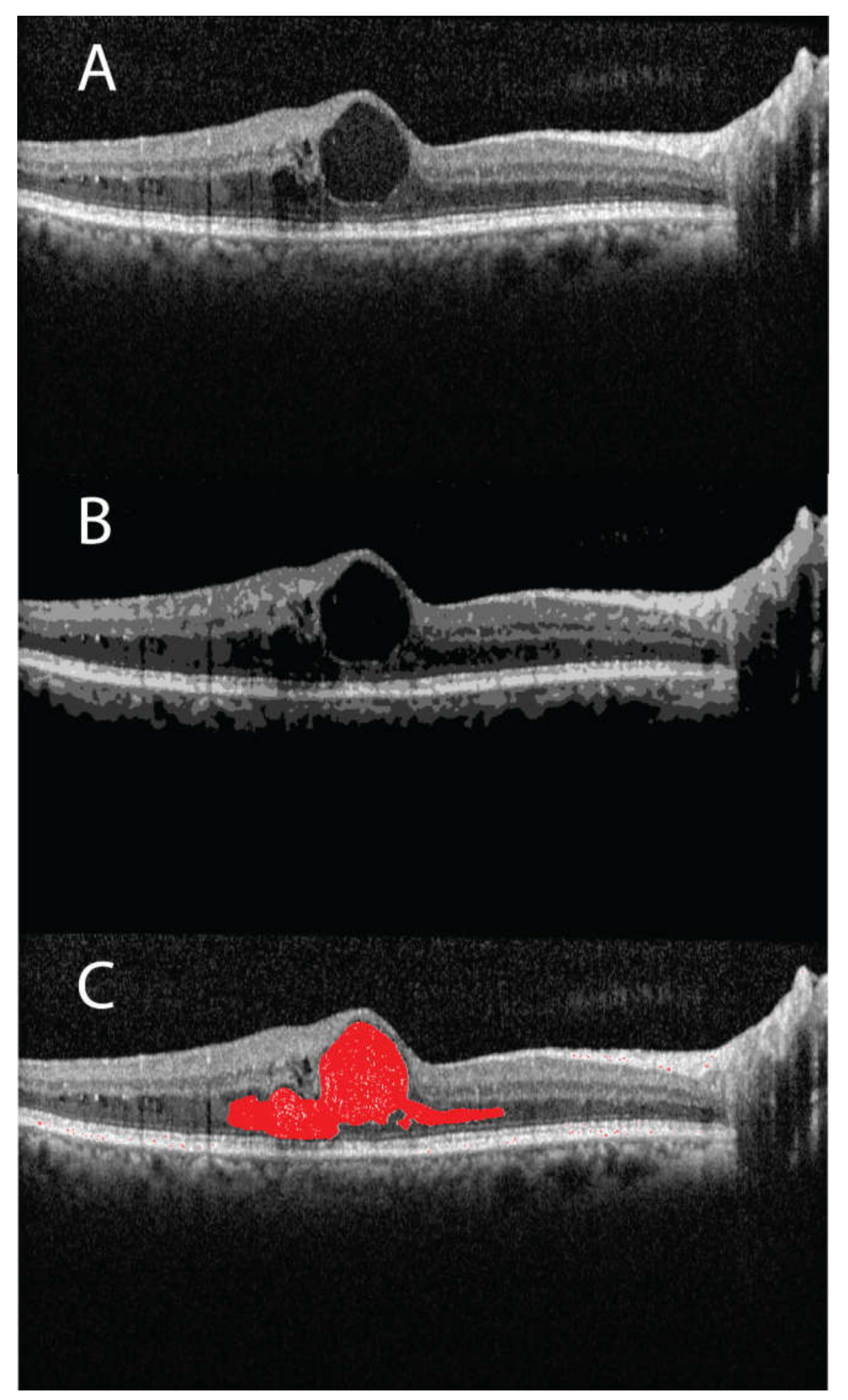
3.1. Segmentation and Edema Volume Calculation
3.2. Tomographic Features vs. DR Grades
3.3. Tomographic Features vs. Treatment Response
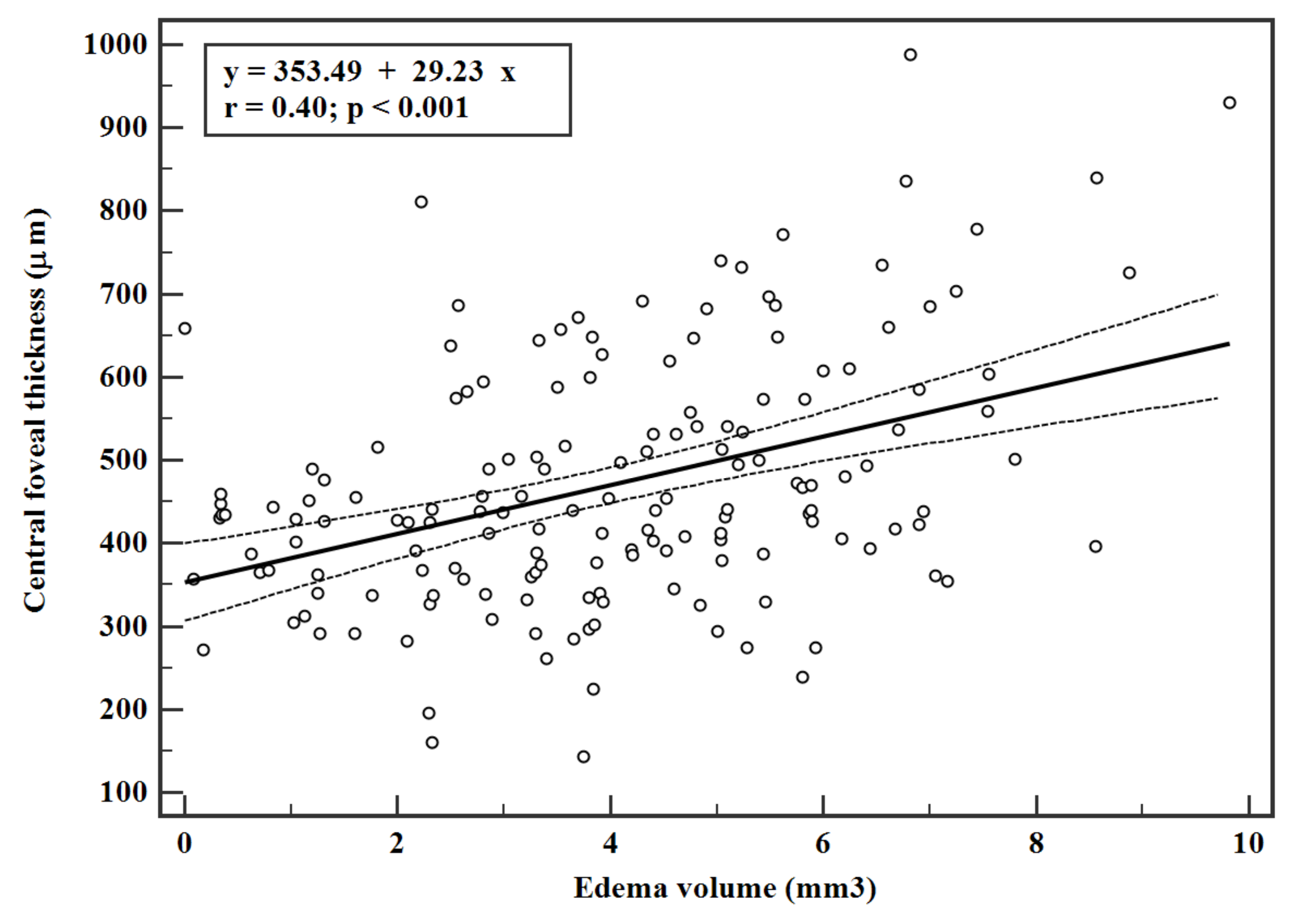
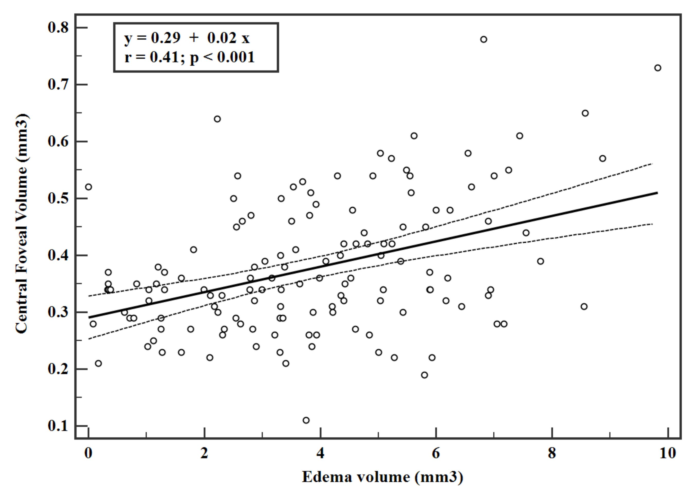
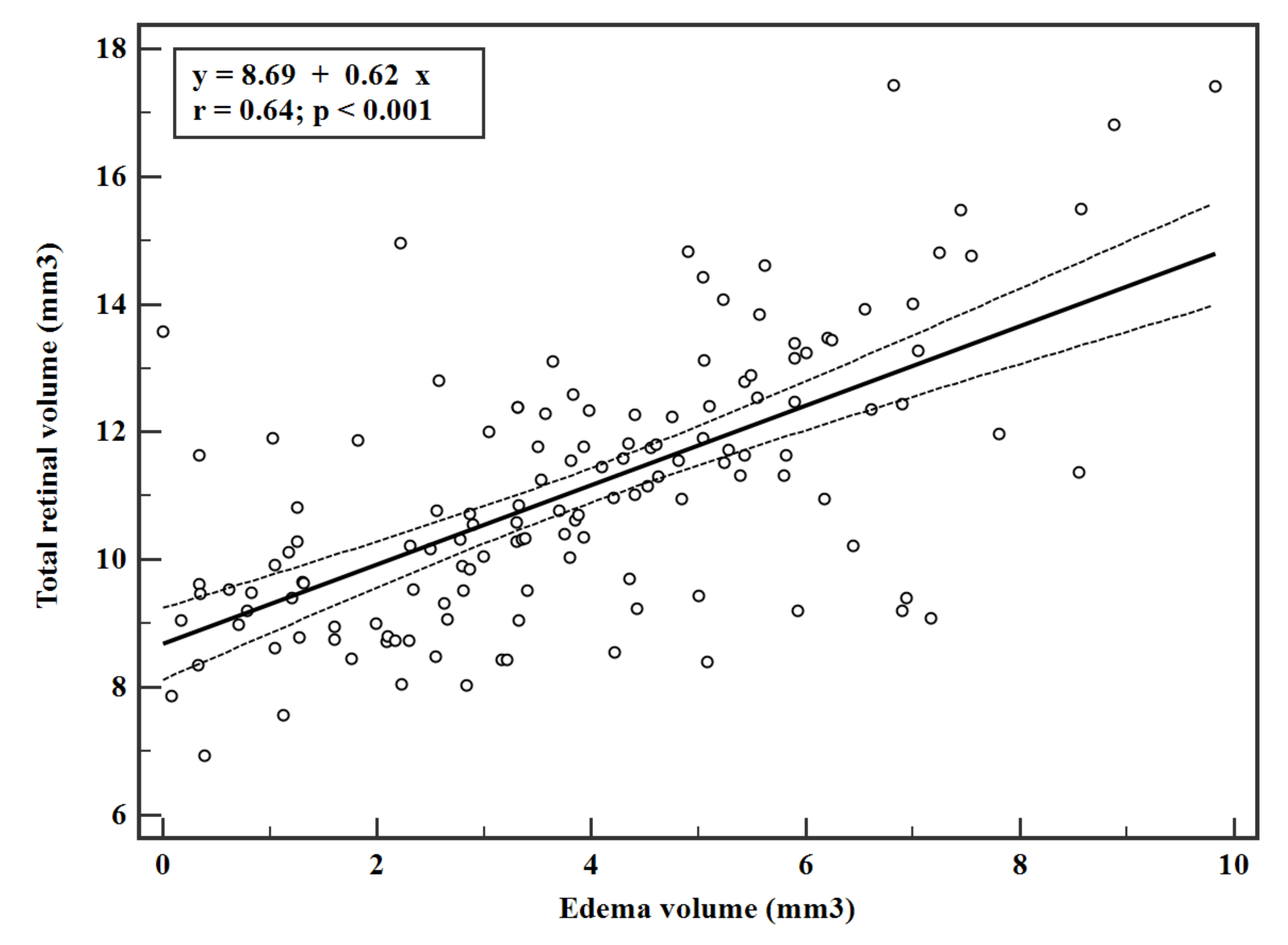
3.4. Correlation among Features
4. Discussion
Author Contributions
Funding
Institutional Review Board Statement
Informed Consent Statement
Data Availability Statement
Acknowledgments
Conflicts of Interest
References
- Nentwich, M.M.; Ulbig, M.W. Diabetic retinopathy-ocular complications of diabetes mellitus. World J. Diabetes 2015, 6, 489–499. [Google Scholar] [CrossRef] [PubMed]
- Ferris, F.L.; Patz, A. Macular edema. A complication of diabetic retinopathy. Surv. Ophthalmol. 1984, 28, 452–461. [Google Scholar] [CrossRef]
- Wu, L.; Fernandez-Loaiza, P.; Sauma, J.; Hernandez-Bogantes, E.; Masis, M. Classification of diabetic retinopathy and diabetic macular edema. World J. Diabetes 2013, 4, 290–294. [Google Scholar] [CrossRef] [PubMed]
- Strøm, C.; Sander, B.; Larsen, N.; Larsen, M.; Lund-Andersen, H. Diabetic macular edema assessed with optical coherence tomography and stereo fundus photography. Investig. Ophthalmol. Vis. Sci. 2002, 43, 241–245. [Google Scholar]
- Sánchez-Tocino, H.; Alvarez-Vidal, A.; Maldonado, M.J.; Moreno-Montañés, J.; García-Layana, A. Retinal thickness study with optical coherence tomography in patients with diabetes. Investig. Ophthalmol. Vis. Sci. 2002, 43, 1588–1594. [Google Scholar]
- Hee, M.R.; Puliafito, C.A.; Duker, J.S.; Reichel, E.; Coker, J.G.; Wilkins, J.R.; Schuman, J.S.; Swanson, E.A.; Fujimoto, J.G. Topography of diabetic macular edema with optical coherence tomography. Ophthalmology 1998, 105, 360–370. [Google Scholar] [CrossRef] [Green Version]
- Cabrera Fernández, D.; Salinas, H.M.; Puliafito, C.A. Automated detection of retinal layer structures on optical coherence tomography images. Opt. Express 2005, 13, 10200–10216. [Google Scholar] [CrossRef]
- Helmy, Y.M.; Atta Allah, H.R. Optical coherence tomography classification of diabetic cystoid macular edema. Clin. Ophthalmol. 2013, 7, 1731–1737. [Google Scholar]
- Srinivasan, P.P.; Kim, L.A.; Mettu, P.S.; Cousins, S.W.; Comer, G.M.; Izatt, J.A.; Farsiu, S. Fully automated detection of diabetic macular edema and dry age-related macular degeneration from optical coherence tomography images. Biomed. Opt. Express 2014, 5, 3568–3577. [Google Scholar] [CrossRef] [Green Version]
- Hassan, B.; Raja, G.; Hassan, T.; Usman Akram, M. Structure tensor based automated detection of macular edema and central serous retinopathy using optical coherence tomography images. J. Opt. Soc. Am. A Opt. Image Sci. Vis. 2016, 33, 455–463. [Google Scholar] [CrossRef]
- Chan, A.; Duker, J.S.; Ko, T.H.; Fujimoto, J.G.; Schuman, J.S. Normal macular thickness measurements in healthy eyes using Stratus optical coherence tomography. Arch. Ophthalmol. 2006, 124, 193–198. [Google Scholar] [CrossRef]
- Bressler, S.B.; Ayala, A.R.; Bressler, N.M.; Melia, M.; Qin, H.; Ferris, F.L., III; Flaxel, C.J.; Friedman, S.M.; Glassman, A.R.; Jampol, L.M.; et al. Persistent macular thickening after ranibizumab treatment for diabetic macular edema with vision impairment. JAMA Ophthalmol. 2016, 134, 278–285. [Google Scholar] [CrossRef] [Green Version]
- Bressler, N.M.; Beaulieu, W.T.; Glassman, A.R.; Blinder, K.J.; Bressler, S.B.; Jampol, L.M.; Melia, M.; Wells, J.A., III. Persistent macular thickening following intravitreous aflibercept, bevacizumab, or ranibizumab for central-involved diabetic macular edema with vision impairment: A secondary analysis of a randomized clinical trial. JAMA Ophthalmol. 2018, 136, 257–269. [Google Scholar] [CrossRef] [Green Version]
- Wilkins, G.R.; Houghton, O.M.; Oldenburg, A.L. Automated segmentation of intraretinal cystoid fluid in optical coherence tomography. IEEE Trans. Biomed. Eng. 2012, 59, 1109–1114. [Google Scholar] [CrossRef] [Green Version]
- Soille, P. Morphological Image Analysis: Principles and Applications; Springer: Berlin/Heidelberg, Germany, 2003; pp. 1–368. [Google Scholar]
- Das, R.; Spence, G.; Hogg, R.E.; Stevenson, M.; Chakravarthy, U. Disorganization of Inner Retina and Outer Retinal Morphology in Diabetic Macular Edema. JAMA Ophthalmol. 2018, 136, 202–208. [Google Scholar] [CrossRef]
- Wilkinson, C.P.; Ferris, F.L., III; Klein, R.E.; Lee, P.P.; Agardh, C.D.; Davis, M.; Dills, D.; Kampik, A.; Pararajasegaram, R.; Verdaguer, J.T.; et al. Proposed international clinical diabetic retinopathy and diabetic macular edema disease severity scales. Ophthalmology 2003, 110, 1677–1682. [Google Scholar] [CrossRef]
- Ebneter, A.; Wolf, S.; Abhishek, J.; Zinkernagel, M.S. Retinal layer response to ranibizumab during treatment of diabetic macular edema. Retina 2016, 36, 1314–1323. [Google Scholar] [CrossRef] [Green Version]
- Schlegl, T.; Waldstein, S.M.; Bogunovic, H.; Endstraßer, F.; Sadeghipour, A.; Philip, A.M.; Podkowinski, D.; Gerendas, B.S.; Langs, G.; Schmidt-Erfurth, U. Fully Automated Detection and Quantification of Macular Fluid in OCT Using Deep Learning. Ophthalmology 2018, 125, 549–558. [Google Scholar] [CrossRef] [Green Version]
- Roberts, P.K.; Vogl, W.D.; Gerendas, B.S.; Glassman, A.R.; Bogunovic, H.; Jampol, L.M.; Schmidt-Erfurth, U.M. Quantification of Fluid Resolution and Visual Acuity Gain in Patients With Diabetic Macular Edema Using Deep Learning: A Post Hoc Analysis of a Randomized Clinical Trial. JAMA Ophthalmol. 2020, 138, 945–953. [Google Scholar] [CrossRef]
- Gerendas, B.S.; Bogunovic, H.; Sadeghipour, A.; Schlegl, T.; Langs, G.; Waldstein, S.M.; Schmidt-Erfurth, U. Computational image analysis for prognosis determination in DME. Vis. Res. 2017, 139, 204–210. [Google Scholar] [CrossRef]
- Chen, X.; Niemeijer, M.; Zhang, L.; Lee, K.; Abramoff, M.D.; Sonka, M. Three-dimensional segmentation of fluid-associated abnormalities in retinal OCT: Probability constrained graph-search-graph-cut. IEEE Trans. Med. Imaging 2012, 31, 1521–1531. [Google Scholar] [CrossRef] [PubMed] [Green Version]
- Xu, X.; Lee, K.; Zhang, L.; Sonka, M.; Abramoff, M.D. Stratified sampling voxel classification for segmentation of intraretinal and sub retinal fluid in longitudinal clinical OCT data. IEEE Trans. Med. Imaging 2015, 34, 1616–1623. [Google Scholar] [CrossRef] [PubMed]

| Mild NPDR | Mod NPDR | Severe NPDR | PDR | p-Value | |
|---|---|---|---|---|---|
| Age (years) | 66 (61.4 to 64.8) | 64 (64.00 to 66.00) | 62 (61.00 to 65.76) | 62.5 (57.33 to 65.00) | 0.5 |
| CDVA (LogMAR) | 0.3 (0.36 to 0.52) | 0.18 (0.16 to 0.30) | 0.3 (0.18 to 0.48) | 0.477 (0.33 to 0.48) | 0.02 |
| Edema Volume (mm3) | 2.86 (0.47 to 3.84) | 2.60 (2.17 to 3.69) | 3.85 (3.34 to 4.70) | 4.011 (3.31 to 4.76) | 0.17 |
| Total Retinal Volume (mm3) | 9.05 (8.40 to 9.79) | 9.52 (9.0 to 10.15) | 11.82 (10.63 to 12.4) | 11.16 (10.75 to 11.80) | <0.001 |
| Central Foveal Volume (mm3) | 0.33 (0.28 to 0.35) | 0.34 (0.32 to 0.38) | 0.42 (0.31 to 0.48) | 0.34 (0.31 to 0.36) | 0.42 |
| Central Foveal Thickness (µm) | 416 (359.24 to 443.11) | 434.5 (402.8 to 471.93) | 461 (400.03 to 536.78) | 435 (398.27 to 452.04) | 0.58 |
| Non-Responder | Recurrent | Responder | Treatment Naïve | p-Value | |
|---|---|---|---|---|---|
| Age (years) | 64 (61.00 to 65.24) | 65 (61.00 to 69.05) | 64 (59.46 to 66.00) | 62 (55.00 to 66.00) | 0.56 |
| CDVA (LogMAR) | 0.48 (0.42 to 0.60) | 0.6 (0.18 to 1.09) | 0.3 (0.18 to 0.48) | 0.477 (0.18 to 0.48) | 0.05 |
| Edema Volume (mm3) | 3.93 (3.31 to 4.49) | 4.675 (2.83 to 5.41) | 3.65 (2.69 to 3.96) | 1.604 (0.37 to 3.82) | 0.11 |
| Total Retinal Volume (mm3) | 10.59 (9.82 to 11.39) | 11.64 (10.81 to 12.42) | 10.74 (10.28 to 11.78) | 10.23 9 (32 to 11.94) | 0.47 |
| Central Foveal Volume (mm3) | 0.34 (0.32 to 0.36) | 0.395 (0.35 to 0.46) | 0.33 (0.280 to 0.40) | 0.34 (0.31 to 0.37) | 0.11 |
| Central Foveal Thickness (µm) | 431 (401.69 to 459.80) | 494.5 (439.55 to 577.51) | 415 (364.07 to 470.43) | 438 (397.51 to 457.57) | 0.18 |
Publisher’s Note: MDPI stays neutral with regard to jurisdictional claims in published maps and institutional affiliations. |
© 2021 by the authors. Licensee MDPI, Basel, Switzerland. This article is an open access article distributed under the terms and conditions of the Creative Commons Attribution (CC BY) license (https://creativecommons.org/licenses/by/4.0/).
Share and Cite
Gadde, S.G.K.; Kshirsagar, A.; Anegondi, N.; Mochi, T.B.; Heymans, S.; Ghosh, A.; Roy, A.S. Correlation of Volume of Macular Edema with Retinal Tomography Features in Diabetic Retinopathy Eyes. J. Pers. Med. 2021, 11, 1337. https://doi.org/10.3390/jpm11121337
Gadde SGK, Kshirsagar A, Anegondi N, Mochi TB, Heymans S, Ghosh A, Roy AS. Correlation of Volume of Macular Edema with Retinal Tomography Features in Diabetic Retinopathy Eyes. Journal of Personalized Medicine. 2021; 11(12):1337. https://doi.org/10.3390/jpm11121337
Chicago/Turabian StyleGadde, Santosh Gopi Krishna, Arpita Kshirsagar, Neha Anegondi, Thirumalesh B. Mochi, Stephane Heymans, Arkasubhra Ghosh, and Abhijit Sinha Roy. 2021. "Correlation of Volume of Macular Edema with Retinal Tomography Features in Diabetic Retinopathy Eyes" Journal of Personalized Medicine 11, no. 12: 1337. https://doi.org/10.3390/jpm11121337
APA StyleGadde, S. G. K., Kshirsagar, A., Anegondi, N., Mochi, T. B., Heymans, S., Ghosh, A., & Roy, A. S. (2021). Correlation of Volume of Macular Edema with Retinal Tomography Features in Diabetic Retinopathy Eyes. Journal of Personalized Medicine, 11(12), 1337. https://doi.org/10.3390/jpm11121337