Development and Initial Validation of the Novel Computational Method for Dynamic Intracardiac Blood Flow Evaluation

 , , , , , ,
Abstract
1. Introduction
2. Materials and Methods
- •
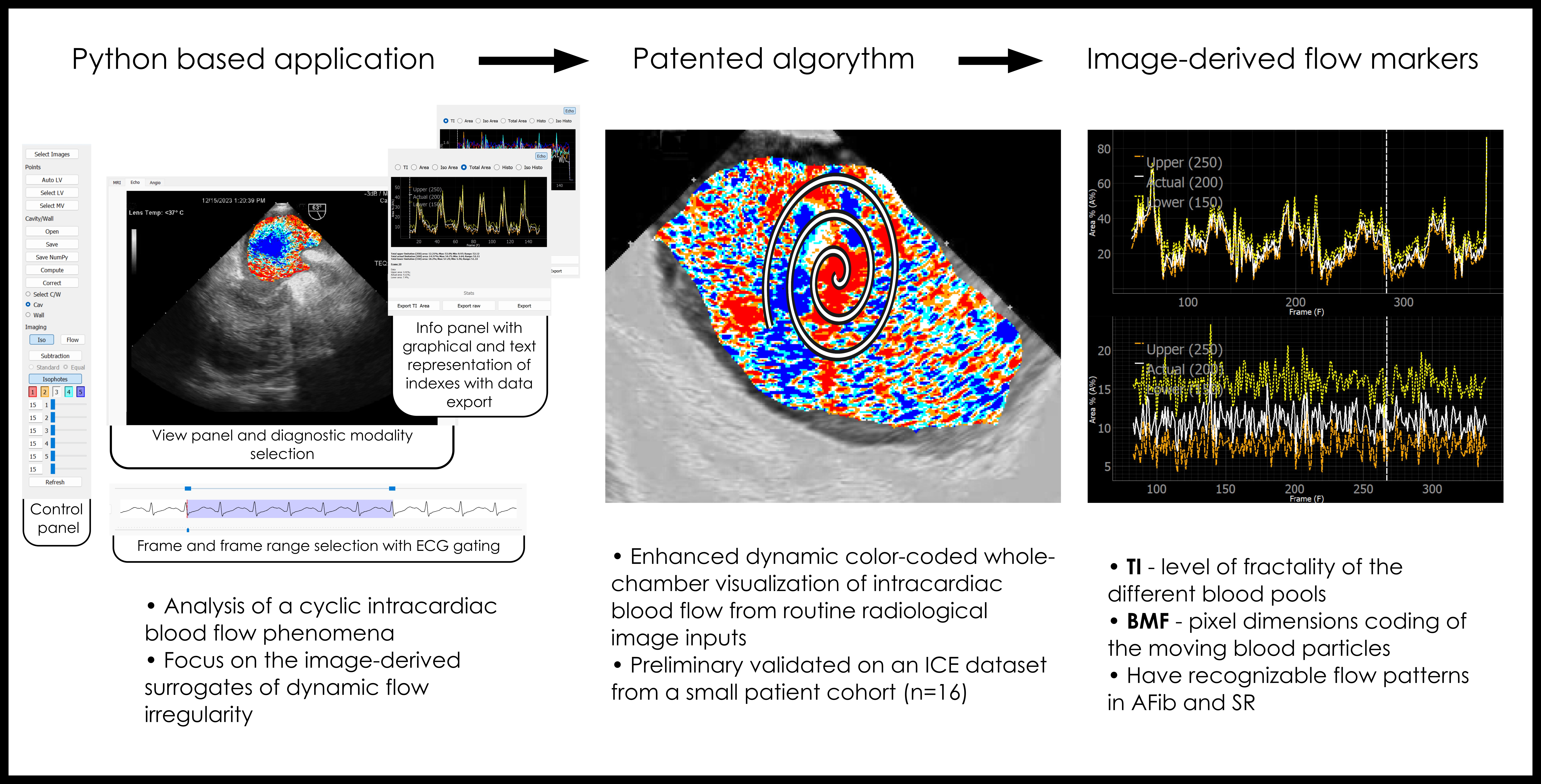
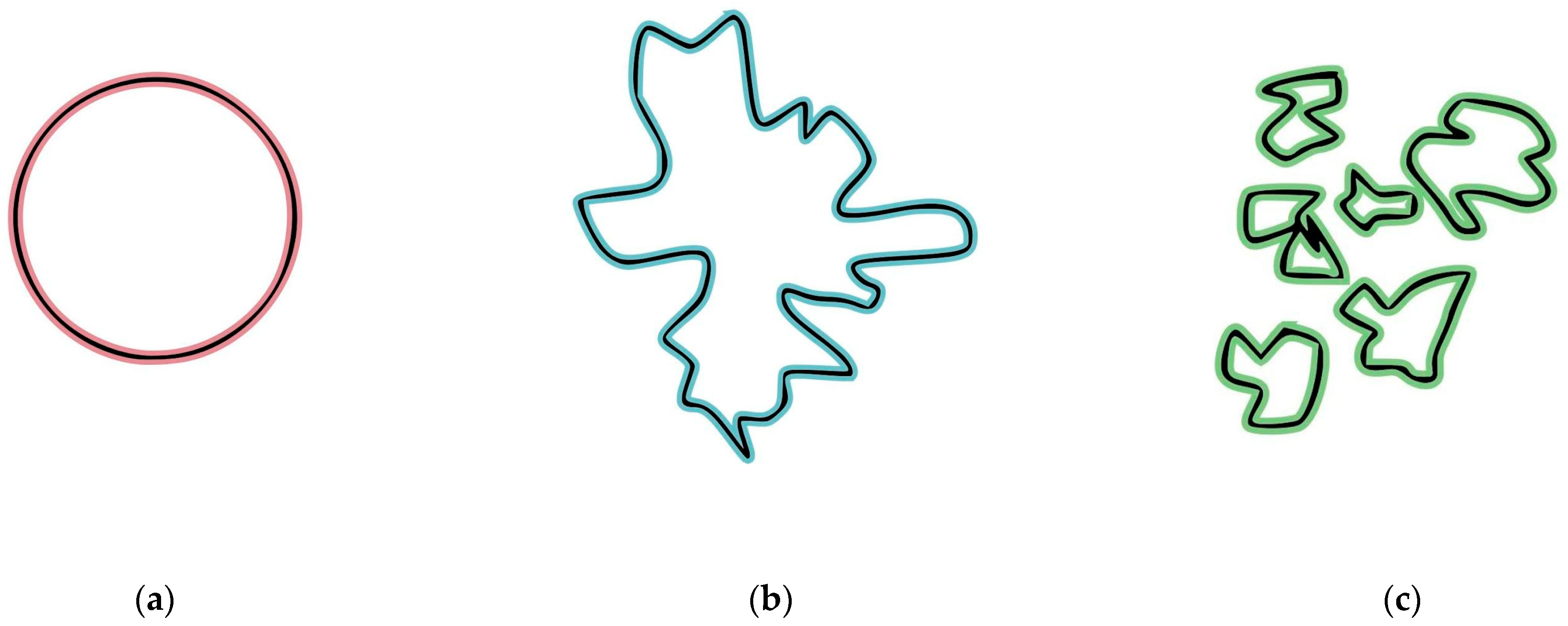
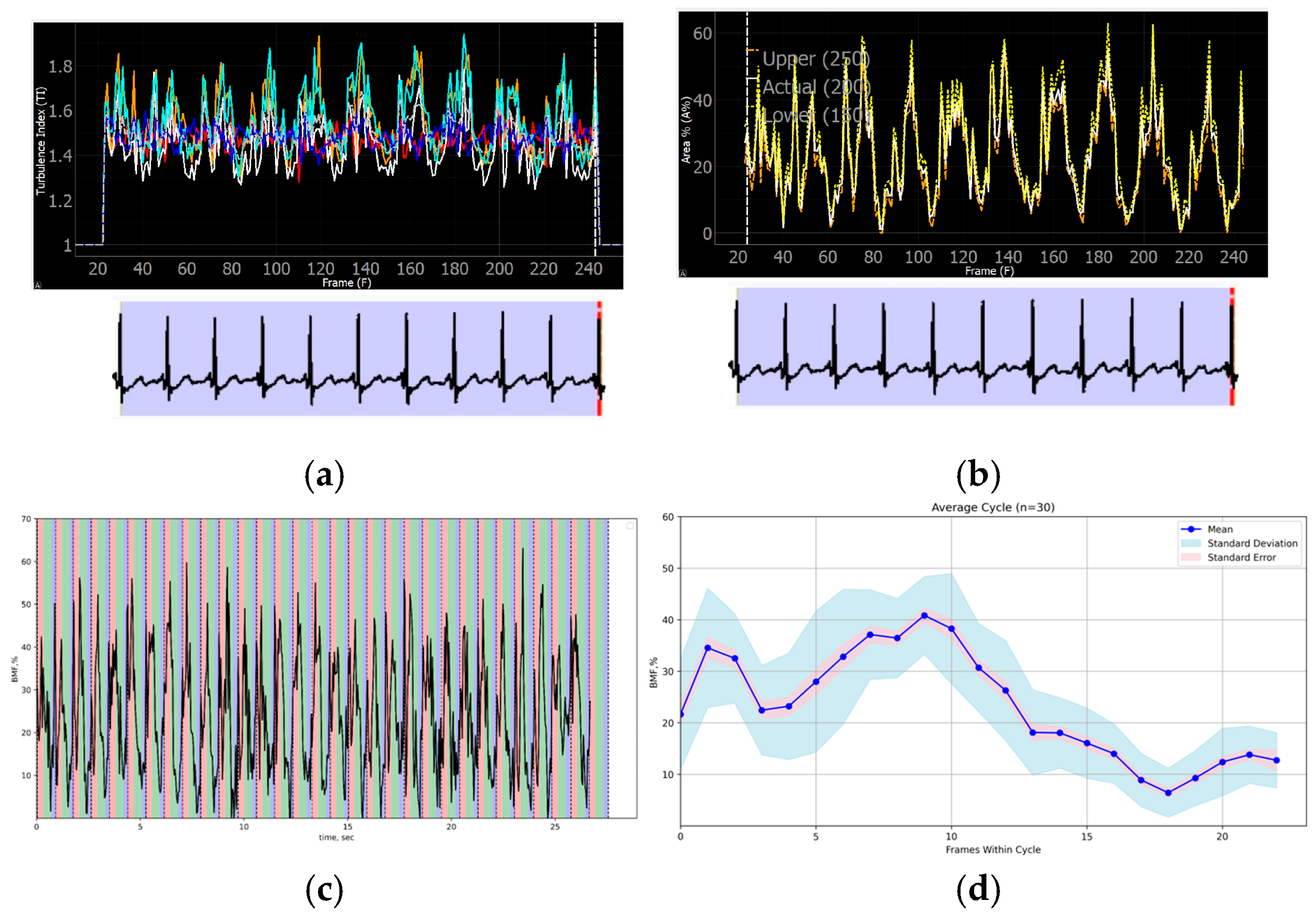
- Turbulence index (TI)—fractality level of the different blood pools;
- •
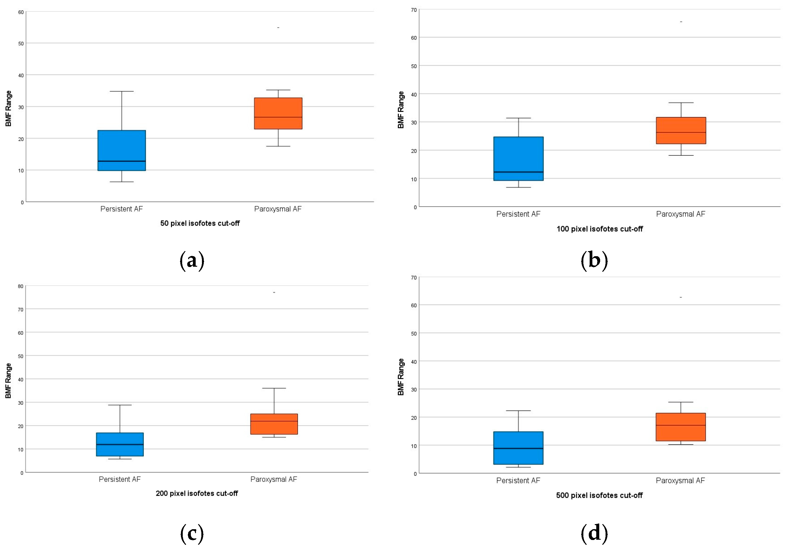
- Blood mobility fraction (BMF)—pixel dimensions coding of the moving blood particles.
3. Results
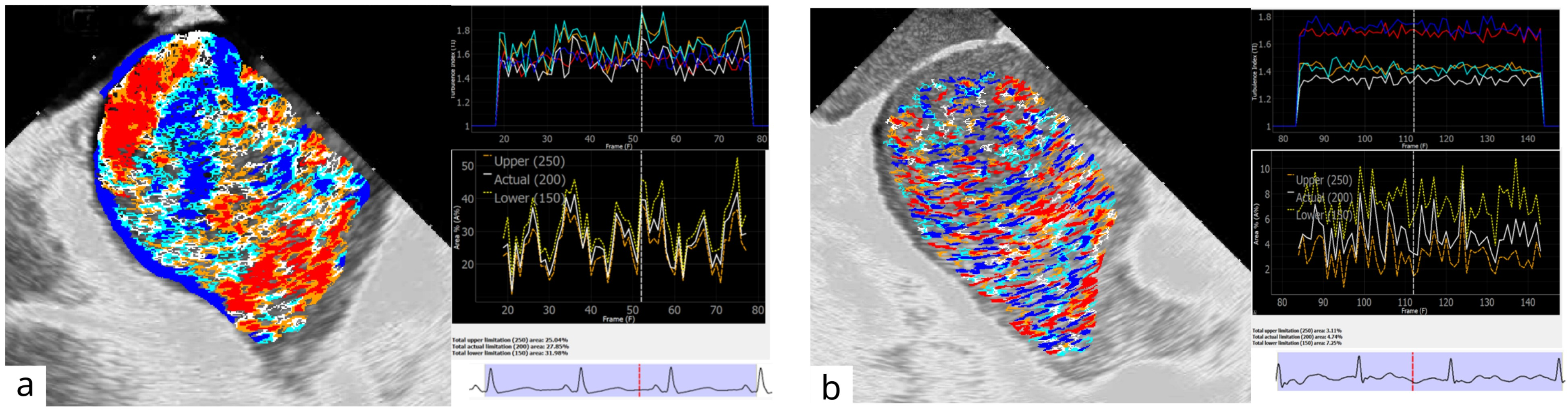
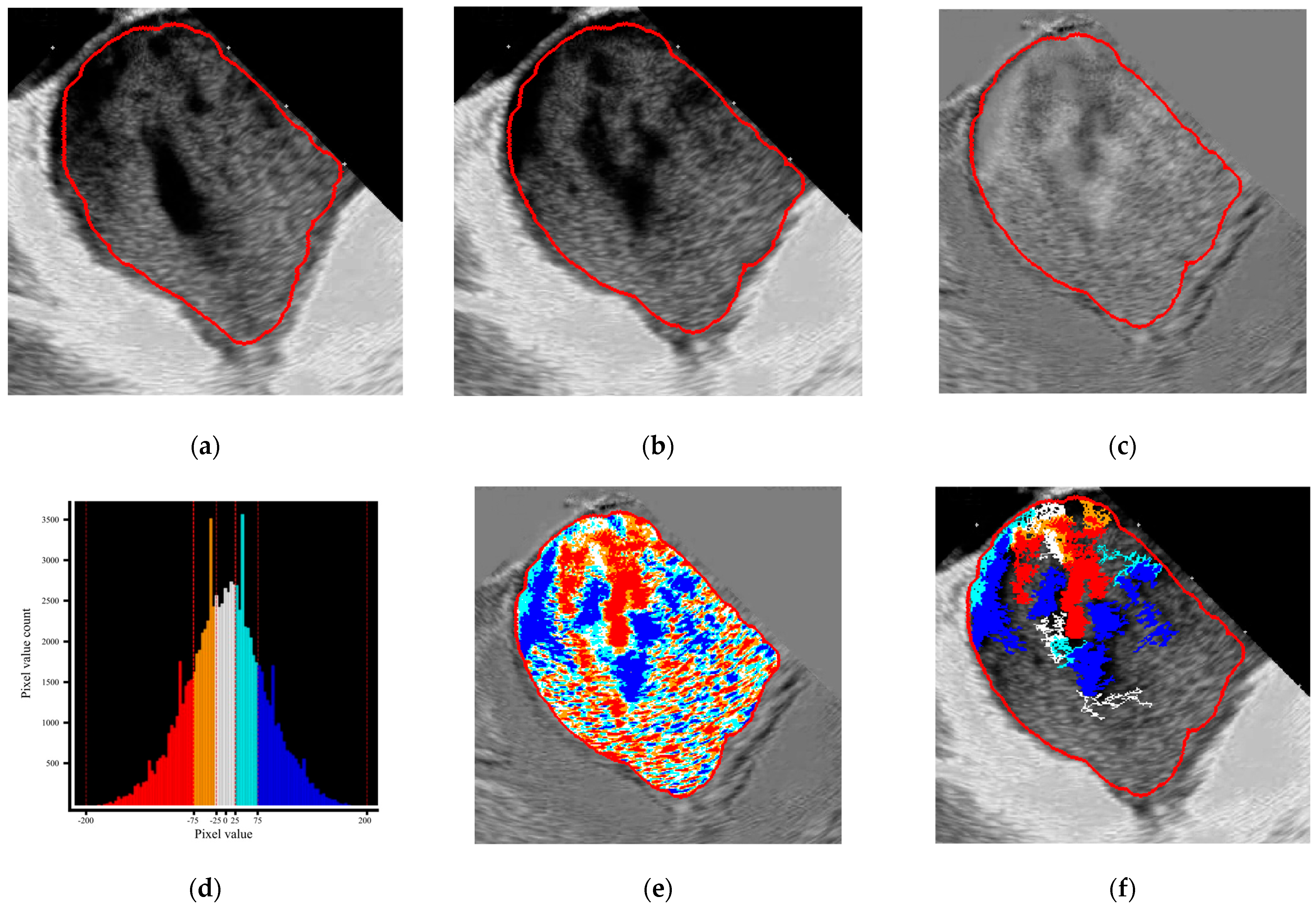
3.1. Visual Representations of the Flow Patterns
3.2. Program-Derived Data Repetitiveness and Correlation with Cardiac Cycle Phases and Other Parameters
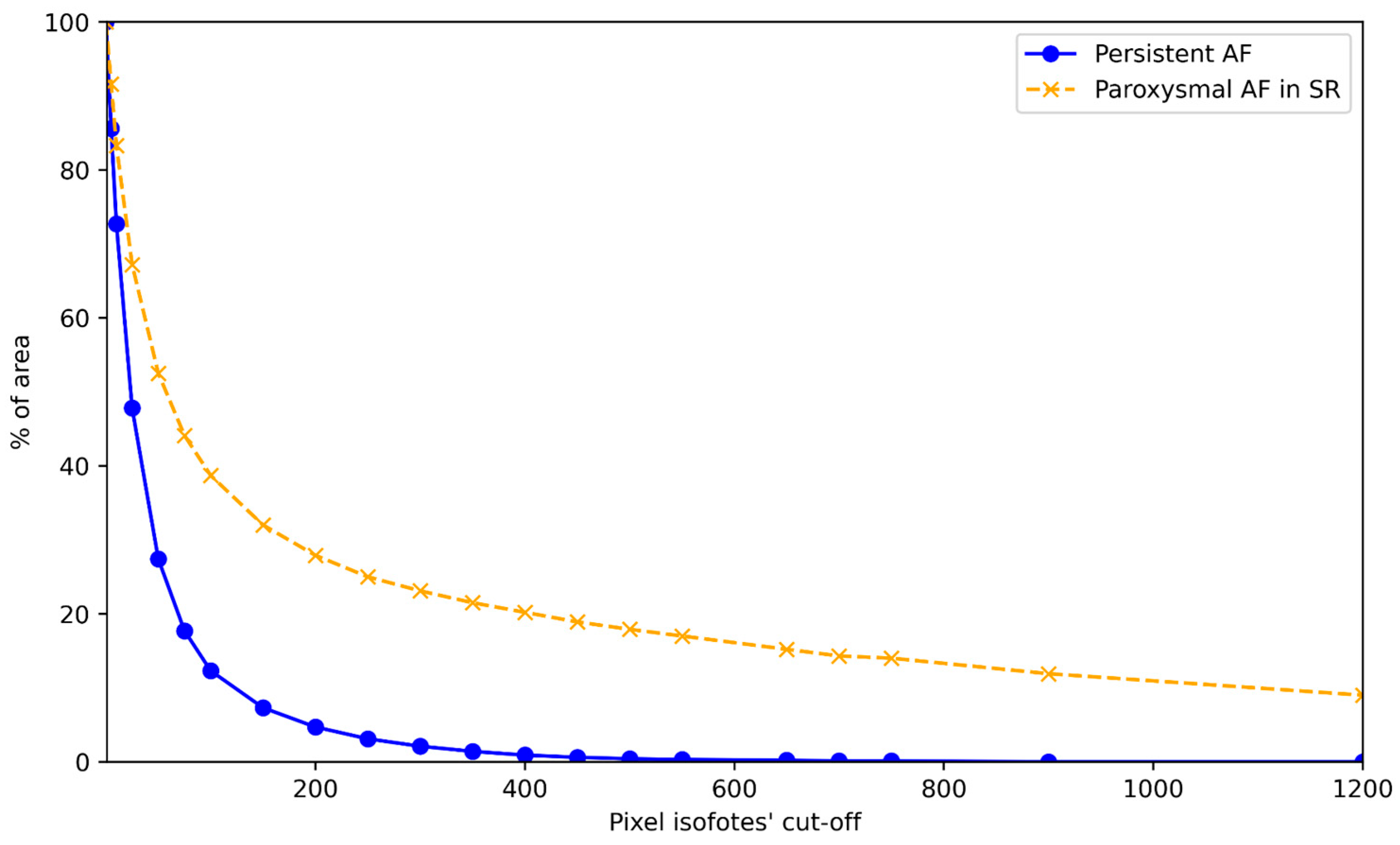
3.3. Comparison of the Blood Flow Patterns Among Study Patients’ Groups
4. Discussion
5. Conclusions
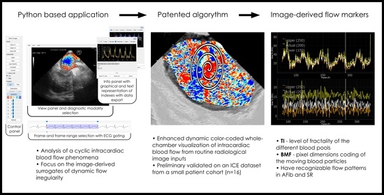
- A Python-based computer program was created to analyze cyclic intracardiac blood flow phenomena with a focus on the image-derived surrogates of dynamic flow irregularity.
- The program creates an enhanced, dynamic, and color-coded, whole-chamber visualization of intracardiac blood flow from routine radiological image inputs, and the qualitative feasibility was explored across several imaging modalities.
- The newly proposed TI and BMF were preliminary validated on a small ICE dataset and should currently be interpreted as exploratory, image-derived surrogate markers of flow behavior that have recognizable patterns and warrant broader technical and clinical validation.
6. Patents
Supplementary Materials
Author Contributions
Funding
Institutional Review Board Statement
Informed Consent Statement
Data Availability Statement
Conflicts of Interest
Abbreviations
| AF | Atrial fibrillation |
| AI | Artificial intelligence |
| BMF (n) | Blood mobility fraction with n-pixel cutoff |
| CW | Clockwise |
| CCW | Counterclockwise |
| ECG | Electrocardiogram |
| ICE | Intracardiac echocardiography |
| LA | Left atrium, left atrial |
| LAA | Left atrial appendage |
| LV | Left ventricle, left ventricular |
| MR | Mitral regurgitation |
| MRI | Magnetic resonance imaging |
| PAF | Paroxysmal AF |
| PersAF | Persistent AF |
| PV(s) | Pulmonary vein(s) |
| RA | Right atrium, right atrial |
| ROI | Region of interest |
| TI | Turbulence index |
| TTE | Transthoracic echocardiography |
| TEE | Transesophageal echocardiography |
| TR | Tricuspid regurgitation |
References
- January, C.T.; Wann, L.S.; Calkins, H.; Chen, L.Y.; Cigarroa, J.E.; Cleveland, J.C., Jr.; Ellinor, P.T.; Ezekowitz, M.D.; Field, M.E.; Furie, K.L.; et al. 2019 AHA/ACC/HRS Focused Update of the 2014 AHA/ACC/HRS Guideline for the Management of Patients with Atrial Fibrillation: A Report of the American College of Cardiology/American Heart Association Task Force on Clinical Practice Guidelines and the Heart Rhythm Society in Collaboration with the Society of Thoracic Surgeons. Circulation 2019, 140, e125–e151. [Google Scholar]
- Donal, E.; Lip, G.Y.; Galderisi, M.; Goette, A.; Shah, D.; Marwan, M.; Lederlin, M.; Mondillo, S.; Edvardsen, T.; Sitges, M.; et al. EACVI/EHRA Expert Consensus Document on the role of multi-modality imaging for the evaluation of patients with atrial fibrillation. Eur. Heart J.–Cardiovasc. Imaging 2016, 17, 355–383. [Google Scholar] [CrossRef] [Scilit]
- Bosi, G.M.; Cook, A.; Rai, R.; Menezes, L.J.; Schievano, S.; Torii, R.; Burriesci, G. Computational Fluid Dynamic Analysis of the Left Atrial Appendage to Predict Thrombosis Risk. Front. Cardiovasc. Med. 2018, 5, 34. [Google Scholar] [CrossRef] [Scilit]
- Spartera, M.; Pessoa-Amorim, G.; Stracquadanio, A.; Von Ende, A.; Fletcher, A.; Manley, P.; Neubauer, S.; Ferreira, V.M.; Casadei, B.; Hess, A.T.; et al. Left atrial 4D flow cardiovascular magnetic resonance: A reproducibility study in sinus rhythm and atrial fibrillation. J. Cardiovasc. Magn. Reson. 2021, 23, 29. [Google Scholar] [CrossRef] [Scilit]
- Klabunde, R.E. Cardiovascular Physiology Concepts, 3rd ed.; LWW: Philadelphia, PA, USA, 2021; 272p. [Google Scholar]
- Kundu, P.K.; Cohen, I.M.; Dowling, D.R. Fluid Mechanics, 6th ed.; Elsevier Inc.: Amsterdam, The Netherlands, 2016; 784p. [Google Scholar]
- Kaufmann, T.A.S.; Graefe, R.; Hormes, M.; Schmitz-Rode, T.; Steinseiferand, U. Computational Fluid Dynamics: Theory, Analysis and Applications; Nova Science Publishers: Hauppauge, NY, USA, 2011; pp. 109–136. [Google Scholar]
- Baillargeon, B.; Rebelo, N.; Fox, D.D.; Taylor, R.L.; Kuhl, E. The Living Heart Project: A robust and integrative simulator for human heart function. Eur. J. Mech.—A/Solids 2014, 48, 38–47. [Google Scholar] [CrossRef] [Scilit]
- Qin, J.J.; Indja, B.; Gholipour, A.; Gök, M.; Grieve, S.M. Evaluation of Left Ventricular Function Using Four-Dimensional Flow Cardiovascular Magnetic Resonance: A Systematic Review. J. Cardiovasc. Dev. Dis. 2022, 9, 304. [Google Scholar] [CrossRef] [Scilit] [PubMed]
- Dyverfeldt, P.; Bissell, M.; Barker, A.J.; Bolger, A.F.; Carlhäll, C.J.; Ebbers, T.; Francios, C.J.; Frydrychowicz, A.; Geiger, J.; Giese, D.; et al. 4D flow cardiovascular magnetic resonance consensus statement. J. Cardiovasc. Magn. Reson. 2015, 17, 72. [Google Scholar] [CrossRef] [Scilit]
- Fiorencis, A.; Pepe, M.; Smarrazzo, V.; Martini, M.; Severino, S.; Pergola, V.; Evangelista, M.; Incarnato, P.; Previtero, M.; Maglione, M.; et al. Noninvasive Evaluation of Intraventricular Flow Dynamics by the HyperDoppler Technique: First Application to Normal Subjects, Athletes, and Patients with Heart Failure. J. Clin. Med. 2022, 11, 2216. [Google Scholar] [CrossRef] [Scilit] [PubMed]
- Hong, G.R.; Kim, M.; Pedrizzetti, G.; Vannan, M.A. Current clinical application of intracardiac flow analysis using echocardiography. J. Cardiovasc. Ultrasound 2013, 21, 155–162. [Google Scholar] [CrossRef] [Scilit]
- Mele, D.; Beccari, R.; Pedrizzetti, G. Effect of Aging on Intraventricular Kinetic Energy and Energy Dissipation. J. Cardiovasc. Dev. Dis. 2023, 10, 308. [Google Scholar] [CrossRef] [Scilit]
- Zajac, J.; Eriksson, J.; Dyverfeldt, P.; Bolger, A.F.; Ebbers, T.; Carlhäll, C.J. Turbulent kinetic energy in normal and myopathic left ventricles. J. Magn. Reson. Imaging 2015, 41, 1021–1029. [Google Scholar] [CrossRef] [Scilit] [PubMed]
- Volkov, D.; Batsak, B.; Skoryi, D. Blood flow turbulence index as a new parameter for cardiac function assessment in various cardiac imaging modalities. Eur. J. Heart Fail. 2022, 24, 84–85. [Google Scholar]
- Bäck, S.; Skoda, I.; Lantz, J.; Henriksson, L.; Karlsson, L.O.; Persson, A.; Carlhäll, C.J.; Ebbers, T. Elevated atrial blood stasis in paroxysmal atrial fibrillation during sinus rhythm: A patient-specific computational fluid dynamics study. Front. Cardiovasc. Med. 2023, 10, 1219021. [Google Scholar] [CrossRef] [Scilit] [PubMed]
- Mele, D.; Trevisan, F.; Fiorencis, A.; Smarrazzo, V.; Bertini, M.; Ferrari, R. Current Role of Echocardiography in Cardiac Resynchronization Therapy: From Cardiac Mechanics to Flow Dynamics Analysis. Curr. Heart Fail. Rep. 2020, 17, 384–396. [Google Scholar] [CrossRef] [Scilit]
- Russo, C.; Jin, Z.; Liu, R.; Iwata, S.; Tugcu, A.; Yoshita, M.; Homma, S.; Elkind, M.S.; Rundek, T.; DeCarli, C.; et al. LA volumes and reservoir function are associated with subclinical cerebrovascular disease: The CABL (Cardiovascular Abnormalities and Brain Lesions) study. JACC Cardiovasc. Imaging 2013, 6, 313–323. [Google Scholar] [CrossRef] [Scilit]
- Fyrenius, A.; Wigström, L.; Ebbers, T.; Karlsson, M.; Engvall, J.; Bolger, A.F. Three dimensional flow in the human left atrium. Heart 2001, 86, 448–455. [Google Scholar] [CrossRef] [Scilit] [PubMed]
- Park, K.H.; Son, J.W.; Park, W.J.; Lee, S.H.; Kim, U.; Park, J.S.; Shin, D.G.; Kim, Y.J.; Choi, J.H.; Houle, H.; et al. Characterization of the left atrial vortex flow by two-dimensional transesophageal contrast echocardiography using particle image velocimetry. Ultrasound Med. Biol. 2013, 39, 62–71. [Google Scholar] [CrossRef] [Scilit]
- Lantz, J.; Gupta, V.; Henriksson, L.; Karlsson, M.; Persson, A.; Carlhäll, C.J.; Ebbers, T. Impact of Pulmonary Venous Inflow on Cardiac Flow Simulations: Comparison with In Vivo 4D Flow MRI. Ann. Biomed. Eng. 2019, 47, 413–424. [Google Scholar] [CrossRef] [Scilit]
- Zhong, J.; Long, D.; Chu, H.; Fu, H.; Han, X.; Jaing, C.; Li, Y.; Li, X.; Tang, M.; Wang, Z.; et al. Intracardiac echocardiography Chinese expert consensus. Front. Cardiovasc. Med. 2022, 9, 1012731. [Google Scholar] [CrossRef] [Scilit]
- Volkov, D.; Boyko, V.; Bakai, A. A61B6/032, A61B8/06, A61B8/0883 Method and System for Processing of Medical Images for Generating of Prognosis of Cardiac Function. U.S. Patent 10,631,811 B2; U.S. Patent Application No. 15/060,970, 28 April 2020. [Google Scholar]
- Pedrizzetti, G.; La Canna, G.; Alfieri, O.; Tonti, G. The vortex—An early predictor of cardiovascular outcome? Nat. Rev. Cardiol. 2014, 11, 545–553. [Google Scholar] [CrossRef] [Scilit]
- Spartera, M.; Stracquadanio, A.; Pessoa-Amorim, G.; Von Ende, A.; Fletcher, A.; Manley, P.; Ferreira, V.M.; Hess, A.T.; Hopewell, J.C.; Neubauer, S.; et al. The impact of atrial fibrillation and stroke risk factors on left atrial blood flow characteristics. Eur. Heart J.—Cardiovasc. Imaging 2022, 23, 115–123. [Google Scholar] [CrossRef] [Scilit]
- Cimino, S.; Pedrizzetti, G.; Tonti, G.; Canali, E.; Petronilli, V.; De Luca, L.; Iacoboni, C.; Agati, L. In vivo analysis of intraventricular fluid dynamics in healthy hearts. Eur. J. Mech.—B/Fluids 2012, 35, 40–46. [Google Scholar] [CrossRef] [Scilit]
- Fadnes, S.; Wigen, M.S.; Nyrnes, S.A.; Lovstakken, L. In Vivo Intracardiac Vector Flow Imaging Using Phased Array Transducers for Pediatric Cardiology. IEEE Trans. Ultrason. Ferroelectr. Freq. Control 2017, 64, 1318–1326. [Google Scholar] [CrossRef] [Scilit]
- Sun, Y.; Ling, Y.; Chen, Z.; Wang, Z.; Li, T.; Tong, Q.; Qian, Y. Finding low CHA2DS2-VASc scores unreliable? Why not give morphological and hemodynamic methods a try? Front. Cardiovasc. Med. 2023, 9, 1032736. [Google Scholar] [CrossRef] [Scilit] [PubMed]
- Nakagami, H.; Yamamoto, K.; Ikeda, U.; Mitsuhashi, T.; Goto, T.; Shimada, K. Mitral regurgitation reduces the risk of stroke in patients with nonrheumatic atrial fibrillation. Am. Heart J. 1998, 136, 528–532. [Google Scholar] [CrossRef] [Scilit]
- Melduni, R.; Nkomo, V.T.; Wysokinski, W.; Gersh, B.J.; Deshmukh, A.; Padang, R.; Greene, E.L.; Oh, J.K.; Lee, H.C. Risk of left atrial appendage thrombus and stroke in patients with atrial fibrillation and mitral regurgitation. Heart 2022, 108, 29–36. [Google Scholar] [CrossRef] [Scilit] [PubMed]
- Van Laer, S.L.; Verreyen, S.; Winkler, K.M.; Miljoen, H.; Sarkozy, A.; Heuten, H.; Saenen, J.; Van Herck, P.; Van de Heyning, C.M.; Heidbuchel, H.; et al. Effect of Mitral Regurgitation on Thrombotic Risk in Patients With Nonrheumatic Atrial Fibrillation: A New CHA2DS2-VASc Score Risk Modifier? Am. J. Cardiol. 2021, 145, 69–76. [Google Scholar] [CrossRef] [Scilit]
- Spartera, M.; Stracquadanio, A.; Pessoa-Amorim, G.; Harston, G.; Mazzucco, S.; Young, V.; Von Ende, A.; Hess, A.T.; Ferreira, V.M.; Kennedy, J.; et al. Reduced Left Atrial Rotational Flow Is Independently Associated with Embolic Brain Infarcts. JACC Cardiovasc. Imaging 2023, 16, 1149–1159. [Google Scholar] [CrossRef] [Scilit]





| Patients, n = 16 | Paroxysmal AF, n = 8 | Persistent AF, n = 8 | p-Value |
|---|---|---|---|
| Age | 59 ± 19 | 74 ± 8 | 0.0551 |
| BMI | 26.4 | 29.3 | 0.1856 |
| CHA2DS2-VASC score | 2.0 | 3.9 | 0.0205 |
| LVEF, % | 58 ± 7 | 60 ± 10 | 0.5971 |
| LA dimensions, mm | 40 ± 3.8 | 43 ± 3.6 | 0.0523 |
| Mitral max velocity, m/s | 0.66 ± 0.25 | 0.89 ± 0.36 | 0.0657 |
| Mitral deceleration, ms | 198 ± 35 | 183 ± 28 | 0.5619 |
| MR, grade | 1.1 ± 0.4 | 1.4 ± 0.7 | 0.1108 |
| Patients | Paroxysmal AF, n = 8 | Persistent AF, n = 8 | p-Value |
|---|---|---|---|
| ECG with SR | |||
| HR, bpm | 62 ± 15 | 70 ± 17 | 0.5287 |
| P wave, ms | 106 ± 15 | 104 ± 17 | 0.7948 |
| PR, ms | 151 ± 30 | 158 ± 23 | 0.6383 |
| QRS, ms | 97 ± 26 | 90 ± 21 | 0.5287 |
| QT, ms | 412 ± 56 | 397 + 45 | 0.9601 |
| Peak velocities (ICE), m/s | |||
| LAA | 0.48 ± 0.13 | 0.3 ± 0.11 | 0.0106 |
| Left PVs | 0.47 ± 0.15 | 0.36 ± 0.06 | 0.0929 |
| Right PVs | 0.31 ± 0.16 | 0.4 ± 0.18 | 0.0404 |
| Transmitral flow | 0.46 ± 0.07 | 0.58 ± 0.1 | 0.0209 |
| Mean LA pressure | 13 ± 5 | 19 ± 7 | 0.0930 |
| Patients | BMF Maximum Value | BMF Minimum Value | BMF Min-Max Range | |||
|---|---|---|---|---|---|---|
| PesrAF | PAF | PesrAF | PAF | PersAF | PAF | |
| 1 | 15.4 | 41.8 | 2.8 | 5.8 | 12.6 | 36 |
| 2 | 17.9 | 16.3 | 1.28 | 0.5 | 16.6 | 15.8 |
| 3 | 8.4 | 30.4 | 2.7 | 2.4 | 5.7 | 28 |
| 4 | 19.6 | 21.9 | 2.4 | 0 | 17.2 | 21.9 |
| 5 | 14.2 | 78.6 | 3 | 1.6 | 11.2 | 77 |
| 6 | 9.1 | 15.9 | 2.2 | 0.9 | 6.9 | 15 |
| 7 | 8.7 | 18.7 | 1.8 | 1.9 | 7 | 16.8 |
| 8 | 29.3 | 25.1 | 0.5 | 3.1 | 28.8 | 22 |
| Avg | 15.3 | 31.1 | 2.1 | 2 | 13.3 | 29.1 |
| SD | 7.1 | 21 | 0.8 | 1.8 | 7.7 | 20.6 |
| p-value | 0.0238 | 0.6383 | 0.0315 | |||
Disclaimer/Publisher’s Note: The statements, opinions and data contained in all publications are solely those of the individual author(s) and contributor(s) and not of MDPI and/or the editor(s). MDPI and/or the editor(s) disclaim responsibility for any injury to people or property resulting from any ideas, methods, instructions or products referred to in the content. |
© 2026 by the authors. Licensee MDPI, Basel, Switzerland. This article is an open access article distributed under the terms and conditions of the Creative Commons Attribution (CC BY) license.
Share and Cite
Volkov, D.; Skoryi, D.; Batsak, B.; Karpenko, I.; Sidhu, A.K.; Gianni, C.; La Fazia, V.M.; MacDonald, B.; Gallinghouse, J.; Horton, R.; et al. Development and Initial Validation of the Novel Computational Method for Dynamic Intracardiac Blood Flow Evaluation. Diagnostics 2026, 16, 1352. https://doi.org/10.3390/diagnostics16091352
Volkov D, Skoryi D, Batsak B, Karpenko I, Sidhu AK, Gianni C, La Fazia VM, MacDonald B, Gallinghouse J, Horton R, et al. Development and Initial Validation of the Novel Computational Method for Dynamic Intracardiac Blood Flow Evaluation. Diagnostics. 2026; 16(9):1352. https://doi.org/10.3390/diagnostics16091352
Chicago/Turabian StyleVolkov, Dmytro, Dmytro Skoryi, Bogdan Batsak, Iurii Karpenko, Alamjeet Kaur Sidhu, Carola Gianni, Vincenzo Mirko La Fazia, Bryan MacDonald, Joseph Gallinghouse, Rodney Horton, and et al. 2026. "Development and Initial Validation of the Novel Computational Method for Dynamic Intracardiac Blood Flow Evaluation" Diagnostics 16, no. 9: 1352. https://doi.org/10.3390/diagnostics16091352
APA StyleVolkov, D., Skoryi, D., Batsak, B., Karpenko, I., Sidhu, A. K., Gianni, C., La Fazia, V. M., MacDonald, B., Gallinghouse, J., Horton, R., Mohanty, S., & Natale, A. (2026). Development and Initial Validation of the Novel Computational Method for Dynamic Intracardiac Blood Flow Evaluation. Diagnostics, 16(9), 1352. https://doi.org/10.3390/diagnostics16091352