Article

Morphological Variability of Sphenoid Sinus Pneumatization and Its Impact on Adjacent Neurovascular Structures

by
Panagiotis Papadopoulos-Manolarakis
1,2,
George Triantafyllou
1,
Christos Georgalas
3,4,
Ioannis Paschopoulos
1,
George Stranjalis
5 and
Maria Piagkou
1,*
1
Department of Anatomy, School of Medicine, Faculty of Health Sciences, National and Kapodistrian University of Athens, 11527 Athens, Greece
2
Department of Neurosurgery, General Hospital of Nikaia-Piraeus, 18454 Athens, Greece
3
Department of Otorhinolaryngology Head and Neck Surgery, University of Nicosia Medical School, 2408 Nicosia, Cyprus
4
Endoscopic Skull Base Centre Athens, Hygeia Hospital, 15123 Athens, Greece
5
Department of Neurosurgery, Evangelismos Hospital, School of Medicine, Faculty of Health Sciences, National and Kapodistrian University of Athens, 10676 Athens, Greece
*
Author to whom correspondence should be addressed.
Diagnostics 2026, 16(5), 809; https://doi.org/10.3390/diagnostics16050809

Submission received: 2 February 2026 / Revised: 2 March 2026 / Accepted: 6 March 2026 / Published: 9 March 2026
(This article belongs to the Special Issue Brain/Neuroimaging 2025–2026)

Abstract

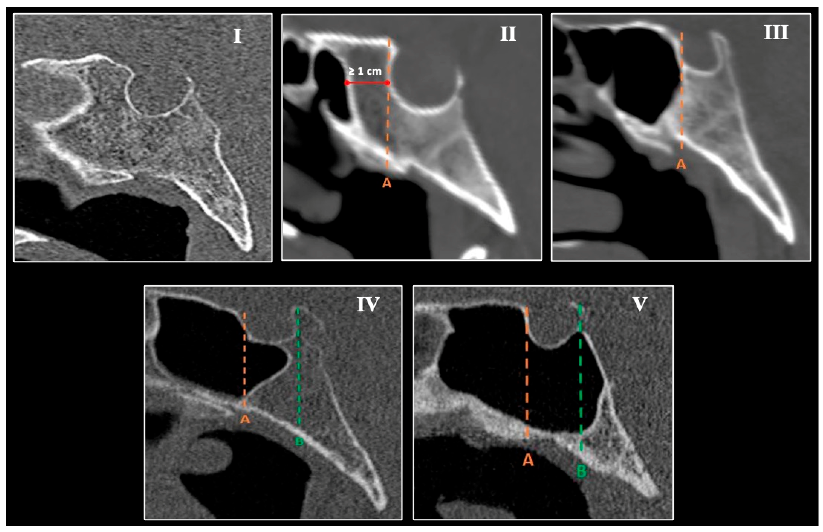
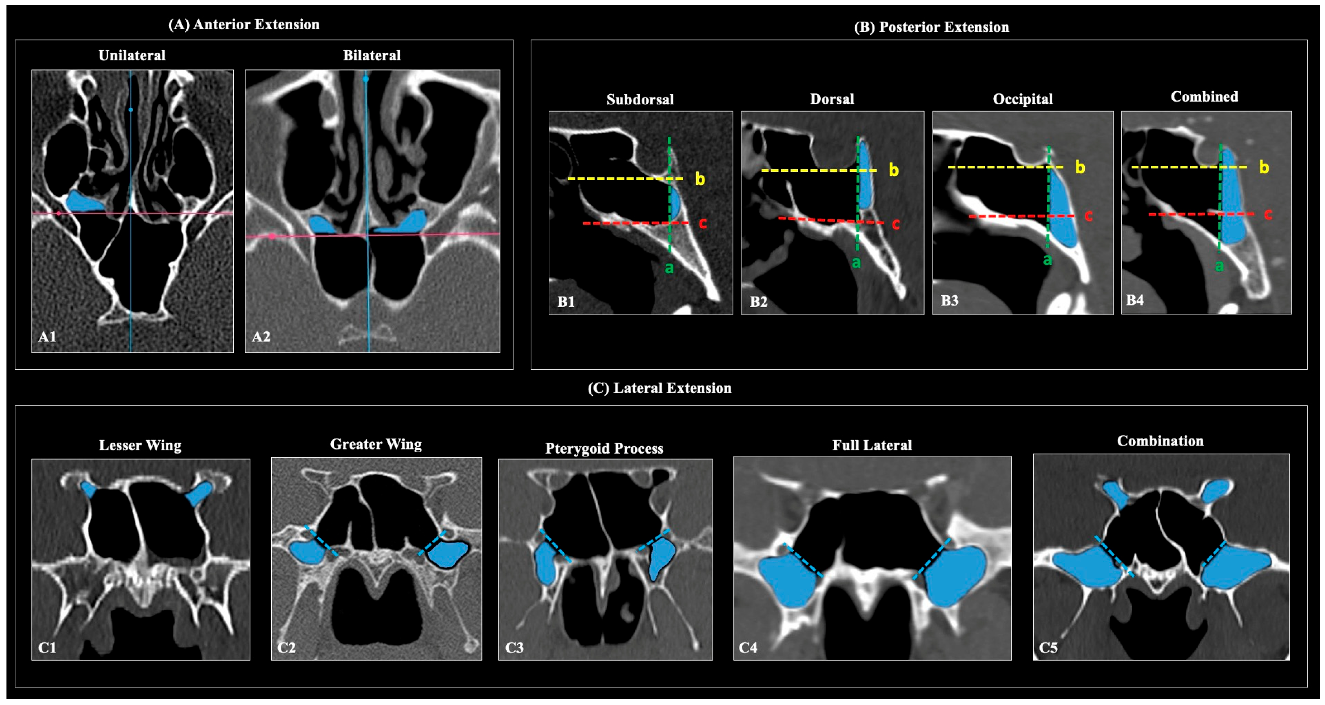
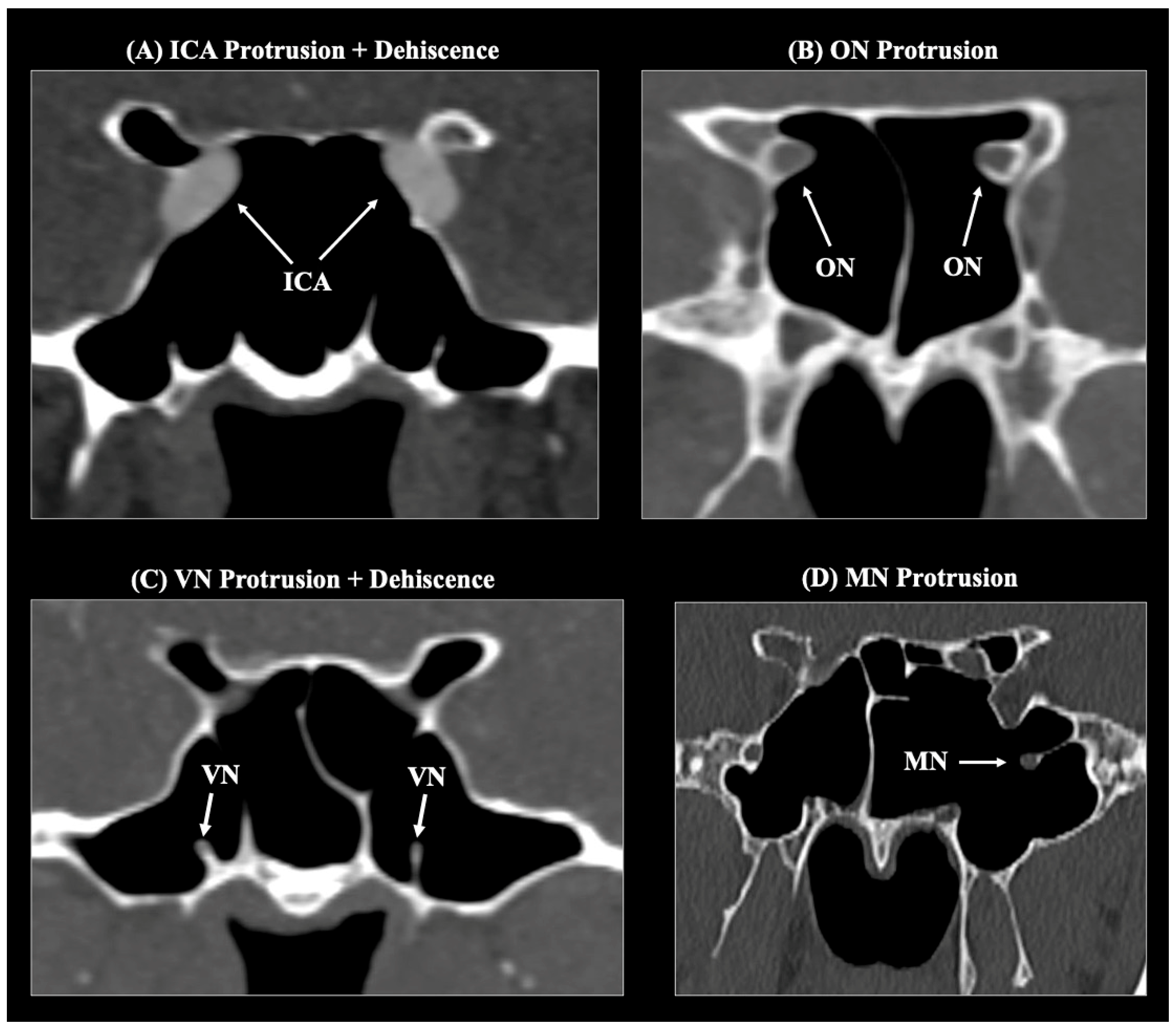
Background/Objectives: The sphenoid sinus (SS) exhibits marked morphological variability, influencing the relationship of critical neurovascular skull base structures. This study aimed to characterize sphenoid sinus pneumatization (SSP) patterns and assess their impact on the course of the internal carotid artery (ICA), optic nerve (ON), Vidian nerve (VN), and maxillary nerve (MN) within a Greek adult population. Methods: A retrospective analysis of 253 adult skull base computed tomography (CT) scans was performed. The degree and direction of SSP were classified according to established radiological criteria. Anterior, lateral, and posterior extensions were evaluated. The course of adjacent neurovascular structures was categorized as typical, protruding, or dehiscent. Associations between pneumatization types and neurovascular variants were analyzed. Results: The sellar complete type was the predominant SS pattern (63.2%), followed by sellar incomplete (27.7%) and presellar (8.7%) types; agenesis was rare (0.4%). Posterior (63.6%) and lateral (46.6%) extensions were most common. Lateral and posterior pneumatization significantly correlated with protrusion and/or dehiscence of adjacent neurovascular structures, particularly the ICA, ON, and VN. LW extension was strongly associated with ON protrusion (96%), while PP and full-lateral extensions correlated with VN protrusion (56.1% and 79.9%, respectively). No significant sex- or side-related differences were identified. Conclusions: SSP demonstrates extensive morphological variability that significantly affects the anatomical course and osseous coverage of neighboring neurovascular structures. Comprehensive preoperative CT evaluation of SS anatomy is essential for planning endoscopic transsphenoidal and extended skull base procedures to minimize the risk of neurovascular injury.
Keywords: sphenoid sinus; pneumatization; internal carotid artery; optic nerve; Vidian nerve; maxillary nerve; variation; endoscopic transsphenoidal surgery sphenoid sinus; pneumatization; internal carotid artery; optic nerve; Vidian nerve; maxillary nerve; variation; endoscopic transsphenoidal surgery