Echocardiographic Assessment After Heart Transplant
 , , ,
, , ,
Abstract
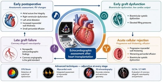
1. Introduction
1.1. Typical Echocardiographic Findings in Heart Transplant
1.2. Early Graft Dysfunction Following Heart Transplantation
1.3. Acute Allograft Rejection
- •
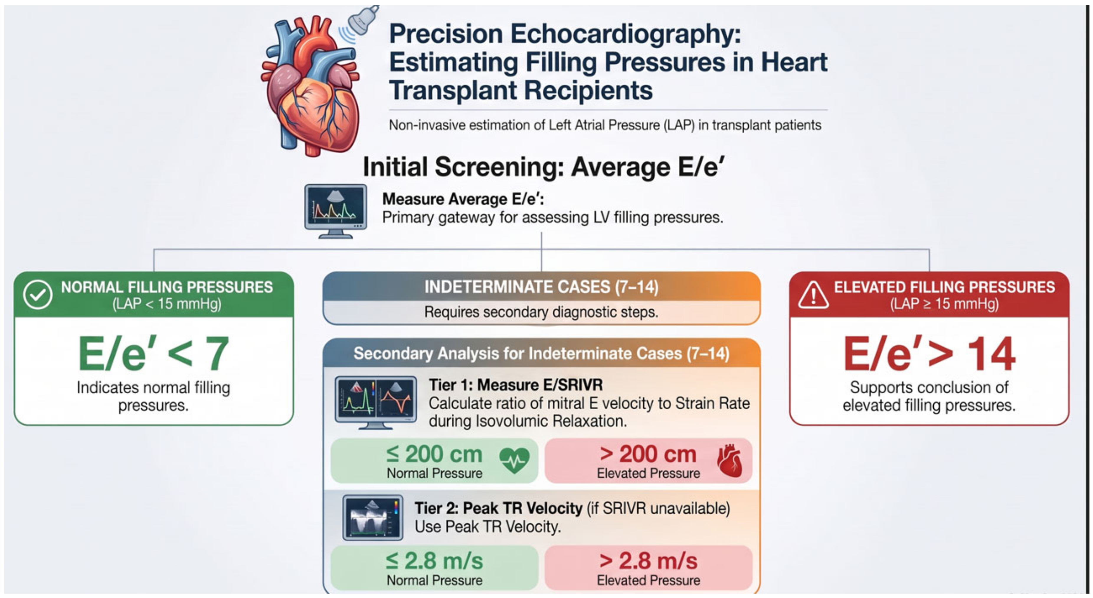
- Average E/e′ < 7: normal LV filling pressures.
- •
- Average E/e′ > 14: elevated LV filling pressures.
- •
- Average E/e′ 7–14: measure LV strain rate during isovolumic relaxation (SRIVR) from all three apical views.
- ∘
- Mitral E/SRIVR ≤ 200 cm: normal LV filling pressures.
- ∘
- Mitral E/SRIVR > 200 cm: elevated LV filling pressures.
- •
- If SRIVR is unavailable: use peak tricuspid regurgitation (TR) velocity.
- ∘
- Peak TR velocity ≤ 2.8 m/s: normal LV filling pressures.
- ∘
- Peak TR velocity > 2.8 m/s: elevated LV filling pressures.
1.4. Late Graft Failure Due to Cardiac Allograft Vasculopathy
1.5. New Echocardiographic Techniques
1.6. Speckle-Tracking Echocardiography
1.7. Three-Dimensional Echocardiography
1.8. Discussion and Future Directions
2. Conclusions
Author Contributions
Funding
Institutional Review Board Statement
Informed Consent Statement
Data Availability Statement
Acknowledgments
Conflicts of Interest
References
- Costanzo, M.R.; Dipchand, A.; Starling, R.; Anderson, A.; Chan, M.; Desai, S.; Fedson, S.; Fisher, P.; Gonzales-Stawinski, G.; Martinelli, L.; et al. The International Society of Heart and Lung Transplantation Guidelines for the care of heart transplant recipients. J. Heart Lung Transplant. 2010, 29, 914–956. [Google Scholar] [CrossRef] [Scilit]
- Luckraz, H.; Sharples, L.D.; Charman, S.C.; Tsui, S.S.; Wallwork, J.; Parameshwar, J.; Large, S.R. Does heart transplantation confer survival benefit in all risk groups? J. Heart Lung Transplant. 2005, 24, 1231–1234. [Google Scholar] [CrossRef] [Scilit]
- Stehlik, J.; Edwards, L.B.; Kucheryavaya, A.Y.; Benden, C.; Christie, J.D.; Dipchand, A.I.; Dobbels, F.; Kirk, R.; Rahmel, A.O.; Hertz, M.I.; et al. The Registry of the International Society for Heart and Lung Transplantation: 29th official adult heart transplant report--2012. J. Heart Lung Transplant. 2012, 31, 1052–1064. [Google Scholar] [CrossRef] [Scilit]
- Masarone, D.; Kittleson, M.; Gravino, R.; Valente, F.; Petraio, A.; Pacileo, G. The Role of Echocardiography in the Management of Heart Transplant Recipients. Diagnostics 2021, 11, 2338. [Google Scholar] [CrossRef] [Scilit]
- Peteiro, J.; Calvino, R.; Redondo, F.; Castro, A. Unreliability of echocardiographic and Doppler indexes in the diagnosis of heart transplant acute rejection. Rev. Port. Cardiol. 1996, 15, 548, 575–581. [Google Scholar]
- Badano, L.P.; Miglioranza, M.H.; Edvardsen, T.; Colafranceschi, A.S.; Muraru, D.; Bacal, F.; Nieman, K.; Zoppellaro, G.; Braga, F.G.M.; Binder, T.; et al. European Association of Cardiovascular Imaging/Cardiovascular Imaging Department of the Brazilian Society of Cardiology recommendations for the use of cardiac imaging to assess and follow patients after heart transplantation. Eur. Heart J. Cardiovasc. Imaging 2015, 16, 919–948. [Google Scholar] [CrossRef] [Scilit]
- Miniati, D.N.; Robbins, R.C. Techniques in orthotopic cardiac transplantation: A review. Cardiol. Rev. 2001, 9, 131–136. [Google Scholar] [CrossRef] [Scilit]
- Davies, R.R.; Russo, M.J.; Morgan, J.A.; Sorabella, R.A.; Naka, Y.; Chen, J.M. Standard versus bicaval techniques for orthotopic heart transplantation: An analysis of the United Network for Organ Sharing database. J. Thorac. Cardiovasc. Surg. 2010, 140, 700–708.e2. [Google Scholar] [CrossRef] [Scilit]
- Dell’Aquila, A.M.; Mastrobuoni, S.; Bastarrika, G.; Praschker, B.L.; Aguero, P.A.; Castano, S.; Herreros, J.; Rabago, G. Bicaval versus standard technique in orthotopic heart transplant: Assessment of atrial performance at magnetic resonance and transthoracic echocardiography. Interact. Cardiovasc. Thorac. Surg. 2012, 14, 457–462. [Google Scholar] [CrossRef] [Scilit]
- Kim, H.R.; Kim, H.J.; Lee, S.E.; Jung, S.H.; Yun, T.J.; Kim, J.J.; Lee, J.W. Prevalence and Risk Factors of Post-heart Transplant Tricuspid Regurgitation. Transplantation 2022, 106, e297–e303. [Google Scholar] [CrossRef] [Scilit]
- Locali, R.F.; Matsuoka, P.K.; Cherbo, T.; Gabriel, E.A.; Buffolo, E. Should biatrial heart transplantation still be performed?: A Meta-analysis. Arq. Bras. Cardiol. 2010, 94, 829–840. [Google Scholar]
- Bacal, F.; Pires, P.V.; Moreira, L.F.; Silva, C.P.; Filho, J.R.; Costa, U.M.; Rosário-Neto, M.A.; Ávila, V.M.; Cruz, F.D.; Guimarães, G.V.; et al. Normalization of right ventricular performance and remodeling evaluated by magnetic resonance imaging at late follow-up of heart transplantation: Relationship between function, exercise capacity and pulmonary vascular resistance. J. Heart Lung Transplant. 2005, 24, 2031–2036. [Google Scholar] [CrossRef] [Scilit]
- Goland, S.; Siegel, R.J.; Burton, K.; De Robertis, M.A.; Rafique, A.; Schwarz, E.; Zivari, K.; Mirocha, J.; Trento, A.; Czer, L.S.C. Changes in left and right ventricular function of donor hearts during the first year after heart transplantation. Heart 2011, 97, 1681–1686. [Google Scholar] [CrossRef] [Scilit]
- Stobierska-Dzierzek, B.; Awad, H.; Michler, R.E. The evolving management of acute right-sided heart failure in cardiac transplant recipients. J. Am. Coll. Cardiol. 2001, 38, 923–931. [Google Scholar] [CrossRef] [Scilit]
- Bhatia, S.J.; Kirshenbaum, J.M.; Shemin, R.J.; Cohn, L.H.; Collins, J.J.; Di Sesa, V.J.; Young, P.J.; Mudge, G.H.; Sutton, M.G. Time course of resolution of pulmonary hypertension and right ventricular remodeling after orthotopic cardiac transplantation. Circulation 1987, 76, 819–826. [Google Scholar] [CrossRef] [Scilit]
- Mastouri, R.; Batres, Y.; Lenet, A.; Gradus-Pizlo, I.; O’Donnell, J.; Feigenbaum, H.; Sawada, S.G. Frequency, time course, and possible causes of right ventricular systolic dysfunction after cardiac transplantation: A single center experience. Echocardiography 2013, 30, 9–16. [Google Scholar] [CrossRef] [Scilit]
- Lang, R.M.; Badano, L.P.; Mor-Avi, V.; Afilalo, J.; Armstrong, A.; Ernande, L.; Flachskampf, F.A.; Foster, E.; Goldstein, S.A.; Kuznetsova, T.; et al. Recommendations for cardiac chamber quantification by echocardiography in adults: An update from the American Society of Echocardiography and the European Association of Cardiovascular Imaging. Eur. Heart J. Cardiovasc. Imaging 2015, 16, 233–270. [Google Scholar] [CrossRef] [Scilit]
- Ingvarsson, A.; Werther Evaldsson, A.; Waktare, J.; Nilsson, J.; Smith, G.J.; Stagmo, M.; Roijer, A.; Rådegran, G.; Meurling, C.J. Normal Reference Ranges for Transthoracic Echocardiography Following Heart Transplantation. J. Am. Soc. Echocardiogr. 2018, 31, 349–360. [Google Scholar] [CrossRef] [Scilit]
- Russo, M.J.; Iribarne, A.; Hong, K.N.; Ramlawi, B.; Chen, J.M.; Takayama, H.; Mancini, D.M.; Naka, Y. Factors associated with primary graft failure after heart transplantation. Transplantation 2010, 90, 444–450. [Google Scholar] [CrossRef] [Scilit]
- Pardo-Mindan, F.J.; Lozano, M.D. “Quilty effect” in heart transplantation: Is it related to acute rejection? J. Heart Lung Transplant. 1991, 10, 937–941. [Google Scholar]
- Mondillo, S.; Maccherini, M.; Galderisi, M. Usefulness and limitations of transthoracic echocardiography in heart transplantation recipients. Cardiovasc. Ultrasound 2008, 6, 2. [Google Scholar] [CrossRef] [Scilit]
- Sagar, K.B.; Hastillo, A.; Wolfgang, T.C.; Lower, R.R.; Hess, M.L. Left ventricular mass by M-mode echocardiography in cardiac transplant patients with acute rejection. Circulation 1981, 64, II217–II220. [Google Scholar]
- Sun, J.P.; Abdalla, I.A.; Asher, C.R.; Greenberg, N.L.; Popovic, Z.B.; Taylor, D.O.; Starling, R.C.; Thomas, J.D.; Garcia, M.J. Non-invasive evaluation of orthotopic heart transplant rejection by echocardiography. J. Heart Lung Transplant. 2005, 24, 160–165. [Google Scholar] [CrossRef] [Scilit]
- Ciliberto, G.R.; Anjos, M.C.; Gronda, E.; Bonacina, E.; Danzi, G.; Colombo, P.; Mangiavacchi, M.; Alberti, A.; Frigerio, M.; De Vita, C. Significance of pericardial effusion after heart transplantation. Am. J. Cardiol. 1995, 76, 297–300. [Google Scholar] [CrossRef] [Scilit]
- Nagueh, S.F.; Sanborn, D.Y.; Oh, J.K.; Anderson, B., 3rd; Billick, K.; Derumeaux, G.; Klein, A.; Koulogiannis, K.; Mitchell, C.; Shah, A. Recommendations for the Evaluation of Left Ventricular Diastolic Function by Echocardiography and for Heart Failure With Preserved Ejection Fraction Diagnosis: An Update From the American Society of Echocardiography. J. Am. Soc. Echocardiogr. 2025. [Google Scholar] [CrossRef] [Scilit]
- Mehra, M.R.; Crespo-Leiro, M.G.; Dipchand, A.; Ensminger, S.M.; Hiemann, N.E.; Kobashigawa, J.A.; Madsen, J.; Parameshwar, J.; Starling, R.C.; Uber, P.A. International Society for Heart and Lung Transplantation working formulation of a standardized nomenclature for cardiac allograft vasculopathy-2010. J. Heart Lung Transplant. 2010, 29, 717–727. [Google Scholar] [CrossRef] [Scilit]
- Spes, C.H.; Klauss, V.; Rieber, J.; Schnaack, S.D.; Tammen, A.R.; Uberfuhr, P.; Reichart, B.; Theisen, K.; Angermann, C.E.; Mudra, H. Functional and morphological findings in heart transplant recipients with a normal coronary angiogram: An analysis by dobutamine stress echocardiography, intracoronary Doppler and intravascular ultrasound. J. Heart Lung Transplant. 1999, 18, 391–398. [Google Scholar] [CrossRef] [Scilit]
- Spes, C.H.; Mudra, H.; Schnaack, S.D.; Klauss, V.; Reichle, F.M.; Uberfuhr, P.; Theisen, K.; Angermann, C.E. Dobutamine stress echocardiography for noninvasive diagnosis of cardiac allograft vasculopathy: A comparison with angiography and intravascular ultrasound. Am. J. Cardiol. 1996, 78, 168–174. [Google Scholar] [CrossRef] [Scilit]
- Kamel, M.A.; Scalia, I.G.; Badr, A.T.; Baba Ali, N.; Farina, J.M.; Pereyra, M.; Abbas, M.T.; Mahmoud, A.K.; Scott, R.L.; Steidley, D.E.; et al. Cardiac Allograft Vasculopathy: Challenges and Advances in Invasive and Non-Invasive Diagnostic Modalities. J. Cardiovasc. Dev. Dis. 2024, 11, 95. [Google Scholar] [CrossRef] [Scilit]
- Ahn, Y.; Koo, H.J.; Hyun, J.; Lee, S.E.; Jung, S.H.; Park, D.W.; Ahn, J.M.; Kang, D.Y.; Park, S.J.; Hwang, H.S.; et al. CT Coronary Angiography and Dynamic CT Myocardial Perfusion for Detection of Cardiac Allograft Vasculopathy. J. Am. Coll. Cardiol. Imaging 2023, 16, 934–947. [Google Scholar] [CrossRef] [Scilit]
- Thorn, E.M.; de Filippi, C.R. Echocardiography in the cardiac transplant recipient. Heart Fail. Clin. 2007, 3, 51–67. [Google Scholar] [CrossRef] [Scilit]
- Collings, C.A.; Pinto, F.J.; Valantine, H.A.; Popylisen, S.; Puryear, J.V.; Schnittger, I. Exercise echocardiography in heart transplant recipients: A comparison with angiography and intracoronary ultrasonography. J. Heart Lung Transplant. 1994, 13, 604–613. [Google Scholar]
- Estep, J.D.; Shah, D.J.; Nagueh, S.F.; Mahmarian, J.J.; Torre-Amione, G.; Zoghbi, W.A. The role of multimodality cardiac imaging in the transplanted heart. JACC Cardiovasc. Imaging 2009, 2, 1126–1140. [Google Scholar] [CrossRef] [Scilit]
- Kociolek, L.K.; Bierig, S.M.; Herrmann, S.C.; Labovitz, A.J. Efficacy of atropine as a chronotropic agent in heart transplant patients undergoing dobutamine stress echocardiography. Echocardiography 2006, 23, 383–387. [Google Scholar] [CrossRef] [Scilit]
- Bacal, F.; Moreira, L.; Souza, G.; Rodrigues, A.C.; Fiorelli, A.; Stolf, N.; Bocchi, E.; Bellotti, G.; Ramires, J.F. Dobutamine stress echocardiography predicts cardiac events or death in asymptomatic patients long-term after heart transplantation: 4-year prospective evaluation. J. Heart Lung Transplant. 2004, 23, 1238–1244. [Google Scholar] [CrossRef] [Scilit]
- Ciliberto, G.R.; Massa, D.; Mangiavacchi, M.; Danzi, G.B.; Pirelli, S.; Faletra, F.; Frigerio, M.; Gronda, E.; DE Vita, C. High-dose dipyridamole echocardiography test in coronary artery disease after heart transplantation. Eur. Heart J. 1993, 14, 48–52. [Google Scholar] [CrossRef] [Scilit]
- Zhu, S.; Li, M.; Tian, F.; Wang, S.; Li, Y.; Yin, P.; Zhang, L.; Xie, M. Diagnostic value of myocardial strain using two-dimensional speckle-tracking echocardiography in acute cardiac allograft rejection: A systematic review and meta-analysis. Echocardiography 2020, 37, 561–569. [Google Scholar] [CrossRef] [Scilit]
- Tseng, A.S.; Gorsi, U.S.; Barros-Gomes, S.; Miller, F.A.; Pellikka, P.A.; Clavell, A.L.; Villarraga, H.R. Use of speckle-tracking echocardiography-derived strain and systolic strain rate measurements to predict rejection in transplant hearts with preserved ejection fraction. BMC Cardiovasc. Disord. 2018, 18, 241. [Google Scholar] [CrossRef] [Scilit]
- Balinisteanu, A.; Duchenne, J.; Puvrez, A.; Wouters, L.; Bézy, S.; Youssef, A.; Minten, L.; Bekhuis, Y.; Van Langenhoven, L.; Papangelopoulou, K.; et al. Vendor differences in 2D-speckle tracking global longitudinal strain: An update on a 10-year standardization effort. Eur. Heart J. Cardiovasc. Imaging 2025, 26, 1360–1373. [Google Scholar] [CrossRef] [Scilit]
- Clemmensen, T.S.; Eiskjaer, H.; Logstrup, B.B.; Mellemkjaer, S.; Andersen, M.J.; Tolbod, L.P.; Harms, H.J.; Poulsen, S.H. Clinical features, exercise hemodynamics, and determinants of left ventricular elevated filling pressure in heart-transplanted patients. Transpl. Int. 2016, 29, 196–206. [Google Scholar] [CrossRef] [Scilit]
- Eroglu, E.; D’Hooge, J.; Sutherland, G.R.; Marciniak, A.; Thijs, D.; Droogne, W.; Herbots, L.; Van Cleemput, J.; Claus, P.; Bijnens, B.; et al. Quantitative dobutamine stress echocardiography for the early detection of cardiac allograft vasculopathy in heart transplant recipients. Heart 2008, 94, e3. [Google Scholar] [CrossRef] [Scilit]
- Mingo-Santos, S.; Monivas-Palomero, V.; Garcia-Lunar, I.; Mitroi, C.D.; Goirigolzarri-Artaza, J.; Rivero, B.; Oteo, J.F.; Castedo, E.; González-Mirelis, J.; Cavero, M.A.; et al. Usefulness of Two-Dimensional Strain Parameters to Diagnose Acute Rejection after Heart Transplantation. J. Am. Soc. Echocardiogr. 2015, 28, 1149–1156. [Google Scholar] [CrossRef] [Scilit]
- Sato, T.; Kato, T.S.; Komamura, K.; Hashimoto, S.; Shishido, T.; Mano, A.; Oda, N.; Takahashi, A.; Ishibashi-Ueda, H.; Nakatani, T.; et al. Utility of left ventricular systolic torsion derived from 2-dimensional speckle-tracking echocardiography in monitoring acute cellular rejection in heart transplant recipients. J. Heart Lung Transplant. 2011, 30, 536–543. [Google Scholar] [CrossRef] [Scilit]
- Eleid, M.F.; Caracciolo, G.; Cho, E.J.; Scott, R.L.; Steidley, D.E.; Wilansky, S.; Arabia, F.A.; Khandheria, B.K.; Sengupta, P.P. Natural history of left ventricular mechanics in transplanted hearts: Relationships with clinical variables and genetic expression profiles of allograft rejection. JACC Cardiovasc. Imaging 2010, 3, 989–1000. [Google Scholar]
- Sharma, D.; Subramaniam, G.; Sharma, N.; Sharma, P.; Sharma, P. Insight into Noninvasive Radiological Modalities to Detect Heart Transplant Rejection. Indian J. Radiol. Imaging 2021, 31, 946–955. [Google Scholar] [CrossRef] [Scilit]
- Cruz, C.; Hajjar, L.A.; Bacal, F.; Lofrano-Alves, M.S.; Lima, M.S.M.; Abduch, M.C.; Vieira, M.L.; Chiang, H.P.; Salviano, J.B.; da Silva Costa, I.B.S.; et al. Usefulness of speckle tracking echocardiography and biomarkers for detecting acute cellular rejection after heart transplantation. Cardiovasc. Ultrasound 2021, 19, 6. [Google Scholar] [CrossRef] [Scilit]
- Xourgia, E.; Brignoli, K.; Linder, O.; Neagoe, A.M.; Capek, L.; Bruno, J.; Strickler, E.; Bakula, A.; Pavlicek-Bahlo, M.; Fürholz, M.; et al. Speckle-tracking echocardiography of left and right ventricle and acute cellular rejection in orthotopic heart transplantation: A systematic review and meta-analysis. Int. J. Cardiovasc. Imaging 2024, 41, 669–679. [Google Scholar] [CrossRef] [Scilit]
- Ji, X.; Zhang, J.; Xie, Y.; Wang, W.; Zhang, Y.; Xie, M.; Zhang, L. Speckle-Tracking Echocardiography in Right Ventricular Function of Clinically Well Patients with Heart Transplantation. Diagnostics 2024, 14, 1305. [Google Scholar] [CrossRef] [Scilit]
- Roemer, S.; Jaglan, A.; Santos, D.; Umland, M.; Jain, R.; Tajik, A.J.; Khandheria, B.K. The Utility of Myocardial Work in Clinical Practice. J. Am. Soc. Echocardiogr. 2021, 34, 807–818. [Google Scholar] [CrossRef] [Scilit]
- Otto, M.E.B.; Martins, A.M.A.; Campos Dall’Orto, A.O.M.; Leite, S.F.; de Queiroz Mauricio Filho, M.A.F.; Martins, N.T.; de Araújo, S.R.; Almeida, S.V.; Paiva, M.U.B.; Atik, F.A. Acute Cellular Rejection in Heart Transplant Patients: Insights of Global Longitudinal Strain, Myocardial Work, and an Exclusive Group of Chagas Disease. Front. Cardiovasc. Med. 2022, 9, 841698. [Google Scholar] [CrossRef] [Scilit]
- Manrique Antón, R.; Pascual Izco, M.; Díaz Dorronsoro, A.; Ezponda, A.; de la Torre Carazo, F.; Salteráin, N.; Jimeno-San Martín, L.; Martín-Calvo, N.; Manrique Antón, Á.; Iribarren, M.J.; et al. Non-Invasive Myocardial Work Detects Extensive Coronary Disease in Orthotopic Heart Transplant Patients. Med. Sci. 2025, 13, 212. [Google Scholar] [CrossRef] [Scilit]
- D’Andrea, A.; Riegler, L.; Nunziata, L.; Scarafile, R.; Gravino, R.; Salerno, G.; Amarelli, C.; Maiello, C.; Limongelli, G.; Di Salvo, G.; et al. Right heart morphology and function in heart transplantation recipients. J. Cardiovasc. Med. 2013, 14, 648–658. [Google Scholar] [CrossRef] [Scilit]
- Myhre, P.L.; Grenne, B.; Asch, F.M.; Delgado, V.; Khera, R.; Lafitte, S.; Lang, R.M.; Pellikka, P.A.; Sengupta, P.P.; Vemulapalli, S.; et al. Artificial intelligence-enhanced echocardiography in cardiovascular disease management. Nat. Rev. Cardiol. 2026, 23, 164–182. [Google Scholar] [CrossRef] [Scilit]



| Technique | Main Advantages | Main Limitations | Potential Role After HTx |
|---|---|---|---|
| Conventional 2D TTE | Widely available; bedside; repeatable; evaluates chambers, valves, pericardium and global systolic function. | Low sensitivity for mild or subclinical rejection; measurements are affected by transplant-specific geometry and loading. | Routine surveillance; postoperative complications; serial comparison with individual baseline. |
| Doppler/tissue Doppler | Provides hemodynamic and diastolic information; estimates filling pressures and pulmonary pressures. | Denervation-related tachycardia may fuse E and A waves; e′ and other indices vary with postoperative recovery and loading. | Integrated assessment of filling pressures and graft hemodynamics. |
| Speckle-tracking echocardiography | Detects subclinical LV and RV dysfunction despite preserved EF; useful for serial assessment; potential rule-out value for rejection. | Load- and image-quality dependent; residual inter-vendor/software variability; transplant-specific cutoffs are not fully standardized. | Rejection surveillance; CAV assessment; longitudinal graft-function monitoring. |
| Stress echocardiography | Non-invasive functional assessment; provides prognostic information; may reveal inducible wall-motion abnormalities. | Chronotropic incompetence may limit exercise or dobutamine stress; variable sensitivity for diffuse CAV. | Non-invasive surveillance for CAV, particularly when invasive testing is not immediately indicated. |
| Three-dimensional echocardiography | More accurate chamber volumes and RV geometry; improved anatomical assessment; avoids geometric assumptions. | Dependent on acoustic window, temporal/spatial resolution and expertise; limited transplant-specific outcome data. | Quantification of LV/RV volumes and function; anatomical assessment; potential adjunct during stress. |
| Myocardial work | Integrates strain with estimated LV pressure and partially accounts for afterload; may add information beyond GLS. | Requires high-quality strain and blood-pressure data; limited validation and reference ranges in HTx. | Emerging marker for rejection and CAV; research and advanced surveillance. |
| Parameter/Consideration | Non-Transplanted Heart | Heart Transplant Recipient | Practical Implication |
|---|---|---|---|
| Heart rate/innervation | Normal autonomic innervation and heart-rate variability. | Cardiac denervation causes higher resting heart rate and altered chronotropic response. | Interpret Doppler filling and stress response in the context of denervation. |
| Atrial morphology | Normal atrial anatomy. | Atrial enlargement and suture-line morphology depend on biatrial vs. bicaval surgical technique. | Do not misinterpret the atrial suture line as thrombus or mass; account for surgical technique. |
| LV systolic function | Reference EF and GLS values derived from general populations. | EF may remain preserved while GLS is mildly reduced even in stable recipients. | Serial change from the patient’s own baseline is important; subtle strain reduction is not diagnostic of rejection in isolation. |
| RV size and function | Standard guideline reference ranges generally apply. | RV size is often larger and longitudinal indices such as TAPSE and RV strain may be lower after HTx. | General-population thresholds may overcall RV dysfunction; integrate FAC, S′, strain, 3D assessment and clinical context. |
| Diastolic assessment | Standard algorithms generally applicable. | Tachycardia, E/A fusion, postoperative edema and altered tissue velocities complicate interpretation. | Use an integrated approach to filling pressures; avoid relying on a single diastolic parameter. |
| Tricuspid regurgitation | Usually reflects primary/secondary valve disease. | Common after HTx and influenced by surgical technique, RV loading and repeated biopsies. | Assess mechanism and severity longitudinally rather than interpreting TR in isolation. |
| Pericardial effusion/wall thickness | New changes usually prompt evaluation for conventional causes. | Small effusion and transient wall thickening can be postoperative findings; interval worsening may raise concern for rejection. | Comparison with prior studies and timing after transplantation are essential. |
| Longitudinal follow-up | Population reference ranges are often sufficient. | Marked inter-individual variability makes a stable post-transplant baseline particularly valuable. | Use standardized acquisition and compare serial studies with the same patient’s baseline whenever possible. |
Disclaimer/Publisher’s Note: The statements, opinions and data contained in all publications are solely those of the individual author(s) and contributor(s) and not of MDPI and/or the editor(s). MDPI and/or the editor(s) disclaim responsibility for any injury to people or property resulting from any ideas, methods, instructions or products referred to in the content. |
© 2026 by the authors. Licensee MDPI, Basel, Switzerland. This article is an open access article distributed under the terms and conditions of the Creative Commons Attribution (CC BY) license.
Share and Cite
Cruz, C.B.B.V.; Sobral-Alves, J.B.; Pio, S.M.; Salviano, J.B.C.; Lofrano-Alves, M.S.; Rassi, D.d.C.; Hajjar, L.A.; Vieira, M.L.C. Echocardiographic Assessment After Heart Transplant. Diagnostics 2026, 16, 2970. https://doi.org/10.3390/diagnostics16182970
Cruz CBBV, Sobral-Alves JB, Pio SM, Salviano JBC, Lofrano-Alves MS, Rassi DdC, Hajjar LA, Vieira MLC. Echocardiographic Assessment After Heart Transplant. Diagnostics. 2026; 16(18):2970. https://doi.org/10.3390/diagnostics16182970
Chicago/Turabian StyleCruz, Cecília Beatriz Bittencourt Viana, Juliana Barbosa Sobral-Alves, Stephan Milhorini Pio, Juliana Bittencourt Cruz Salviano, Marco Stephan Lofrano-Alves, Daniela do Carmo Rassi, Ludhmila Abrahão Hajjar, and Marcelo Luiz Campos Vieira. 2026. "Echocardiographic Assessment After Heart Transplant" Diagnostics 16, no. 18: 2970. https://doi.org/10.3390/diagnostics16182970
APA StyleCruz, C. B. B. V., Sobral-Alves, J. B., Pio, S. M., Salviano, J. B. C., Lofrano-Alves, M. S., Rassi, D. d. C., Hajjar, L. A., & Vieira, M. L. C. (2026). Echocardiographic Assessment After Heart Transplant. Diagnostics, 16(18), 2970. https://doi.org/10.3390/diagnostics16182970