Article

Clinicopathological and Prognostic Significance of TROP2 and HER3 Expression in Early-Stage Cervical Carcinoma

1
Department of Obstetrics and Gynecology, Sivas Cumhuriyet University School of Medicine, Sivas 58140, Turkey
2
Department of Pathology, Sivas Cumhuriyet University School of Medicine, Sivas 58140, Turkey
*
Author to whom correspondence should be addressed.
Diagnostics 2026, 16(17), 2871; https://doi.org/10.3390/diagnostics16172871
Submission received: 22 July 2026 / Revised: 31 August 2026 / Accepted: 1 September 2026 / Published: 7 September 2026

Abstract

Background: Our objective was to evaluate the prognostic value of TROP2 and HER3 expression in patients with early-stage cervical carcinoma undergoing primary surgical treatment. Methods: We retrospectively analyzed 54 patients with FIGO 2018 stage IA–IIA cervical carcinoma who underwent radical surgery between January 2016 and December 2025. TROP2 and HER3 expression were assessed using immunohistochemistry and a standardized immunoreactivity scoring system. Associations with clinicopathological characteristics were examined, and survival outcomes were analyzed using Kaplan–Meier estimates and Cox proportional hazards models. Results: High TROP2 and HER3 expression was identified in 63.0% and 37.0% of tumors, respectively. Neither biomarker was associated with the conventional clinicopathological variables. After a median follow-up of 88.2 months, 22 patients (40.7%) developed recurrent disease and 18 (33.3%) died. Patients with high TROP2 expression had significantly shorter overall and disease-free survival than those with low expression. After adjustment for established prognostic factors, high TROP2 expression remained independently associated with disease recurrence (HR 5.00, 95% CI 1.28–19.57; p = 0.021). In contrast, HER3 expression was not independently associated with overall or disease-free survival rates. Older age and advanced FIGO stage were independently associated with poorer outcomes. Conclusions: TROP2 expression identifies a subgroup of patients with early-stage cervical carcinoma who are at an increased risk of recurrence despite primary surgical treatment. Unlike HER3, TROP2 provided prognostic information beyond conventional clinicopathological factors, supporting its potential role in postoperative risk stratification. These findings also reinforce the rationale for further investigation of TROP2 as a therapeutic target in cervical carcinoma.

1. Introduction

Cervical carcinoma remains one of the most common malignancies and is a leading cause of cancer-related death among women worldwide [1]. Although the widespread implementation of human papillomavirus (HPV) vaccination and cervical screening programs has substantially reduced disease incidence in many high-income countries, cervical cancer continues to impose a considerable health burden in low- and middle-income regions, where access to preventive and early diagnostic services remains limited [2]. Persistent infection with high-risk HPV types, particularly HPV16 and HPV18, is the primary driver of cervical cancer. The viral oncoproteins E6 and E7 promote malignant transformation by inactivating the tumor suppressor proteins p53 and retinoblastoma (Rb), leading to genomic instability, dysregulated cell cycle control, and uncontrolled cellular proliferation [3].
Most patients with early-stage cervical carcinoma achieve favorable outcomes after radical surgery, with or without adjuvant treatment. Nevertheless, disease recurrence remains the leading cause of treatment failure and cancer-related mortality. Despite apparently curative treatment, some patients experience locoregional or distant relapse, highlighting the considerable biological heterogeneity of this disease. At present, postoperative risk stratification is primarily based on established clinicopathological factors, including FIGO stage, tumor size, histological subtype, depth of stromal invasion, lymphovascular space invasion, lymph node involvement, and surgical margin status [4]. Although these parameters are essential for guiding adjuvant treatment and estimating prognosis, they do not fully explain the variability in clinical outcomes observed among patients with similar pathological characteristics. Consequently, there is growing interest in identifying reliable molecular biomarkers that can complement conventional prognostic factors, improve risk stratification, and support more individualized management of patients with early-stage cervical carcinoma [5].
The growing emphasis on precision oncology has increased interest in biomarkers that can improve prognostic assessment and serve as potential therapeutic targets. One such biomarker is the trophoblast cell surface antigen 2 (TROP2), a transmembrane glycoprotein encoded by the TACSTD2 gene. TROP2 plays an important role in tumor progression by promoting cell proliferation, migration, invasion, and epithelial–mesenchymal transition through the activation of several oncogenic signaling pathways, including PI3K/AKT, MAPK/ERK, and NF-κB [6]. TROP2 overexpression has been reported in a wide range of epithelial malignancies and has consistently been associated with aggressive tumor behavior, increased metastatic potential, and poor clinical outcomes [7,8,9]. More recently, the clinical success of TROP2-targeted antibody–drug conjugates has further highlighted its therapeutic relevance, making TROP2 an attractive biomarker for both prognostic evaluation and targeted treatment strategies in cervical carcinoma [10,11].
Another biomarker of growing interest is the human epidermal growth factor receptor 3 (HER3, ERBB3), a member of the ERBB receptor family. Although HER3 has minimal intrinsic kinase activity, it exerts biological effects by forming heterodimers with other ERBB receptors, particularly HER2, leading to the activation of the PI3K/AKT signaling pathway. Through these interactions, HER3 contributes to tumor cell proliferation, survival, invasion, and resistance to anticancer therapy [12]. Increased HER3 expression has been linked to unfavorable clinicopathological features and poor prognosis in several solid malignancies [13,14,15,16]. However, its prognostic significance in cervical carcinoma remains uncertain, as published studies have reported conflicting results [17].
Given their distinct but complementary roles in tumor progression, TROP2 and HER3 may provide prognostic information beyond that offered by conventional clinicopathological factors. A better understanding of their clinical significance could improve risk stratification and help identify patients at an increased risk of recurrence after primary surgery. Therefore, the present study evaluated the immunohistochemical expression of TROP2 and HER3 in a cohort of patients with early-stage cervical carcinoma and investigated their associations with clinicopathological characteristics, disease recurrence, progression-free survival, and overall survival. We also sought to determine whether these biomarkers provide independent prognostic information that could support more individualized postoperative management and guide future biomarker-driven therapeutic strategies in patients.

2. Materials and Methods

2.1. Study Design and Patient Selection

This retrospective cohort study was conducted at the Departments of Pathology and Obstetrics and Gynecology of Sivas Cumhuriyet University Faculty of Medicine. The study included patients who underwent primary surgical treatment for cervical carcinoma between January 2016 and December 2025.
A total of 75 consecutive patients with histopathologically confirmed cervical carcinoma were initially identified through the institutional electronic pathology archive and hospital information system. Clinical records, operative reports, pathology reports, radiological examinations, treatment records, and follow-up data were comprehensively reviewed.
Patients were eligible if they fulfilled all of the following criteria:
  • Histologically confirmed primary cervical carcinoma;
  • FIGO 2018 stage IA–IIA disease;
  • Primary treatment with radical hysterectomy and pelvic lymph node dissection, with or without para-aortic lymphadenectomy;
  • Availability of adequate formalin-fixed paraffin-embedded (FFPE) tissue for immunohistochemical analysis;
  • Complete clinicopathological and follow-up data.
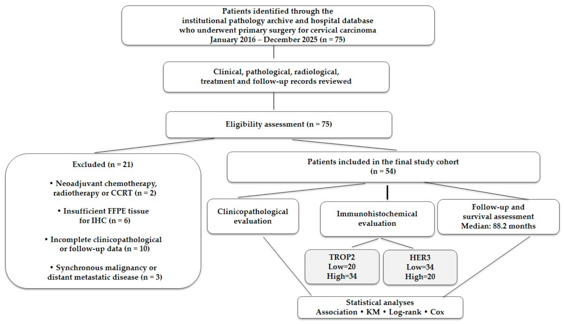
Patients were excluded if they had received neoadjuvant chemotherapy, radiotherapy, or concurrent chemoradiotherapy before surgery (n = 2); had insufficient tumor tissue available for immunohistochemical staining (n = 6); had incomplete clinicopathological or follow-up information (n = 10); or had synchronous malignancies or distant metastatic disease at diagnosis (n = 3).
After applying these eligibility criteria, 54 patients were included in the final study cohort and underwent clinicopathological, immunohistochemical, and survival analyses. All patient data were anonymized before statistical analysis to ensure confidentiality (Figure 1).

2.2. Follow-Up and Outcome Assessment

All patients were managed according to the contemporary National Comprehensive Cancer Network (NCCN) guidelines and institutional protocols for early-stage cervical carcinoma [18]. Primary treatment consisted of radical hysterectomy with pelvic lymph node dissection, with or without para-aortic lymphadenectomy. Postoperative adjuvant radiotherapy, concurrent platinum-based chemoradiotherapy, or observation was administered according to pathological risk factors and multidisciplinary tumor board recommendations.
The primary study endpoints were disease-free survival (DFS) and overall (OS) survival. DFS was defined as the interval from primary surgery to the first documented recurrence, disease progression, death from any cause, or last follow-up. OS was defined as the interval from primary surgery to death from any cause or the last follow-up. Mortality was evaluated based on death from any cause rather than cancer-specific death. Patients without recurrence who were alive at the last follow-up were censored on the date of their most recent clinical assessment. All survival analyses were based exclusively on data derived from the study cohort, without the incorporation of any publicly available or external datasets.

2.3. Histopathological Evaluation

All surgical specimens were immediately fixed in 10% neutral-buffered formalin for 24–48 h after surgical resection and subsequently processed according to standardized pathology protocols. Tissue samples were processed using an automated tissue processor, embedded in paraffin, sectioned to 4-μm thickness, and stained with hematoxylin and eosin (H&E).
Histopathological diagnosis was confirmed according to the 2020 World Health Organization (WHO) Classification of Female Genital Tumors [19], whereas pathological staging was reassessed according to the 2018 International Federation of Gynecology and Obstetrics (FIGO) staging system [20].
The histological subtypes were classified as squamous cell carcinoma or adenocarcinoma. Tumor grade was recorded according to the original pathological diagnosis and re-evaluated when required.

2.4. Immunohistochemical Evaluation of TROP2 and HER3 Expression

Representative formalin-fixed, paraffin-embedded (FFPE) tissue blocks were selected after a comprehensive review of all available hematoxylin and eosin (H&E)-stained slides. For each case, a tissue block containing the largest area of viable invasive carcinoma with optimal fixation and preservation was selected for immunohistochemical analysis. Areas predominantly composed of carcinoma in situ, necrosis, hemorrhage, fibrosis, inflammatory infiltrates, cautery artifacts, or tissue folds were excluded. In tumors showing intratumoral heterogeneity, multiple blocks were reviewed, and the block most representative of the invasive tumor component was selected for the analysis.
Immunohistochemical staining was performed on 4-µm-thick sections using a fully automated immunostaining platform (Ventana Benchmark Ultra; Roche Diagnostics, Basel, Switzerland). The sections were incubated with primary antibodies against TROP2 (mouse monoclonal antibody, Clone B-9, 1:100 dilution, Santa Cruz Biotechnology, Santa Cruz, CA, USA) and HER3 (rabbit monoclonal antibody, clone D22C5, 1:100 dilution; Cell Signaling Technology, Danvers, MA, USA). Appropriate positive controls were included in each staining run, consisting of normal breast epithelium for TROP2 and breast carcinoma tissue for HER3, according to the manufacturers’ recommendations. Negative controls were prepared by omitting the primary antibodies.
All stained slides were independently evaluated by experienced gynecologic pathologists (R.O.Y.) who were blinded to the clinicopathological data and the patient outcomes. For TROP2, membranous and/or cytoplasmic staining of invasive tumor cells was assessed using a semi-quantitative method. Staining intensity was graded as 0 (absent), 1 (weak), 2 (moderate), or 3 (strong), while the proportion of positive tumor cells was scored as 0 (0%), 1 (1–19%), 2 (20–50%), and 3 (>50%). The immunoreactivity score (IRS) was calculated by multiplying the intensity and proportion scores, yielding a total score ranging from 0 to 9 [21]. Tumors with an IRS < 6 were classified as having low TROP2 expression, whereas those with an IRS ≥ 6 were classified as having high TROP2 expression.
For HER3, only membranous staining was considered positive. Staining intensity was scored as 0 (absent), 1 (weak), 2 (moderate), or 3 (strong), and the proportion of positive tumor cells was graded as 0 (0%), 1 (1–10%), 2 (11–30%), 3 (31–50%), and 4 (>50%). The immunoreactivity score (IRS) was calculated by multiplying the intensity and proportion scores [22]. Tumors with an IRS < 6 were classified as having low HER3 expression, whereas those with an IRS ≥ 6 were considered to have high HER3 expression.
Although the proportion scores differed between TROP2 and HER3, the scoring systems were based on previously published biomarker-specific methods rather than on the distribution of expression in our cohort [21,22]. These differences reflect the established immunohistochemical assessment criteria for each marker. However, an IRS of ≥6 was used to define high expression for both biomarkers. The scoring criteria and cutoffs were therefore defined a priori and were not selected or modified according to the survival outcomes or biomarker distribution in the present cohort.

2.5. Statistical Analysis

Statistical analyses were performed using IBM SPSS Statistics version 27.0 (IBM Corp., Armonk, NY, USA) and R software version 4.5.2 (R Foundation for Statistical Computing, Vienna, Austria). The normality of continuous variables was assessed using the Shapiro–Wilk test. Normally distributed variables are presented as mean ± standard deviation (SD), whereas non-normally distributed variables are expressed as median (interquartile range [IQR]). Categorical variables were summarized as frequencies and percentages.
Comparisons between biomarker expression groups were performed using the independent-samples Student’s t-test or Mann–Whitney U test, as appropriate. Categorical variables were compared using Pearson’s chi-square or Fisher’s exact tests.
Disease-free survival (DFS) and overall survival (OS) were estimated using the Kaplan–Meier method and compared using the log-rank test. Prognostic factors associated with DFS and OS were initially evaluated using univariate Cox proportional hazards regression analysis. Variables with p < 0.10 in the univariable analysis, together with clinically relevant variables, were included in the multivariable Cox regression models.
For each survival endpoint, two multivariate models were constructed separately for TROP2 and HER3. Model 1 included age, menopausal status, FIGO stage, histological subtype, tumor size, lymphovascular space invasion (LVSI), and adjuvant treatment status. Model 2 included all variables in Model 1, together with either TROP2 or HER3 expression, to determine the independent prognostic contribution of each biomarker.
Hazard ratios (HRs) and 95% confidence intervals (95% CIs) were calculated for all Cox models. The proportional hazards assumption was evaluated using scaled Schoenfeld residuals for each covariate as well as for the overall model. A p value > 0.05 was considered to indicate no significant departure from proportionality. Both individual covariate and global tests were examined, and no significant violations of the proportional hazards assumption were identified. Statistical significance was set at p < 0.05.

3. Results

This study included 54 patients with early-stage cervical carcinoma. The mean age was 57.5 ± 12.2 years, and the median gravida and parity were 3 (IQR, 2–4). Most patients were postmenopausal (72.2%), and 51.9% had at least one comorbidity. HPV18 (44.4%) and HPV16 (33.3%) were the predominant HPV genotypes. Squamous cell carcinoma accounted for 81.5% of cases, and 38.9% demonstrated lymphovascular space invasion. Primary treatment consisted of radical hysterectomy. Eight patients underwent surgery without pelvic lymph node dissection and had no evidence of nodal involvement at initial evaluation. Para-aortic lymph node involvement was present in 12 patients and received adjuvant treatment (68.5% of patients). Chemotherapy was administered to 94.4% of patients (Table 1).
Table 1. Baseline clinicopathological characteristics of patients with early-stage cervical carcinoma.
CharacteristicOverall (n = 54)
Age, years57.5 ± 12.2
Gravida3 (2–4)
Parity3 (2–4)
Menopausal status
Premenopausal15 (27.8)
Postmenopausal39 (72.2)
Comorbidity
Absent26 (48.1)
Present28 (51.9)
HPV status
Negative10 (18.5)
HPV1618 (33.3)
HPV1824 (44.4)
Other2 (3.7)
FIGO 2018 stage
IA110 (18.5)
IA215 (27.8)
IB17 (13.0)
IB24 (7.4)
IB310 (18.5)
IIA14 (7.4)
IIA24 (7.4)
Tumor size
<2 cm24 (44.4)
2–4 cm12 (22.2)
>4 cm18 (33.3)
Histological subtype
Squamous cell carcinoma44 (81.5)
Adenocarcinoma10 (18.5)
LVSI
Absent33 (61.1)
Present21 (38.9)
Surgical procedure
Radical hysterectomy8 (14.8)
Radical hysterectomy + pelvic lymph node dissection46 (85.2)
Adjuvant treatment
No17 (31.5)
Yes37 (68.5)
Chemotherapy
No3 (5.6)
Yes51 (94.4)
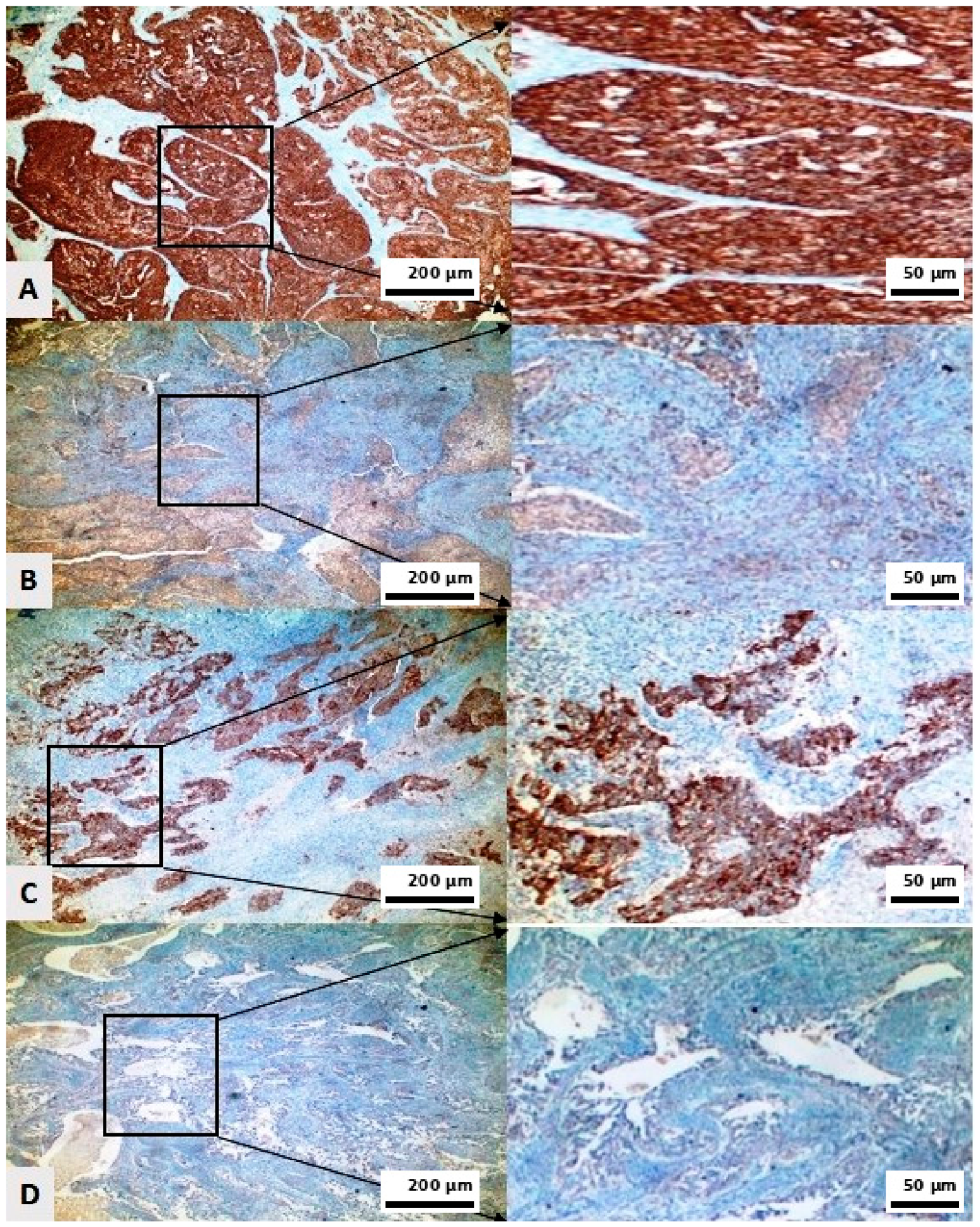
Among the 54 patients included in the study, low TROP2 expression was observed in 20 (37.0%), whereas high TROP2 expression was detected in 34 (63.0%). In contrast, HER3 expression was classified as low in 34 patients (63.0%) and high in 20 patients (37.0%) (Figure 2).
The associations between TROP2 and HER3 expression and clinicopathological characteristics are shown in Table 2. Patients with low and high TROP2 expression levels showed comparable demographic and clinicopathological characteristics. The mean age was similar between the TROP2-low and TROP2-high groups (57.2 ± 10.2 vs. 57.7 ± 13.4 years, p = 0.897). No significant differences were observed in gravida, parity, menopausal status, comorbidity, HPV genotype, FIGO 2018 stage, tumor size, histological subtype, or lymphovascular space invasion (p > 0.05). Similarly, the distribution of surgical procedures and adjuvant treatments, including radiotherapy and chemotherapy, did not differ significantly between the groups.
Similarly, HER3 expression was not significantly associated with any baseline clinicopathological characteristics. Patients with high and low HER3 expression had comparable age, reproductive history, menopausal status, comorbidity status, HPV subtype distribution, FIGO stage, tumor size, histological subtype, LVSI status, and surgical management (p > 0.05). Although patients with low HER3 expression more frequently received adjuvant radiotherapy (76.5% vs. 55.0%, p = 0.091), these differences did not reach statistical significance. Similarly, adjuvant chemotherapy was administered to nearly all patients in both HER3 expression groups (97.1% vs. 90.0%, p = 0.306).
During a median follow-up of 88.2 months, 18 (33.3%) patients died and 36 (66.7%) remained alive. Disease recurrence occurred in 22 patients (40.7%), whereas 32 patients (59.3%) remained recurrence free. The median overall survival (OS) for the entire cohort was 89.1 months (95% CI: 75.2–103.1), and the median disease-free survival (DFS) was 80.5 months (95% CI: 66.0–95.2).
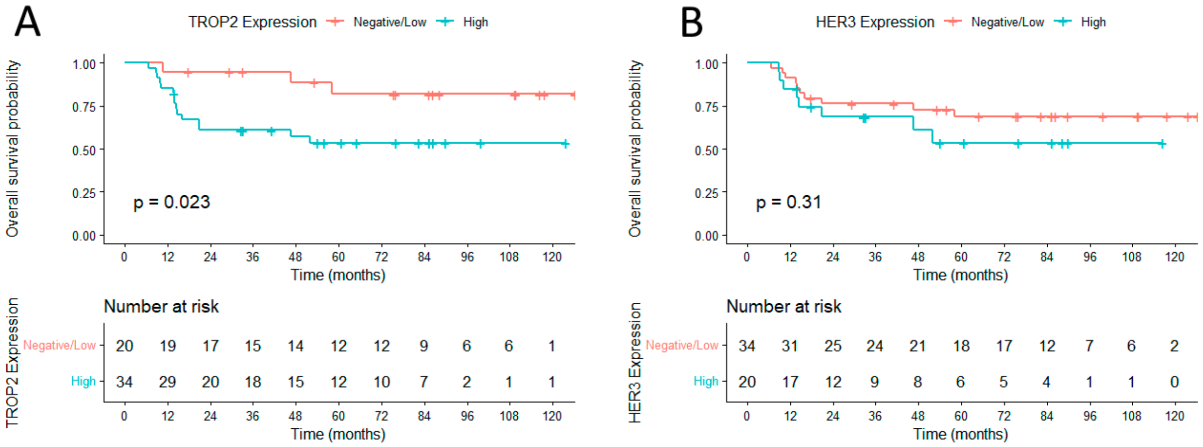
Kaplan–Meier survival analysis demonstrated that patients with negative/low TROP2 expression had a significantly longer median OS than those with high TROP2 expression (110.9 months [95% CI: 94.8–127.0] vs. 75.1 months [95% CI: 56.7–93.4], respectively; log-rank p = 0.023), indicating that high TROP2 expression was associated with a poorer overall survival. Regarding HER3 expression, the median OS was 94.3 months (95% CI: 77.5–111.1) in the negative/low HER3 expression group and 73.9 months (95% CI: 51.5–96.2) in the high HER3 expression group. However, this difference was not statistically significant (log-rank p = 0.310) (Figure 3).
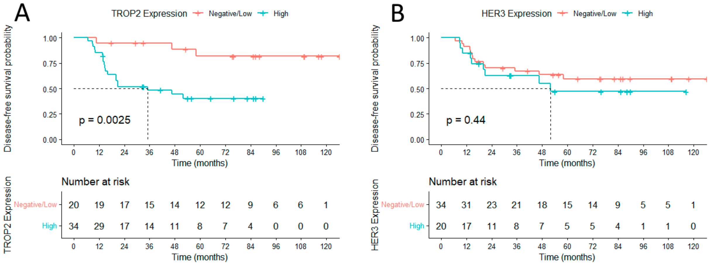
Kaplan–Meier analysis further demonstrated that patients with negative/low TROP2 expression had a significantly longer median DFS than those with high TROP2 expression (110.9 months [95% CI: 94.8–127.0] vs. 48.4 months [95% CI: 36.0–60.7], respectively; log-rank p = 0.003), indicating that elevated TROP2 expression was associated with an increased risk of disease recurrence. In a separate analysis of HER3 expression, the median DFS was 84.7 months (95% CI: 66.9–102.5) in the negative/low HER3 expression group and 68.0 months (95% CI: 45.4–90.7) in the high HER3 expression group. However, the difference between the two groups was not statistically significant (log-rank p = 0.440) (Figure 4).
Univariable Cox proportional hazards regression analyses were performed to identify clinicopathological factors associated with overall survival (OS) and disease-free survival (DFS). For OS, high TROP2 expression was significantly associated with an increased risk of mortality (HR = 2.01, 95% CI: 1.12–3.58, p = 0.019). HER3 expression demonstrated a trend toward worse OS but did not reach statistical significance (HR = 1.69, 95% CI: 0.96–2.98, p = 0.071). FIGO stage also showed a borderline association with OS (HR = 1.17, 95% CI: 1.00–1.36, p = 0.051). No significant associations were observed between OS and age, gravida, parity, menopausal status, comorbidity, HPV status, tumor size, histological subtype, lymphovascular invasion, surgical approach, adjuvant chemotherapy, and adjuvant radiotherapy (p > 0.05). For DFS, elevated TROP2 expression was identified as a significant predictor of disease recurrence (HR = 2.97, 95% CI: 1.56–5.64, p < 0.001). Although the surgical approach showed a borderline association with DFS (HR = 2.28, 95% CI: 0.94–5.56, p = 0.069), no statistically significant relationships were observed for HER3 expression or the remaining clinicopathological variables (p > 0.05) (Table 3).
Multivariable Cox proportional hazards regression analyses were performed using three sequential models to evaluate the independent prognostic value of TROP2 and HER3 expression for overall survival (OS). In Model 1, which included clinicopathological variables only, older age (HR = 1.07, 95% CI: 1.02–1.13, p = 0.011), advanced FIGO stage (HR = 1.80, 95% CI: 1.33–2.43, p < 0.001), and lymphovascular invasion (HR = 1.06, 95% CI: 0.01–1.31, p < 0.001) were independently associated with OS. After the addition of TROP2 expression in Model 2, age and FIGO stage remained significant independent predictors, whereas high TROP2 expression was not independently associated with OS (HR = 2.40, 95% CI: 0.61–9.47, p = 0.213). In Model 3, which additionally incorporated HER3 expression, the associations between age, FIGO stage, and lymphovascular invasion remained unchanged. Neither TROP2 (HR = 2.42, 95% CI: 0.59–10.00, p = 0.221) nor HER3 expression (HR = 0.96, 95% CI: 0.27–3.39, p = 0.950) emerged as independent prognostic factors for OS after adjustment for clinicopathological variables (Table 4).
Multivariable Cox proportional hazards regression analyses were performed using three sequential models to determine the independent prognostic significance of TROP2 and HER3 expression for disease-free survival (DFS). In Model 1, older age (HR = 1.06, 95% CI: 1.01–1.11, p = 0.026), advanced FIGO stage (HR = 1.56, 95% CI: 1.18–2.06, p = 0.002), and lymphovascular invasion (HR = 1.09, 95% CI: 0.02–1.35, p < 0.001) were identified as independent predictors of DFS. Following the inclusion of TROP2 expression in Model 2, age and FIGO stage remained independently associated with DFS, while high TROP2 expression emerged as an independent adverse prognostic factor, conferring a 4.66-fold increased risk of disease recurrence (HR = 4.66, 95% CI: 1.26–17.17, p = 0.021). In Model 3, after additional adjustment for HER3 expression, the prognostic significance of TROP2 remained unchanged (HR = 5.00, 95% CI: 1.28–19.57, p = 0.021), whereas HER3 expression was not independently associated with DFS (HR = 0.80, 95% CI: 0.25–2.52, p = 0.701). These findings indicate that TROP2, but not HER3, provides independent prognostic information for disease recurrence beyond established clinicopathological variables (Table 5).

4. Discussion

In this study, we examined the clinicopathological and prognostic significance of TROP2 and HER3 expression in early-stage cervical carcinoma. Neither marker was significantly associated with the clinicopathological characteristics evaluated. High TROP2 expression, however, was associated with poorer overall and disease-free survival and with an increased risk of recurrence after adjustment for clinicopathological factors. HER3 expression showed no independent association with survival outcomes. These findings point to a potentially greater prognostic relevance of TROP2 in early-stage cervical carcinoma.
The relationship between TROP2 expression and recurrence, in the absence of clear associations with conventional pathological features, may reflect differences in the underlying biology of these tumors. Experimental studies have linked TROP2 to tumor cell proliferation, invasion, migration, and epithelial–mesenchymal transition through signaling pathways such as PI3K/AKT and MAPK/ERK [23,24]. These biological effects may partly account for the poorer disease-free survival observed in patients with high TROP2 expression. TROP2 may therefore capture aspects of tumor behavior that are not fully represented by routine clinicopathological parameters.
Our findings are broadly consistent with those of Hiller et al., who reported an association between TROP2 overexpression and unfavorable survival in cervical cancer [25]. In contrast to their study, we did not observe a significant relationship between TROP2 expression and clinicopathological features. Differences in patient characteristics and methodological factors, including immunohistochemical assessment and scoring criteria, may explain these differences [26]. Despite these variations, the association between TROP2 expression and recurrence in our cohort warrants further investigation of its potential role in postoperative risk assessment.
The findings for HER3 were less conclusive. Although high HER3 expression showed a tendency toward poorer overall survival, it was not independently associated with clinical outcomes. This differs from the findings of Chang et al., who identified HER3 overexpression as an independent predictor of both disease-free and overall survival in cervical cancer [27]. Differences in patient populations, treatment approaches, antibodies, scoring methods, and cut-off definitions may contribute to these conflicting results. The prognostic significance of HER3 in cervical cancer therefore remains uncertain and requires confirmation in independent cohorts.
TROP2 has also received increasing attention as a therapeutic target, particularly with the development of TROP2-directed antibody–drug conjugates, some of which have shown antitumor activity in recurrent or metastatic cervical cancer [28]. Our study, however, focused on the prognostic significance of TROP2 and did not evaluate treatment response. The present findings therefore cannot establish a predictive role for TROP2 or demonstrate benefit from TROP2-directed therapy.
This study had several limitations. The retrospective single-center design may have introduced a selection bias and limited the generalizability of the findings. The relatively small sample size, particularly for subgroup analyses, may have reduced the statistical power to detect modest associations, especially for HER3. The limited number of recurrence and death events also increased the potential for overfitting in the multivariable Cox models and may have affected the stability of the hazard ratio estimates. Therefore, these results, particularly those with wide confidence intervals, should be interpreted cautiously. Furthermore, our cohort was limited to surgically treated patients with early-stage cervical carcinoma; therefore, the findings may not be applicable to patients with more advanced disease. Finally, the lack of external validation and functional molecular analyses precluded confirmation of the biological mechanisms underlying the prognostic role of TROP2 and HER3. Future multicenter prospective studies with larger cohorts are required to validate these findings.

5. Conclusions

In summary, our findings indicate that TROP2 may have prognostic value in early-stage cervical carcinoma. High TROP2 expression was independently associated with an increased risk of recurrence, whereas HER3 expression showed no significant association with clinical outcomes. These findings suggest that TROP2 may help identify patients at higher risk of recurrence. However, treatment response was not assessed in this study, and our results therefore do not establish a predictive or therapeutic role for TROP2. Prospective studies with larger cohorts and treatment-response data are needed to confirm its prognostic value and explore its potential relevance as a therapeutic biomarker.

Author Contributions

Conceptualization, D.K. and N.Y.; methodology, D.K.; software, D.K.; validation, D.K., N.Y. and R.O.Y.; formal analysis, R.O.Y.; investigation, D.K. and R.O.Y. resources, D.K.; data curation, D.K., N.Y. and R.O.Y.; writing—original draft preparation, D.K., N.Y. and R.O.Y.; writing—review and editing, D.K., N.Y. and R.O.Y.; visualization, R.O.Y.; supervision, D.K.; project administration, D.K.; funding acquisition, D.K. All authors have read and agreed to the published version of the manuscript.

Funding

This research received no external funding.

Institutional Review Board Statement

The study was approved by the Institutional Sivas Cumhuriyet University Ethics Committee (Approval No: 2026-04/55; 16 April 2026) and was conducted in accordance with the Declaration of Helsinki (1964, revised 2024).

Informed Consent Statement

Patient consent was waived due to the fact that this study was conducted on a retrospective file.

Data Availability Statement

The datasets used in this study can be made available by the corresponding author upon reasonable request, with permission from the Obstetrics and Gynecology Department of Sivas Cumhuriyet University School of Medicine.

Conflicts of Interest

The authors declare no conflicts of interest.

Abbreviations

ADCantibody–drug conjugate
CIconfidence interval
DFSdisease-free survival
FFPEformalin-fixed paraffin-embedded
FIGOInternational Federation of Gynecology and Obstetrics
HER3human epidermal growth factor receptor 3
HRhazard ratio
IHCimmunohistochemistry
IRSimmunoreactivity score
LVSIlymphovascular space invasion
OSoverall survival
PI3Kphosphatidylinositol 3-kinase
AKTprotein kinase B
MAPKmitogen-activated protein kinase
ERKextracellular signal-regulated kinase
TROP2trophoblast cell surface antigen 2

References

  1. Wu, J.; Jin, Q.; Zhang, Y.; Ji, Y.; Li, J.; Liu, X.; Duan, H.; Feng, Z.; Liu, Y.; Zhang, Y.; et al. Global burden of cervical cancer: Current estimates, temporal trend and future projections based on the GLOBOCAN 2022. J. Natl. Cancer Cent. 2025, 5, 322–329. [Google Scholar] [CrossRef] [Scilit] [PubMed]
  2. Zhang, S.; Xu, H.; Zhang, L.; Qiao, Y. Cervical cancer: Epidemiology, risk factors and screening. Chin. J. Cancer Res. 2020, 32, 720–728. [Google Scholar] [CrossRef] [Scilit] [PubMed]
  3. Pal, A.; Kundu, R. Human Papillomavirus E6 and E7: The Cervical Cancer Hallmarks and Targets for Therapy. Front. Microbiol. 2020, 10, 3116. [Google Scholar] [CrossRef] [Scilit] [PubMed]
  4. Santoro, A.; Inzani, F.; Angelico, G.; Arciuolo, D.; Bragantini, E.; Travaglino, A.; Valente, M.; D’Alessandris, N.; Scaglione, G.; Sfregola, S.; et al. Recent Advances in Cervical Cancer Management: A Review on Novel Prognostic Factors in Primary and Recurrent Tumors. Cancers 2023, 15, 1137. [Google Scholar] [CrossRef] [Scilit] [PubMed]
  5. Guimarães, Y.M.; Godoy, L.R.; Longatto-Filho, A.; Reis, R.D. Management of Early-Stage Cervical Cancer: A Literature Review. Cancers 2022, 14, 575. [Google Scholar] [CrossRef] [Scilit] [PubMed]
  6. Li, X.; Teng, S.; Zhang, Y.; Zhang, W.; Zhang, X.; Xu, K.; Yao, H.; Yao, J.; Wang, H.; Liang, X.; et al. TROP2 promotes proliferation, migration and metastasis of gallbladder cancer cells by regulating PI3K/AKT pathway and inducing EMT. Oncotarget 2017, 8, 47052–47063. [Google Scholar] [CrossRef] [Scilit] [PubMed]
  7. Wen, Y.; Ouyang, D.; Zou, Q.; Chen, Q.; Luo, N.; He, H.; Anwar, M.; Yi, W. A literature review of the promising future of TROP2: A potential drug therapy target. Ann. Transl. Med. 2022, 10, 1403. [Google Scholar] [CrossRef] [Scilit] [PubMed]
  8. Loriot, Y.; Balar, A.V.; Petrylak, D.P.; Kalebasty, A.R.; Grivas, P.; Fléchon, A.; Jain, R.K.; Swami, U.; Bupathi, M.; Barthélémy, P.; et al. Sacituzumab Govitecan Demonstrates Efficacy across Tumor Trop-2 Expression Levels in Patients with Advanced Urothelial Cancer. Clin. Cancer Res. 2024, 30, 3179–3188. [Google Scholar] [CrossRef] [Scilit] [PubMed]
  9. Yuceer, R.O.; Aydin, S.; Ozer, H.; Aydin, A.A.; Yilmaz, M.; Kaya, S.; Koc, T. The prognostic and therapeutic potential of Claudin-6 and Trop-2 expression as targeted biomarkers in serous ovarian cancer: An observational study. Medicine 2025, 104, e43110. [Google Scholar] [CrossRef] [Scilit] [PubMed]
  10. Chiba, Y.; Kojima, Y.; Yazaki, S.; Yoshida, H.; Takamizawa, S.; Kitadai, R.; Saito, A.; Okuma, H.S.; Nishikawa, T.; Shimoi, T.; et al. Trop-2 expression and the tumor immune microenvironment in cervical cancer. Gynecol. Oncol. 2024, 187, 51–57. [Google Scholar] [CrossRef] [Scilit] [PubMed]
  11. Nagdev, P.; Chittilla, M. Advances in Screening, Immunotherapy, Targeted Agents, and Precision Surgery in Cervical Cancer: A Comprehensive Clinical Review (2018–2025). Curr. Oncol. 2026, 33, 48. [Google Scholar] [CrossRef] [Scilit] [PubMed]
  12. Pan, L.; Li, J.; Xu, Q.; Gao, Z.; Yang, M.; Wu, X.; Li, X. HER2/PI3K/AKT pathway in HER2-positive breast cancer: A review. Medicine 2024, 103, e38508. [Google Scholar] [CrossRef] [Scilit] [PubMed]
  13. Majumder, A. HER3: Toward the Prognostic Significance, Therapeutic Potential, Current Challenges, and Future Therapeutics in Different Types of Cancer. Cells 2023, 12, 2517. [Google Scholar] [CrossRef] [Scilit] [PubMed]
  14. Kim, M.; Ju, H.M.; Song, J.Y.; Sampson, J.; Bayliss, R.; Choi, J. HER3 overexpression: A predictive marker for poor prognosis in advanced ALK-positive non-small cell lung cancer treated with ALK inhibitors. Transl. Lung Cancer Res. 2024, 13, 321–333. [Google Scholar] [CrossRef] [Scilit] [PubMed]
  15. Garrett, J.T.; Tendler, S.; Feroz, W.; Kilroy, M.K.; Yu, H. Emerging importance of HER3 in tumorigenesis and cancer therapy. Nat. Rev. Clin. Oncol. 2025, 22, 348–370. [Google Scholar] [CrossRef] [Scilit] [PubMed]
  16. Hassanzadeh Makoui, M.; Fekri, S.; Jafari, Z.; Hassanzadeh Makoui, R.; Ansari, N.; Shokrzadeh, S. Effect of HER3 Overexpression on Survival Outcomes in Pancreatic Cancer Patients: A Systematic Review and Meta-Analysis. Asian Pac. J. Cancer Prev. 2025, 26, 3175–3183. [Google Scholar] [CrossRef] [Scilit] [PubMed]
  17. Yang, X.; Chen, Y.; Li, M.; Zhu, W. ERBB3 methylation and immune infiltration in tumor microenvironment of cervical cancer. Sci. Rep. 2022, 12, 8112. [Google Scholar] [CrossRef] [Scilit] [PubMed]
  18. Abu-Rustum, N.R.; Campos, S.M.; Amarnath, S.; Arend, R.; Barber, E.; Bradley, K.; Brooks, R.; Chino, J.; Chon, H.S.; Crispens, M.A.; et al. Cervical Cancer, Version 2.2026, NCCN Clinical Practice Guidelines In Oncology. J. Natl. Compr. Cancer Netw. 2025, 23, 549–573. [Google Scholar] [CrossRef] [Scilit] [PubMed]
  19. Höhn, A.K.; Brambs, C.E.; Hiller, G.G.R.; May, D.; Schmoeckel, E.; Horn, L.C. 2020 WHO Classification of Female Genital Tumors. Geburtshilfe Und Frauenheilkd. 2021, 81, 1145–1153. [Google Scholar] [CrossRef] [Scilit] [PubMed]
  20. Tang, X.; Guo, C.; Liu, S.; Guo, J.; Hua, K.; Qiu, J. A novel prognostic nomogram utilizing the 2018 FIGO staging system for cervical cancer: A large multicenter study. Int. J. Gynaecol. Obstet. 2021, 155, 86–94. [Google Scholar] [CrossRef] [Scilit] [PubMed]
  21. Foersch, S.; Schmitt, M.; Litmeyer, A.S.; Tschurtschenthaler, M.; Gress, T.; Bartsch, D.K.; Pfarr, N.; Steiger, K.; Denkert, C.; Jesinghaus, M. TROP2 in colorectal carcinoma: Associations with histopathology, molecular phenotype, and patient prognosis. J. Pathol. Clin. Res. 2024, 10, e12394. [Google Scholar] [CrossRef] [Scilit] [PubMed]
  22. Becker, A.K.; Puladi, B.; Xie, K.; Cassataro, A.; Götzl, R.; Hölzle, F.; Beier, J.P.; Knüchel-Clarke, R.; Braunschweig, T. HER3 (ERBB3) amplification in liposarcoma—A putative new therapeutic target? World J. Surg. Oncol. 2024, 22, 131. [Google Scholar] [CrossRef] [Scilit] [PubMed]
  23. Liu, T.; Liu, Y.; Bao, X.; Tian, J.; Liu, Y.; Yang, X. Overexpression of TROP2 predicts poor prognosis of patients with cervical cancer and promotes the proliferation and invasion of cervical cancer cells by regulating ERK signaling pathway. PLoS ONE 2013, 8, e75864. [Google Scholar] [CrossRef] [Scilit] [PubMed]
  24. Liu, Y.; Huang, W.; Saladin, R.J.; Hsu, J.C.; Cai, W.; Kang, L. Trop2-Targeted Molecular Imaging in Solid Tumors: Current Advances and Future Outlook. Mol. Pharm. 2024, 21, 5909–5928. [Google Scholar] [CrossRef] [Scilit] [PubMed]
  25. Hiller, G.G.R.; Wolf, B.; Forberger, M.; Freude, A.; Brambs, C.E.; Droste, S.; Horn, L.C.; Höhn, A.K. Trop2 expression, p16 expression status, and histologic subtype in carcinoma of the uterine cervix. Pathol. Oncol. Res. POR 2025, 31, 1612252. [Google Scholar] [CrossRef] [Scilit] [PubMed]
  26. Ulvang, M.L.; Kvile, O.F.; Berg, H.F.; Woie, K.; Haldorsen, I.S.; Santin, A.D.; Bertelsen, B.I.; Krakstad, C.; Halle, M.K. Co-expression of tissue factor, TROP2, and NECTIN4 in primary and matched metastatic cervical cancer lesions. Transl. Oncol. 2025, 59, 102453. [Google Scholar] [CrossRef] [Scilit] [PubMed]
  27. Chang, C.S.; Shim, J.I.; Byeon, S.J.; Lee, E.J.; Lee, Y.Y.; Kim, T.J.; Lee, J.W.; Kim, B.G.; Choi, C.H. Prognostic Significance of HER3 Expression in Patients with Cervical Cancer. Cancers 2022, 14, 2139. [Google Scholar] [CrossRef] [Scilit] [PubMed]
  28. Ettorre, V.M.; Demirkiran, C.; Bellone, S.; Hartwich, T.M.P.; Greenman, M.; McNamara, B.; Sethi, N.; Palmieri, L.; Santin, A.D. Effective preclinical activity of datopotamab deruxtecan (Dato-DXd), an ADC targeting trophoblast cell-surface antigen 2 (TROP2), against primary cervical carcinoma cell lines and xenografts. Gynecol. Oncol. 2025, 201, 195–202. [Google Scholar] [CrossRef] [Scilit] [PubMed]
Figure 1. Study flow diagram illustrating patient selection and study design.
Diagnostics 16 02871 g001
Figure 2. Representative immunohistochemical staining patterns of TROP2 and HER3 in cervical carcinoma. (A) High TROP2 expression with strong membranous and cytoplasmic staining in tumor cells. (B) Negative/low TROP2 expression with absent or minimal staining. (C) High HER3 expression with prominent membranous staining in tumor cells. (D) Negative/low HER3 expression with absent or weak membranous staining. For each case, the left panel shows the low-magnification image (scale bar, 200 µm), with the boxed area shown at higher magnification in the corresponding right panel (scale bar, 50 µm).
Diagnostics 16 02871 g002
Figure 3. Kaplan–Meier curves for overall survival according to TROP2 and HER3 expression. (A) Overall survival according to TROP2 expression (negative/low, n = 20; high, n = 34). (B) Overall survival according to HER3 expression (negative/low, n = 34; high, n = 20). Numbers below each curve indicate the number of patients at risk at the corresponding time points. Differences between groups were assessed using the log-rank test.
Diagnostics 16 02871 g003
Figure 4. Kaplan–Meier curves for disease-free survival according to TROP2 and HER3 expression. (A) Disease-free survival according to TROP2 expression (negative/low, n = 20; high, n = 34). (B) Disease-free survival according to HER3 expression (negative/low, n = 34; high, n = 20). Numbers below each curve indicate the number of patients at risk at the corresponding time points. Differences between groups were assessed using the log-rank test.
Diagnostics 16 02871 g004
Table 2. Association of TROP2 and HER3 expression with clinicopathological characteristics.
VariableTROP2 Low
(n = 20)
TROP2 High
(n = 34)
pHER3 Low
(n = 34)
HER3 High
(n = 20)
p
Age, years57.2 ± 10.257.7 ± 13.40.89758.2 ± 11.356.3 ± 13.90.577
Gravida3 (2–4)3 (2–5)0.3313.5 (2–5)3 (2–4)0.298
Parity3 (2–3)3 (2–5)0.1763 (2–4)3 (2–3)0.214
Menopausal status
Premenopausal5 (25.0)10 (29.4)0.4918 (23.5)7 (35.0)0.274
Postmenopausal15 (75.0)24 (70.6) 26 (76.5)13 (65.0)
Comorbidity
Absent12 (60.0)14 (41.2)0.14615 (44.1)11 (55.0)0.312
Present8 (40.0)20 (58.8) 19 (55.9)9 (45.0)
HPV status
Negative4 (20.0)6 (17.7) 5 (14.7)5 (25.0)
HPV168 (40.0)10 (29.4)0.56912 (35.3)6 (30.0)0.569
HPV187 (35.0)17 (50.0) 16 (47.1)8 (40.0)
Other1 (5.0)1 (2.9) 1 (2.9)1 (5.0)
FIGO 2018 stage
IA15 (25.0)5 (14.6) 7 (20.7)3 (15.0)
IA25 (25.0)10 (29.4) 10 (29.4)5 (25.0)
IB13 (15.0)4 (11.8) 5 (14.7)2 (10.0)
IB22 (10.0)2 (5.9)0.1713 (8.8)1 (5.0)0.153
IB35 (25.0)5 (14.7) 6 (17.6)4 (20.0)
IIA104 (11.8) 2 (5.9)2 (10.0)
IIA204 (11.8) 1 (2.9)3 (15.0)
Tumor size
<2 cm10 (50.0)14 (41.2) 16 (47.0)8 (40.0)
2–4 cm4 (20.0)8 (23.5)0.5717 (20.6)5 (25.0)0.697
>4 cm6 (30.0)12 (35.3) 11 (32.4)7 (35.0)
Histological subtype
Squamous cell carcinoma15 (75.0)29 (85.3)0.27828 (82.4)16 (80.0)0.550
Adenocarcinoma5 (25.0)5 (14.7) 6 (17.6)4 (20.0)
LVSI
Absent11 (55.0)22 (64.7)0.33720 (58.8)13 (65.0)0.439
Present9 (45.0)12 (35.3) 14 (41.2)7 (35.0)
Surgical procedure
Radical hysterectomy5 (25.0)3 (8.8)0.1136 (17.6)2 (10.0)0.366
Radical hysterectomy + pelvic lymph node dissection15 (75.0)31 (91.2) 28 (82.4)18 (90.0)
Adjuvant radiotherapy
No7 (35.0)10 (29.4)0.4478 (23.5)9 (45.0)0.091
Yes13 (65.0)24 (70.6) 26 (76.5)11 (55.0)
Adjuvant chemotherapy
No1 (5.0)2 (5.9)0.6941 (2.9)2 (10.0)0.306
Yes19 (95.0)32 (94.1) 33 (97.1)18 (90.0)
Table 3. Univariable Cox proportional hazards regression analyses for overall survival and disease-free survival.
VariableOverall Survival HR (95% CI)pDisease-Free Survival HR (95% CI)p
Age1.01 (0.98–1.04)0.4311.01 (0.98–1.04)0.427
Gravida1.04 (0.89–1.20)0.6451.03 (0.89–1.19)0.684
Parity1.08 (0.90–1.29)0.3991.10 (0.92–1.30)0.291
Menopausal status0.68 (0.37–1.24)0.2090.75 (0.41–1.36)0.339
Comorbidity1.49 (0.85–2.60)0.1621.30 (0.75–2.24)0.352
HPV status0.98 (0.69–1.37)0.8881.10 (0.79–1.54)0.579
FIGO stage1.17 (1.00–1.36)0.0511.10 (0.94–1.29)0.225
Adjuvant chemotherapy0.87 (0.48–1.57)0.6401.05 (0.58–1.90)0.868
Adjuvant radiotherapy0.92 (0.28–3.00)0.8961.04 (0.32–3.37)0.954
Lymphovascular invasion1.77 (0.44–2.34)0.3521.71 (0.41–2.24)0.229
Surgical approach1.86 (0.81–4.26)0.1432.28 (0.94–5.56)0.069
Tumor size1.13 (0.82–1.55)0.4541.12 (0.81–1.55)0.479
Histological subtype1.03 (0.51–2.08)0.9381.12 (0.55–2.28)0.763
TROP2 expression2.01 (1.12–3.58)0.0192.97 (1.56–5.64)<0.001
HER3 expression1.69 (0.96–2.98)0.0711.45 (0.83–2.54)0.197
Table 4. Multivariable Cox proportional hazards regression analyses for overall survival.
VariableModel 1 HR (95% CI)pModel 2 HR (95% CI)pModel 3 HR (95% CI)p
Age1.07 (1.02–1.13)0.0111.07 (1.01–1.13)0.0131.07 (1.01–1.13)0.015
Menopausal status0.46 (0.09–2.39)0.3580.53 (0.11–2.70)0.4480.54 (0.11–2.74)0.455
FIGO stage1.80 (1.33–2.43)<0.0011.70 (1.25–2.30)<0.0011.70 (1.24–2.33)<0.001
Histological subtype0.31 (0.03–2.88)0.3030.38 (0.04–3.64)0.4000.38 (0.04–3.77)0.411
Tumor size1.22 (0.68–2.21)0.5021.34 (0.73–2.48)0.3441.35 (0.70–2.60)0.365
Lymphovascular invasion1.06 (0.01–1.31)<0.0011.06 (0.01–1.32)0.0011.06 (0.01–1.32)0.001
Adjuvant chemotherapy2.00 (0.61–6.52)0.2522.23 (0.66–7.56)0.1992.20 (0.62–7.84)0.223
TROP2——2.40 (0.61–9.47)0.2132.42 (0.59–10.00)0.221
HER3————0.96 (0.27–3.39)0.950
Table 5. Multivariable Cox proportional hazards regression analyses for disease-free survival.
VariableModel 1 HR (95% CI)pModel 2 HR (95% CI)pModel 3 HR (95% CI)p
Age1.06 (1.01–1.11)0.0261.06 (1.01–1.11)0.0251.05 (1.01–1.11)0.028
Menopausal status0.65 (0.16–2.62)0.5490.82 (0.21–3.17)0.7770.84 (0.22–3.25)0.802
FIGO stage1.56 (1.18–2.06)0.0021.42 (1.08–1.87)0.0121.45 (1.08–1.93)0.012
Histological subtype0.64 (0.12–3.35)0.6020.94 (0.17–5.02)0.9390.99 (0.18–5.38)0.988
Tumor size1.23 (0.74–2.06)0.4281.37 (0.81–2.31)0.2371.45 (0.80–2.65)0.224
Lymphovascular invasion1.09 (0.02–1.35)<0.0011.08 (0.02–1.36)0.0011.08 (0.02–1.36)0.001
Adjuvant chemotherapy2.03 (0.69–5.96)0.1972.50 (0.81–7.73)0.1122.40 (0.76–7.61)0.136
TROP2——4.66 (1.26–17.17)0.0215.00 (1.28–19.57)0.021
HER3————0.80 (0.25–2.52)0.701
Disclaimer/Publisher’s Note: The statements, opinions and data contained in all publications are solely those of the individual author(s) and contributor(s) and not of MDPI and/or the editor(s). MDPI and/or the editor(s) disclaim responsibility for any injury to people or property resulting from any ideas, methods, instructions or products referred to in the content.