Feasibility of AI-Denoised Ultra-Low-Dose CT for Detection of Acute Pelvic and Hip Fractures: A Pilot Multi-Reader Study
 , ,
, ,
Abstract
1. Background
2. Methods
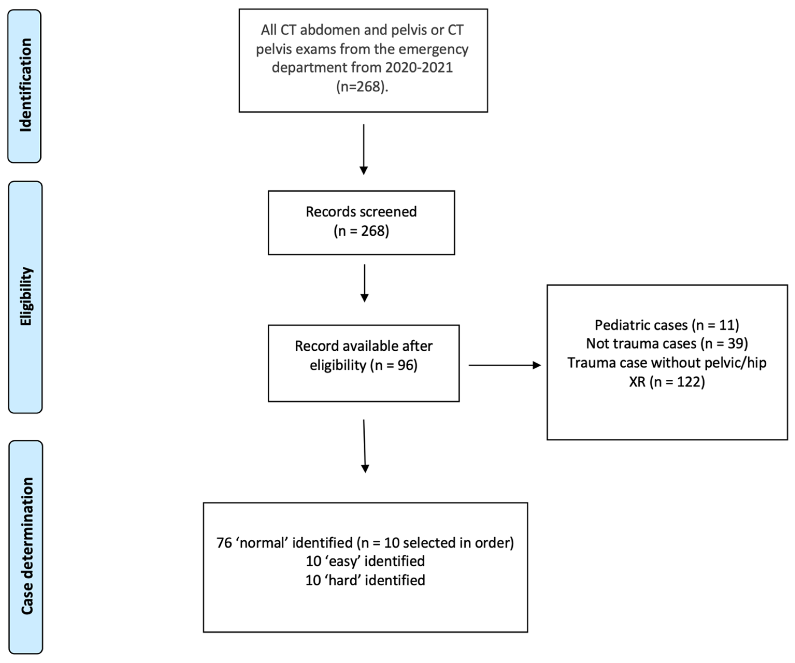
2.1. Inclusion/Exclusion Criteria
2.2. Case Selection
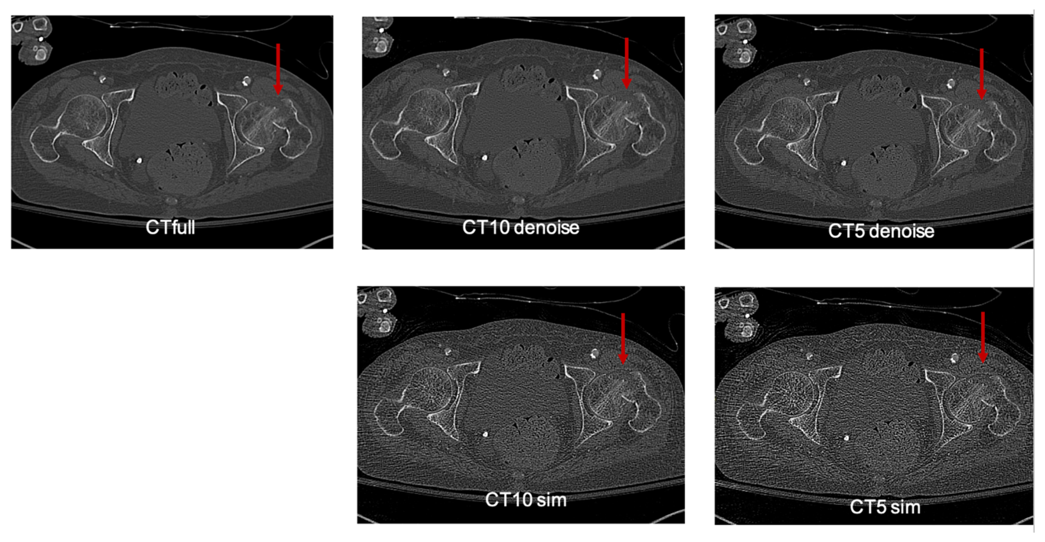
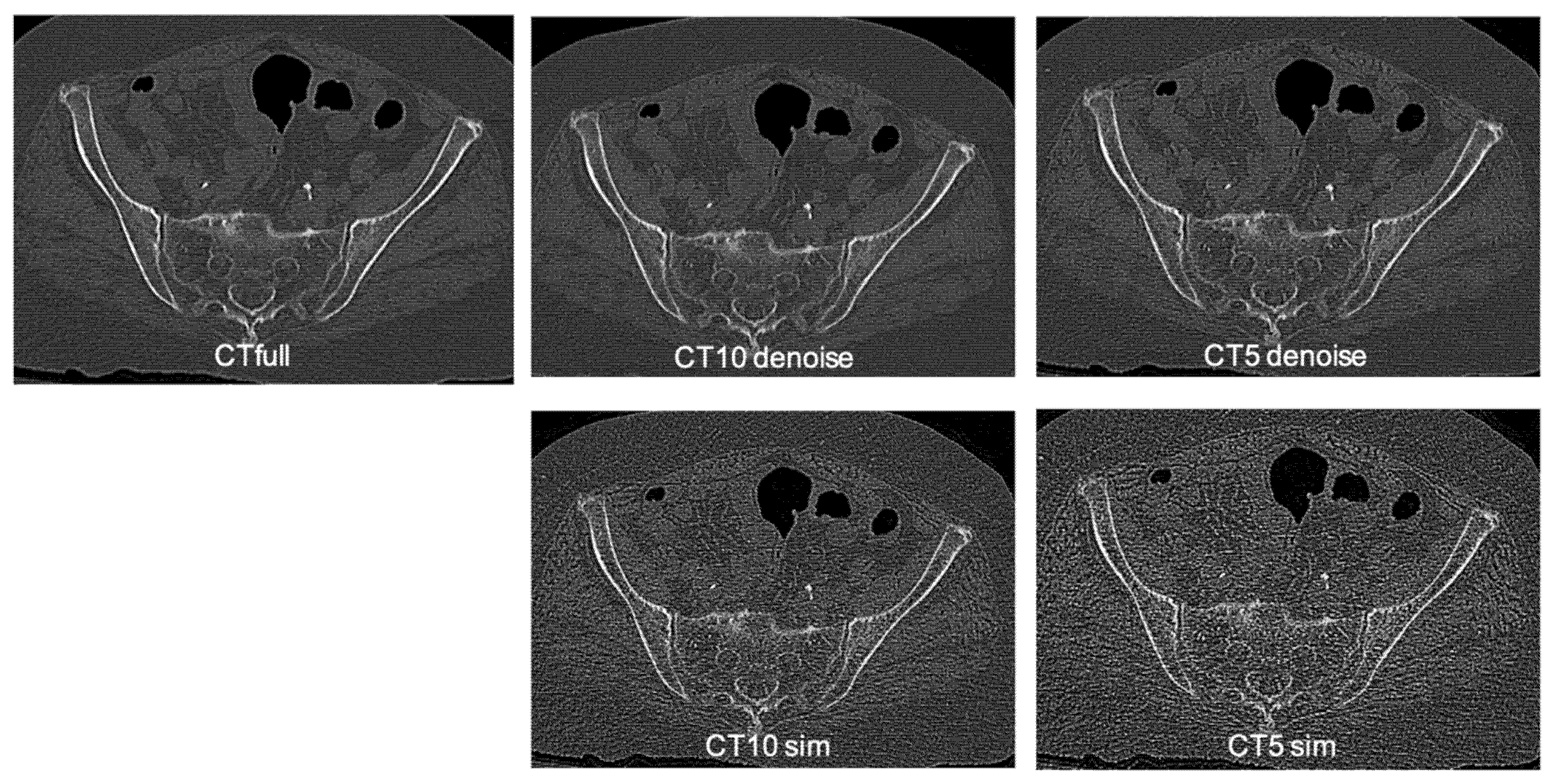
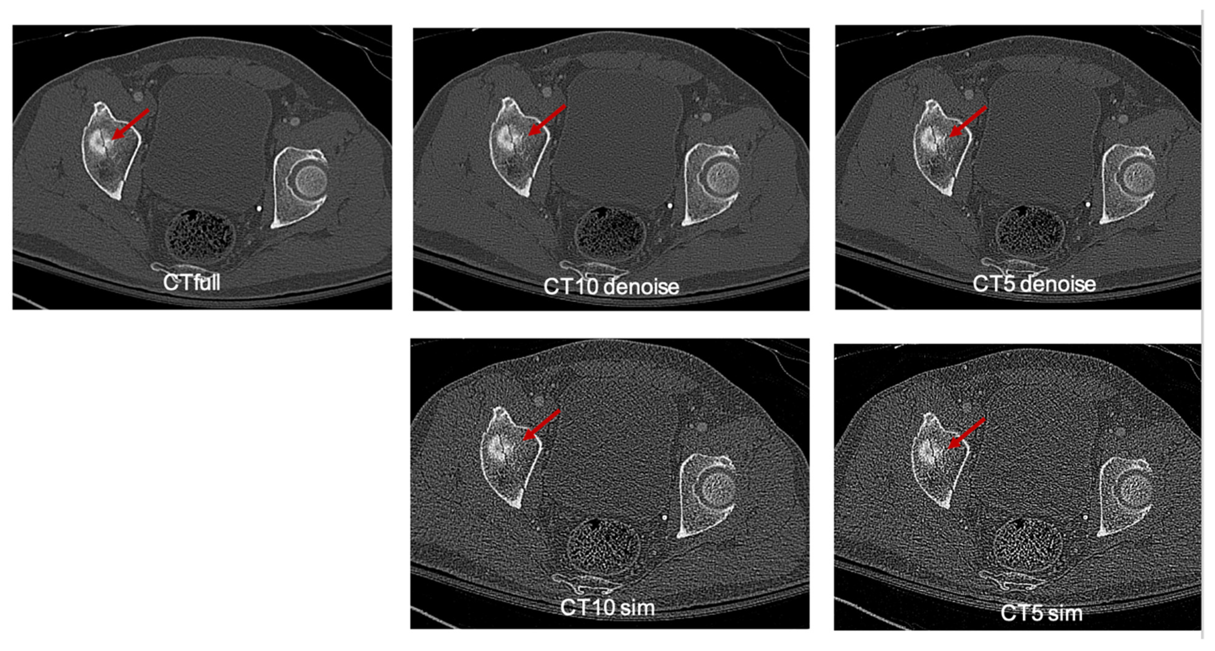
2.3. Reduced-Dose Simulation and Denoising Algorithm
2.4. Deep Learning Denoising Model
2.5. Readers
2.6. Radiation Dose Calculation and Conversion
2.7. Statistical Analysis
3. Results
4. Discussion and Conclusions
Supplementary Materials
Author Contributions
Funding
Institutional Review Board Statement
Informed Consent Statement
Data Availability Statement
Acknowledgments
Conflicts of Interest
Abbreviations
| BMI | Body Mass Index |
| CI | Confidence Interval |
| CNN | Convolutional Neural Network |
| CT | Computed Tomography |
| CT5sim | Computed Tomography with 5% radiation dose |
| CT5dld | Computed Tomography with 5% radiation dose, denoised |
| CT10sim | Computed Tomography with 10% radiation dose |
| CT10dld | Computed Tomography with 10% radiation dose, denoised |
| CTdld | Computed Tomography denoised low dose |
| CTfull | Full-dose Computed Tomography |
| DICOM | Digital Imaging and Communications in Medicine |
| DLD | Deep Learning-based Denoising |
| DLR | Deep Learning Reconstruction |
| ED | Emergency Department |
| IRB | Institutional Review Board |
| MSK | Musculoskeletal |
| mSv | Millisievert (unit of radiation dose) |
| PACS | Picture Archiving and Communication System |
| PGY | Postgraduate Year (Residency Level) |
| ULD-CT | Ultra-Low Dose Computed Tomography |
| XR | X-ray (Radiograph) |
References
- Voth, M.; Lustenberger, T.; Auner, B.; Frank, J.; Marzi, I. What Injuries Should We Expect in the Emergency Room? Injury 2017, 48, 2119–2124. [Google Scholar] [CrossRef] [Scilit] [PubMed]
- Moreland, B.L.; Legha, J.K.; Thomas, K.E.; Burns, E.R. Hip Fracture-Related Emergency Department Visits, Hospitalizations and Deaths by Mechanism of Injury among Adults Aged 65 and Older, United States 2019. J. Aging Health 2023, 35, 345–355. [Google Scholar] [CrossRef] [Scilit] [PubMed]
- Ring, J.; Talbot, C.; Cross, C.; Hinduja, K. NHSLA Litigation in Hip Fractures: Lessons Learnt from NHSLA Data. Injury 2017, 48, 1853–1857. [Google Scholar] [CrossRef] [Scilit] [PubMed]
- Pinto, A.; Berritto, D.; Russo, A.; Riccitiello, F.; Caruso, M.; Belfiore, M.P.; Papapietro, V.R.; Carotti, M.; Pinto, F.; Giovagnoni, A.; et al. Traumatic Fractures in Adults: Missed Diagnosis on Plain Radiographs in the Emergency Department. Acta Biomed. Atenei Parm. 2018, 89, 111–123. [Google Scholar] [CrossRef] [Scilit] [PubMed]
- Thabet, A.M.; Adams, A.; Jeon, S.; Pisquiy, J.; Gelhert, R.; DeCoster, T.A.; Abdelgawad, A. Malpractice Lawsuits in Orthopedic Trauma Surgery: A Meta-Analysis of the Literature. OTA Int. 2022, 5, e199. [Google Scholar] [CrossRef] [Scilit] [PubMed]
- Shah, K.H.; Slovis, B.H.; Runde, D.; Godbout, B.; Newman, D.H.; Lee, J. Radiation Exposure among Patients with the Highest CT Scan Utilization in the Emergency Department. Emerg. Radiol. 2013, 20, 485–491. [Google Scholar] [CrossRef] [Scilit] [PubMed]
- Brenner, D.J.; Hall, E.J. Computed Tomography—An Increasing Source of Radiation Exposure. N. Engl. J. Med. 2007, 357, 2277–2284. [Google Scholar] [CrossRef] [Scilit] [PubMed]
- Kempter, F.; Heye, T.; Vosshenrich, J.; Ceresa, B.; Jäschke, D. Trends in CT Examination Utilization in the Emergency Department during and after the COVID-19 Pandemic. BMC Med. Imaging 2024, 24, 283. [Google Scholar] [CrossRef] [Scilit] [PubMed]
- Hess, E.P.; Haas, L.R.; Shah, N.D.; Stroebel, R.J.; Denham, C.R.; Swensen, S.J. Trends in Computed Tomography Utilization Rates: A Longitudinal Practice-Based Study. J. Patient Saf. 2014, 10, 52–58. [Google Scholar] [CrossRef] [Scilit] [PubMed]
- Larson, D.B.; Johnson, L.W.; Schnell, B.M.; Salisbury, S.R.; Forman, H.P. National Trends in CT Use in the Emergency Department: 1995-2007. Radiology 2011, 258, 164–173. [Google Scholar] [CrossRef] [Scilit] [PubMed]
- Sagara, Y.; Hara, A.K.; Pavlicek, W.; Silva, A.C.; Paden, R.G.; Wu, Q. Abdominal CT: Comparison of Low-Dose CT with Adaptive Statistical Iterative Reconstruction and Routine-Dose CT with Filtered Back Projection in 53 Patients. AJR Am. J. Roentgenol. 2010, 195, 713–719. [Google Scholar] [CrossRef] [Scilit] [PubMed]
- Huber, A.; Landau, J.; Ebner, L.; Bütikofer, Y.; Leidolt, L.; Brela, B.; May, M.; Heverhagen, J.; Christe, A. Performance of Ultralow-Dose CT with Iterative Reconstruction in Lung Cancer Screening: Limiting Radiation Exposure to the Equivalent of Conventional Chest X-Ray Imaging. Eur. Radiol. 2016, 26, 3643–3652. [Google Scholar] [CrossRef] [Scilit] [PubMed]
- Zhang, J.; Gong, W.; Ye, L.; Wang, F.; Shangguan, Z.; Cheng, Y. A Review of Deep Learning Methods for Denoising of Medical Low-Dose CT Images. Comput. Biol. Med. 2024, 171, 108112. [Google Scholar] [CrossRef] [Scilit] [PubMed]
- Jain, V.; Seung, H.S. Natural Image Denoising with Convolutional Networks. In Proceedings of the 22nd International Conference on Neural Information Processing Systems, Vancouver, BC, Canada, 8–10 December 2008; Curran Associates Inc.: Red Hook, NY, USA, 2008. [Google Scholar]
- Liu, Q.; Liu, H.; Mirian, N.; Ren, S.; Viswanath, V.; Karp, J.; Surti, S.; Liu, C. A Personalized Deep Learning Denoising Strategy for Low-Count PET Images. Phys. Med. Biol. 2022, 67, 145014. [Google Scholar] [CrossRef] [Scilit] [PubMed]
- Kang, E.; Min, J.; Ye, J.C. A Deep Convolutional Neural Network Using Directional Wavelets for Low-dose X-ray CT Reconstruction. Med. Phys. 2017, 44, e360–e375. [Google Scholar] [CrossRef] [Scilit] [PubMed]
- Chen, H.; Zhang, Y.; Zhang, W.; Liao, P.; Li, K.; Zhou, J.; Wang, G. Low-Dose CT via Convolutional Neural Network. BioMed Opt. Express 2017, 8, 679–694. [Google Scholar] [CrossRef] [Scilit] [PubMed]
- Gholizadeh-Ansari, M.; Alirezaie, J.; Babyn, P. Deep Learning for Low-Dose CT Denoising Using Perceptual Loss and Edge Detection Layer. J. Digit Imaging 2020, 33, 504–515. [Google Scholar] [CrossRef] [Scilit] [PubMed]
- Hepp, T.; Othman, A.; Liebgott, A.; Kim, J.H.; Pfannenberg, C.; Gatidis, S. Effects of Simulated Dose Variation on Contrast-Enhanced CT-Based Radiomic Analysis for Non-Small Cell Lung Cancer. Eur. J. Radiol. 2020, 124, 108804. [Google Scholar] [CrossRef] [Scilit] [PubMed]
- Won Kim, C.; Kim, J.H. Realistic Simulation of Reduced-Dose CT with Noise Modeling and Sinogram Synthesis Using DICOM CT Images. Med. Phys. 2014, 41, 011901. [Google Scholar] [CrossRef] [Scilit] [PubMed]
- 510(k) Premarket Notification (Device Name ClariCT.AI). FDA. 2019. Available online: https://www.accessdata.fda.gov/cdrh_docs/pdf18/K183460.pdf (accessed on 12 February 2026).
- Ahn, C.K.; Heo, C.; Kim, J.H. Combined Low-Dose Simulation and Deep Learning for CT Denoising: Application in Ultra-Low-Dose Chest CT. In Proceedings of the International Forum on Medical Imaging in Asia 2019; Fujita, H., Lin, F., Kim, J.H., Eds.; SPIE: Singapore, 2019; p. 43. [Google Scholar]
- Hong, J.H.; Park, E.-A.; Lee, W.; Ahn, C.; Kim, J.-H. Incremental Image Noise Reduction in Coronary CT Angiography Using a Deep Learning-Based Technique with Iterative Reconstruction. Korean J. Radiol. 2020, 21, 1165. [Google Scholar] [CrossRef] [Scilit] [PubMed]
- Nam, J.G.; Ahn, C.; Choi, H.; Hong, W.; Park, J.; Kim, J.H.; Goo, J.M. Image Quality of Ultralow-Dose Chest CT Using Deep Learning Techniques: Potential Superiority of Vendor-Agnostic Post-Processing over Vendor-Specific Techniques. Eur. Radiol. 2021, 31, 5139–5147. [Google Scholar] [CrossRef] [Scilit] [PubMed]
- Choi, H.U.; Cho, J.; Hwang, J.; Lee, S.; Chang, W.; Park, J.H.; Lee, K.H. Diagnostic Performance and Image Quality of an Image-Based Denoising Algorithm Applied to Radiation Dose-Reduced CT in Diagnosing Acute Appendicitis. Abdom. Radiol. 2024, 49, 1839–1849. [Google Scholar] [CrossRef] [Scilit] [PubMed]
- McCollough, C.; Cody, D.; Edyvean, S.; Geise, R.; Gould, B.; Keat, N.; Huda, W.; Judy, P.; Kalender, W.; McNitt-Gray, M.; et al. The Measurement, Reporting, and Management of Radiation Dose in CT; American Association of Physicists in Medicine: Alexandria, VA, USA, 2008. [Google Scholar]
- Li, X.; Hirsch, J.A.; Rehani, M.M.; Yang, K.; Liu, B. Effective Dose Assessment for Patients Undergoing Contemporary Fluoroscopically Guided Interventional Procedures. AJR Am. J. Roentgenol. 2020, 214, 158–170. [Google Scholar] [CrossRef] [Scilit] [PubMed]
- Landis, J.R.; Koch, G.G. The Measurement of Observer Agreement for Categorical Data. Biometrics 1977, 33, 159. [Google Scholar] [CrossRef] [Scilit]
- Addala, T.E.; Greffier, J.; Hamard, A.; Snene, F.; Bobbia, X.; Bastide, S.; Belaouni, A.; de Forges, H.; Larbi, A.; de la Coussaye, J.-E.; et al. Early Results of Ultra-Low-Dose CT-Scan for Extremity Traumas in Emergency Room. Quant. Imaging Med. Surg. 2022, 12, 4248–4258. [Google Scholar] [CrossRef] [Scilit] [PubMed]
- Hamard, A.; Greffier, J.; Bastide, S.; Larbi, A.; Addala, T.; Sadate, A.; Beregi, J.-P.; Frandon, J. Ultra-Low-Dose CT versus Radiographs for Minor Spine and Pelvis Trauma: A Bayesian Analysis of Accuracy. Eur. Radiol. 2021, 31, 2621–2633. [Google Scholar] [CrossRef] [Scilit] [PubMed]
- Wassipaul, C.; Janata-Schwatczek, K.; Domanovits, H.; Tamandl, D.; Prosch, H.; Scharitzer, M.; Polanec, S.; Schernthaner, R.E.; Mang, T.; Asenbaum, U.; et al. Ultra-Low-Dose CT vs. Chest X-Ray in Non-Traumatic Emergency Department Patients—A Prospective Randomised Crossover Cohort Trial. eClinicalMedicine 2023, 65, 102267. [Google Scholar] [CrossRef] [Scilit] [PubMed]
- Luís Duarte, M.; Dos Santos, L.R.; Oliveira, A.S.B.; Iared, W.; Peccin, M.S. Computed Tomography with Low-Dose Radiation versus Standard-Dose Radiation for Diagnosing Fractures: Systematic Review and Meta-Analysis. Sao Paulo Med. J. 2021, 139, 388–397. [Google Scholar] [CrossRef] [Scilit] [PubMed]
- Nicolan, B.; Greffier, J.; Dabli, D.; de Forges, H.; Arcis, E.; Al Zouabi, N.; Larbi, A.; Beregi, J.-P.; Frandon, J. Diagnostic Performance of Ultra-Low Dose versus Standard Dose CT for Non-Traumatic Abdominal Emergencies. Diagn. Interv. Imaging 2021, 102, 379–387. [Google Scholar] [CrossRef] [Scilit] [PubMed]
- Veltre, V.; Feher, G.; Pecoraro, C.; Elias, J.; Mahany, P.; Goldschmiedt, J. Ultra-Low-Dose CT in Extremity Fracture Detection: A Systematic Review and Meta-Analysis. Emerg. Radiol. 2025, 33, 129–139. [Google Scholar] [CrossRef] [Scilit] [PubMed]
- Yu, L.; Shiung, M.; Jondal, D.; McCollough, C.H. Development and Validation of a Practical Lower-Dose-Simulation Tool for Optimizing Computed Tomography Scan Protocols. J. Comput. Assist. Tomogr. 2012, 36, 477–487. [Google Scholar] [CrossRef] [Scilit] [PubMed]
- Alsaihati, N.; Solomon, J.; McCrum, E.; Samei, E. Development, Validation, and Application of a Generic Image-Based Noise Addition Method for Simulating Reduced Dose Computed Tomography Images. Med. Phys. 2025, 52, 171–187. [Google Scholar] [CrossRef] [Scilit] [PubMed]



| Stratum | Modality | Correct/Total | Accuracy (%) | 95% CI |
|---|---|---|---|---|
| Overall Cases | ||||
| CTfull | 97/120 | 80.8% | [72.9, 86.9] | |
| CT10dld | 94/120 | 78.3% | [70.1, 84.8] | |
| CT5dld | 93/120 | 77.5% | [69.2, 84.1] | |
| Easy Cases | ||||
| CTfull | 35/40 | 87.5% | [73.9, 94.5] | |
| CT10dld | 37/40 | 92.5% | [80.1, 97.4] | |
| CT5dld | 37/40 | 92.5% | [80.1, 97.4] | |
| Normal Cases | ||||
| CTfull | 39/40 | 97.5% | [87.1, 99.6] | |
| CT10dld | 39/40 | 97.5% | [87.1, 99.6] | |
| CT5dld | 39/40 | 97.5% | [87.1, 99.6] | |
| Hard Cases | ||||
| CTfull | 23/40 | 57.5% | [42.2, 71.5] | |
| CT10dld | 18/40 | 45.0% | [30.7, 60.2] | |
| CT5dld | 17/40 | 42.5% | [28.5, 57.8] | |
| Modality | Reader | Median (IQR) | Mean ± SD |
|---|---|---|---|
| All Readers | |||
| CTfull | All | 5 (4–5) | 4.61 ± 0.63 |
| CT10dld | All | 5 (4–5) | 4.45 ± 0.71 |
| CT5dld | All | 5 (4–5) | 4.41 ± 0.72 |
| ED Radiologists | |||
| CTfull | ED | 5 (5–5) | 4.91 ± 0.28 |
| CT10dld | ED | 5 (5–5) | 4.80 ± 0.46 |
| CT5dld | ED | 5 (5–5) | 4.77 ± 0.49 |
| MSK Radiologists | |||
| CTfull | MSK | 4 (4–5) | 4.30 ± 0.73 |
| CT10dld | MSK | 4 (4–4) | 4.10 ± 0.74 |
| CT5dld | MSK | 4 (4–4) | 4.05 ± 0.80 |
| Modality | Sufficient | Rate (%) | 95% CI |
|---|---|---|---|
| CTfull | 110/120 | 91.7% | [85.3, 95.4] |
| CT10dld | 105/120 | 87.5% | [80.4, 92.3] |
| CT5dld | 106/120 | 88.3% | [81.4, 92.9] |
| Stratum | Modality | Median (IQR) | Mean ± SD |
|---|---|---|---|
| Overall Cases | |||
| CTfull | 4 (4–5) | 4.21 ± 0.83 | |
| CT10dld | 3 (3–4) | 3.47 ± 0.97 | |
| CT5dld | 3 (3–4) | 3.23 ± 0.98 | |
| Easy Cases | |||
| CTfull | 4 (4–5) | 4.17 ± 0.87 | |
| CT10dld | 3 (3–4) | 3.27 ± 0.96 | |
| CT5dld | 3 (2–4) | 3.15 ± 0.98 | |
| Normal Cases | |||
| CTfull | 4 (3–5) | 4.17 ± 0.84 | |
| CT10dld | 4 (3–4) | 3.60 ± 0.96 | |
| CT5dld | 3 (3–3) | 3.15 ± 0.876 | |
| Hard Cases | |||
| CTfull | 4 (4–5) | 4.28 ± 0.78 | |
| CT10dld | 3 (3–4) | 3.52 ± 0.99 | |
| CT5dld | 3 (3–4) | 3.40 ± 0.82 | |
| Modality | Reader Pair | Cohen’s κ [95% C.I] | Interpretation | Specialty Pair |
|---|---|---|---|---|
| CTfull | ||||
| ED1 vs. ED2 | 0.286 [0.0, 0.571] | Fair | ED–ED | |
| ED1 vs. MSK1 | 0.259 [0.0, 0.783] | Fair | ED–MSK | |
| ED1 vs. MSK2 | 0.429 [0.0, 0.839] | Moderate | ED–MSK | |
| ED2 vs. MSK1 | 0.127 [0.0, 0.420] | Slight | ED–MSK | |
| ED2 vs. MSK2 | 0.462 [0.156, 0.757] | Moderate | ED–MSK | |
| MSK1 vs. MSK2 | 0.429 [0.0, 0.870] | Moderate | MSK–MSK | |
| CT10dld | ||||
| ED1 vs. ED2 | 0.351 [0.0, 0.672] | Fair | ED–ED | |
| ED1 vs. MSK1 | 0.520 [0.0, 0.889] | Moderate | ED–MSK | |
| ED1 vs. MSK2 | 0.520 [0.0, 0.889] | Moderate | ED–MSK | |
| ED2 vs. MSK1 | 0.514 [0.194, 0.889] | Moderate | ED–MSK | |
| ED2 vs. MSK2 | 0.351 [0.0, 0.672] | Fair | ED–MSK | |
| MSK1 vs. MSK2 | 0.760 [0.286, 1.00] | Substantial | MSK–MSK | |
| CT5dld | ||||
| ED1 vs. ED2 | 0.333 [0.0, 0.667] | Fair | ED–ED | |
| ED1 vs. MSK1 | 0.524 [0.0, 0.870] | Moderate | ED–MSK | |
| ED1 vs. MSK2 | 0.510 [0.07, 0.870] | Moderate | ED–MSK | |
| ED2 vs. MSK1 | 0.294 [0.0, 0.630] | Fair | ED–MSK | |
| ED2 vs. MSK2 | 0.432 [0.067, 0.769] | Moderate | ED–MSK | |
| MSK1 vs. MSK2 | 0.672 [0.242, 1.00] | Substantial | MSK–MSK | |
Disclaimer/Publisher’s Note: The statements, opinions and data contained in all publications are solely those of the individual author(s) and contributor(s) and not of MDPI and/or the editor(s). MDPI and/or the editor(s) disclaim responsibility for any injury to people or property resulting from any ideas, methods, instructions or products referred to in the content. |
© 2026 by the authors. Licensee MDPI, Basel, Switzerland. This article is an open access article distributed under the terms and conditions of the Creative Commons Attribution (CC BY) license.
Share and Cite
Nguyen, D.; Shiang, T.; Ge, C.; Watts, G.; Sereni, C.; Radcliffe, D.; Kotecha, H.; Nunez, G.S.; Kim, Y.H. Feasibility of AI-Denoised Ultra-Low-Dose CT for Detection of Acute Pelvic and Hip Fractures: A Pilot Multi-Reader Study. Diagnostics 2026, 16, 2862. https://doi.org/10.3390/diagnostics16172862
Nguyen D, Shiang T, Ge C, Watts G, Sereni C, Radcliffe D, Kotecha H, Nunez GS, Kim YH. Feasibility of AI-Denoised Ultra-Low-Dose CT for Detection of Acute Pelvic and Hip Fractures: A Pilot Multi-Reader Study. Diagnostics. 2026; 16(17):2862. https://doi.org/10.3390/diagnostics16172862
Chicago/Turabian StyleNguyen, Daniel, Tina Shiang, Connie Ge, George Watts, Christopher Sereni, David Radcliffe, Hemang Kotecha, Gabriela Santos Nunez, and Young H. Kim. 2026. "Feasibility of AI-Denoised Ultra-Low-Dose CT for Detection of Acute Pelvic and Hip Fractures: A Pilot Multi-Reader Study" Diagnostics 16, no. 17: 2862. https://doi.org/10.3390/diagnostics16172862
APA StyleNguyen, D., Shiang, T., Ge, C., Watts, G., Sereni, C., Radcliffe, D., Kotecha, H., Nunez, G. S., & Kim, Y. H. (2026). Feasibility of AI-Denoised Ultra-Low-Dose CT for Detection of Acute Pelvic and Hip Fractures: A Pilot Multi-Reader Study. Diagnostics, 16(17), 2862. https://doi.org/10.3390/diagnostics16172862