Interesting Images

Inclusion-Body Myopathy with Paget Disease of the Bone and Frontotemporal Dementia (IBMPFD) with SCN4A Mutation: Modifying Factor or Not?


1
Department of Rehabilitation Medicine, Fukui General Hospital, Fukui 910-8561, Japan
2
Graduate School of Health Science, Fukui Health Science University, Fukui 910-3190, Japan
3
Department of Neurology, Fukui Prefectural Hospital, Fukui 910-8526, Japan
4
Department of Neuromuscular Research, National Institute of Neuroscience (NIN), National Center of Neurology and Psychiatry (NCNP), Tokyo 187-8502, Japan
*
Author to whom correspondence should be addressed.
These authors contributed equally to this work.
Diagnostics 2026, 16(17), 2833; https://doi.org/10.3390/diagnostics16172833

Submission received: 10 June 2026 / Revised: 11 August 2026 / Accepted: 1 September 2026 / Published: 3 September 2026
(This article belongs to the Special Issue Advances in the Diagnosis of Nervous System Diseases—3rd Edition)

Abstract

A 56-year-old man with a family history of myopathy developed generalized muscle weakness at age 45. At age 52, he was diagnosed with inclusion-body myopathy with Paget disease of bone and frontotemporal dementia (IBMPFD), confirmed by muscle pathology and a pathogenic VCP mutation (c.464G>A, p.R155H). Concurrent testing identified a heterozygous SCN4A variant (c.215C>T, p.P72L), a known modifier in myotonic dystrophy type 2 (DM2). At age 56, neurological examination showed proximal muscle weakness (MMT grade 3), distal lower extremity weakness (grade 4–5), and areflexia. He ambulated independently with knee locking, though squatting was impossible. No myotonia or periodic paralysis was observed. Multimodal imaging captured classic IBMPFD hallmarks: skeletal muscle computed tomography (CT) showed advanced fatty degeneration of paraspinal and limb muscles; brain magnetic resonance imaging (MRI) revealed mild frontal lobe atrophy; and bone scintigraphy showed increased uptake in the lumbar spine and iliums. Although the SCN4A p.P72L variant exacerbates DM2, the patient’s three-year progression from independent walking to cane use remained consistent with the natural history of IBMPFD. This case suggests that in the presence of a robustly penetrant VCP phenotype, the SCN4A variant does not necessarily exert a clinical modifying effect.

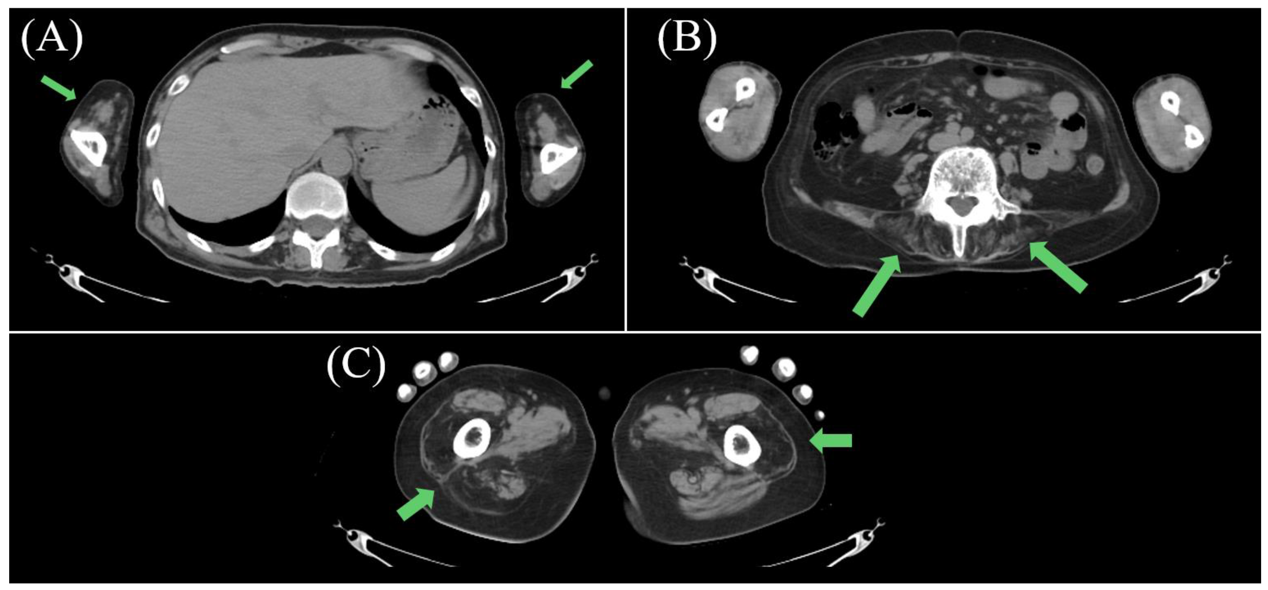
Figure 1. Muscle computed tomography (CT) findings. The CT scan showing muscle atrophy predominantly in the proximal muscles, including the upper arms (A), paraspinal muscles (B), and thighs (C) (green arrows).
Diagnostics 16 02833 g001
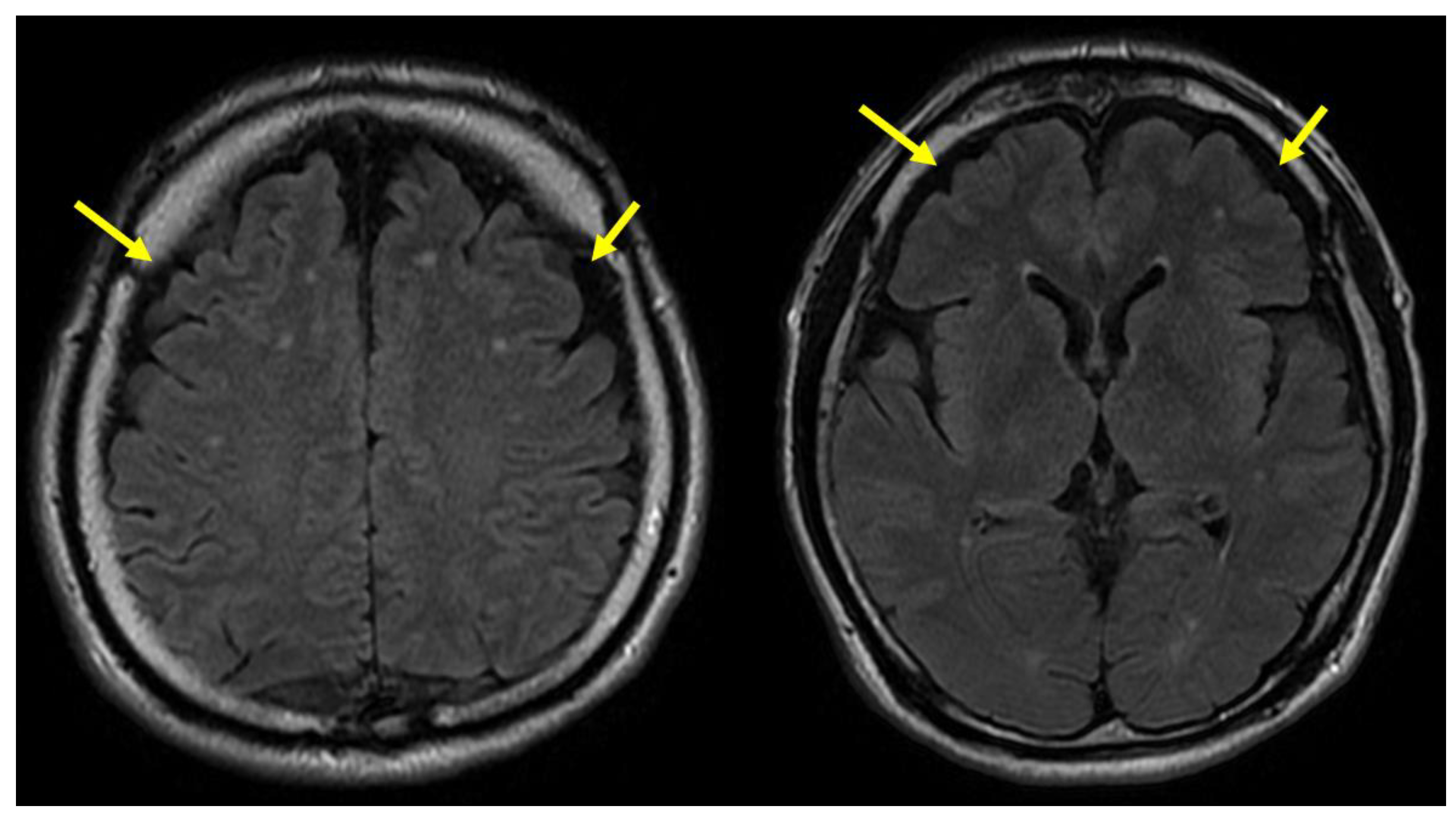
Figure 2. Brain magnetic resonance imaging (MRI). T2-weighted fluid-attenuated inversion recovery (FLAIR) images showing mild atrophy in the bilateral frontal lobes (yellow arrows).
Diagnostics 16 02833 g002
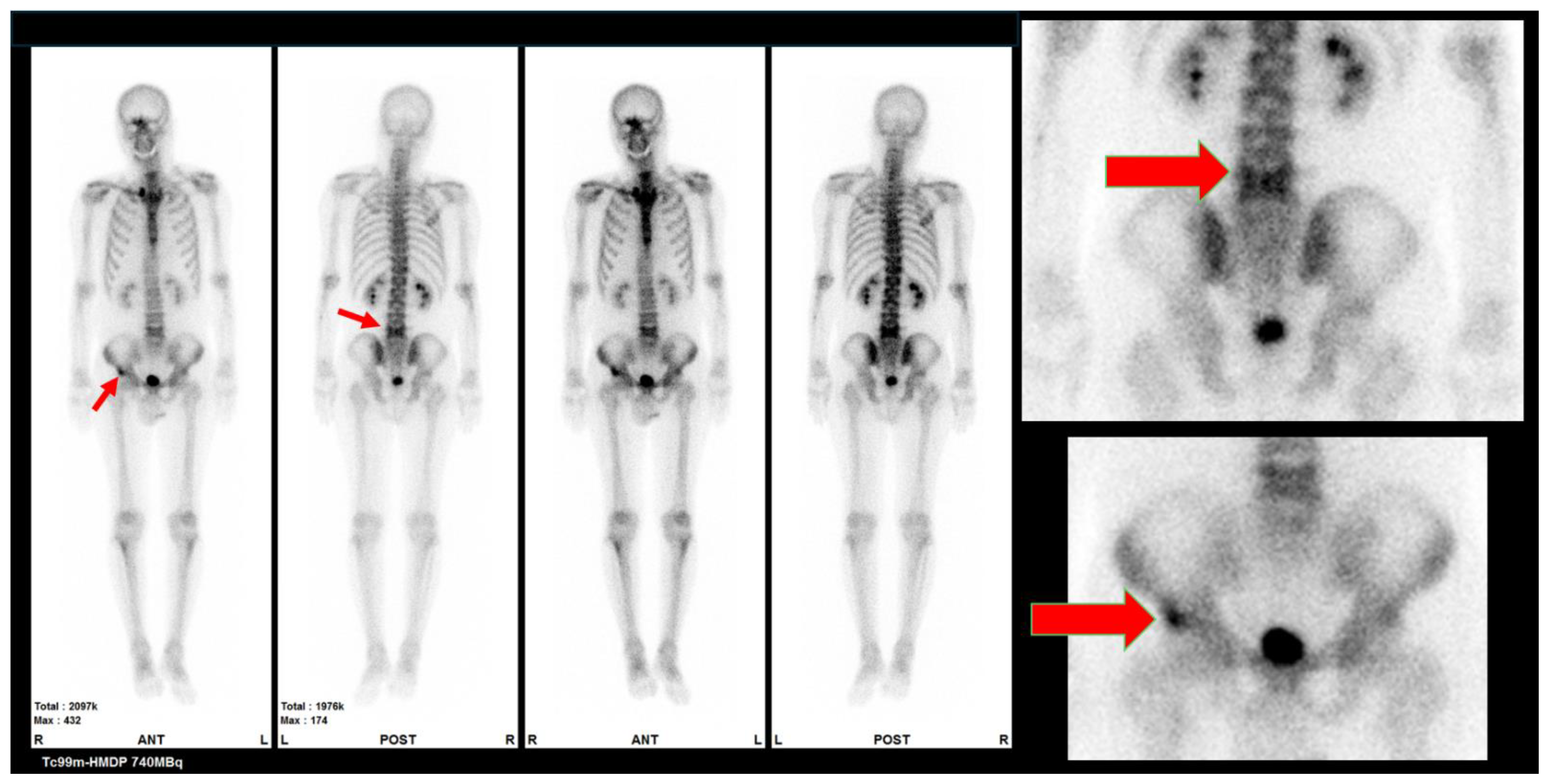
Figure 3. 99mTc-HMDP bone scintigraphy. Increased uptake suggestive of Paget disease of bone is observed in the pelvis and vertebral bodies (red arrows).
Diagnostics 16 02833 g003
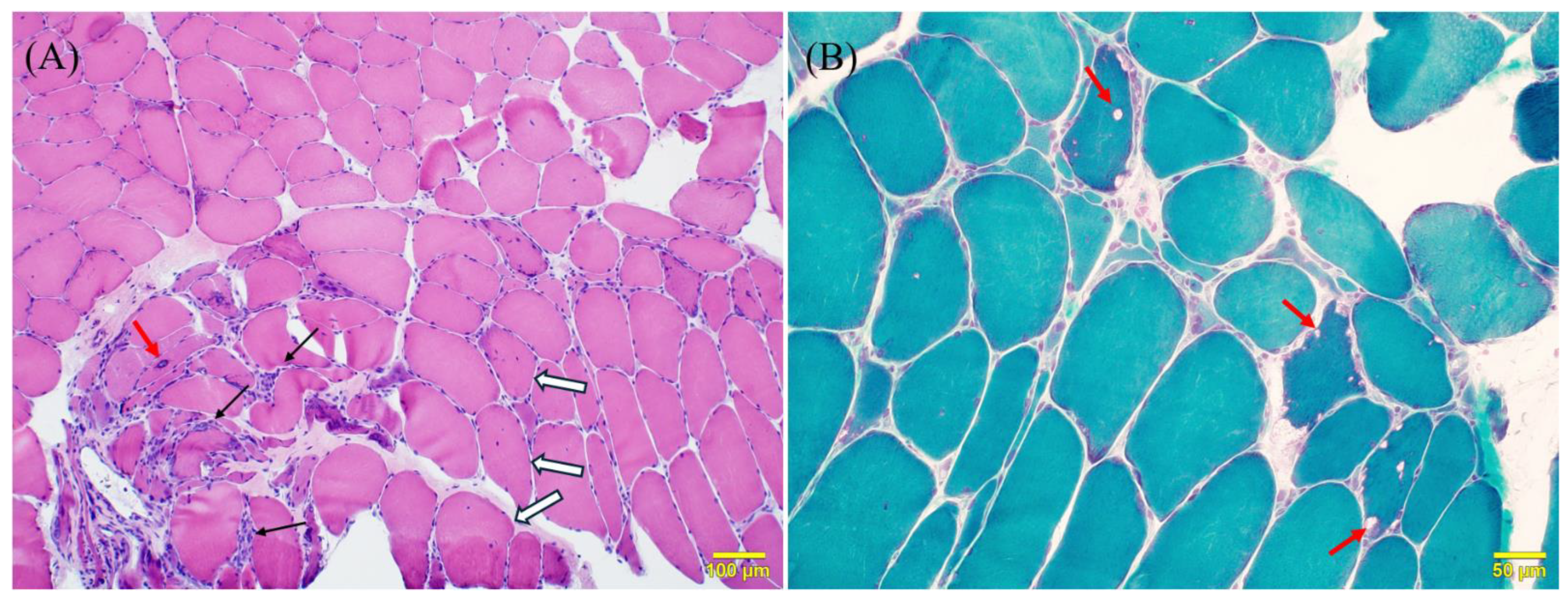
Figure 4. Muscle biopsy findings. (A) On hematoxylin and eosin (H&E) staining, mild variation in muscle fiber size is observed, together with endomysial fibrosis (black arrows), fibers with centrally located nuclei (white arrows), and fibers containing vacuoles (red arrows). (B) On modified Gomori trichrome staining, fibers with rimmed vacuoles are noted (red arrows). The histopathological findings in Figure 4, including variation in muscle fiber size and internalized nuclei, reflect a chronic myopathic process [1,2]. The hallmark feature is the presence of rimmed vacuoles (RVs), which represent a fundamental failure in autophagosome maturation [3,4]. Unlike sporadic inclusion-body myositis (sIBM), this case lacks significant inflammatory infiltration, a key diagnostic separator for VCP-associated disease [1,2].
Diagnostics 16 02833 g004
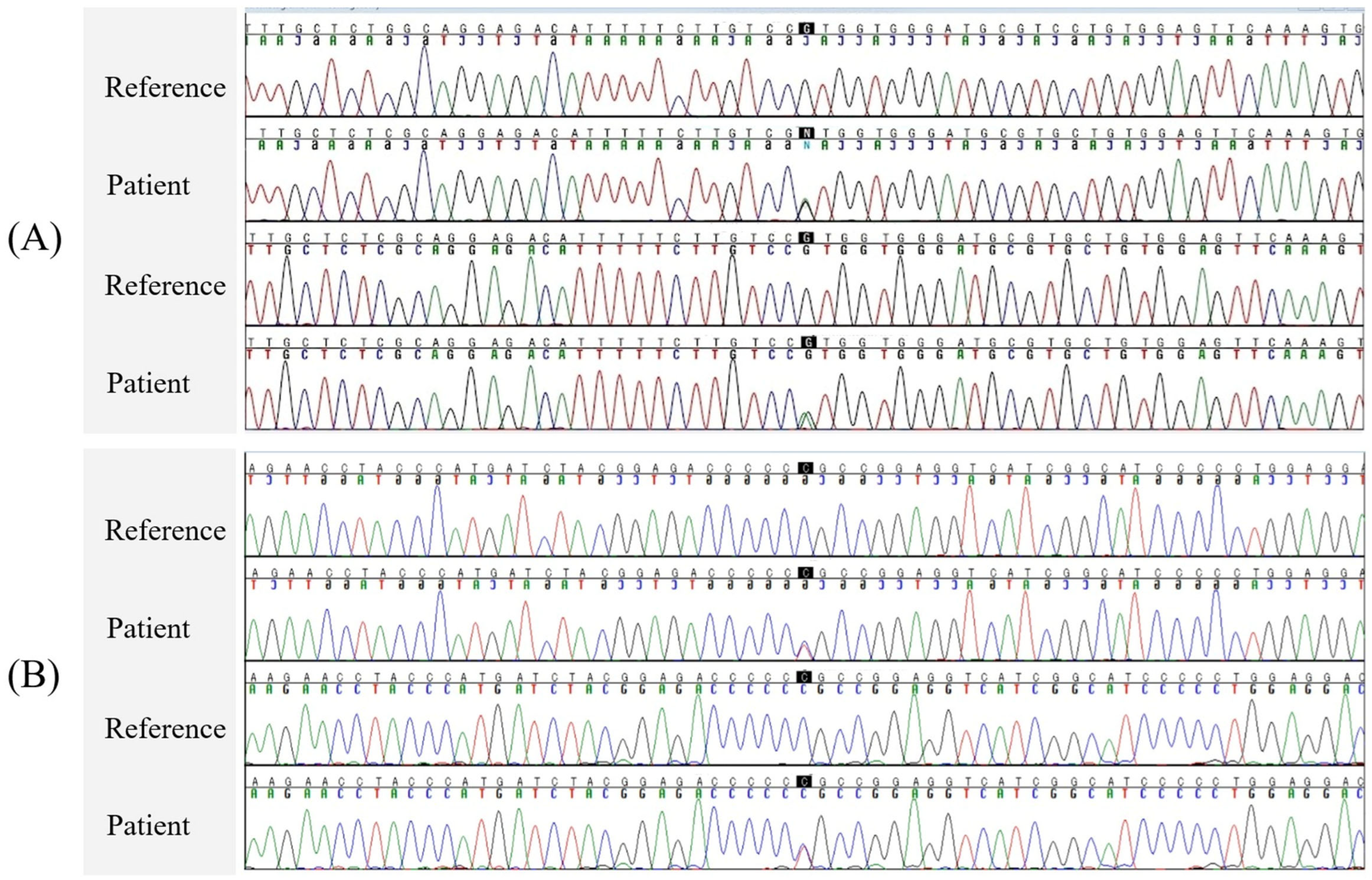
Figure 5. Results of Sanger sequencing. (A) Analysis of the VCP gene revealed a heterozygous c.464G>A variant in exon 5 (NM_007126.5), which results in a p.R155H amino acid substitution. This p.R155H variant is a well-established pathogenic variant strongly associated with VCP-associated multisystem proteinopathy, including inclusion-body myopathy with Paget disease of the bone and frontotemporal dementia (IBMPFD) or the scapuloperoneal syndrome [1,4,5,6,7,8,9,10,11,12,13]. (B) Analysis of the SCN4A gene identified a heterozygous c.215C>T (p.P72L) variant in exon 1 (NM_000334.4). According to gnomAD [14], this variant is extremely rare (allele frequency: 0.006%) and is currently classified as a variant of uncertain significance (VUS) based on the American College of Medical Genetics and Genomics (ACMG) criteria [15]. Although the amino acid substitution preserves the nonpolar property (proline to leucine), it eliminates the structural constraint unique to proline residues, potentially affecting local protein folding. Functional studies of the SCN4A p.P72L variant demonstrated a hyperpolarizing shift in the voltage dependence of channel activation, suggesting increased membrane excitability [16]. Although the role of SCN4A variants as clinical modifiers has been established in myotonic dystrophy type 2 (DM2) by enhancing membrane hyperexcitability, their impact on other neuromuscular disorders remains to be elucidated [16,17]. Crucially, the modifier effect of SCN4A in DM2 relies on an additive interaction with an underlying channelopathy background (i.e., CLC-1 dysfunction) [17]. In contrast, IBMPFD is fundamentally a degenerative proteinopathy driven by impaired protein homeostasis and autophagy, lacking a primary ion channel defect [1,2,3,5,7,11,18,19]. Therefore, the increased electrical excitability conferred by the SCN4A variant is unlikely to interact with or accelerate the proteotoxic and vacuolar degenerative pathways characteristic of VCP disease. In our patient, despite the presence of the SCN4A p.P72L variant, the clinical phenotype was entirely representative of classic IBMPFD, with no additional myotonic or atypical features. This suggests that in the context of VCP-associated disease, this SCN4A variant represents an incidental co-occurrence rather than a substantial clinical modifier. Ultimately, this case underscores the indispensable role of comprehensive clinical and radiological phenotyping in interpreting complex genetic findings in the genomic era. Further studies are warranted to accumulate and evaluate the potential modifying effects of SCN4A variants across a broader range of non-myotonic muscle diseases.
Diagnostics 16 02833 g005

Author Contributions

Conceptualization, S.T. and K.H.; investigation, K.H., T.H., W.Y. and I.N.; writing—original draft preparation, S.T. and K.H.; writing—review and editing, M.S., T.H., A.S., Y.N., T.M., W.Y., I.N. and Y.K.; supervision, M.S. and Y.K. All authors have read and agreed to the published version of the manuscript.

Funding

This study was supported partly by Intramural Research Grant (8-13, 5-6) for Neurological and Psychiatric Disorders of NCNP.

Institutional Review Board Statement

The study was conducted in accordance with the Declaration of Helsinki, and approved by the Institutional Review Board of the National Center of Neurology and Psychiatry (protocol code B2024-130, approval date 7 March 2025).

Informed Consent Statement

Informed consent was obtained from all subjects involved in the study. Written informed consent has been obtained from the patient to publish this paper.

Data Availability Statement

The data presented in this study are available on request from the corresponding author. Due to patient privacy and ethical considerations, the data are not publicly accessible.

Acknowledgments

The authors thank the patient for granting permission to publish this information. During the preparation of this manuscript, the authors used ChatGPT (GPT-4.1) for the purposes of language editing only. No data or conclusions were generated by AI. The authors have reviewed and edited the output and take full responsibility for the content of this publication.

Conflicts of Interest

The authors declare no conflicts of interest.

References

  1. Schiava, M.; Parkhurst, Y.; Henderson, M.; Polvikoski, T.; Valtcheva, M.V.; Nishino, I.; Inoue, M.; Nishimori, Y.; Saito, Y.; Stojkovic, T.; et al. Muscle biopsy findings in valosin-containing protein multisystem proteinopathy. Neurol. Genet. 2025, 11, e200265. [Google Scholar] [CrossRef] [Scilit] [PubMed]
  2. Tresse, E.; Salomons, F.A.; Vesa, J.; Bott, L.C.; Kimonis, V.; Yao, T.-P.; Dantuma, N.P.; Taylor, J.P. VCP/p97 is essential for maturation of ubiquitin-containing autophagosomes and this function is impaired by mutations that cause IBMPFD. Autophagy 2010, 6, 217–227. [Google Scholar] [CrossRef] [Scilit] [PubMed]
  3. Ju, J.S.; Weihl, C.C. p97/VCP at the intersection of the autophagy and the ubiquitin proteasome system. Autophagy 2010, 6, 283–285. [Google Scholar] [CrossRef] [Scilit] [PubMed]
  4. Jerath, N.U. Resolving a multi-generational neuromuscular mystery in a family presenting with a variable scapuloperoneal syndrome in a c.464G>A, p.Arg155His VCP mutation. Case Rep. Genet. 2019, 2019, 2403024. [Google Scholar] [CrossRef] [Scilit] [PubMed]
  5. Iannibelli, E.; Gibertini, S.; Cheli, M.; Blasevich, F.; Cavaliere, A.; Riolo, G.; Ruggieri, A.; Maggi, L. VCP-related myopathy: A case series and a review of literature. Acta Myol. 2023, 42, 2–13. [Google Scholar] [CrossRef] [Scilit] [PubMed]
  6. Columbres, R.A.; Chin, Y.; Pratti, S.; Quinn, C.; Gonzalez-Cuyar, L.F.; Weiss, M.; Quintero-Rivera, F.; Kimonis, V. Novel Variants in the VCP Gene Causing Multisystem Proteinopathy 1. Genes 2023, 14, 676. [Google Scholar] [CrossRef] [Scilit] [PubMed]
  7. Ferrari, V.; Cristofani, R.; Tedesco, B.; Crippa, V.; Chierichetti, M.; Casarotto, E.; Cozzi, M.; Mina, F.; Piccolella, M.; Galbiati, M.; et al. Valosin Containing Protein (VCP): A Multistep Regulator of Autophagy. Int. J. Mol. Sci. 2022, 23, 1939. [Google Scholar] [CrossRef] [Scilit] [PubMed]
  8. Shmara, A.; Perez-Rosendahl, M.; Murphy, K.; Kwon, A.; Smith, C.; Kimonis, V. A clinicopathologic study of malignancy in VCP-associated multisystem proteinopathy. Orphanet J. Rare Dis. 2022, 17, 272. [Google Scholar] [CrossRef] [Scilit] [PubMed]
  9. Chan, J.M.; Romano, C.; Lee, A.Y.; Wang, S.; Lombardo, D.; Kamdar, F.; Dora, M.; Khan, S.; Mammen, P.; Kimonis, V. Cardiomyopathy in valosin-containing protein multisystem proteinopathy: Evaluation, diagnosis, and management. Am. Heart J. Plus 2025, 60, 100644. [Google Scholar] [CrossRef] [Scilit] [PubMed]
  10. Wan, Y.; Wang, Q.; Zheng, Y.; Yu, M.; Xie, Z.; Ling, C.; Meng, L.; Yu, J.; Zheng, Y.; Wang, Y.; et al. Novel variants, muscle imaging, and myopathological changes in Chinese patients with VCP-related multisystem proteinopathy. Mol. Genet. Genom. Med. 2023, 11, e2176. [Google Scholar] [CrossRef] [Scilit] [PubMed]
  11. Luzzi, A.; Wang, F.; Li, S.; Iacovino, M.; Chou, T.F. Skeletal muscle cell protein dysregulation highlights the pathogenesis mechanism of myopathy-associated p97/VCP R155H mutations. Front. Neurol. 2023, 14, 1211635. [Google Scholar] [CrossRef] [Scilit] [PubMed]
  12. Pfeffer, G.; Lee, G.; Pontifex, C.S.; Fanganiello, R.D.; Peck, A.; Weihl, C.C.; Kimonis, V. Multisystem proteinopathy due to VCP mutations: A review of clinical heterogeneity and genetic diagnosis. Genes 2022, 13, 963. [Google Scholar] [CrossRef] [Scilit] [PubMed]
  13. Takata, T.; Kimura, Y.; Ohnuma, Y.; Kawawaki, J.; Kakiyama, Y.; Tanaka, K.; Kakizuka, A. Rescue of growth defects of yeast cdc48 mutants by pathogenic IBMPFD-VCPs. J. Struct. Biol. 2012, 179, 93–103. [Google Scholar] [CrossRef] [Scilit] [PubMed][Green Version]
  14. Karczewski, K.J.; Francioli, L.C.; Tiao, G.; Cummings, B.B.; Alfoldi, J.; Wang, Q.; Collins, R.L.; Laricchia, K.M.; Ganna, A.; Birnbaum, D.P.; et al. The mutational constraint spectrum quantified from variation in 141,456 humans. Nature 2020, 581, 434–443. [Google Scholar] [CrossRef] [Scilit] [PubMed]
  15. Richards, S.; Aziz, N.; Bale, S.; Bick, D.; Das, S.; Gastier-Foster, J.; Grody, W.W.; Hegde, M.; Lyon, E.; Spector, E.; et al. Standards and guidelines for the interpretation of sequence variants: A joint consensus recommendation of the American College of Medical Genetics and Genomics and the Association for Molecular Pathology. Genet Med. 2015, 17, 405–424. [Google Scholar] [CrossRef] [Scilit] [PubMed]
  16. Binda, A.; Renna, L.V.; Bosè, F.; Brigonzi, E.; Botta, A.; Valaperta, R.; Fossati, B.; Rivolta, I.; Meola, G.; Cardani, R. SCN4A as modifier gene in patients with myotonic dystrophy type 2. Sci. Rep. 2018, 8, 11058. [Google Scholar] [CrossRef] [Scilit] [PubMed]
  17. Bugiardini, E.; Rivolta, I.; Binda, A.; Caminero, A.S.; Cirillo, F.; Cinti, A.; Giovannoni, R.; Botta, A.; Cardani, R.; Wicklund, M.; et al. SCN4A mutation as modifying factor of myotonic dystrophy type 2 phenotype. Neuromuscul. Disord. 2015, 25, 301–307. [Google Scholar] [CrossRef] [Scilit] [PubMed]
  18. Chae, J.; Park, S.H.; Sung, J.J. VCP-related multisystem proteinopathy presenting with lobulated myofibers. Korean J. Neuromuscul. Disord. 2024, 16, 17–19. [Google Scholar] [CrossRef] [Scilit]
  19. Jang, H.; Jang, E.R.; Wilson, P.G.; Anderson, D.; Galperin, E. VCP/p97 controls signals of the ERK1/2 pathway transmitted via the Shoc2 scaffolding complex: Novel insights into IBMPFD pathology. Mol. Biol. Cell 2019, 30, 1655–1663. [Google Scholar] [CrossRef] [Scilit] [PubMed]
Disclaimer/Publisher’s Note: The statements, opinions and data contained in all publications are solely those of the individual author(s) and contributor(s) and not of MDPI and/or the editor(s). MDPI and/or the editor(s) disclaim responsibility for any injury to people or property resulting from any ideas, methods, instructions or products referred to in the content.