Predictive Model for Critical Illness Infection in Hospitalized Children with RSV Infection: A Retrospective Study

Abstract
1. Introduction
2. Materials and Methods
2.1. Study Design and Data Collection
2.2. Patient Inclusion
2.3. Statistical Analysis
3. Results
3.1. Description of Study Population
3.2. A Comparison of Laboratory Test Results
3.3. Risk Factors for Critical Illness Infection Outcomes
3.4. Construction of a Prediction Nomogram for Critical Illness Infection
3.5. Internal Validation of the Nomogram Prediction Model
4. Discussion
Supplementary Materials
Author Contributions
Funding
Institutional Review Board Statement
Informed Consent Statement
Data Availability Statement
Conflicts of Interest
Abbreviations
| RSV | Respiratory Syncytial Virus |
| ROC | Receiver Operating Characteristic |
| DCA | Decision Curve Analysis |
| APTT | Activated Partial Thromboplastin Time |
| PT | Prothrombin Time |
| hs-CRP | High-sensitivity C-reactive Protein |
| PaCO2 | Partial Pressure of Carbon Dioxide |
| PaO2 | Partial Pressure of Oxygen |
| SBE | Standard Base Excess |
| Hb | Hemoglobin Concentration |
| WBC | White Blood Cell Count |
| PLT | Platelet Count |
| CK | Creatine Kinase |
| CK-MB | Creatine Kinase Isoenzyme |
| ALT | Alanine Aminotransferase |
| AST | Aspartate Aminotransferase |
| TBIL | Total Bilirubin |
| BUN | Blood Urea Nitrogen |
| PCT | Procalcitonin |
| SpO2 | Peripheral Capillary Oxygen Saturation |
| CPAP | Continuous Positive Airway Pressure |
| RC | Regression Coefficient |
| SE | Standard Error |
References
- World Health Organization. WHO (Coronavirus) Respiratory Syncytial Virus (RSV) Dashboard. 2023. Available online: https://www.who.int/news-room/fact-sheets/detail/respiratory-syncytial-virus-(rsv) (accessed on 25 May 2026).
- Mazur, N.I.; Caballero, M.T.; Nunes, M.C. Severe respiratory syncytial virus infection in children: Burden, management, and emerging therapies. Lancet 2024, 404, 1143–1156. [Google Scholar] [CrossRef] [Scilit]
- Oppenlander, K.E.; Chung, A.A.; Clabaugh, D. Respiratory Syncytial Virus Bronchiolitis: Rapid Evidence Review. Am. Fam. Physician 2023, 108, 52–57. [Google Scholar]
- Hon, K.L.; Leung, A.K.C.; Wong, A.H.C.; Dudi, A.; Leung, K.K.Y. Respiratory Syncytial Virus is the Most Common Causative Agent of Viral Bronchiolitis in Young Children: An Updated Review. Curr. Pediatr. Rev. 2023, 19, 139–149. [Google Scholar] [CrossRef] [Scilit] [PubMed]
- Baraldi, E.; Checcucci Lisi, G.; Costantino, C.; Heinrichs, J.H.; Manzoni, P.; Riccò, M.; Roberts, M.; Vassilouthis, N. RSV disease in infants and young children: Can we see a brighter future? Hum. Vaccin. Immunother. 2022, 18, 2079322. [Google Scholar] [CrossRef] [Scilit]
- Makhni, S.; Cerrato, P.; Rico, J.; Niazi, S.; O’Horo, J.; Peters, S.; Shah, V.; Halamka, J. Meeting the challenges of electronic health record (EHR) optimization. npj Digit. Med. 2025, 9, 8. [Google Scholar] [CrossRef] [Scilit]
- Zhang, R.; Long, F.; Zhao, Z.; Wu, J.; Tan, R.; Xu, W.; Li, L.; Long, Y.; Qu, H. Machine learning predicts sepsis deterioration trajectories. npj Digit. Med. 2026, 9, 385. [Google Scholar] [CrossRef] [Scilit] [PubMed]
- Belgacem, A.; Khoudi, A.; Boudane, F.; Berrichi, A. (Eds.) Machine Learning in the Medical Field: A Comprehensive Overview. In Proceedings of the 2023 International Conference on Decision Aid Sciences and Applications (DASA), Annaba, Algeria, 16–17 September 2023; IEEE: New York, NY, USA; pp. 103–108.
- Shehab, M.; Abualigah, L.; Shambour, Q.; Abu-Hashem, M.A.; Shambour, M.K.Y.; Alsalibi, A.I.; Gandomi, A.H. Machine learning in medical applications: A review of state-of-the-art methods. Comput. Biol. Med. 2022, 145, 105458. [Google Scholar] [CrossRef] [Scilit] [PubMed]
- Agnello, L.; Vidali, M.; Padoan, A.; Lucis, R.; Mancini, A.; Guerranti, R.; Plebani, M.; Ciaccio, M.; Carobene, A. Machine learning algorithms in sepsis. Clin. Chim. Acta 2024, 553, 117738. [Google Scholar] [CrossRef] [Scilit]
- Liu, J.; Bao, B.; Zhang, T.; Jia, W.; Guo, C.; Song, C. Comparative analysis of the clinical characteristics of severe Mycoplasma pneumoniae pneumonia and severe bacterial pneumonia in children. BMC Pediatr. 2025, 25, 439. [Google Scholar] [CrossRef] [Scilit]
- Huang, Y.; Chen, Y.; Liu, M.; Tang, L.-F.; Tang, L.-F. Comparative analysis of Chlamydia pneumoniae pneumonia (CPP) and Mycoplasma pneumoniae pneumonia in children and risk factors of severe CPP. BMC Infect. Dis. 2025, 25, 993. [Google Scholar] [CrossRef] [Scilit]
- Li, Z.; Gu, W.; Zhu, F.; Han, E.; Yan, Y.; Sun, H.; Xu, W.; Zhang, X.; Huang, L.; Gao, S.; et al. Clinical characteristics and risk factors of severe pneumonia caused by human bocavirus in children. BMC Infect. Dis. 2025, 25, 58. [Google Scholar] [CrossRef] [Scilit]
- Wang, Y.; Xiang, Y.; Jin, R.; Li, B.; Feng, X.; Han, Q.; Deng, X.; Lu, Y.; Zhang, C.; Xiao, S. Risk factors for severe lower respiratory tract infection among children under 5 years of age hospitalized with respiratory syncytial virus. Eur. J. Clin. Microbiol. Infect. Dis. 2025, 44, 2239–2251. [Google Scholar] [CrossRef] [Scilit]
- Esposito, S.; Fainardi, V.; Titolo, A.; Lazzara, A.; Menzella, M.; Campana, B.; Argentiero, A.; Principi, N. How air pollution fuels respiratory infections in children: Current insights. Front. Public Health 2025, 13, 1567206. [Google Scholar] [CrossRef] [Scilit]
- Yagi, K.; Asai, N.; Huffnagle, G.B.; Lukacs, N.W.; Fonseca, W. Early-Life Lung and Gut Microbiota Development and Respiratory Syncytial Virus Infection. Front. Immunol. 2022, 13, 877771. [Google Scholar] [CrossRef] [Scilit]
- Georgakopoulou, V.E.; Pitiriga, V.C. Immunomodulation in Respiratory Syncytial Virus Infection: Mechanisms, Therapeutic Targets, and Clinical Implications. Microorganisms 2025, 13, 1876. [Google Scholar] [CrossRef] [Scilit]
- Elbadawy, H.M.; Khattab, A.; El-Agamy, D.S.; Eltahir, H.M.; Alhaddad, A.; Aljohani, F.D.; Almuzaini, T.M.; Abouzied, M.M.; Aldhafiri, A. IL-6 at the center of cytokine storm: Circulating inflammation mediators as biomarkers in hospitalized COVID-19 patients. J. Clin. Lab. Anal. 2023, 37, e24881. [Google Scholar] [CrossRef] [Scilit] [PubMed]
- Tanaka, T.; Narazaki, M.; Kishimoto, T. IL-6 in inflammation, immunity, and disease. Cold Spring Harb. Perspect. Biol. 2014, 6, a016295. [Google Scholar] [CrossRef] [Scilit]
- Aliyu, M.; Zohora, F.T.; Anka, A.U.; Ali, K.; Maleknia, S.; Saffarioun, M.; Azizi, G. Interleukin-6 cytokine: An overview of the immune regulation, immune dysregulation, and therapeutic approach. Int. Immunopharmacol. 2022, 111, 109130. [Google Scholar] [CrossRef] [Scilit]
- Muszynski, J.A.; Nofziger, R.; Greathouse, K.; Steele, L.; Hanson-Huber, L.; Nateri, J.; Hall, M.W. Early adaptive immune suppression in children with septic shock: A prospective observational study. Crit. Care 2014, 18, R145. [Google Scholar] [CrossRef] [Scilit]
- Hall, M.W.; Carcillo, J.A.; Cornell, T. on behalf of the Pediatric Organ Dysfunction Information Update Mandate (PODIUM) Collaborative. Immune System Dysfunction Criteria in Critically Ill Children: The PODIUM Consensus Conference. Pediatrics 2022, 149, S91–S98. [Google Scholar] [CrossRef] [Scilit]
- Delshad, M.; Tavakolinia, N.; Pourbagheri-Sigaroodi, A.; Safaroghli-Azar, A.; Bagheri, N.; Bashash, D. The contributory role of lymphocyte subsets, pathophysiology of lymphopenia and its implication as prognostic and therapeutic opportunity in COVID-19. Int. Immunopharmacol. 2021, 95, 107586. [Google Scholar] [CrossRef] [Scilit]
- Zorio, V.; Venet, F.; Delwarde, B.; Floccard, B.; Marcotte, G.; Textoris, J.; Monneret, G.; Rimmelé, T. Assessment of sepsis-induced immunosuppression at ICU discharge and 6 months after ICU discharge. Ann. Intensive Care 2017, 7, 80. [Google Scholar] [CrossRef] [Scilit]
- Zinellu, A.; Sotgia, S.; Fois, A.G.; Mangoni, A.A. Serum CK-MB, COVID-19 severity and mortality: An updated systematic review and meta-analysis with meta-regression. Adv. Med. Sci. 2021, 66, 304–314. [Google Scholar] [CrossRef] [Scilit]
- Essone, P.N.; Adegbite, B.R.; Mbadinga, M.J.M.; Mbouna, A.V.; Lotola-Mougeni, F.; Alabi, A.; Edoa, J.R.; Lell, B.; Alabi, A.S.; Adegnika, A.A.; et al. Creatine kinase-(MB) and hepcidin as candidate biomarkers for early diagnosis of pulmonary tuberculosis: A proof-of-concept study in Lambaréné, Gabon. Infection 2022, 50, 897–905. [Google Scholar] [CrossRef] [Scilit]
- Li, W.; Qiao, X. Rhabdomyolysis due to influenza A and respiratory syncytial virus co-infection: A case report. J. Med. Case Rep. 2026, 20, 68. [Google Scholar] [CrossRef] [Scilit]
- Langedijk, A.C.; Bont, L.J. Respiratory syncytial virus infection and novel interventions. Nat. Rev. Microbiol. 2023, 21, 734–749. [Google Scholar] [CrossRef] [Scilit]
- Ivey, K.S.; Edwards, K.M.; Talbot, H.K. Respiratory Syncytial Virus and Associations With Cardiovascular Disease in Adults. J. Am. Coll. Cardiol. 2018, 71, 1574–1583. [Google Scholar] [CrossRef] [Scilit] [PubMed]
- AbdelMassih, A.F.; Ismail, H.A.; Nabil, F.M.; Mabrouk, A.I.; Abdel Aziz, F. RSV-induced LV dysfunction, what can be the underlying cause? Pediatr. Pulmonol. 2023, 58, 991–995. [Google Scholar] [CrossRef] [Scilit]
- Sorrentino, L.; Toscanelli, W.; Fracella, M.; De Angelis, M.; Frasca, F.; Scagnolari, C.; Petrarca, L.; Nenna, R.; Midulla, F.; Palamara, A.T.; et al. NRF2 Antioxidant Response and Interferon-Stimulated Genes Are Differentially Expressed in Respiratory-Syncytial-Virus- and Rhinovirus-Infected Hospitalized Children. Pathogens 2023, 12, 577. [Google Scholar] [CrossRef] [Scilit]
- Rossi, M.L.; Hadley, S.M.; Randanne, P.C.; Escobar-Diaz, M.C.; Camprubi, M.C.; Jordan, I.; Sanchez-de-Toledo, J. Cardiac function in bronchiolitis: Not only a right ventricle matter. Pediatr. Pulmonol. 2023, 58, 288–296. [Google Scholar] [CrossRef] [Scilit]
- Berend, K. Diagnostic Use of Base Excess in Acid-Base Disorders. N. Engl. J. Med. 2018, 378, 1419–1428. [Google Scholar] [CrossRef] [Scilit]
- Kofstad, J. Base excess: A historical review-has the calculation of base excess been more standardised the last 20 years? Clin. Chim. Acta 2001, 307, 193–195. [Google Scholar] [CrossRef] [Scilit]
- Yang, D.; Zhang, W.; Cao, Y.; Zhang, Y.; Yin, C. Association between standard base excess and acute kidney injury in intensive care unit patients: A study based on the medical information mart for intensive care database. BMC Nephrol. 2025, 26, 350. [Google Scholar] [CrossRef] [Scilit]
- Li, Y.; Pillai, P.; Miyake, F.; Nair, H. The role of viral co-infections in the severity of acute respiratory infections among children infected with respiratory syncytial virus (RSV): A systematic review and meta-analysis. J. Glob. Health 2020, 10, 010426. [Google Scholar] [CrossRef] [Scilit]
- Torres, A.R.; Gaio, V.; Melo, A.; Lança, M.; Barreto, M.; Gomes, L.; Azevedo, I.; Bandeira, T.; Lito, D.; Guiomar, R.; et al. RSV-Bacterial Co-Infection Is Associated With Increased Illness Severity in Hospitalized Children—Results From a Prospective Sentinel Surveillance Study. J. Med. Virol. 2025, 97, e70209. [Google Scholar] [CrossRef] [Scilit]
- Theofilis, P.; Sagris, M.; Oikonomou, E.; Antonopoulos, A.S.; Siasos, G.; Tsioufis, C.; Tousoulis, D. Inflammatory Mechanisms Contributing to Endothelial Dysfunction. Biomedicines 2021, 9, 781. [Google Scholar] [CrossRef] [Scilit]
- Robinot, R.; Hubert, M.; de Melo, G.D.; Lazarini, F.; Bruel, T.; Smith, N.; Levallois, S.; Larrous, F.; Fernandes, J.; Gellenoncourt, S.; et al. SARS-CoV-2 infection induces the dedifferentiation of multiciliated cells and impairs mucociliary clearance. Nat. Commun. 2021, 12, 4354. [Google Scholar] [CrossRef] [Scilit]
- Pezato, R.; David, A.G.; Boggi, A.C.; Melo, B.; Rosalino, C.M.V.; Rodriguez, A.P.; Thamboo, A.; Voegels, R.L. Upper Airway Mucociliary Clearance is Impaired in Dyspneic COVID-19 Patients. Indian J. Otolaryngol. Head. Neck Surg. 2023, 75, 772–776. [Google Scholar] [CrossRef] [Scilit]
- Kappler, U.; Henningham, A.; Nasreen, M.; Yamamoto, A.; Buultjens, A.H.; Stinear, T.P.; Sly, P.; Fantino, E. Tolerance to Haemophilus influenzae infection in human epithelial cells: Insights from a primary cell-based model. PLoS Pathog. 2024, 20, e1012282. [Google Scholar] [CrossRef] [Scilit]
- Cheng, X.F.; Wei, S.; Xia, J.Q.; Zhou, K.; Sun, D. Predictive Model for Critical Illness Infection in Hospitalized Children with RSV Infection: A Retrospective Study-–supplementary dataset. Preprints 2026. [Google Scholar] [CrossRef] [Scilit]



| Characteristics | Critical Illness (n = 30) | Non-Critical Illness (n = 90) | t/χ2/z | p |
|---|---|---|---|---|
| Age (month) | 8.61 (3.75, 24.86) | 16.84 (4.88, 33.67) | −1.349 | 0.177 |
| Sex | 100.800 | 0.349 | ||
| male | 15 (50.00) | 60 (66.67) | ||
| female | 15 (50.00) | 30 (33.33) | ||
| BW (kg) | 8.50 (5.22, 11.50) | 11.00 (7.50, 14.00) | −2.096 | 0.036 |
| HT (cm) | 69.50 (60.00, 86.25) | 81.50 (65.75, 97.00) | −1.861 | 0.063 |
| BMI | 16.34 (14.87, 17.35) | 15.69 (14.72, 17.71) | −0.288 | 0.773 |
| Co-infection | 194.255 | 0.441 | ||
| Single RSV infection | 7 (23.3%) | 35 (38.9%) | ||
| Bacterial co-infection | 6 (20%) | 13 (14.4%) | ||
| Viral co-infection | 3 (10%) | 16 (17.8%) | ||
| Mycoplasma | 1 (3.3%) | 10 (11.1%) | ||
| Three or more pathogenic types co-infection | 13 (43.3%) | 16 (17.8%) |
| Characteristics | Critical Illness (n = 30) | Non-Critical Illness (n = 90) | t/χ2/z | p |
|---|---|---|---|---|
| APTT (s) | 29.80 (26.95, 34.60) | 33.65 (29.20, 37.62) | −2.246 | 0.025 |
| PT (s) | 11.20 (10.50, 12.60) | 11.35 (10.60, 12.30) | −0.382 | 0.702 |
| Fibrinogen (g/L) | 2.88 ± 1.21 | 3.13 ± 1.17 | −1.005 | 0.317 |
| D-dimer (mg/L) | 0.54 (0.32, 1.20) | 0.42 (0.28, 0.64) | −1.791 | 0.073 |
| hs-CRP (mg/L) | 9.55 (2.43, 23.93) | 9.65 (2.39, 29.30) | −0.076 | 0.940 |
| SpO2 (%) | 91.00 (87.00, 96.00) | 93.00 (91.00, 96.00) | −1.851 | 0.064 |
| PaCO2 (mmHg) | 38.50 (33.00, 45.75) | 36.00 (31.00, 40.25) | −1.935 | 0.053 |
| PaO2 (mmHg) | 63.50 (54.00, 81.00) | 65.50 (60.00, 76.50) | −1.071 | 0.284 |
| SBE (mmol/L) | 0.00 (−0.75, 2.00) | 0.00 (−3.08, 1.00) | −2.014 | 0.044 |
| pH | 7.38 (7.33, 7.45) | 7.41 (7.37, 7.43) | −1.291 | 0.197 |
| IL-6 (pg/mL) | 42.45 (21.71, 91.28) | 12.10 (8.15, 25.20) | −5.545 | <0.001 |
| IL-10 (pg/mL) | 8.25 (5.95, 11.96) | 13.35 (7.51, 22.84) | −2.658 | 0.008 |
| TNF-α (pg/mL) | 3.49 (2.86, 6.25) | 3.33 (2.20, 5.36) | −1.015 | 0.310 |
| Hb (g/L) | 117.00 (104.25, 125.00) | 114.00 (104.00, 121.00) | −1.258 | 0.208 |
| WBC (109/L) | 7.53 (5.76, 11.81) | 8.61 (6.62, 11.39) | −0.864 | 0.388 |
| PLT (109/L) | 397.77 ± 196.88 | 340.01 ± 133.20 | 1.810 | 0.073 |
| Lymphocyte Percentage (%) | 36.66 ± 18.41 | 46.90 ± 21.68 | −2.323 | 0.022 |
| Neutrophil Percentage (%) | 65.12 ± 12.32 | 39.89 ± 20.27 | 6.422 | <0.001 |
| CK (U/L) | 104.00 (58.75, 147.25) | 82.00 (59.00, 119.50) | −1.267 | 0.205 |
| CK-MB (U/L) | 44.65 (35.00, 79.00) | 28.00 (23.00, 38.50) | −4.477 | <0.001 |
| ALT (U/L) | 20.50 (13.75, 29.75) | 18.00 (11.75, 32.00) | −0.995 | 0.320 |
| AST (U/L) | 44.50 (29.50, 54.25) | 40.50 (32.75, 55.00) | −0.297 | 0.766 |
| TBIL (μmol/L) | 6.80 (3.48, 8.20) | 4.90 (3.50, 7.60) | −1.121 | 0.262 |
| Creatinine (μmol/L) | 22.00 (18.95, 27.02) | 22.60 (19.17, 28.80) | −0.485 | 0.628 |
| BUN (mmol/L) | 3.00 (2.18, 3.75) | 2.96 (2.39, 3.73) | −0.100 | 0.920 |
| PCT (ng/mL) | 0.20 (0.11, 0.35) | 0.18 (0.11, 0.50) | −0.055 | 0.956 |
| Characteristics | RC | SE | z | OR (95% CI) | p |
|---|---|---|---|---|---|
| Age | 0.003 | 0.009 | 0.294 | 1.003 (0.985, 1.021) | 0.769 |
| Sex (female) | −0.693 | 0.428 | −1.619 | 0.500 (0.216, 1.157) | 0.105 |
| BW | 0.025 | 0.04 | 0.618 | 1.025 (0.948, 1.109) | 0.537 |
| HT | 0.013 | 0.011 | 1.217 | 1.013 (0.991, 1.035) | 0.224 |
| BMI | 0.037 | 0.086 | 0.427 | 1.038 (0.877, 1.228) | 0.669 |
| Single RSV infection | - | - | - | - | - |
| Two pathogenic types co-infection | 0.248 | 0.545 | 0.456 | 1.281 (0.440, 3.729) | 0.648 |
| Three or more pathogenic types co-infection | 1.402 | 0.558 | 2.514 | 4.063 (1.361, 12.130) | 0.012 |
| APTT | 0.05 | 0.034 | 1.446 | 1.051 (0.983, 1.124) | 0.148 |
| PT | 0.008 | 0.154 | 0.050 | 1.008 (0.745, 1.363) | 0.960 |
| Fibrinogen | 0.191 | 0.19 | 1.004 | 1.210 (0.834, 1.757) | 0.315 |
| D-dimer | 0.04 | 0.095 | 0.421 | 1.041 (0.864, 1.254) | 0.674 |
| hs-CRP | 0.005 | 0.008 | 0.622 | 1.005 (0.989, 1.021) | 0.534 |
| SpO2 | 0.008 | 0.05 | 0.150 | 1.008 (0.914, 1.112) | 0.880 |
| PaCO2 | −0.048 | 0.023 | −2.038 | 0.953 (0.911, 0.997) | 0.042 |
| PaO2 | 0.023 | 0.015 | 1.518 | 1.023 (0.994, 1.054) | 0.129 |
| SBE | −0.136 | 0.06 | −2.243 | 0.873 (0.776, 0.982) | 0.025 |
| pH | 5.305 | 3.362 | 1.578 | 201.341 (0.277, 146,461.811) | 0.115 |
| IL-6 | 0.107 | 0.033 | 3.237 | 1.113 (1.043, 1.187) | 0.001 |
| IL-10 | 0.038 | 0.023 | 1.654 | 1.039 (0.993, 1.087) | 0.098 |
| TNF-α | 0.002 | 0.004 | 0.446 | 1.002 (0.994, 1.010) | 0.655 |
| Hb | 0.075 | 0.018 | 4.182 | 1.078 (1.041, 1.117) | <0.001 |
| WBC | 0.012 | 0.037 | 0.334 | 1.012 (0.941, 1.088) | 0.738 |
| PLT | −0.002 | 0.001 | −1.772 | 0.998 (0.996, 1.000) | 0.076 |
| Lymphocyte percentage | 0.024 | 0.011 | 2.245 | 1.024 (1.002, 1.047) | 0.025 |
| Neutrophil percentage | 0.025 | 0.011 | 2.394 | 1.025 (1.003, 1.048) | 0.017 |
| CK | −0.003 | 0.002 | −1.678 | 0.997 (0.993, 1.001) | 0.093 |
| CK-MB | −0.005 | 0.008 | −0.716 | 0.995 (0.980, 1.011) | 0.474 |
| ALT | 0.005 | 0.009 | 0.493 | 1.005 (0.987, 1.023) | 0.622 |
| AST | −0.001 | 0.008 | −0.147 | 0.999 (0.983, 1.015) | 0.883 |
| TBIL | −0.007 | 0.029 | −0.229 | 0.993 (0.938, 1.051) | 0.819 |
| Creatinine | 0.013 | 0.031 | 0.425 | 1.013 (0.953, 1.077) | 0.671 |
| BUN | −0.05 | 0.177 | −0.282 | 0.951 (0.672, 1.346) | 0.778 |
| PCT | 0.12 | 0.191 | 0.630 | 1.127 (0.775, 1.639) | 0.529 |
| Characteristics | RC | SE | z | OR (95% CI) | p |
|---|---|---|---|---|---|
| Constant term | −7.818 | 1.548 | −5.050 | 0.000 (0.000, 0.008) | <0.001 |
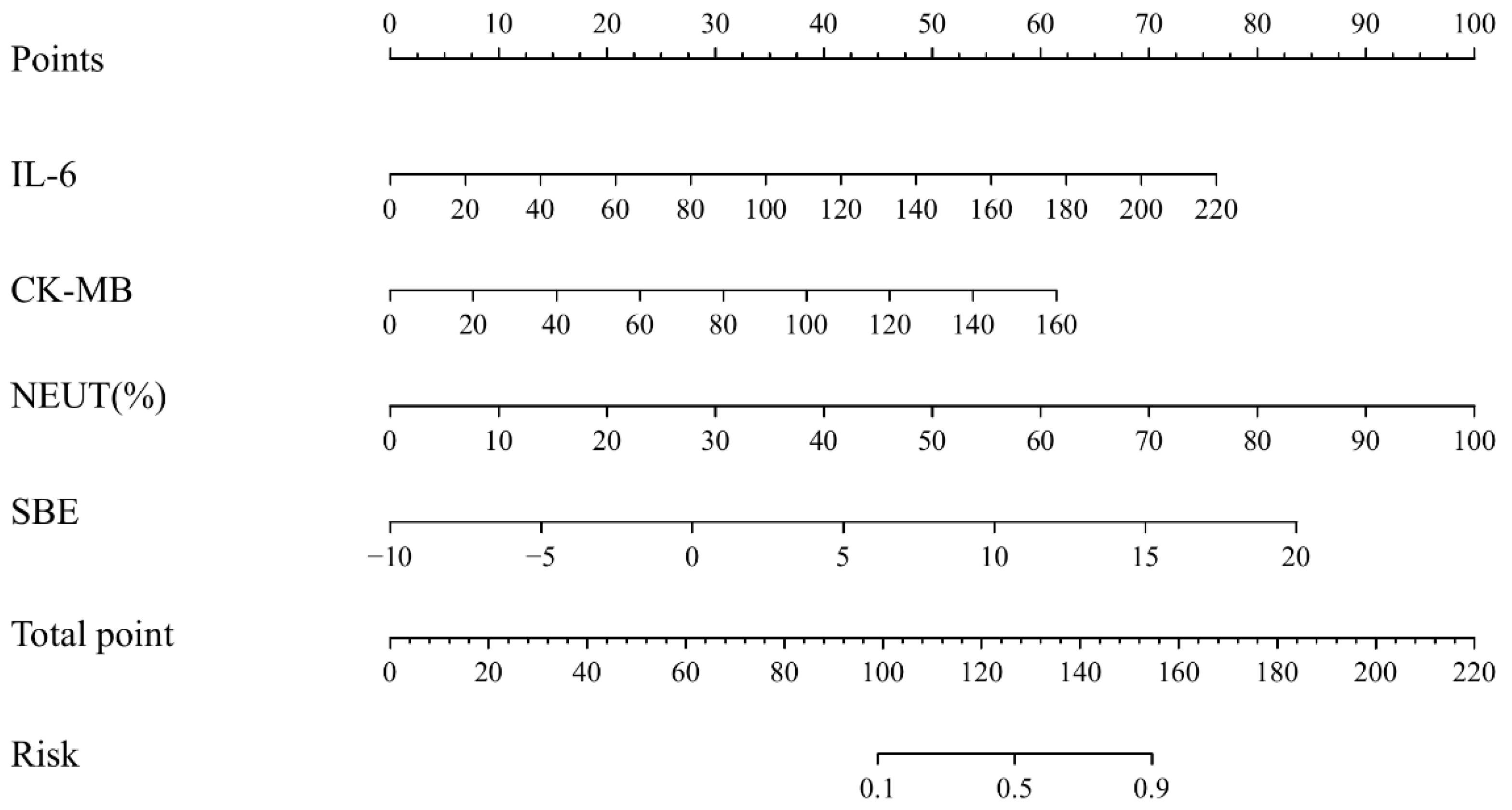
| Neutrophil percentage | 0.079 | 0.02 | 4.049 | 1.082 (1.041, 1.125) | <0.001 |
| IL-6 | 0.027 | 0.008 | 3.541 | 1.027 (1.011, 1.044) | <0.001 |
| CK-MB | 0.03 | 0.012 | 2.492 | 1.030 (1.007, 1.055) | 0.013 |
| SBE | 0.22 | 0.107 | 2.051 | 1.246 (1.010, 1.537) | 0.040 |
Disclaimer/Publisher’s Note: The statements, opinions and data contained in all publications are solely those of the individual author(s) and contributor(s) and not of MDPI and/or the editor(s). MDPI and/or the editor(s) disclaim responsibility for any injury to people or property resulting from any ideas, methods, instructions or products referred to in the content. |
© 2026 by the authors. Licensee MDPI, Basel, Switzerland. This article is an open access article distributed under the terms and conditions of the Creative Commons Attribution (CC BY) license.
Share and Cite
Cheng, X.; Wei, S.; Xia, J.; Zhou, K.; Sun, D. Predictive Model for Critical Illness Infection in Hospitalized Children with RSV Infection: A Retrospective Study. Diagnostics 2026, 16, 1701. https://doi.org/10.3390/diagnostics16111701
Cheng X, Wei S, Xia J, Zhou K, Sun D. Predictive Model for Critical Illness Infection in Hospitalized Children with RSV Infection: A Retrospective Study. Diagnostics. 2026; 16(11):1701. https://doi.org/10.3390/diagnostics16111701
Chicago/Turabian StyleCheng, Xingfeng, Sha Wei, Jinquan Xia, Kai Zhou, and Dan Sun. 2026. "Predictive Model for Critical Illness Infection in Hospitalized Children with RSV Infection: A Retrospective Study" Diagnostics 16, no. 11: 1701. https://doi.org/10.3390/diagnostics16111701
APA StyleCheng, X., Wei, S., Xia, J., Zhou, K., & Sun, D. (2026). Predictive Model for Critical Illness Infection in Hospitalized Children with RSV Infection: A Retrospective Study. Diagnostics, 16(11), 1701. https://doi.org/10.3390/diagnostics16111701