Interesting Images

Computed Tomography, Coronary Angiography, and Intravascular Ultrasound in the Diagnosis of Left Anterior Descending Stenosis in a 38-Year-Old Woman with a Calcium Score of Zero
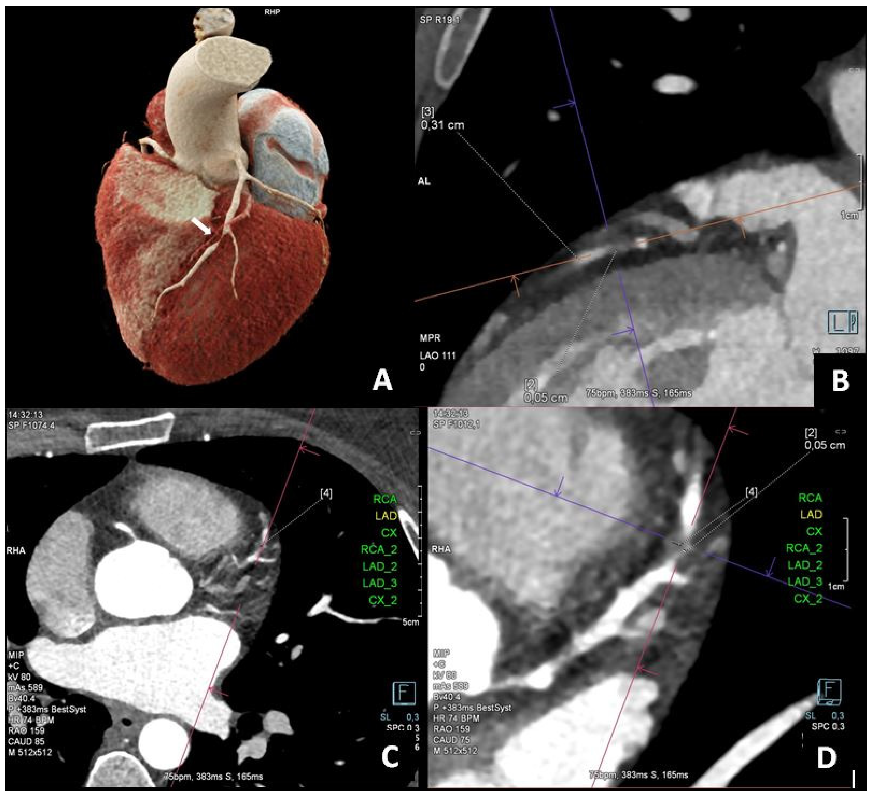
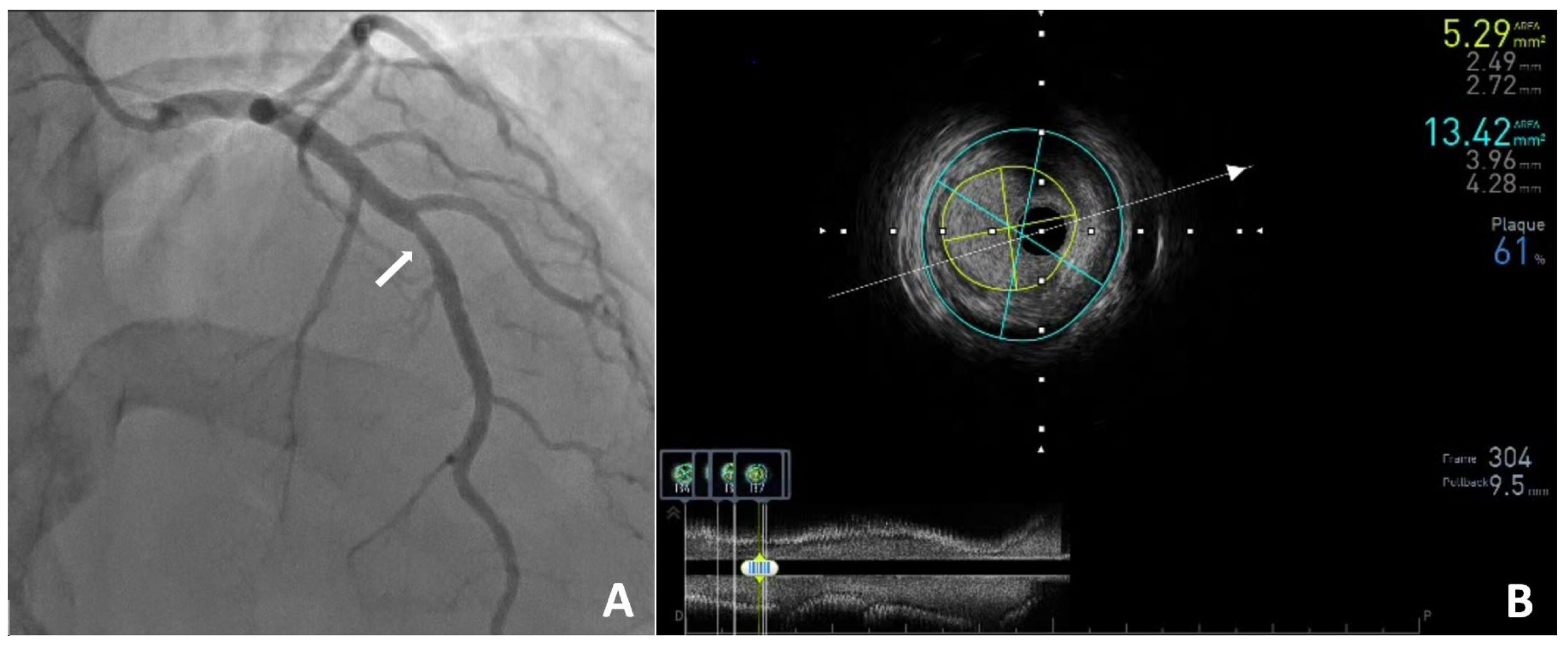
by
Malgorzata Zalewska-Adamiec
* and
Slawomir Dobrzycki
Department of Invasive Cardiology, Medical University of Bialystok, 15-089 Bialystok, Poland
*
Author to whom correspondence should be addressed.
Diagnostics 2025, 15(9), 1169; https://doi.org/10.3390/diagnostics15091169
Submission received: 18 April 2025 / Revised: 29 April 2025 / Accepted: 3 May 2025 / Published: 4 May 2025
(This article belongs to the Special Issue Clinical Diagnosis and Management in Cardiology)

Abstract

:
Cardiovascular diseases, including coronary artery disease, are the leading cause of mortality in the general population. The likelihood of significant coronary artery disease occurring in young women (<40 years of age) is very low. Diagnosis is challenging and often delayed, treatment tends to be suboptimal, and the long-term prognosis is poor. We present the case of a 38-year-old woman with typical anginal chest pain whose coronary computed tomography angiography (CCTA) revealed significant narrowing in the left anterior descending artery (LAD) despite a coronary artery calcium score (CACS) of zero. To assess the significance of the lesion, coronary angiography and intravascular ultrasound (IVUS) were performed, which revealed borderline narrowing. Conservative treatment was recommended.

Figure 1. A 38-year-old woman who was diagnosed with hypercholesterolemia, was a former smoker, and had a history of typical exertional angina for about six months was admitted to the clinic for coronary angiography. No ischemic abnormalities were detected in the baseline ECG recordings. An exercise stress test performed on an outpatient basis was normal at a workload of 10 METs. A coronary computed tomography angiography (CCTA) was performed, revealing a coronary artery calcium score (CACS) of 0 according to the Agatston scale. In the mid-segment of the LAD (left anterior descending), a non-calcified, hypodense atherosclerotic plaque measuring up to 10 mm was visualized, causing significant/critical stenosis of approximately 80–90% ((AD), white arrow). Features of positive vessel remodeling were observed at the plaque level. No stenoses were found in the proximal or distal segments of the LAD or the remaining coronary arteries.
Diagnostics 15 01169 g001
Figure 2. In the clinic, a transthoracic echocardiogram was performed and showed normal valve function and left ventricular contractility with an ejection fraction of 60%. Subsequently, coronary angiography revealed a 50% stenosis in the LAD ((A), white arrow), with no other changes in the remaining coronary arteries. The angiographic findings were further evaluated using IVUS (intravascular ultrasound), which showed borderline stenosis in the LAD with the following measurements: MLA (minimal lumen area)—5.29 mm2; CSA (cross-sectional area)—13.42 mm2; and PB (plaque burden)—61% (B). Laboratory investigations performed during hospitalization were within normal limits. Results included the following: total cholesterol—104 mg/dL, LDL cholesterol—43 mg/dL, HDL cholesterol—52 mg/dL, triglycerides—22 mg/dL, lipoprotein(a)—7.7 mg/dL, HbA1c—4.7%, and NT–proBNP—67 pg/mL. No elevation in inflammatory markers was observed. The patient was qualified for conservative management, with pharmacotherapy recommendations including continued use of acetylsalicylic acid (75 mg/day) and atorvastatin (40 mg/day). Nebivolol, which was used previously, was replaced with amlodipine (2.5 mg/day) and trimetazidine (35 mg twice daily) due to a possible vasospastic component. Three months later, the patient returned for a follow-up visit at the cardiology outpatient clinic. She reported feeling well and denied any substernal chest pain. Continuation of the current treatment regimen and regular cardiology follow-up were recommended. In our patient, the probability of significant coronary artery disease was very low [1]. As part of the initial diagnostic evaluation, the patient underwent a stress test. However, in this group of patients, an ischemia imaging test, such as stress cardiac magnetic resonance (CMR) or stress echocardiography, could have been considered [2]. The CACS in our patient was 0, whereas CCTA revealed a lesion in the proximal segment that required further evaluation via coronary angiography and intravascular ultrasound [3]. This case illustrates that, in young individuals, a zero CACS does not exclude the presence of significant coronary artery disease [4,5,6,7]. Therefore, in this population, a personalized diagnostic approach should be applied, taking into account risk factors and clinical symptoms.
Diagnostics 15 01169 g002

Author Contributions

Conceptualization, M.Z.-A. and S.D.; investigation, M.Z.-A.; data curation, M.Z.-A.; writing—original draft preparation, M.Z.-A.; writing—review and editing, M.Z.-A. and S.D. All authors have read and agreed to the published version of the manuscript.

Funding

This research received no external funding.

Institutional Review Board Statement

Not applicable.

Informed Consent Statement

We have received verbal consent for publication from our patient. The decision to publish this case was made after the patient’s hospitalization and follow-up visit, which is why we obtained her consent for publication during a phone conversation.

Data Availability Statement

Original data supporting the reported results are available by contacting the authors.

Conflicts of Interest

The authors declare no conflicts of interest.

References

  1. Vrints, C.; Andreotti, F.; Koskinas, K.C.; Rossello, X.; Adamo, M.; Ainslie, J.; Banning, A.P.; Budaj, A.; Buechel, R.R.; Chiariello, G.A.; et al. 2024 ESC Guidelines for the management of chronic coronary syndromes. Eur. Heart J. 2024, 45, 3415–3537. [Google Scholar] [CrossRef] [PubMed]
  2. Morrone, D.; Gentile, F.; Aimo, A.; Cameli, M.; Barison, A.; Picoi, M.E.; Guglielmo, M.; Villano, A.; DeVita, A.; Mandoli, G.E.; et al. Coronary Physiopathology and Microcirculation Working Group and Cluster Imaging of the Italian Society of Cardiology (SIC). Perspectives in noninvasive imaging for chronic coronary syndromes. Int. J. Cardiol. 2022, 365, 19–29. [Google Scholar] [CrossRef] [PubMed]
  3. Pawłowski, T.; Legutko, J.; Kochman, J.; Roleder, T.; Pręgowski, J.; Chmielak, Z.; Kubica, J.; Ochała, A.; Parma, R.; Grygier, M.; et al. Clinical use of intracoronary imaging modalities in Poland. Expert opinion of the Association of Cardiovascular Interventions of the Polish Cardiac Society. Pol. Heart J. 2022, 80, 509–519. [Google Scholar] [CrossRef] [PubMed]
  4. Wojciechowski, A.; Zarotyńska, M.; Chyrchel, B.; Terlecki, M.; Wojciechowska, W.; Rajzer, M. Zero calcium score: A potential pitfall in the diagnosis of coronary artery disease. Pol. Arch. Intern. Med. 2025, 135, 16936. [Google Scholar] [CrossRef] [PubMed]
  5. Gać, P.; Jaworski, A.; Parfianowicz, A.; Karwacki, J.; Wysocki, A.; Poręba, R. Discrepancies between Coronary Artery Calcium Score and Coronary Artery Disease Severity in Computed Tomography Angiography Studies. Diagnostics 2024, 14, 1928. [Google Scholar] [CrossRef] [PubMed]
  6. Mortensen, M.B.; Gaur, S.; Frimmer, A.; Bøtker, H.E.; Sørensen, H.T.; Kragholm, K.H.; Niels Peter, S.R.; Steffensen, F.H.; Jensen, R.V.; Mæng, M.; et al. Association of Age with the Diagnostic Value of Coronary Artery Calcium Score for Ruling Out Coronary Stenosis in Symptomatic Patients. JAMA Cardiol. 2022, 7, 36–44. [Google Scholar] [CrossRef] [PubMed]
  7. Bergström, G.; Persson, M.; Adiels, M.; Björnson, E.; Bonander, C.; Ahlström, H.; Alfredsson, J.; Angerås, O.; Berglund, G.; Blomberg, A.; et al. Prevalence of Subclinical Coronary Artery Atherosclerosis in the General Population. Circulation 2021, 144, 916–929. [Google Scholar] [CrossRef] [PubMed]
Disclaimer/Publisher’s Note: The statements, opinions and data contained in all publications are solely those of the individual author(s) and contributor(s) and not of MDPI and/or the editor(s). MDPI and/or the editor(s) disclaim responsibility for any injury to people or property resulting from any ideas, methods, instructions or products referred to in the content.

