
Inflammatory Pseudotumor of the Anal Canal Mimicking Colorectal Cancer: Case Report and Hints to Improve a Patient’s Fitness for Treatment and Prevention

 ,
,  , , , and
Abstract
1. Introduction
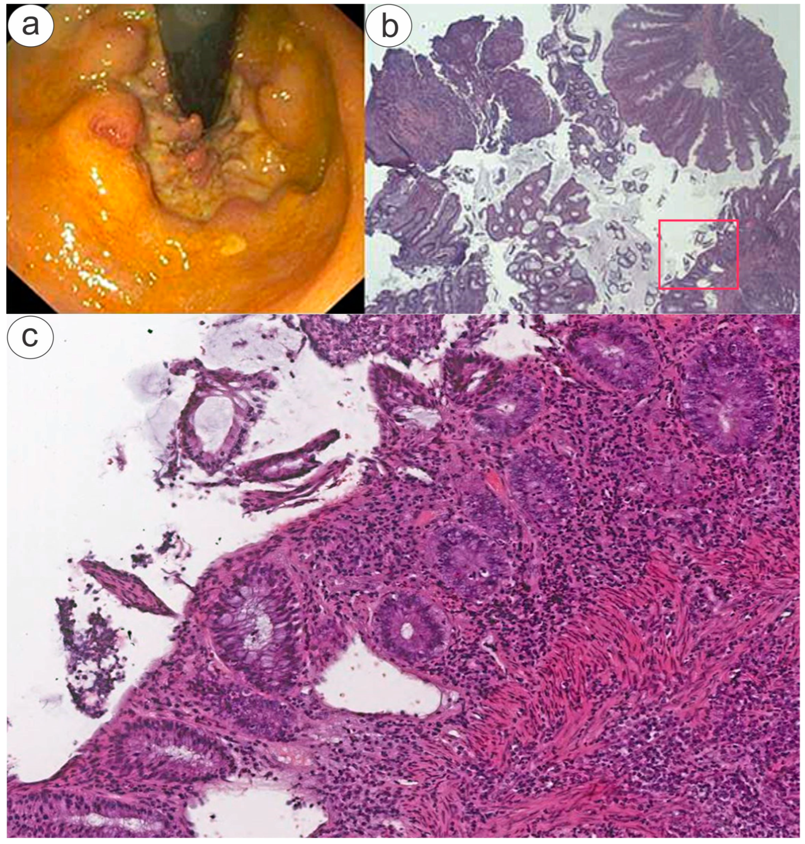
2. Case Report
3. Discussion
4. Conclusions
Author Contributions
Funding
Institutional Review Board Statement
Informed Consent Statement
Data Availability Statement
Acknowledgments
Conflicts of Interest
References
- Berlingieri, F.; Kovacic, M. Health and relationship quality of sexual minorities in Europe. J. Popul. Econ. 2025, 38, 15. [Google Scholar] [CrossRef]
- Ryan, P.; Dolengevich-Segal, H.; Ramos-Ruperto, L.; Cabello, A.; Sanchez-Conde, M.; Vergas, J.; Valencia, J.; Cuevas, G.; Sanz, J.; Curto-Ramos, J.; et al. Patterns of Sexualized Drug Use among Gay, Bisexual, and Other Men Who Have Sex with Men Living with HIV: Insights from a Comprehensive Study—The U-SEX-2 GESIDA 9416 Study. J. Clin. Med. 2023, 12, 7293. [Google Scholar] [CrossRef] [PubMed]
- Martin, J.H. No Cock Needed: Exploring the Hapto-Erotic Assemblage of Fist-Play in Gay Men’s Anal Fisting. J. Homosex. 2023, 71, 2974–2996. [Google Scholar] [CrossRef] [PubMed]
- Preuss, V.; Wöllner, K.; Vennemann, B.; Fieguth, A.; Hagemeier, L.; Klintschar, M. Fatal anogenital exenteration of the intestine. Forensic Sci. Med. Pathol. 2022, 18, 64–68. [Google Scholar] [CrossRef]
- Martin, J.H. Fisting Subjectivity: Narratives of Sexual Subjectivity Among Gay Fist-Fuckers. J. Sex Res. 2024, 62, 398–410. [Google Scholar] [CrossRef]
- Cappelletti, S.; Aromatario, M.; Bottoni, E.; Fiore, P.A.; Fineschi, V.; di Luca, N.M.; Ciallella, C. Variability in findings of anogenital injury in consensual and non-consensual fisting intercourse: A systematic review. J. Forensic Leg. Med. 2016, 44, 58–62. [Google Scholar] [CrossRef]
- Ciccarese, G.; Di Biagio, A.; Bruzzone, B.; Guadagno, A.; Taramasso, L.; Oddenino, G.; Brucci, G.; Labate, L.; De Pace, V.; Mastrolonardo, M.; et al. Monkeypox outbreak in Genoa, Italy: Clinical, laboratory, histopathologic features, management, and outcome of the infected patients. J. Med. Virol. 2023, 95, e28560. [Google Scholar] [CrossRef] [PubMed]
- Workowski, K.A.; Bachmann, L.H.; Chan, P.A.; Johnston, C.M.; Muzny, C.A.; Park, I.; Reno, H.; Zenilman, J.M.; Bolan, G.A. Sexually Transmitted Infections Treatment Guidelines, 2021. MMWR. Recomm. Rep. 2021, 70, 1–187. [Google Scholar] [CrossRef] [PubMed] [PubMed Central]
- Twisk, D.E.; van der Sande, M.A.; van Eeden, A.; Heideman, D.A.; van der Klis, F.R.; de Vries, H.J.; Schim van der Loeff, M.F. Detection of Incident Anal High-Risk Human Papillomavirus DNA in Men Who Have Sex With Men: Incidence or Reactivation? J. Infect. Dis. 2018, 218, 1018–1026. [Google Scholar] [CrossRef]
- Available online: https://intranet.policlinico.pa.it/pub/documenti/browse.do?dispatch=documentRead&documentId=1323c0545872ac910158731923c50019 (accessed on 2 February 2025).
- Wong, I.K.J.; Poynten, I.M.; Cornall, A.; Templeton, D.J.; Molano, M.; Garland, S.M.; Fairley, C.K.; Law, C.; Hillman, R.J.; Polizzotto, M.N.; et al. SPANC study team. Sexual behaviours associated with incident high-risk anal human papillomavirus among gay and bisexual men. Sex. Transm. Infect. 2022, 98, 101–107. [Google Scholar] [CrossRef] [PubMed] [PubMed Central]
- Fasciana, T.; Capra, G.; Di Carlo, P.; Calà, C.; Vella, M.; Pistone, G.; Colomba, C.; Giammanco, A. Socio-Demographic Characteristics and Sexual Behavioral Factors of Patients with Sexually Transmitted Infections Attending a Hospital in Southern Italy. Int. J. Environ. Res. Public Health 2021, 18, 4722. [Google Scholar] [CrossRef] [PubMed]
- Pipitò, L.; Ganci, I.; Cicero, A.; Medaglia, A.A.; D’avenia, S.; Mandalà, E.; Calà, C.; Di Carlo, P.; Cascio, A. A Case of Syphilis With Ocular Involvement: Persistent Negative Serology in a Patient With Multiple Sclerosis. Open Forum Infect. Dis. 2024, 11, ofae563. [Google Scholar] [CrossRef] [PubMed]
- Bosco, L.; Serra, N.; Fasciana, T.; Pistoia, D.; Vella, M.; Di Gregorio, L.; Schillaci, R.; Perino, A.; Calagna, G.; Firenze, A.; et al. Potential impact of a nonavalent anti HPV vaccine in Italian men with and without clinical manifestations. Sci. Rep. 2021, 11, 4096. [Google Scholar] [CrossRef]
- Preuss, J.; Strehler, M.; Dettmeyer, R.; Madea, B. Death after anal “fisting”. Arch Kriminol. 2008, 221, 28–35. [Google Scholar]
- Orr, C.J.; Clark, M.A.; Hawley, D.A.; Pless, J.E.; Tate, L.R.; Fardal, P.M. Fatal anorectal injuries: A series of four cases. J. Forensic Sci. 1995, 40, 219–221. [Google Scholar]
- Shook, L.L.; Whittle, R.; Rose, E.F. Rectal fist insertion. An unusual form of sexual behavior. Am. J. Forensic. Med. Pathol. 1985, 6, 319–324. [Google Scholar]
- Bitzer, J. Sexual and Reproductive Healthcare for LGBTI. In Textbook of Contraception, Sexual and Reproductive Health; Cambridge University Press: Cambridge, UK, 2024; pp. 347–352. [Google Scholar]
- Peuchant, O.; Laurier-Nadalié, C.; Albucher, L.; Balcon, C.; Dolzy, A.; Hénin, N.; Touati, A.; Bébéar, C. Anorectal lymphogranuloma venereum among men who have sex with men: A 3-year nationwide survey, France, 2020 to 2022. Euro Surveill. 2024, 29, 2300520. [Google Scholar] [CrossRef]
- Taj, S.; Austin, C.; Hussain, A.; Babar, M.S.; Sanekommu, H.; Imburgio, S.; Johal, A.; CruzPonce, A.; Vedire, A.; Liu, E. Severe Gastrointestinal Hemorrhage due to Monkeypox Virus-Associated Proctocolitis. Case Rep. Gastrointest. Med. 2023, 2023, 9981631. [Google Scholar] [CrossRef] [PubMed] [PubMed Central]
- Sergi, C.; Magener, A.; Ehemann, V.; De Villiers, E.M.; Sinn, H.P. Stage IIa cervix carcinoma with metastasis to the heart: Report of a case with immunohistochemistry, flow cytometry, and virology findings. Gynecol. Oncol. 2000, 76, 133–138. [Google Scholar] [CrossRef]
- ARC Working Group on the Evaluation of Carcinogenic Risks to Humans. Human Papillomaviruses; International Agency for Research on Cancer: Lyon, France, 2007; (IARC Monographs on the Evaluation of Carcinogenic Risks to Humans, No. 90). Available online: https://www.ncbi.nlm.nih.gov/books/NBK321760/ (accessed on 8 March 2024).
- Cabibi, D.; Napolitano, C.; Giannone, A.G.; Micciulla, M.C.; Porcasi, R.; Lo Coco, R.; Bosco, L.; Vinciguerra, M.; Capra, G. Predictive Role of the p16 Immunostaining Pattern in Atypical Cervical Biopsies with Less Common High Risk HPV Genotypes. Diagnostics 2021, 11, 1947. [Google Scholar] [CrossRef]
- van Bogaert, L.J. Cervical preneoplasia biomarkers: A conundrum for the community based gynecologic surgical pathologist. J. Gynecol. Oncol. 2014, 25, 3–5. [Google Scholar] [CrossRef] [PubMed]
- Liu, Y.; Alqatari, M.; Sultan, K.; Ye, F.; Gao, D.; Sigel, K.; Zhang, D.; Kalir, T. Using p16 immunohistochemistry to classify morphologic cervical intraepithelial neoplasia 2: Cor-relation of ambiguous staining patterns with HPV subtypes and clinical outcome. Hum. Pathol. 2017, 66, 144–151. [Google Scholar] [CrossRef] [PubMed] [PubMed Central]
- Soliman, S.; Dogbey, P.; Pan, S. Lymphogranuloma Venereum Mimicking Locally Metastatic Rectal Cancer in an HIV-Negative Man. Cureus 2021, 13, e20216. [Google Scholar] [CrossRef] [PubMed] [PubMed Central]
- Masiá, M.; Fernández-González, M.; García, J.A.; Padilla, S.; García-Payá, E.; Gutiérrez, A.; Ortiz de la Tabla, V.; García-Abellán, J.; Agulló, V.; Gutiérrez, F. Infection with Chlamydia trachomatis Increases the Risk of High-grade Anal Intraepithelial Neoplasia in People Living With Human Immunodeficiency Virus. Clin. Infect. Dis. 2020, 70, 2161–2167. [Google Scholar] [CrossRef]
- Lin, C.; Franceschi, S.; Clifford, G.M. Human papillomavirus types from infection to cancer in the anus, according to sex and HIV status: A systematic review and meta-analysis. Lancet Infect. Dis. 2018, 18, 198–206. [Google Scholar] [CrossRef] [PubMed]
- WHO Classification of Tumours Editorial Board. Digestive System Tumours, 5th ed.; WHO Classification of Tumours Series; International Agency for Research on Cancer: Lyon, France, 2019; Volume 1, Available online: https://publications.iarc.fr/579 (accessed on 24 March 2025).
- Darragh, T.M.; Colgan, T.J.; Cox, J.T.; Heller, D.S.; Henry, M.R.; Luff, R.D.; McCalmont, T.; Nayar, R.; Palefsky, J.M.; Stoler, M.H.; et al. The Lower Anogenital Squamous Terminology Standardization Project for HPV-Associated Lesions: Background and consensus recommendations from the College of American Pathologists and the American Society for Colposcopy and Cervical Pathology. Arch. Pathol. Lab. Med. 2012, 136, 1266–1297. [Google Scholar] [CrossRef] [PubMed]
- Lim, J.; Rezaie, A. Irritable Bowel Syndrome-Like Symptoms in Quiescent Inflammatory Bowel Disease: A Practical Approach to Diagnosis and Treatment of Organic Causes. Dig. Dis. Sci. 2023, 68, 4081–4097. [Google Scholar] [CrossRef]
- Maverakis, E.; Ma, C.; Shinkai, K.; Fiorentino, D.; Callen, J.P.; Wollina, U.; Marzano, A.V.; Wallach, D.; Kim, K.; Schadt, C.; et al. Diagnostic Criteria of Ulcerative Pyoderma Gangrenosum: A Delphi Consensus of International Experts. JAMA Dermatol. 2018, 154, 461–466. [Google Scholar] [CrossRef]
- Nepal, S.; Navaneethan, U.; Bennett, A.E.; Shen, B. De Novo Inflammatory Bowel Disease and Its Mimics After Organ Transplantation. Inflamm. Bowel Dis. 2013, 19, 1518–1527. [Google Scholar] [CrossRef]
- Dapaah, J.M.; Senah, K.A. HIV/AIDS clients, privacy and confidentiality; the case of two health centres in the Ashanti Region of Ghana. BMC Med. Ethics 2016, 17, 41. [Google Scholar] [CrossRef]
- Gazzetta, S.; Valent, F.; Sala, A.; Driul, L.; Brunelli, L. Sexually transmitted infections and the HPV-related burden: Evolution of Italian epidemiology and policy. Front. Public Health 2024, 12, 1336250. [Google Scholar] [CrossRef]
- Ranabhotu, A.; Habibian, N.; Patel, B.; Farrell, E.; Do, J.; Sedghi, S.; Sedghi, L. Case Report: Resolution of high grade anal squamous intraepithelial lesion with antibiotics proposes a new role for syphilitic infection in potentiation of HPV-associated ASCC. Front. Oncol. 2023, 13, 1226202. [Google Scholar] [CrossRef]
- Stier, E.A.; Clarke, M.A.; Deshmukh, A.A.; Wentzensen, N.; Liu, Y.; Poynten, I.M.; Cavallari, E.N.; Fink, V.; Barroso, L.F.; Clifford, G.M.; et al. International Anal Neoplasia Society’s consensus guidelines for anal cancer screening. Int. J. Cancer 2024, 154, 1694–1702. [Google Scholar] [CrossRef] [PubMed]
- Assi, R.; Hashim, P.W.; Reddy, V.B.; Einarsdottir, H.; Longo, W.E. Sexually transmitted infections of the anus and rectum. World J. Gastroenterol. 2014, 20, 15262–15268. [Google Scholar] [CrossRef] [PubMed] [PubMed Central]
- Hamlyn, E.; Taylor, C. Sexually transmitted proctitis. Postgrad. Med. J. 2006, 82, 733–736. [Google Scholar] [CrossRef] [PubMed] [PubMed Central]
- Bassett, S.S.; Delaney, D.J.; Moore, A.M.; Clair-Michaud, M.; Clarke, J.G.; Stein, L. Motivational interviewing to reduce risky sexual behaviors among at-risk male youth: A randomized controlled pilot study. Psychol. Serv. 2022, 19, 167. [Google Scholar] [CrossRef]
- Andersen, R.S.; Risør, M.B. The importance of contextualization. Anthropological reflections on descriptive analysis, its limitations and implications. Anthr. Med. 2014, 21, 345–356. [Google Scholar] [CrossRef] [PubMed]
- Bernstein, B. Class, codes and control. In The Structuring of Pedagogic Discourse; Routledge: London, UK, 1990; Volume IV. [Google Scholar]
- Eerdmans, S.; Prevignano, C.; Thibault, P. Language and Interaction. Discussions with J. J. Gumperz; Benjamins: Amsterdam, The Netherlands, 2002. [Google Scholar]
- Gumperz, J.J. Discourse Strategies; Cambridge University Press: Cambridge, UK, 1982. [Google Scholar]
- Gumperz, J.J. (Ed.) Language and Social Identity; Cambridge University Press: Cambridge, UK, 1982. [Google Scholar]
- Ishida, H. Learners’ perception and interpretation of contextualization cues in spontaneous Japanese conversation: Back-channel cue Uun. J. Pragmat. 2006, 38, 1943–1981. [Google Scholar] [CrossRef]
- Masuda, K. Style-shifting in student-professor interactions. J. Pragmat. 2016, 101, 101–117. [Google Scholar] [CrossRef]
- Sohrabi, C.; Mathew, G.; Maria, N.; Kerwan, A.; Franchi, T.; Agha, R.A.; Collaborators. The SCARE 2023 guideline: Updating consensus Surgical CAse REport (SCARE) guidelines. Int. J. Surg. 2023, 109, 1136–1140. [Google Scholar] [CrossRef] [PubMed] [PubMed Central]


| HR HPV Genotypes | LR HPV Genotypes |
|---|---|
| 16 | 6 |
| 18 | 11 |
| 26 * | 40 |
| 31 | 42 |
| 33 | 43 |
| 35 | 44 |
| 39 | 54 |
| 45 | 55 |
| 51 | 61 |
| 52 | 62 |
| 53 | 64 |
| 56 | 71 |
| 58 | 72 |
| 59 | 81 |
| 66 | 83 |
| 67 | 84 |
| 68a | 87 |
| 68b | 89 |
| 69 | 90 |
| 70 | |
| 73 | |
| 82 |
| Description | AIN | LAST | P16 IHC |
|---|---|---|---|
| Mild dysplasia | AIN 1 | Low grade SIL | Negative or weak staining |
| Moderate dysplasia | AIN 2 | High grade SIL | Positive and sometimes patchy staining |
| Severe dysplasia | AIN 3 | High grade SIL | Diffuse, strong staining; nuclear and cytoplasmic “block-like” |
| Carcinoma in situ | AIN 3 | High grade SIL | Diffuse, strong staining; nuclear and cytoplasmic “block-like” |
| Screening Test | Triage Test | LEV | Special Considerations |
|---|---|---|---|
| Cytology | None HR-HPV (±genotyping) | BII | Anal cytology is the most widely used and evaluated test for anal cancer screening. |
| CII | HR-HPV testing to triage ASC-US cytology could be used to reduce HR referral rates. | ||
| HR-HPV (±genotyping) | None | BII | The efficiency of primary testing with a pooled HR-HPV test is limited in populations with high HPV prevalence (e.g., MSM with HIV). This strategy is useful in settings with no cytological infrastructure, or to reduce HRA (for HR-HPV negative patients) in practices providing HRA on all patients. Additional triage may be needed. Use of HR-HPV genotyping, specifically for HPV16, may help identify patients with high risk of HSIL or cancer. Performance does not seem to improve with the addition of HPV18. |
| Cytology | CII | HRHPV testing to triage ASC-US cytology could be used to reduce HRA referral rates. | |
| HR-HPV (±genotyping) | None | BII | The efficiency of primary testing with a pooled HR-HPV test is limited in populations with high HPV prevalence (e.g., MSM with HIV). This strategy could be considered in settings with no cytological infrastructure, or to reduce HRA (in HR-HPV negative patients) in practices providing HRA on all patients. In most settings, additional triage will be needed for HR-HPV positive individuals. Use of HR-HPV genotyping may help identify patients with high risk of HSIL or cancer. |
| Cytology/HR-HPV co-test (±genotyping) | None | BII | Anal co-testing does not provide any benefit over primary HR-HPV testing for anal HSIL. Anal co-testing may be especially beneficial for its negative predictive value. Co-testing may be less efficient in populations with high HR-HPV prevalence. |
| DARE | None | BII | All populations at-risk for anal cancer receive DARE at time of screening tests (or in lieu of screening tests in absence of HRA availability). |
Disclaimer/Publisher’s Note: The statements, opinions and data contained in all publications are solely those of the individual author(s) and contributor(s) and not of MDPI and/or the editor(s). MDPI and/or the editor(s) disclaim responsibility for any injury to people or property resulting from any ideas, methods, instructions or products referred to in the content. |
© 2025 by the authors. Licensee MDPI, Basel, Switzerland. This article is an open access article distributed under the terms and conditions of the Creative Commons Attribution (CC BY) license (https://creativecommons.org/licenses/by/4.0/).
Share and Cite
Rodolico, V.; Di Carlo, P.; Geraci, G.; Capra, G.; Calà, C.; Costantino, C.; Meli, M.; Sergi, C.M. Inflammatory Pseudotumor of the Anal Canal Mimicking Colorectal Cancer: Case Report and Hints to Improve a Patient’s Fitness for Treatment and Prevention. Diagnostics 2025, 15, 885. https://doi.org/10.3390/diagnostics15070885
Rodolico V, Di Carlo P, Geraci G, Capra G, Calà C, Costantino C, Meli M, Sergi CM. Inflammatory Pseudotumor of the Anal Canal Mimicking Colorectal Cancer: Case Report and Hints to Improve a Patient’s Fitness for Treatment and Prevention. Diagnostics. 2025; 15(7):885. https://doi.org/10.3390/diagnostics15070885
Chicago/Turabian StyleRodolico, Vito, Paola Di Carlo, Girolamo Geraci, Giuseppina Capra, Cinzia Calà, Claudio Costantino, Maria Meli, and Consolato M. Sergi. 2025. "Inflammatory Pseudotumor of the Anal Canal Mimicking Colorectal Cancer: Case Report and Hints to Improve a Patient’s Fitness for Treatment and Prevention" Diagnostics 15, no. 7: 885. https://doi.org/10.3390/diagnostics15070885
APA StyleRodolico, V., Di Carlo, P., Geraci, G., Capra, G., Calà, C., Costantino, C., Meli, M., & Sergi, C. M. (2025). Inflammatory Pseudotumor of the Anal Canal Mimicking Colorectal Cancer: Case Report and Hints to Improve a Patient’s Fitness for Treatment and Prevention. Diagnostics, 15(7), 885. https://doi.org/10.3390/diagnostics15070885